Fratura da Cabeça Radial
Patients › Elbow
Radial head fractures — Mason classification, conservative management, and indications for fixation or replacement.
O que você está sentindo
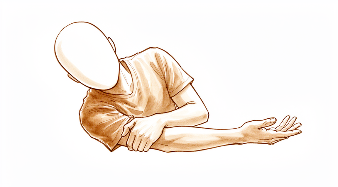
Você provavelmente sentirá dor logo na parte externa do cotovelo. Essa dor frequentemente se intensifica quando você tenta girar o antebraço, como ao abrir uma porta ou usar uma chave de fenda. Tarefas simples, como alcançar as costas para fechar um sutiã ou guardar a camisa dentro da calça, podem se tornar muito difíceis. Você pode ter dificuldade para levantar objetos ou carregar sacolas de compras porque a articulação parece instável.
Seus sintomas podem parecer piores à noite ou logo pela manhã. Muitos pacientes relatam que dormir do lado lesionado é impossível devido à dor. A dor frequentemente retorna após o uso do braço nas atividades diárias. Embora algumas fraturas sejam estáveis e cicatrizem bem sem cirurgia, outras causam desconforto significativo que limita seus movimentos.
Se sua lesão envolver mais do que apenas o osso, você pode sentir uma sensação de soltura ou instabilidade no cotovelo. Isso pode acontecer se outras estruturas ao redor da articulação também estiverem danificadas. Seu cirurgião examinará cuidadosamente essas áreas para garantir que tudo seja reparado corretamente. Sem o tratamento adequado, essa instabilidade pode levar a resultados ruins a longo prazo. A maioria das pessoas com fraturas leves descobre que o repouso e os movimentos suaves ajudam a aliviar a dor com o tempo.
O que está realmente acontecendo
Quando você fratura a parte superior do osso do antebraço, frequentemente danifica os tecidos moles que atuam como uma junta ao redor da sua articulação do cotovelo. Essa lesão pode romper os ligamentos que mantêm sua articulação unida, assim como um elástico esticado perde sua elasticidade. Se o osso se quebrar em muitos pedaços, a estabilidade de todo o seu braço está em risco, e outras estruturas também podem se tornar instáveis.
Seu cirurgião analisa cuidadosamente o alinhamento da articulação para decidir se ela pode cicatrizar por conta própria ou precisa de reparo. Se o osso se quebrar em três ou mais pedaços, seu cirurgião pode substituir a parte quebrada por um implante metálico. Essa peça metálica atua como um novo amortecedor de choque para manter o movimento do seu braço suave. Em muitos casos, os pacientes têm um bom resultado com essa substituição, e os resultados a longo prazo mostram que 96% dos pacientes têm resultados funcionais satisfatórios.
Mesmo que o osso cicatrize bem, você pode notar algum rigidez ou alterações na articulação ao longo do tempo. Os raios-X frequentemente mostram artrite por desgaste na maioria dos casos após a remoção precoce do osso quebrado. No entanto, se seu cirurgião puder reparar o osso ou substituí-lo adequadamente, você pode recuperar uma boa função. A maioria dos pacientes com lesões complexas pode alcançar bons resultados, embora alguns ainda possam ficar insatisfeitos com sua recuperação.
O que podemos fazer a respeito
Para muitas fraturas estáveis, você pode iniciar com autocuidados e fisioterapia. Seu cirurgião pode recomendar repouso do braço e uso de uma atadura por um curto período. A fisioterapia visa restaurar a amplitude de movimento e a força do seu cotovelo. A maioria dos pacientes com fraturas isoladas e estáveis obtém excelentes resultados com essa abordagem não cirúrgica. Mesmo fraturas com pequenos deslocamentos de 2 a 3 mm frequentemente cicatrizam bem sem cirurgia. Você deve dar uma chance justa a esse manejo conservador antes de considerar outras medidas.
Se a dor persistir, seu cirurgião pode discutir opções médicas para ajudá-lo a gerenciar os sintomas. Embora as evidências indiquem que injeções de anestésico local após a aspiração articular não oferecem benefício adicional, outros tratamentos podem ser usados para problemas específicos, como artrite. Para alguns pacientes com artrite por desgaste após uma fratura, injeções de cortisona ou ácido hialurônico podem proporcionar alívio. Essas injeções visam reduzir a inflamação ou lubrificar a articulação para diminuir a dor. O efeito desses tratamentos varia, mas eles são frequentemente utilizados quando a função geral é limitada pelo desconforto, e não pela própria fratura.
A cirurgia é considerada quando o tratamento conservador atinge seu limite ou quando o osso está muito fraturado para ser reparado. Seu cirurgião pode recomendar a substituição do osso fraturado por uma prótese metálica se ele não puder ser reconstruído. Essa opção é frequentemente escolhida para fraturas complexas com mais de três fragmentos ou quando os ligamentos também estão lesados. Em casos de lesões do tríade terrível, seu cirurgião pode optar por reparar o osso se uma fixação estável for possível. O objetivo da cirurgia é restaurar a estabilidade e a função quando os métodos não cirúrgicos não são suficientes.
Quando procurar ajuda médica
Consulte o seu médico de família se tiver dor persistente que não melhora com o repouso, ou se sentir fraqueza e instabilidade no cotovelo. Solicite uma avaliação por um especialista se o cotovelo bloquear ou ceder, ou se os sintomas interferirem no seu sono ou trabalho. Procure ajuda imediatamente se notar uma piora súbita dos sintomas. O diagnóstico atempado é fundamental para evitar complicações como a instabilidade do antebraço. A maioria das lesões menores identificadas em exames de imagem não causa sintomas, mas se a dor persistir, é necessária uma avaliação adicional para garantir que as estruturas corretas sejam tratadas.
Evidence & references
Overview
- Medium-term data suggest that patients with comminuted radial head fractures do well with radial head replacement [1].
- Long-term patient-reported outcomes were excellent following the nonoperative management of isolated stable fractures of the radial head or neck [2].
- No clinical benefit with ORIF could be found compared to nonoperative management of isolated partial articular radial head fractures with displacement of greater than 2 mm but less than 5 mm at short-term followup [4].
- Radial head fractures treated by early resection arthroplasty offer satisfactory functional results in 96% of patients at long-term follow-up [5].
- Radiographic degenerative changes are present in the great majority of cases following early resection arthroplasty for radial head fractures [5].
- The complications of radial head fractures are characteristic to their classification [6].
- At the time of short-term followup, arthroplasty with a metal radial head implant was found to be a safe and effective treatment option for patients with an unreconstructible radial head fracture [8].
- Radial head implants offer a reliable treatment for complex Mason type III and IV fractures, with good functional and survival outcomes and a low incidence of complications [10].
- Treatment of radial head fractures may have an independent effect on outcome in the context of terrible triad injuries [11].
- The authors recommend reconstruction of comminuted radial head fractures in the context of a terrible triad injury, providing stable fixation can be achieved [11].
- The clinical and radiographic outcomes of revision surgery of radial head prostheses are favorable [14].
- Radial head treatment was associated with increased reoperation risk in terrible triad injuries [19].
- No patient- or injury-related factors were associated with the reoperation risk in terrible triad injuries [19].
- Radial head arthroplasty is a reliable procedure for complex radial head fractures not amenable to reconstruction [21].
- Radial head arthroplasty is particularly indicated for complex radial head fractures when associated with unstable elbows or forearm injuries [21].
- Radial head arthroplasty requires that concomitant injuries be addressed [21].
- Fracture displacement of 2 to 3 mm is not necessarily an indication for surgical fixation in isolated fractures of the radial head [26].
Anatomy & Pathophysiology
- Isolated displaced type II partial articular radial head fractures are associated with lateral ulnar collateral ligament tears [9].
- Concurrent injuries and specific complications related to treatment modalities must be assessed in comminuted radial head fractures rather than relying solely on general elbow function outcomes [20].
- The coronoid opening angle is a radiographic technique used to assess bone loss in coronoid trauma and serves as an adjunct to 3-dimensional imaging for clinical decision making [27].
- Understanding relevant anatomy and factors associated with elbow stability allows for the application of a systematic treatment algorithm in terrible triad injuries to ensure sufficient stability for early motion [29].
- An anatomic model of the terrible triad of the elbow was created by exerting axial compression on an elbow in 15° flexion and maximal pronation at speeds of 100 and 10 mm/min [30].
- Detailed knowledge of fracture characteristics and their association with specific patterns of traumatic elbow instability assists in decision making and preoperative planning [32].
- Disruptions in any of the forearm structures may lead to forearm instability with consequences at the remaining structures [34].
- Understanding patterns of traumatic elbow instability helps surgeons counsel and manage patients with these injuries [35].
- The presence of nerve injury and intra-articular involvement predisposes floating elbow injuries to worse clinical outcomes [38].
- Trans-olecranon fracture posterior dislocation is a rare injury with unique characteristics involving complex elbow instability [48].
- Long-term outcomes with surgical management of complex elbow injuries are unknown [42].
Classification
- Complications of radial head fractures are characteristic to their classification [6].
- Radial head and neck fractures have distinct epidemiological characteristics [7].
- Consideration for osteoporosis is recommended in a subset of patients with radial head and neck fractures [7].
- Apparently isolated, stable partial fractures of the radial head are infrequently displaced [15].
- Observers have moderate disagreement regarding the diagnosis of displacement in apparently isolated, stable partial fractures of the radial head [15].
- Displacement of apparently isolated, stable partial fractures of the radial head is likely overdiagnosed [15].
- Fracture maps demonstrate no association between fracture line distribution and location of displaced partial articular fractures of the radial head and overall specific patterns of traumatic elbow instability [18].
- A common fracture mechanism involving the anterolateral part of the radial head occurs in most patients with displaced partial articular fractures [18].
- The most common location of displaced articular fractures of part of the radial head (Mason type 2) is the anterolateral quadrant with the forearm in neutral rotation [28].
- Recognition of the triad of triceps avulsion, radial head fracture, and medial collateral ligament rupture is key to appropriate treatment [25].
- Treatment of radial head fractures may have an independent effect on outcome in terrible triad injuries of the elbow [11].
- Reconstruction of comminuted radial head fractures is recommended in the context of a terrible triad injury, provided stable fixation can be achieved [11].
- The authors created a comprehensive classification of complex fracture-dislocations of the elbow that appeared to be reproducible [45].
- The comprehensive classification of complex fracture-dislocations of the elbow may represent a useful tool for the management of such difficult injuries [45].
- Selected Mason III radial head fractures and fracture dislocations could be stabilized satisfactorily with internal fixation [50].
- None of the radial head prostheses functioned as well as the native radial head [52].
- Open reduction and internal fixation to restore radial head anatomy is preferable to replacement when possible [52].
Clinical Presentation
- Patients with comminuted radial head fractures may achieve good medium-term outcomes with radial head replacement [1].
- Long-term patient-reported outcomes are excellent following nonoperative management of isolated stable fractures of the radial head or neck [2].
- Conservative management of isolated Mason type II radial head fractures yields favorable therapeutic outcomes with a low incidence of complications [3].
- There is no clinical benefit with ORIF compared to nonoperative management for isolated partial articular radial head fractures with displacement greater than 2 mm but less than 5 mm at short-term follow-up [4].
- Radial head fractures treated by early resection arthroplasty offer satisfactory functional results in 96% of patients at long-term follow-up [5].
- Complications of radial head fractures are characteristic to their classification [6].
- Radial head and neck fractures have distinct epidemiological characteristics, and consideration for osteoporosis is recommended in a subset of patients [7].
- Arthroplasty with a metal radial head implant is a safe and effective treatment option for unreconstructible radial head fractures at short-term follow-up [8].
- It is important to determine which structures need to be repaired in isolated displaced type II partial articular radial head fractures to avoid complications leading to elbow instability [9].
- Treatment of radial head fractures may have an independent effect on outcome in terrible triad injuries, with a recommendation for reconstruction of comminuted fractures if stable fixation can be achieved [11].
- Most injuries found with MRI in patients with radial head fractures are not symptomatic or of clinical importance in short-term follow-up [12].
- A 69-year clinical and radiologic follow-up of a previously unknown radial head prosthesis has been reported [13].
- The clinical and radiographic outcomes of revision surgery of radial head prostheses are favorable [14].
- Displacement of apparently isolated, stable partial fractures of the radial head is likely overdiagnosed due to infrequent actual displacement and moderate disagreement among observers regarding diagnosis [15].
- The outcome of patients undergoing treatment for terrible triad injuries is similar whether the radial head was excised or replaced [16].
- Fracture maps demonstrate no association between fracture line distribution and location of displaced partial articular fractures of the radial head and overall specific patterns of traumatic elbow instability [18].
- Radial head treatment is associated with increased reoperation risk in terrible triad injuries, leading to a recommendation for fixation when feasible [19].
- Forearm instability results from traumatic disruption of the radial head, interosseous membrane, and triangular fibrocartilage complex [22].
- Delayed recognition and treatment of forearm instability lead to poor outcomes, making timely diagnosis and appropriate initial intervention imperative [22].
- Nonsurgical management of minimally displaced radial neck fractures produces excellent results in most pediatric patients [23].
- Open reduction of proximal radius fractures in children has been associated with particularly poor outcomes [23].
- Recognition of the triad of triceps avulsion, radial head fracture, and medial collateral ligament rupture is key to appropriate treatment [25].
Investigations
- Radial head and neck fractures have distinct epidemiological characteristics, and consideration for osteoporosis in a subset of patients is recommended [7].
- Most injuries found with MRI in patients with radial head fractures are not symptomatic or of clinical importance in short-term follow-up [12].
- Elbow arthroscopy has a significant diagnostic value in radial head fractures when compared to standard radiological imaging and revealed concomitant injuries even in patients with ununeventful MRI/CT [39].
- The coronoid opening angle can be of value alongside 3-dimensional imaging in evaluating elbow injuries and used as an adjunct in clinical decision making [27].
- Because apparently isolated, stable partial fractures of the radial head are infrequently displaced and observers have moderate disagreement regarding the diagnosis of displacement, it is likely that displacement is overdiagnosed [15].
- It is important to determine which structures need to be repaired to avoid complications that could lead to elbow instability in isolated displaced type II partial articular radial head fractures [9].
- Fracture maps demonstrated no association between fracture line distribution and location of displaced partial articular fractures of the radial head and overall specific patterns of traumatic elbow instability, suggesting a common fracture mechanism that involves the anterolateral part of the radial head in most patients [18].
- Quantitative analysis confirms that the most common location of displaced articular fractures of part of the radial head (Mason type 2) is the anterolateral quadrant with the forearm in neutral rotation [28].
- The complications of radial head fractures are characteristic to their classification [6].
- It emphasizes the importance of assessing for concurrent injuries and specific complications related to each treatment modality rather than solely relying on general elbow function outcomes in comminuted radial head fractures [20].
Treatment
- Medium-term data suggest that patients with comminuted radial head fractures do well with radial head replacement [1].
- Long-term patient-reported outcomes were excellent following the nonoperative management of isolated stable fractures of the radial head or neck [2].
- Conservative management of isolated Mason II radial head fractures yields favorable therapeutic outcomes with a low incidence of complications [3].
- No clinical benefit with ORIF could be found compared to nonoperative management of isolated partial articular radial head fractures with displacement of greater than 2 mm but less than 5 mm at short-term followup [4].
- Radial head fractures treated by early resection arthroplasty offer satisfactory functional results in 96% of patients at long-term follow-up, in spite of the radiographic degenerative changes present in the great majority of cases [5].
- Arthroplasty with a metal radial head implant was found to have been a safe and effective treatment option for patients with an unreconstructible radial head fracture at short-term followup [8].
- Radial head implants offer a reliable treatment for complex Mason type III and IV fractures, with good functional and survival outcomes and a low incidence of complications [10].
- Treatment of radial head fractures may have an independent effect on outcome; reconstruction of comminuted radial head fractures is recommended in the context of a terrible triad injury if stable fixation can be achieved [11].
- The clinical and radiographic outcomes of revision surgery of radial head prostheses are favorable [14].
- The outcome of patients undergoing treatment for terrible triad injuries is similar whether the patient's radial head was excised or replaced [16].
- Patients with radial head arthroplasty (RHA) implanted within 4 weeks for nonreconstructable fractures of the radial head sustained a limited number of failures and obtained a good long-term clinical outcome despite a relatively high rate of post-traumatic arthritis [17].
- Radial head treatment was associated with increased reoperation risk in terrible triad injuries, leading to a recommendation for fixation when feasible [19].
- It is important to assess for concurrent injuries and specific complications related to each treatment modality rather than solely relying on general elbow function outcomes [20].
- Nonsurgical management of minimally displaced radial neck fractures produces excellent results in most patients [23].
- Open reduction of proximal radius fractures in children has been associated with particularly poor outcomes [23].
- Fracture displacement of 2 to 3 mm is not necessarily an indication for surgical fixation in isolated fractures of the radial head [26].
- No significant difference was found between monopolar and bipolar radial head prostheses in terms of efficacy and safety [43].
- For displaced fractures with greater than 3 fragments, radial head replacement is recommended [46].
- Arthroplasty may be preferred over tenuous fracture fixation in the setting of associated ligament injuries [46].
- Intra-articular use of local anaesthetic after joint aspiration does not offer any benefit over aspiration alone in the treatment of undisplaced radial head fractures [51].
Complications
- Patients with comminuted radial head fractures treated with radial head replacement demonstrate good medium-term outcomes [1].
- Nonoperative management of isolated stable fractures of the radial head or neck results in excellent long-term patient-reported outcomes [2].
- Conservative management of isolated Mason II radial head fractures yields favorable therapeutic outcomes with a low incidence of complications [3].
- Operative fixation (ORIF) provides no clinical benefit compared to nonoperative management for isolated partial articular radial head fractures with displacement greater than 2 mm but less than 5 mm at short-term follow-up [4].
- Early resection arthroplasty for radial head fractures offers satisfactory functional results in 96% of patients at long-term follow-up [5].
- Radiographic degenerative changes are present in the great majority of cases following early resection arthroplasty for radial head fractures [5].
- Complications of radial head fractures are characteristic to their classification [6].
- Osteoporosis should be considered in a subset of patients with radial head and neck fractures [7].
- Arthroplasty with a metal radial head implant is a safe and effective treatment option for unreconstructible radial head fractures at short-term follow-up [8].
- Long-term follow-up data for metal radial head implants in unreconstructible fractures were noted as still needed at the time of the 2001 study [8].
- Determining which structures require repair is important to avoid complications that could lead to elbow instability in isolated displaced type II partial articular radial head fractures [9].
- Radial head implants offer reliable treatment for complex Mason type III and IV fractures with good functional and survival outcomes and a low incidence of complications [10].
- Most injuries found with MRI in patients with radial head fractures are not symptomatic or of clinical importance in short-term follow-up [12].
- A 69-year clinical and radiologic follow-up of a previously unknown radial head prosthesis has been reported [13].
- Patients with radial head arthroplasty (RHA) implanted within 4 weeks for nonreconstructable fractures sustained a limited number of failures [17].
- Despite a relatively high rate of post-traumatic arthritis, patients with RHA for nonreconstructable fractures obtained good long-term clinical outcomes [17].
- Longer-term studies are required to ascertain whether the benefits of radial head arthroplasty are offset by late complications such as loosening [24].
- The incidence of neurologic complications associated with surgical treatment of complex elbow fractures requiring radial head prosthesis implantation may be underestimated in the literature [53].
Recovery
- Patients with comminuted radial head fractures do well with radial head replacement at medium-term follow-up [1].
- Long-term patient-reported outcomes are excellent following nonoperative management of isolated stable fractures of the radial head or neck [2].
- Conservative management of isolated Mason II radial head fractures yields favorable therapeutic outcomes with a low incidence of complications [3].
- No clinical benefit with ORIF was found compared to nonoperative management of isolated partial articular radial head fractures with displacement greater than 2 mm but less than 5 mm at short-term follow-up [4].
- Radial head fractures treated by early resection arthroplasty offer satisfactory functional results in 96% of patients at long-term follow-up [5].
- Radiographic degenerative changes are present in the great majority of cases following early resection arthroplasty for radial head fractures [5].
- Arthroplasty with a metal radial head implant is a safe and effective treatment option for patients with an unreconstructible radial head fracture at short-term follow-up [8].
- Radial head implants offer reliable treatment for complex Mason type III and IV fractures with good functional and survival outcomes and a low incidence of complications [10].
- Most injuries found with MRI in patients with radial head fractures are not symptomatic or of clinical importance in short-term follow-up [12].
- The outcome of patients undergoing treatment for terrible triad injuries is similar whether the radial head was excised or replaced [16].
- Patients with radial head arthroplasty (RHA) implanted within 4 weeks for nonreconstructable fractures sustained a limited number of failures [17].
- Despite a relatively high rate of post-traumatic arthritis, patients with RHA implanted within 4 weeks for nonreconstructable fractures obtained a good long-term clinical outcome [17].
- At short term, there were no differences between patients treated with ORIF for isolated radial head fractures and those treated for radial head fractures in association with other elbow injuries regarding pain and disability scores [36].
- Two-year Kaplan-Meier survival free of revision or resection estimates and reoperation rates were equivalent between acute and delayed radial head arthroplasty groups [54].
- The delayed group experienced worse Mayo Elbow Performance Score outcomes compared to the acute group [54].
- The delayed group experienced a higher revision or resection rate at 5 years compared to the acute group [54].
- The delayed group experienced an increased rate of radiographic loosening compared to the acute group [54].
Key Evidence
- [L5] Medium-term data suggest that patients with comminuted radial head fractures do well with radial head replacement. [1] (10.1016/j.jhsa.2012.10.001)
- [L4] Long-term patient-reported outcomes were excellent following the nonoperative management of isolated stable fractures of the radial head or neck. [2] (10.2106/jbjs.m.01354)
- [L1] Based on the current evidence, conservative management of isolated Mason II radial head fractures yields favorable therapeutic outcomes with a low incidence of complications. [3] (10.1186/s13018-024-05039-6)
- [L3] No clinical benefit with ORIF could be found compared to nonoperative management of isolated partial articular radial head fractures with displacement of greater than 2 mm but less than 5 mm at short-term followup. [4] (10.1007/s11999-014-3541-x)
- [L4] Radial head fractures treated by early resection arthroplasty offer satisfactory functional results in 96% of patients at long-term follow-up, in spite of the radiographic degenerative changes present in the great majority of cases. [5] (10.1016/j.jse.2010.09.005)
- [L4] The complications of radial head fractures are characteristic to their classification. [6] (10.1016/j.jse.2018.11.047)
- [L4] Radial head and neck fractures have distinct epidemiological characteristics, and consideration for osteoporosis in a subset of patients is recommended. [7] (10.1016/j.jhsa.2011.09.034)
- [L4] At the time of short-term followup, arthroplasty with a metal radial head implant was found to have been a safe and effective treatment option for patients with an unreconstructible radial head fracture; however, long-term follow-up is still needed. [8] (10.2106/00004623-200108000-00010)
- [L3] It is important to determine which structures need to be repaired to avoid complications that could lead to elbow instability. [9] (10.1016/j.jse.2019.07.006)
- [L4] Radial head implants offer a reliable treatment for complex Mason type III and IV fractures, with good functional and survival outcomes and a low incidence of complications. [10] (10.1016/j.jse.2025.05.038)
- [L3] Treatment of radial head fractures may have an independent effect on outcome; the authors recommend reconstruction of comminuted radial head fractures in the context of a TTI, providing stable fixation can be achieved. [11] (10.1302/0301-620x.102b12.bjj-2020-2145)
- [L2] Most injuries found with MRI in patients with radial head fractures are not symptomatic or of clinical importance in short-term follow-up. [12] (10.1016/j.jse.2011.06.011)
- [L4] We have reported a 69-year clinical and radiologic follow-up of a previously unknown radial head prosthesis. [13] (10.1016/j.jse.2014.09.030)
- [L4] The clinical and radiographic outcomes of revision surgery of radial head prostheses are favorable. [14] (10.1016/j.jse.2016.09.047)
- [L4] Because apparently isolated, stable partial fractures of the radial head are infrequently displaced and observers have moderate disagreement regarding the diagnosis of displacement, it is likely that displacement is overdiagnosed. [15] (10.1016/j.jse.2006.10.015)
- [L3] The outcome of patients undergoing treatment for terrible triad injuries is similar whether the patient's radial head was excised or replaced. [16] (10.1302/0301-620x.100b11.bjj-2018-0293.r1)
- [L4] However, patients with RHA implanted within 4 weeks for nonreconstructable fractures of the radial head sustained a limited number of failures and, despite a relatively high rate of post-traumatic arthritis, obtained a good long-term clinical outcome. [17] (10.1016/j.jse.2025.06.026)
- [L4] Fracture maps demonstrated no association between fracture line distribution and location of displaced partial articular fractures of the radial head and overall specific patterns of traumatic elbow instability, suggesting a common fracture mechanism that involves the anterolateral part of the radial head in most patients. [18] (10.1016/j.jse.2016.01.030)
- [L3] No patient- or injury-related factors were associated with the reoperation risk, but radial head treatment was associated with increased risk, leading to a recommendation for fixation when feasible. [19] (10.1097/corr.0000000000001391)
- [L4] It emphasizes the importance of assessing for concurrent injuries and specific complications related to each treatment modality rather than solely relying on general elbow function outcomes. [20] (10.1016/j.jse.2011.02.013)
- [L5] Radial head arthroplasty is a reliable procedure for complex radial head fractures not amenable to reconstruction, particularly when associated with unstable elbows or forearm injuries, provided concomitant injuries are addressed. [21] (10.1016/j.jhsa.2009.01.027)
- [L5] Forearm instability results from traumatic disruption of the radial head, interosseous membrane, and triangular fibrocartilage complex; delayed recognition and treatment lead to poor outcomes, making timely diagnosis and appropriate initial intervention imperative. [22] (10.1016/j.jhsa.2013.07.010)
- [L5] Nonsurgical management of minimally displaced radial neck fractures produces excellent results in most patients, whereas open reduction has been associated with particularly poor outcomes. [23] (10.5435/jaaos-d-18-00204)
- [L3] Longer-term studies will be required to ascertain whether the apparent benefits of radial head arthroplasty are offset by late complications of arthroplasty, such as loosening. [24] (10.1007/s11999-013-3331-x)
- [L4] Recognition of the triad of triceps avulsion, radial head fracture, and MCL rupture is the key to appropriate treatment. [25] (10.1016/j.jse.2011.06.017)
- [L2] This retrospective review suggests that fracture displacement of 2 to 3 mm is not necessarily an indication for surgical fixation in isolated fractures of the radial head. [26] (10.1016/j.jse.2013.01.019)
- [L4] It can be of value alongside 3-dimensional imaging in evaluating elbow injuries and used as an adjunct in clinical decision making. [27] (10.1016/j.jse.2021.12.039)
- [L4] This quantitative analysis confirms that the most common location of displaced articular fractures of part of the radial head (Mason type 2) is the anterolateral quadrant with the forearm in neutral rotation. [28] (10.1016/j.jse.2011.08.056)
- [L5] Despite the complexities of this injury, an understanding of the relevant anatomy and the factors associated with elbow stability allows the application of a systematic algorithm for treatment that can help ensure sufficient elbow stability to allow early motion, thereby leading to improved outcomes in most patients. [29] (10.5435/00124635-200903000-00003)
- [L5] The study successfully created and validated an anatomic model of terrible triad of the elbow by exerting axial compression on an elbow in 15° flexion and maximal pronation at speeds of 100 and 10 mm/min. [30] (10.1186/s13018-024-05069-0)
- [L4] Detailed knowledge of fracture characteristics and their association with specific patterns of traumatic elbow instability may assist decision making and preoperative planning. [32] (10.1016/j.jhsa.2014.07.059)
- [L5] Disruptions in any of these structures may lead to forearm instability with consequences at the remaining structures. [34] (10.1016/j.jhsa.2016.10.017)
- [L5] Understanding the patterns of traumatic elbow instability helps the surgeon counsel and manage patients with these injuries. [35] (10.1016/j.jhsa.2010.05.002)
- [L3] At short term, there were no differences between patients treated with ORIF for isolated radial head fractures and those treated for radial head fractures in association with other elbow injuries with regard to pain and disability scores. [36] (10.1007/s11999-014-3519-8)
- [L4] Although the nature of floating elbow injuries is complex, the presence of nerve injury and intra-articular involvement predispose to worse clinical outcomes. [38] (10.1016/j.jse.2012.09.005)
- [L4] Elbow arthroscopy has a significant diagnostic value in radial head fractures when compared to standard radiological imaging and revealed concomitant injuries even in patients with uneventful MRI/CT. [39] (10.1186/s12891-019-2726-6)
- [L5] Long-term outcome with surgical management of complex elbow injuries is unknown. [42] (10.5435/00124635-200605000-00003)
- [L1] No significant difference was found between monopolar and bipolar radial head prostheses in terms of efficacy and safety. [43] (10.1016/j.jse.2021.10.037)
- [L3] The authors created a comprehensive classification of complex fracture-dislocations of the elbow that appeared to be reproducible and may represent a useful tool for the management of such difficult injuries. [45] (10.1016/j.jse.2011.06.003)
- [L5] For displaced fractures with greater than 3 fragments, radial head replacement is recommended, and arthroplasty may be preferred over tenuous fracture fixation in the setting of associated ligament injuries. [46] (10.1016/j.jhsa.2008.12.024)
- [L4] Trans-olecranon fracture posterior dislocation is a rare injury with unique characteristics involving complex elbow instability. [48] (10.1186/s13018-023-03563-5)
- [L3] Selected Mason III radial head fractures and fracture dislocations could be stabilized satisfactorily with internal fixation. [50] (10.1016/j.jhsa.2007.09.016)
- [L1] Intra-articular use of local anaesthetic after joint aspiration does not offer any benefit over aspiration alone in the treatment of undisplaced radial head fractures and its routine application is not supported by the clinical data. [51] (10.1016/j.jse.2009.04.003)
- [L5] However, none of the prostheses functioned as well as the native radial head, suggesting that open reduction and internal fixation to restore radial head anatomy is preferable to replacement when possible. [52] (10.2106/00004623-200112000-00010)
- [L4] The incidence of neurologic complications associated with the surgical treatment of complex elbow fractures requiring implantation of a radial head prosthesis may be underestimated in the literature. [53] (10.1016/j.jse.2020.01.086)
- [L3] Although 2-year Kaplan-Meier survival free of revision or resection estimates and reoperation rates were equivalent between the groups, the delayed group experienced worse Mayo Elbow Performance Score outcomes, a higher revision or resection rate at 5 years, and an increased rate of radiographic loosening. [54] (10.1016/j.jse.2022.07.031)
References
[1] Radial Head Fractures. The Journal of Hand Surgery. 2012. DOI: 10.1016/j.jhsa.2012.10.001 [2] Long-Term Outcomes of Isolated Stable Radial Head Fractures. Journal of Bone and Joint Surgery. 2014. DOI: 10.2106/jbjs.m.01354 [3] Comparison of operatively and nonoperatively treated isolated mason type II radial head fractures: a systematic review and meta-analysis. Journal of Orthopaedic Surgery and Research. 2024. DOI: 10.1186/s13018-024-05039-6 [4] Is ORIF Superior to Nonoperative Treatment in Isolated Displaced Partial Articular Fractures of the Radial Head?. Clinical Orthopaedics & Related Research. 2014. DOI: 10.1007/s11999-014-3541-x [5] Resection arthroplasty for radial head fractures: Long-term follow-up. Journal of Shoulder and Elbow Surgery. 2011. DOI: 10.1016/j.jse.2010.09.005 [6] Surgical revision of radial head fractures: a multicenter retrospective analysis of 466 cases. Journal of Shoulder and Elbow Surgery. 2019. DOI: 10.1016/j.jse.2018.11.047 [7] The Epidemiology of Radial Head and Neck Fractures. The Journal of Hand Surgery. 2012. DOI: 10.1016/j.jhsa.2011.09.034 [8] Arthroplasty with a Metal Radial Head for Unreconstructible Fractures of the Radial Head. The Journal of Bone and Joint Surgery-American Volume. 2001. DOI: 10.2106/00004623-200108000-00010 [9] Isolated displaced type II partial articular radial head fracture: correlation of preoperative imaging with intraoperative findings of lateral ulnar collateral ligament tear. Journal of Shoulder and Elbow Surgery. 2020. DOI: 10.1016/j.jse.2019.07.006 [10] Long-term survival of Acumed anatomical radial head implant for Mason type III-IV fractures: a 15-year follow-up. Journal of Shoulder and Elbow Surgery. 2026. DOI: 10.1016/j.jse.2025.05.038 [11] Infographic: Surgical treatment of the radial head in terrible triad injuries of the elbow. The Bone & Joint Journal. 2020. DOI: 10.1302/0301-620x.102b12.bjj-2020-2145 [12] Magnetic resonance imaging in radial head fractures: most associated injuries are not clinically relevant. Journal of Shoulder and Elbow Surgery. 2011. DOI: 10.1016/j.jse.2011.06.011 [13] Sixty-nine-year follow-up of a McKee radial head arthroplasty. Journal of Shoulder and Elbow Surgery. 2015. DOI: 10.1016/j.jse.2014.09.030 [14] Clinical and radiographic outcome of revision surgery of radial head prostheses: midterm results in 16 patients. Journal of Shoulder and Elbow Surgery. 2017. DOI: 10.1016/j.jse.2016.09.047 [15] Apparently isolated partial articular fractures of the radial head: Prevalence and reliability of radiographically diagnosed displacement. Journal of Shoulder and Elbow Surgery. 2007. DOI: 10.1016/j.jse.2006.10.015 [16] Radial head resection versus prosthetic arthroplasty in terrible triad injury: a retrospective comparative cohort study. The Bone & Joint Journal. 2018. DOI: 10.1302/0301-620x.100b11.bjj-2018-0293.r1 [17] What happens to the elbow 15 years after a radial head prosthesis? A clinical and imaging long-term follow-up study. Journal of Shoulder and Elbow Surgery. 2026. DOI: 10.1016/j.jse.2025.06.026 [18] Fracture mapping of displaced partial articular fractures of the radial head. Journal of Shoulder and Elbow Surgery. 2016. DOI: 10.1016/j.jse.2016.01.030 [19] What Factors Are Associated with Reoperation After Operative Treatment of Terrible Triad Injuries?. Clinical Orthopaedics & Related Research. 2020. DOI: 10.1097/corr.0000000000001391 [20] Comminuted radial head fractures: aspects of current management. Journal of Shoulder and Elbow Surgery. 2011. DOI: 10.1016/j.jse.2011.02.013 [21] Radial Head Implant Arthroplasty. The Journal of Hand Surgery. 2009. DOI: 10.1016/j.jhsa.2009.01.027 [22] Forearm Instability. The Journal of Hand Surgery. 2014. DOI: 10.1016/j.jhsa.2013.07.010 [23] Proximal Radius Fractures in Children. Journal of the American Academy of Orthopaedic Surgeons. 2019. DOI: 10.5435/jaaos-d-18-00204 [24] Fixation Versus Replacement of Radial Head in Terrible Triad: Is There a Difference in Elbow Stability and Prognosis?. Clinical Orthopaedics & Related Research. 2014. DOI: 10.1007/s11999-013-3331-x [25] Triceps avulsion, radial head fracture, and medial collateral ligament rupture about the elbow: a report of 4 cases. Journal of Shoulder and Elbow Surgery. 2012. DOI: 10.1016/j.jse.2011.06.017 [26] A retrospective cohort study of displaced segmental radial head fractures: is 2 mm of articular displacement an indication for surgery?. Journal of Shoulder and Elbow Surgery. 2013. DOI: 10.1016/j.jse.2013.01.019 [27] The coronoid opening angle: a novel radiographic technique to assess bone loss in coronoid trauma. Journal of Shoulder and Elbow Surgery. 2022. DOI: 10.1016/j.jse.2021.12.039 [28] Quantitative measurement of radial head fracture location. Journal of Shoulder and Elbow Surgery. 2012. DOI: 10.1016/j.jse.2011.08.056 [29] Terrible Triad Injury of the Elbow: Current Concepts. Journal of the American Academy of Orthopaedic Surgeons. 2009. DOI: 10.5435/00124635-200903000-00003 [30] Creation of a replicable anatomic model of terrible triad of the elbow. Journal of Orthopaedic Surgery and Research. 2024. DOI: 10.1186/s13018-024-05069-0 [32] Quantitative 3-Dimensional Computed Tomography Measurements of Coronoid Fractures. The Journal of Hand Surgery. 2015. DOI: 10.1016/j.jhsa.2014.07.059 [34] Forearm Instability: Anatomy, Biomechanics, and Treatment Options. The Journal of Hand Surgery. 2017. DOI: 10.1016/j.jhsa.2016.10.017 [35] Traumatic Elbow Instability. The Journal of Hand Surgery. 2010. DOI: 10.1016/j.jhsa.2010.05.002 [36] Open Reduction and Internal Fixation of Radial Head Fractures: Do Outcomes Differ Between Simple and Complex Injuries?. Clinical Orthopaedics & Related Research. 2014. DOI: 10.1007/s11999-014-3519-8 [38] Floating elbow injuries in adults: prognostic factors affecting clinical outcomes. Journal of Shoulder and Elbow Surgery. 2013. DOI: 10.1016/j.jse.2012.09.005 [39] The value of elbow arthroscopy in diagnosing and treatment of radial head fractures. BMC Musculoskeletal Disorders. 2019. DOI: 10.1186/s12891-019-2726-6 [42] Complex Elbow Instability. Journal of the American Academy of Orthopaedic Surgeons. 2006. DOI: 10.5435/00124635-200605000-00003 [43] Efficacy and safety of monopolar versus bipolar radial head arthroplasty: a systematic review and meta-analysis. Journal of Shoulder and Elbow Surgery. 2022. DOI: 10.1016/j.jse.2021.10.037 [45] Complex fracture-dislocations of the proximal ulna and radius in adults: a comprehensive classification. Journal of Shoulder and Elbow Surgery. 2011. DOI: 10.1016/j.jse.2011.06.003 [46] Radial Head Fractures—An Update. The Journal of Hand Surgery. 2009. DOI: 10.1016/j.jhsa.2008.12.024 [48] Trans-olecranon fracture posterior dislocation: a novel type of elbow injury. Journal of Orthopaedic Surgery and Research. 2023. DOI: 10.1186/s13018-023-03563-5 [50] Open Reduction and Internal Fixation of Mason Type III Radial Head Fractures With and Without an Associated Elbow Dislocation. The Journal of Hand Surgery. 2007. DOI: 10.1016/j.jhsa.2007.09.016 [51] Aspiration alone versus aspiration and bupivacaine injection in the treatment of undisplaced radial head fractures: A prospective randomized study. Journal of Shoulder and Elbow Surgery. 2009. DOI: 10.1016/j.jse.2009.04.003 [52] Contribution of Monoblock and Bipolar Radial Head Prostheses to Valgus Stability of the Elbow. The Journal of Bone and Joint Surgery-American Volume. 2001. DOI: 10.2106/00004623-200112000-00010 [53] Neurologic complications after surgical management of complex elbow trauma requiring radial head replacement. Journal of Shoulder and Elbow Surgery. 2020. DOI: 10.1016/j.jse.2020.01.086 [54] Acute versus delayed radial head arthroplasty for the treatment of radial head fractures. Journal of Shoulder and Elbow Surgery. 2022. DOI: 10.1016/j.jse.2022.07.031


