Artrite da articulação interfalângica distal
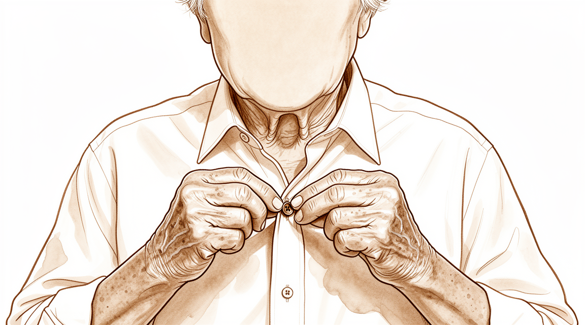
O que você está sentindo
Provavelmente, você sente dor e rigidez exatamente na ponta do dedo, onde a articulação encontra a unha. Esta é a articulação interfalângica distal, ou articulação DIP. A dor frequentemente se intensifica após o uso da mão para tarefas diárias, como alcançar as costas para fechar um sutiã ou guardar a camisa dentro da calça. Você pode notar que a dor é pior ao acordar pela manhã e melhora à medida que move a mão.
Com o tempo, a articulação pode mudar de forma. A ponta do dedo pode se curvar para dentro, criando uma curvatura conhecida como deformidade em pescoço de cisne. Isso ocorre porque a articulação desenvolve uma contratura de flexão, o que significa que fica presa em uma posição flexionada. Se você teve uma lesão prévia, como uma fratura por mallet finger (dedo em martelo), pode apresentar sinais de artrise por desgaste. Esta condição segue um curso semelhante ao envelhecimento natural da articulação e leva a uma diminuição da amplitude de movimento.
A vida diária pode tornar-se difícil quando a articulação incha ou trava. Você pode ter dificuldade para segurar objetos ou realizar movimentos finos. Em alguns casos, a articulação parece instável ou "flutuante", o que pode ser confuso se houver pouca deformidade visível. Embora a dor possa ser incômoda, é importante saber que as alterações visíveis em um raio X nem sempre correspondem ao nível de dor que você sente. Seu cirurgião avaliará seus sintomas específicos para orientar o seu tratamento.
O que está realmente acontecendo
A sua articulação interfalangiana distal (articulação DIP) é a pequena dobradiça na ponta do seu dedo. No interior, a cartilagem lisa atua como um amortecedor, permitindo que os seus ossos deslizem uns sobre os outros sem dor. Quando a artrose por desgaste se desenvolve, este revestimento desgasta-se. Os ossos começam a friccionar diretamente uns contra os outros, causando a sensação de atrito e rigidez que sente. Este dano também pode deslocar a articulação, frequentemente empurrando a ponta do seu dedo para trás.
O tendão que flexiona a ponta do seu dedo é como uma corda forte presa ao osso. Se esta corda se romper ou se a articulação se tornar instável, o tendão não consegue puxar o dedo suavemente. Por vezes, a articulação fica presa numa posição flexionada, conhecida como contratura de flexão. Com o tempo, esta deformidade pode agravar-se, dificultando a extensão do seu dedo ou a sua utilização nas tarefas diárias.
O seu cirurgião avalia quanto da superfície articular está danificada para decidir o melhor caminho a seguir. Se o osso for demasiado pequeno para parafusos padrão, ou se a articulação estiver presa numa posição desfavorável, são necessárias técnicas especiais. O objetivo é aliviar a dor e restaurar a função, seja fundindo os ossos entre si ou utilizando um implante macio para manter algum movimento. Estas opções visam proporcionar-lhe alívio, protegendo ao mesmo tempo a força restante do seu dedo.
O que podemos fazer a respeito
Você pode começar gerenciando a dor por conta própria e trabalhando com um fisioterapeuta. A imobilização da articulação interfalângica distal reduz a dor e melhora a extensão sem causar rigidez ou restringir seus movimentos. No entanto, manter essa articulação imóvel reduzirá sua força de preensão geral, com o efeito tornando-se mais intenso à medida que se avança do indicador ao dedo mínimo. Você deve dar tempo para que esses métodos não cirúrgicos surtam efeito antes de considerar etapas mais invasivas.
Se as medidas simples não forem suficientes, seu cirurgião pode discutir opções médicas para gerenciar seus sintomas. Embora as evidências destaquem alternativas cirúrgicas específicas, elas indicam que injeções, como a colagenase, podem ajudar nas contraturas articulares, embora você deva ponderar o risco de retorno do problema. Para aqueles com dor severa que afeta tanto a articulação média quanto a terminal do mesmo dedo, o tratamento conjunto é frequentemente recomendado. Seu cirurgião também considerará sua saúde pessoal, pois fatores como o diabetes podem aumentar o risco de complicações após o tratamento.
Quando o tratamento conservador atinge seu limite, a cirurgia pode ser considerada para interromper a dor ou restaurar a função. Seu cirurgião pode optar por um procedimento que preserva o movimento para manter sua articulação móvel, ou por uma artrodese para travar a articulação em seu lugar, garantindo estabilidade. Em alguns casos, um implante de silicone é utilizado para proporcionar excelente alívio da dor e uma amplitude de movimento de 30 a 40 graus, com uma taxa geral de complicações baixa de 5%. A abordagem específica depende da condição da sua articulação e dos seus objetivos para a vida diária.
Quando procurar ajuda médica
Consulte o seu médico de família se tiver dor persistente que não melhora com o repouso, ou se o seu dedo parecer fraco e instável. Solicite uma avaliação por um especialista se a articulação bloquear, ceder, ou se os sintomas interferirem no seu sono ou trabalho. Deve também procurar ajuda se notar uma piora súbita dos sintomas. Esteja ciente de que algumas condições, como uma lesão do articulação interfalângica distal flutuante, podem apresentar deformidade mínima inicialmente, mas ainda assim levar à artrite mais tarde. O diagnóstico precoce ajuda a distinguir estes problemas de outras afecções articulares.
Evidence & references
Overview
- Percutaneous DIP joint arthrodesis is advantageous compared with open fusion techniques in select patients [1].
- Swan neck deformity progresses significantly over time due to increasing DIPJ flexion contracture [2].
- Simultaneous surgical intervention is recommended for severe painful osteoarthritis of both the PIP and DIP joints of the same digit [3].
- Denervation with cheilectomy presents a motion-preserving alternative to arthrodesis for symptomatic DIP joint osteoarthritis [4].
- Lateral approach and plate fixation for DIP joint arthrodesis yields results equivalent to traditional methods but with fewer major complications [5].
- The combination of DIP arthrodesis and PIP Swanson arthroplasty results in favorable outcomes regarding simultaneous bony union and flexibility [7].
- Silicone interpositional arthroplasty of the DIP joint is an acceptable alternative to arthrodesis, achieving excellent pain relief and a range of movement of 30–40 degrees [8].
- Silicone interpositional arthroplasty of the DIP joint has a low overall complication rate of 5% [8].
- The smile incision and reverse shotgun approach is a good surgical option for DIPJ arthrodesis when more volar part joint preparation and more volar implant insertion sites are necessary [9].
- The nonaxial multiple small screws (NMSS) technique is a feasible option for DIPJ and thumb IPJ arthrodesis, especially when a small finger is indicated and a significant flexion angle is required [11].
- Customized structural bone grafting addresses bone stock loss and medullary absence in failed DIPJ silicone arthroplasty, achieving reliable union rates and high patient satisfaction [13].
- There is no difference in biomechanical performance between K-wires and compression screws for DIPJ arthrodesis [20].
- Implant selection for DIPJ fusion should consider factors such as cost and complication profiles given the lack of difference in biomechanical performance between K-wires and compression screws [20].
Anatomy & Pathophysiology
- Palmar subluxation of a DIP joint without preexisting arthritic deformity is expected when more than one half of the dorsal articular surface is injured [6].
- Understanding of DIP joint morphology may lend insight into the biomechanics and disease progression within the DIP joints [10].
- A substantial number of distal phalanges are too small to accommodate commonly available headless compression screws, particularly in females and the small finger [31].
- Irreducibility was more commonly seen in dorsal than in volar dislocations of the DIP joint [33].
- Volar dislocations of the DIP joint carried a higher risk of instability immediately after reduction compared to dorsal dislocations [33].
- Biomechanically, dynamic tenodesis for the DIP joint using the remaining FDP tendon results in a flexion angle greater than 30 degrees [23].
- In a cadaveric model, tenodesis successfully restored coordinated interphalangeal joint flexion after a simulated zone I FDP laceration with improvements in DIP joint flexion and composite finger flexion [35].
- Lateral blocking with incremental joint angles allows a safer application of force for the healing tendon during palmar and lateral blocking exercises [26].
Classification
- Swan neck deformity in the DIP joint progresses significantly over time due to increasing DIPJ flexion contracture [2].
- Radiological osteoarthritis following a mallet finger fracture follows a similar course to the natural degenerative process in the DIP joint [12].
- Post-traumatic osteoarthritis of the DIP joint after mallet finger fractures is accompanied by a decrease in range of motion, though this does not clinically affect patient-reported outcome measures (PROMs) [12].
- The interrater reliability of the Kellgren & Lawrence classification system for post-traumatic osteoarthritis in the DIP joint after mallet finger fractures is considerably lower than initially assumed [34].
- The interrater reliability of the OARSI classification system for post-traumatic osteoarthritis in the DIP joint after mallet finger fractures is considerably lower than initially assumed [34].
- Current concepts regarding DIP joint osteoarthritis highlight the roles of cartilage, subchondral bone, and soft tissue structures in etiology, pathogenesis, and evaluation [19].
- Morphological understanding of DIP joint curvatures may provide insight into the biomechanics and disease progression within the DIP joints [10].
- Examination of type I and type II nerve endings provides new information on the sensory systems of the DIP joints and surrounding structures [32].
- Palmar subluxation of a DIP joint without preexisting arthritic deformity is expected when more than one half of the dorsal articular surface is injured [6].
Clinical Presentation
- Swan neck deformity in the DIPJ progresses significantly over time due to increasing DIPJ flexion contracture [2].
- Palmar subluxation of a DIP joint without preexisting arthritic deformity is expected when more than one half of the dorsal articular surface is injured [6].
- Floating DIP joint injuries can be misdiagnosed initially due to minimal deformity [17].
- Radiological osteoarthritis following a mallet finger fracture is similar to the natural degenerative process in the DIP joint [12].
- Radiological osteoarthritis after a mallet finger fracture is accompanied by a decrease in range of motion of the DIP joint [12].
- Radiological osteoarthritis after a mallet finger fracture does not clinically affect patient-reported outcome measures (PROMs) [12].
- Primary synovial chondromatosis of the DIPJ is an extremely rare entity that requires accurate diagnosis to distinguish from other arthropathies [27].
- Understanding the morphology of DIPJ curvatures may lend insight into the biomechanics and disease progression within the DIP joints [10].
- Osteoarthritis of the DIPJ involves roles of cartilage, subchondral bone, and soft tissue structures in its etiology, pathogenesis, and evaluation [19].
Investigations
- Percutaneous DIP joint arthrodesis is advantageous compared with open fusion techniques in select patients [1].
- Swan neck deformity progresses significantly over time due to increasing DIPJ flexion contracture [2].
- Simultaneous surgical intervention is recommended for severe painful osteoarthritis of both the PIP and DIP joints of the same digit [3].
- Denervation with cheilectomy presents a motion-preserving alternative to arthrodesis for symptomatic DIP joint osteoarthritis [4].
- Lateral approach and plate fixation for DIP joint arthrodesis yields results equivalent to traditional methods with fewer major complications [5].
- Palmar subluxation of a DIP joint without preexisting arthritic deformity is expected when more than one half of the dorsal articular surface is injured [6].
- The combination of DIP arthrodesis and PIP Swanson arthroplasty results in favorable outcomes regarding simultaneous bony union and flexibility [7].
- Silicone interpositional arthroplasty of the DIP joint is an acceptable alternative to arthrodesis, achieving excellent pain relief and a range of movement of 30–40 degrees with a low overall complication rate of 5% [8].
- The smile incision and reverse shotgun approach is a good surgical option for DIPJ arthrodesis when more volar part joint preparation and more volar implant insertion sites are necessary [9].
- Understanding the morphology of DIP joints may lend insight into the biomechanics and disease progression within the DIP joints [10].
- The nonaxial multiple small screws (NMSS) technique is a feasible option for DIPJ and thumb IPJ arthrodesis, especially when a small finger is indicated and a significant flexion angle is required [11].
- Radiological osteoarthritis after a mallet finger fracture is similar to the natural degenerative process in the DIP joint and is accompanied by a decrease in range of motion of the DIP joint [12].
- Radiological osteoarthritis after a mallet finger fracture does not clinically affect patient-reported outcome measures (PROMs) [12].
- Customized structural bone grafting addresses bone stock loss and medullary absence in failed DIPJ silicone arthroplasty, achieving reliable union rates and high patient satisfaction [13].
- Immobilization of the distal interphalangeal joint of any finger reduces the overall grip strength of the hand [16].
- The reduction in grip strength from DIP joint immobilization becomes progressively more pronounced from the index to the little fingers [16].
- Floating DIP joint injuries can be misdiagnosed initially due to minimal deformity [17].
- Open reduction and internal fixation is a viable treatment option for chronic floating DIP joint injuries, though osteoarthritis may develop [17].
- Arthrodesis of the distal interphalangeal joint often leads to complications [18].
- Current concepts regarding DIP joint osteoarthritis examine the etiology, pathogenesis, and evaluation of the condition, highlighting the roles of cartilage, subchondral bone, and soft tissue structures [19].
- A size mismatch existed between the anatomic dimensions of the DIP joint and commercially available headless compression screws [21].
- A distinct collagen septum exists between the extensor tendon and skin at the DIP joint [38].
Treatment
- Percutaneous DIP joint arthrodesis is advantageous compared with open fusion techniques in select patients [1].
- Simultaneous surgical intervention is recommended for severe painful osteoarthritis of both the PIP and DIP joints of the same digit [3].
- Denervation with cheilectomy presents a motion-preserving alternative to arthrodesis for symptomatic DIP joint osteoarthritis [4].
- Lateral approach and plate fixation for DIP joint arthrodesis yields results equivalent to traditional methods with fewer major complications [5].
- Simultaneous anterograde screw arthrodesis of the DIP joint and silastic PIP joint replacement results in favorable outcomes regarding bony union and flexibility [7].
- Silicone interpositional arthroplasty of the DIP joint is an acceptable alternative to arthrodesis, achieving excellent pain relief and a range of movement of 30–40 degrees [8].
- Silicone interpositional arthroplasty of the DIP joint has a low overall complication rate of 5% [8].
- The smile incision and reverse shotgun approach is a good surgical option for DIPJ arthrodesis when more volar part joint preparation and more volar implant insertion sites are necessary [9].
- The nonaxial multiple small screws (NMSS) technique is a feasible option for DIPJ and thumb IPJ arthrodesis, especially when a small finger is indicated and a significant flexion angle is required [11].
- Customized structural bone grafting addresses bone stock loss and medullary absence in failed DIPJ silicone arthroplasty, achieving reliable union rates and high patient satisfaction [13].
- Diabetes and surgeon experience are factors increasing the risk of postoperative complications in DIP/thumb IP joint arthrodeses [14].
- Splinting of the DIP joint reduces pain and improves extension at the joint without causing non-compliance, increased stiffness, or restriction of range of motion [15].
- Open reduction and internal fixation is a viable treatment option for chronic floating DIP joint injuries, though osteoarthritis may develop [17].
- Injection with collagenase Clostridium histolyticum is an option for the treatment of DIP joint contractures in Dupuytren disease, though the potential risk for recurrence should be carefully weighed [36].
- Open DIP joint cheilectomy is a safe and effective alternative to DIP joint arthrodesis in patients with symptomatic osteoarthritis who wish to preserve joint motion [37].
Complications
- Swan neck deformity in the DIPJ progresses significantly over time due to increasing DIPJ flexion contracture [2].
- Palmar subluxation of a DIP joint is expected when more than one half of the dorsal articular surface is injured, even without preexisting arthritic deformity [6].
- Radiological osteoarthritis following a mallet finger fracture follows a natural degenerative process and is accompanied by a decrease in DIPJ range of motion [12].
- Diabetes is identified as a factor increasing the risk of postoperative complications in DIP and thumb IP joint arthrodeses [14].
- Surgeon experience is identified as a factor increasing the risk of postoperative complications in DIP and thumb IP joint arthrodeses [14].
- Arthrodesis of the distal interphalangeal joint often leads to complications [18].
- Silicone interpositional arthroplasty of the DIP joint has a low overall complication rate of 5% [8].
- Failed Swanson's arthroplasty of the DIPJ can result in bone stock loss and medullary absence [13].
Recovery
- Percutaneous DIP joint arthrodesis is advantageous compared with open fusion techniques in select patients [1].
- Swan neck deformity progresses significantly over time due to increasing DIPJ flexion contracture [2].
- Simultaneous surgical intervention is recommended for severe painful osteoarthritis of both the PIP and DIP joints of the same digit [3].
- Denervation with cheilectomy presents a motion-preserving alternative to arthrodesis for symptomatic DIP joint osteoarthritis [4].
- Lateral approach and plate fixation for DIP joint arthrodesis yields results equivalent to traditional methods but with fewer major complications [5].
- Palmar subluxation of a DIP joint without preexisting arthritic deformity is expected when more than one half of the dorsal articular surface is injured [6].
- The combination of DIP arthrodesis and PIP Swanson arthroplasty results in favorable outcomes regarding simultaneous bony union and flexibility [7].
- Silicone interpositional arthroplasty of the DIP joint is an acceptable alternative to arthrodesis, achieving excellent pain relief and a range of movement of 30–40 degrees [8].
- Silicone interpositional arthroplasty of the DIP joint has a low overall complication rate of 5% [8].
- Radiological osteoarthritis after a mallet finger fracture is similar to the natural degenerative process in the DIP joint [12].
- Radiological osteoarthritis after a mallet finger fracture is accompanied by a decrease in range of motion of the DIP joint [12].
- The decrease in range of motion of the DIP joint following radiological osteoarthritis from a mallet finger fracture does not clinically affect PROMs [12].
- Customized structural bone grafting addresses bone stock loss and medullary absence in failed DIPJ silicone arthroplasty, achieving reliable union rates and high patient satisfaction [13].
- Diabetes is a factor increasing the risk of postoperative complications in DIP and thumb IP joint arthrodeses [14].
- Surgeon experience is a factor increasing the risk of postoperative complications in DIP and thumb IP joint arthrodeses [14].
- Splinting of the DIP joint reduces pain and improves extension at the joint [15].
- Splinting of the DIP joint does not give rise to non-compliance, increased stiffness, or restriction of range of motion [15].
Key Evidence
- [L4] In select patients, this percutaneous DIP joint arthrodesis is advantageous in comparison with open fusion techniques. [1] (10.1007/s11552-010-9265-9)
- [L5] The swan neck deformity in this individual progressed significantly with time because of increasing DIPJ flexion contracture. [2] (10.1016/j.jht.2009.11.005)
- [L3] The authors recommend simultaneous surgical intervention in case of severe painful OA of the PIP and DIP joints of the same digit. [3] (10.1177/17531934231191255)
- [L4] It presents a compelling motion-preserving alternative to arthrodesis for symptomatic DIP joint osteoarthritis. [4] (10.1016/j.jhsa.2026.01.027)
- [L4] The results obtained in this small series are equivalent to the traditional methods of DIP joint arthrodesis but with fewer major complications. [5] (10.1016/j.jhsa.2007.09.004)
- [L5] Palmar subluxation of a DIP joint without preexisting arthritic deformity is expected when more than one half of the dorsal articular surface is injured. [6] (10.1016/j.jhsa.2007.09.006)
- [L4] The combination of DIP arthrodesis and PIP Swanson arthroplasty resulted in a favourable outcome in terms of simultaneous bony union and flexibility. [7] (10.1177/17531934231215790)
- [L4] The study confirms that silicone interpositional arthroplasty of the DIP joint is an acceptable alternative to arthrodesis, achieving excellent pain relief and a range of movement of 30–40 degrees with a low overall complication rate of 5%. [8] (10.1177/1753193411422679)
- [L4] This technique may be a good surgical option for DIPJ arthrodesis when more volar part joint preparation and more volar implant insertion sites are necessary. [9] (10.1186/s12891-024-08016-6)
- [L5] Our understanding of morphology may lend insight into the biomechanics and disease progression within the DIP joints. [10] (10.1007/s11552-014-9605-2)
- [L4] Thus, the NMSS technique could be used as a feasible option in DIPJ and thumb IPJ arthrodesis, especially when a small finger is indicated and a significant flexion angle is required. [11] (10.1186/s12891-022-05473-9)
- [L4] Radiological OA after an MFF is similar to the natural degenerative process in the DIP joint and is accompanied by a decrease in range of motion of the DIP joint, which does not clinically affect PROMs. [12] (10.1016/j.jhsa.2023.03.027)
- [L4] A customized structural bone graft using the described technique addresses issues of bone stock loss and medullary absence in failed DIPJ silicone arthroplasty, achieving reliable union rates and high patient satisfaction. [13] (10.1177/17531934231151217)
- [L3] Diabetes and surgeon experience were identified as factors increasing the risk of postoperative complications in these DIP/thumb IP joint arthrodeses. [14] (10.1186/s12891-024-07361-w)
- [L2] It does not give rise to non-compliance, increased stiffness or restriction of range of motion. [15] (10.1016/j.jht.2013.08.004)
- [L4] Immobilization of the distal interphalangeal joint of any finger reduces the overall grip strength of the hand, with the effect becoming progressively more pronounced from the index to the little fingers. [16] (10.1177/1753193418765068)
- [Case_report] Floating DIP joint injuries can be misdiagnosed initially due to minimal deformity; open reduction and internal fixation is a viable treatment option for chronic cases, though osteoarthritis may develop. [17] (10.1016/j.jhsa.2010.05.025)
- [L3] Arthrodesis of the distal interphalangeal joint often leads to complications. [18] (10.1177/17531934221111641)
- [L5] This current concepts article examines the recent knowledge base regarding the etiology, pathogenesis, and evaluation of osteoarthritis of the distal interphalangeal joint, highlighting the roles of cartilage, subchondral bone, and soft tissue structures. [19] (10.1016/j.jhsa.2010.09.003)
- [L5] Given the lack of difference in biomechanical performance between K-wires and compression screws, consideration should be given to other factors such as cost and complication profiles when choosing an implant for DIPJ fusion. [20] (10.1177/1558944715627211)
- [L4] A size mismatch existed between the anatomic dimensions of the DIP joint and commercially available headless compression screws. [21] (10.1016/j.jhsa.2014.02.007)
- [L5] Biomechanically, dynamic tenodesis for the DIP joint using the remaining FDP tendon is a valuable procedure because it results in a flexion angle greater than 30 degrees. [23] (10.1016/j.jhsg.2020.08.007)
- [L5] This study supports the concept that lateral blocking with incremental joint angles allows a safer application of force for the healing tendon. [26] (10.1016/j.jht.2020.07.004)
- [Case_report] Primary synovial chondromatosis of the distal interphalangeal joint is an extremely rare entity that requires accurate diagnosis to distinguish from other arthropathies. [27] (10.1177/15589447211049520)
- [L4] A substantial number of distal phalanges are too small to accommodate commonly available headless compression screws, particularly in females and the small finger. [31] (10.1007/s11552-014-9679-x)
- [L5] Our examination of the distribution of type I and type II nerve endings provides new information on the sensory systems of the DIP joints and surrounding structures. [32] (10.1016/j.jhsa.2010.11.050)
- [L4] Irreducibility was more commonly seen in dorsal than in volar dislocations, while volar dislocations carried a higher risk of instability immediately after reduction. [33] (10.1177/1753193415616957)
- [L4] The interrater reliability of the Kellgren & Lawrence and OARSI classification systems for post-traumatic osteoarthritis in the distal interphalangeal joint after mallet finger fractures is considerably lower than initially assumed. [34] (10.1016/j.jhsa.2024.03.012)
- [L5] In this cadaveric model, this tenodesis successfully restored coordinated interphalangeal joint flexion after a simulated zone I FDP laceration with improvements in distal interphalangeal joint flexion and composite finger flexion. [35] (10.1016/j.jhsa.2013.10.009)
- [L4] Injection with CCH is an option for the treatment of DIP joint contractures in Dupuytren disease, though the potential risk for recurrence should be carefully weighed prior to its use. [36] (10.1016/j.jhsa.2018.07.004)
- [L4] Open DIP joint cheilectomy is a safe and effective alternative to DIP joint arthrodesis in patients with symptomatic osteoarthritis who wish to preserve joint motion. [37] (10.1016/j.jhsa.2017.07.006)
- [L5] We confirmed the existence of a distinct collagen septum between the extensor tendon and skin at the DIP joint using MRI and histology. [38] (10.1016/j.jhsa.2008.11.030)
References
[1] Treatment of Symptomatic Distal Interphalangeal Joint Arthritis with Percutaneous Arthrodesis: A Novel Technique in Select Patients. HAND. 2010. DOI: 10.1007/s11552-010-9265-9 [2] Swan Neck Deformity after Distal Interphalangeal Joint Flexion Contractures: A Biomechanical Analysis. Journal of Hand Therapy. 2010. DOI: 10.1016/j.jht.2009.11.005 [3] Does distal interphalangeal joint arthrodesis affect proximal interphalangeal joint arthroplasty outcomes in the same finger?. Journal of Hand Surgery (European Volume). 2023. DOI: 10.1177/17531934231191255 [4] Denervation with Cheilectomy of the Distal Interphalangeal Joint: Technique and Medium-Term Results. The Journal of Hand Surgery. 2026. DOI: 10.1016/j.jhsa.2026.01.027 [5] Alternative to the Distal Interphalangeal Joint Arthrodesis: Lateral Approach and Plate Fixation. The Journal of Hand Surgery. 2008. DOI: 10.1016/j.jhsa.2007.09.004 [6] A Biomechanical Study of Distal Interphalangeal Joint Subluxation After Mallet Fracture Injury. The Journal of Hand Surgery. 2008. DOI: 10.1016/j.jhsa.2007.09.006 [7] Simultaneous anterograde screw arthrodesis of distal interphalangeal joint and silastic proximal interphalangeal joint replacement for osteoarthritis. Journal of Hand Surgery (European Volume). 2023. DOI: 10.1177/17531934231215790 [8] Joint replacement in 131 painful osteoarthritic and post-traumatic distal interphalangeal joints. Journal of Hand Surgery (European Volume). 2011. DOI: 10.1177/1753193411422679 [9] Smile incision and reverse shotgun approach in distal interphalangeal joint arthrodesis. BMC Musculoskeletal Disorders. 2024. DOI: 10.1186/s12891-024-08016-6 [10] Curvatures of the DIP Joints of the Hand. HAND. 2014. DOI: 10.1007/s11552-014-9605-2 [11] Distal interphalangeal joint arthrodesis with nonaxial multiple small screws: a biomechanical analysis with axial headless compression screw and clinical result of 15 consecutive cases. BMC Musculoskeletal Disorders. 2022. DOI: 10.1186/s12891-022-05473-9 [12] Posttraumatic Osteoarthritis of the Distal Interphalangeal Joint: A Follow-Up Study of 12 Years After Nonsurgical Treatment of Mallet Finger Fractures. The Journal of Hand Surgery. 2023. DOI: 10.1016/j.jhsa.2023.03.027 [13] Salvage of failed Swanson’s arthroplasty of the distal interphalangeal joint. Journal of Hand Surgery (European Volume). 2023. DOI: 10.1177/17531934231151217 [14] Arthrodesis of distal interphalangeal and thumb interphalangeal joint: a retrospective cohort study of 149 cases. BMC Musculoskeletal Disorders. 2024. DOI: 10.1186/s12891-024-07361-w [15] Splinting of the Distal Interphalangeal Joint Reduces Pain and Improves Extension at the Joint; Results Front the Splint-OA Study. Journal of Hand Therapy. 2014. DOI: 10.1016/j.jht.2013.08.004 [16] Effect of immobilization of the distal interphalangeal joint of fingers on grip strength. Journal of Hand Surgery (European Volume). 2018. DOI: 10.1177/1753193418765068 [17] Floating Distal Interphalangeal Joint Injury: Case Report. The Journal of Hand Surgery. 2010. DOI: 10.1016/j.jhsa.2010.05.025 [18] Risk factors in distal interphalangeal joint arthrodesis in the hand: a retrospective study of 173 cases. Journal of Hand Surgery (European Volume). 2022. DOI: 10.1177/17531934221111641 [19] Osteoarthritis of the Distal Interphalangeal Joint. The Journal of Hand Surgery. 2010. DOI: 10.1016/j.jhsa.2010.09.003 [20] Biomechanical Analysis of Internal Fixation Methods for Distal Interphalangeal Joint Arthrodesis. HAND. 2016. DOI: 10.1177/1558944715627211 [21] Distal Interphalangeal Joint Bony Dimensions Related to Headless Compression Screw Sizes. The Journal of Hand Surgery. 2014. DOI: 10.1016/j.jhsa.2014.02.007 [23] The Effect of Flexor Digitorum Profundus Dynamic Tenodesis on the Distal Interphalangeal Joint: A Cadaver Study. Journal of Hand Surgery Global Online. 2020. DOI: 10.1016/j.jhsg.2020.08.007 [26] Tensile load on the flexor digitorum profundus tendon during palmar and lateral blocking exercises: Influence on blocking force and distal interphalangeal joint flexion angle. Journal of Hand Therapy. 2021. DOI: 10.1016/j.jht.2020.07.004 [27] Primary Distal Interphalangeal Joint Tenosynovial Chondromatosis of the Small Finger: A Case Report With Literature Review. HAND. 2022. DOI: 10.1177/15589447211049520 [31] Dimensional Analysis of the Distal Phalanx with Consideration of Distal Interphalangeal Joint Arthrodesis Using a Headless Compression Screw. HAND. 2014. DOI: 10.1007/s11552-014-9679-x [32] Distribution of Nerve Endings in Human Distal Interphalangeal Joint and Surrounding Structures. The Journal of Hand Surgery. 2011. DOI: 10.1016/j.jhsa.2010.11.050 [33] Differences between dorsal and volar dislocations of the distal interphalangeal joint of fingers: a report of 30 cases. Journal of Hand Surgery (European Volume). 2016. DOI: 10.1177/1753193415616957 [34] Rater Agreement of Post-Traumatic Osteoarthritis of the Distal Interphalangeal Joint 12 Years After a Mallet Finger Fracture. The Journal of Hand Surgery. 2024. DOI: 10.1016/j.jhsa.2024.03.012 [35] Tenodesis for Restoration of Distal Interphalangeal Joint Flexion in Unrepairable Flexor Digitorum Profundus Injuries. The Journal of Hand Surgery. 2014. DOI: 10.1016/j.jhsa.2013.10.009 [36] Collagenase Clostridium histolyticum for the Treatment of Distal Interphalangeal Joint Contractures in Dupuytren Disease. The Journal of Hand Surgery. 2019. DOI: 10.1016/j.jhsa.2018.07.004 [37] Cheilectomy for Treatment of Symptomatic Distal Interphalangeal Joint Osteoarthritis: A Review of 78 Patients. The Journal of Hand Surgery. 2017. DOI: 10.1016/j.jhsa.2017.07.006 [38] Dorsal Digital Septum of the Distal Interphalangeal Joint. The Journal of Hand Surgery. 2009. DOI: 10.1016/j.jhsa.2008.11.030


