远端指间关节(DIPJ)关节炎
您的感受
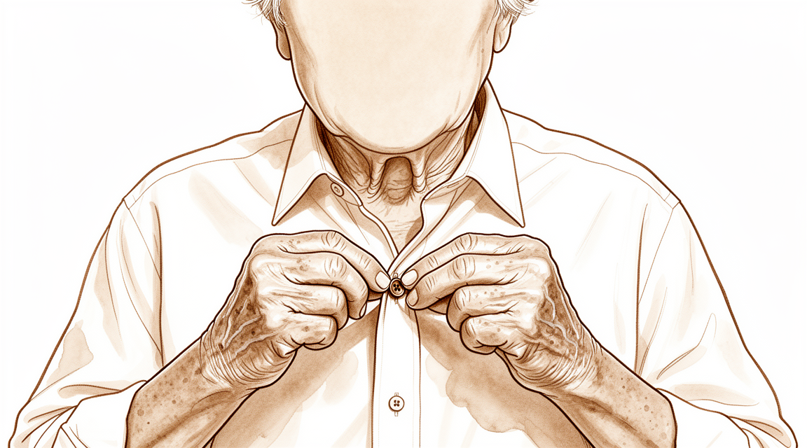
您很可能在手指最尖端感到疼痛和僵硬,即关节与指甲相接处。这是远端指间关节(DIP 关节)。在日常活动中使用手部后,疼痛往往会加剧,例如伸手到背后扣内衣或塞衬衫下摆。您可能会注意到,早晨刚醒来时疼痛最严重,随着手部活动而缓解。
随着时间的推移,关节形状可能会发生改变。指尖可能向内弯曲,形成一种称为天鹅颈畸形的曲线。这是因为关节发生了屈曲挛缩,意味着它卡在弯曲的位置。如果您之前有过锤状指骨折等损伤,您可能会看到磨损性关节炎的迹象。这种情况的发展路径与自然老化类似,会导致活动范围减少。
当关节肿胀或卡住时,日常生活会变得困难。您可能会发现难以抓握物体或进行精细动作。在某些情况下,关节感觉不稳定或“漂浮”,如果肉眼可见的畸形不明显,这可能会令人困惑。虽然疼痛可能令人烦恼,但重要的是要知道,X 光片上的可见变化并不总是与您的疼痛程度相符。您的外科医生将根据您的具体症状来指导治疗。
实际发生了什么
您的远端指间关节(DIP 关节)是指尖处的小型铰链关节。关节内部,光滑的软骨起到减震器的作用,使您的骨骼能够无痛地相互滑动。当发生磨损性关节炎时,这层软骨会逐渐磨损。骨骼开始直接相互摩擦,导致您感受到的研磨感和僵硬感。这种损伤还可能使关节错位,通常会将指尖向后推。
弯曲指尖的肌腱就像一根附着在骨骼上的强韧绳索。如果这根绳索撕裂或关节变得不稳定,肌腱就无法顺畅地拉动手指。有时,关节会卡在弯曲位置,称为屈曲挛缩。随着时间的推移,这种畸形可能会加重,使您难以伸直手指或将其用于日常活动。
您的外科医生会评估关节表面的受损程度,以决定最佳的治疗方案。如果骨骼太小无法使用标准螺钉,或者关节卡在不良位置,则需要采用特殊技术。目标是停止疼痛并恢复功能,无论是通过将骨骼融合在一起,还是使用软组织植入物以保持一定的活动度。这些方案旨在为您提供缓解,同时保护手指剩余的强度。
我们能做什么
您可以从自行管理疼痛并与物理治疗师合作开始。固定远端指间关节可减少疼痛并改善伸展,且不会导致僵硬或限制您的活动。然而,保持该关节静止会降低您的整体握力,且这种影响从食指到小指逐渐增强。在考虑更侵入性的治疗之前,您应给予这些非手术方法足够的时间以发挥作用。
如果简单措施不足以缓解症状,您的外科医生可能会讨论药物选项以管理您的症状。虽然证据强调了特定的手术替代方案,但指出如胶原酶等注射治疗有助于改善关节挛缩,不过您必须权衡问题复发的风险。对于同一手指的中节和末节关节均受严重疼痛影响的患者,通常建议同时治疗这两个关节。您的外科医生还会考虑您的个人健康状况,因为诸如糖尿病等因素会增加治疗后并发症的风险。
当保守治疗达到极限时,可能会考虑手术以缓解疼痛或恢复功能。您的外科医生可能会选择保留关节活动的术式以保持关节活动,或选择关节融合术将关节固定以提供稳定性。在某些情况下,会使用硅胶假体以提供卓越的疼痛缓解,并获得30–40度的活动范围,且总体并发症率仅为5%。具体方案取决于您关节的状况以及您对日常生活的目标。
何时就诊
若疼痛持续且休息后无改善,或手指感觉无力、不稳定,请就诊全科医生(GP)。若关节出现交锁、不稳,或症状影响睡眠或工作,请要求专科医生评估。若症状突然加重,也应寻求医疗帮助。请注意,某些情况(如远端指间关节游离损伤)初期可能畸形不明显,但日后仍可能导致关节炎。早期诊断有助于将这些情况与其他关节问题区分开来。
Evidence & references
Overview
- Percutaneous DIP joint arthrodesis is advantageous compared with open fusion techniques in select patients [1].
- Swan neck deformity progresses significantly over time due to increasing DIPJ flexion contracture [2].
- Simultaneous surgical intervention is recommended for severe painful osteoarthritis of both the PIP and DIP joints of the same digit [3].
- Denervation with cheilectomy presents a motion-preserving alternative to arthrodesis for symptomatic DIP joint osteoarthritis [4].
- Lateral approach and plate fixation for DIP joint arthrodesis yields results equivalent to traditional methods but with fewer major complications [5].
- The combination of DIP arthrodesis and PIP Swanson arthroplasty results in favorable outcomes regarding simultaneous bony union and flexibility [7].
- Silicone interpositional arthroplasty of the DIP joint is an acceptable alternative to arthrodesis, achieving excellent pain relief and a range of movement of 30–40 degrees [8].
- Silicone interpositional arthroplasty of the DIP joint has a low overall complication rate of 5% [8].
- The smile incision and reverse shotgun approach is a good surgical option for DIPJ arthrodesis when more volar part joint preparation and more volar implant insertion sites are necessary [9].
- The nonaxial multiple small screws (NMSS) technique is a feasible option for DIPJ and thumb IPJ arthrodesis, especially when a small finger is indicated and a significant flexion angle is required [11].
- Customized structural bone grafting addresses bone stock loss and medullary absence in failed DIPJ silicone arthroplasty, achieving reliable union rates and high patient satisfaction [13].
- There is no difference in biomechanical performance between K-wires and compression screws for DIPJ arthrodesis [20].
- Implant selection for DIPJ fusion should consider factors such as cost and complication profiles given the lack of difference in biomechanical performance between K-wires and compression screws [20].
Anatomy & Pathophysiology
- Palmar subluxation of a DIP joint without preexisting arthritic deformity is expected when more than one half of the dorsal articular surface is injured [6].
- Understanding of DIP joint morphology may lend insight into the biomechanics and disease progression within the DIP joints [10].
- A substantial number of distal phalanges are too small to accommodate commonly available headless compression screws, particularly in females and the small finger [31].
- Irreducibility was more commonly seen in dorsal than in volar dislocations of the DIP joint [33].
- Volar dislocations of the DIP joint carried a higher risk of instability immediately after reduction compared to dorsal dislocations [33].
- Biomechanically, dynamic tenodesis for the DIP joint using the remaining FDP tendon results in a flexion angle greater than 30 degrees [23].
- In a cadaveric model, tenodesis successfully restored coordinated interphalangeal joint flexion after a simulated zone I FDP laceration with improvements in DIP joint flexion and composite finger flexion [35].
- Lateral blocking with incremental joint angles allows a safer application of force for the healing tendon during palmar and lateral blocking exercises [26].
Classification
- Swan neck deformity in the DIP joint progresses significantly over time due to increasing DIPJ flexion contracture [2].
- Radiological osteoarthritis following a mallet finger fracture follows a similar course to the natural degenerative process in the DIP joint [12].
- Post-traumatic osteoarthritis of the DIP joint after mallet finger fractures is accompanied by a decrease in range of motion, though this does not clinically affect patient-reported outcome measures (PROMs) [12].
- The interrater reliability of the Kellgren & Lawrence classification system for post-traumatic osteoarthritis in the DIP joint after mallet finger fractures is considerably lower than initially assumed [34].
- The interrater reliability of the OARSI classification system for post-traumatic osteoarthritis in the DIP joint after mallet finger fractures is considerably lower than initially assumed [34].
- Current concepts regarding DIP joint osteoarthritis highlight the roles of cartilage, subchondral bone, and soft tissue structures in etiology, pathogenesis, and evaluation [19].
- Morphological understanding of DIP joint curvatures may provide insight into the biomechanics and disease progression within the DIP joints [10].
- Examination of type I and type II nerve endings provides new information on the sensory systems of the DIP joints and surrounding structures [32].
- Palmar subluxation of a DIP joint without preexisting arthritic deformity is expected when more than one half of the dorsal articular surface is injured [6].
Clinical Presentation
- Swan neck deformity in the DIPJ progresses significantly over time due to increasing DIPJ flexion contracture [2].
- Palmar subluxation of a DIP joint without preexisting arthritic deformity is expected when more than one half of the dorsal articular surface is injured [6].
- Floating DIP joint injuries can be misdiagnosed initially due to minimal deformity [17].
- Radiological osteoarthritis following a mallet finger fracture is similar to the natural degenerative process in the DIP joint [12].
- Radiological osteoarthritis after a mallet finger fracture is accompanied by a decrease in range of motion of the DIP joint [12].
- Radiological osteoarthritis after a mallet finger fracture does not clinically affect patient-reported outcome measures (PROMs) [12].
- Primary synovial chondromatosis of the DIPJ is an extremely rare entity that requires accurate diagnosis to distinguish from other arthropathies [27].
- Understanding the morphology of DIPJ curvatures may lend insight into the biomechanics and disease progression within the DIP joints [10].
- Osteoarthritis of the DIPJ involves roles of cartilage, subchondral bone, and soft tissue structures in its etiology, pathogenesis, and evaluation [19].
Investigations
- Percutaneous DIP joint arthrodesis is advantageous compared with open fusion techniques in select patients [1].
- Swan neck deformity progresses significantly over time due to increasing DIPJ flexion contracture [2].
- Simultaneous surgical intervention is recommended for severe painful osteoarthritis of both the PIP and DIP joints of the same digit [3].
- Denervation with cheilectomy presents a motion-preserving alternative to arthrodesis for symptomatic DIP joint osteoarthritis [4].
- Lateral approach and plate fixation for DIP joint arthrodesis yields results equivalent to traditional methods with fewer major complications [5].
- Palmar subluxation of a DIP joint without preexisting arthritic deformity is expected when more than one half of the dorsal articular surface is injured [6].
- The combination of DIP arthrodesis and PIP Swanson arthroplasty results in favorable outcomes regarding simultaneous bony union and flexibility [7].
- Silicone interpositional arthroplasty of the DIP joint is an acceptable alternative to arthrodesis, achieving excellent pain relief and a range of movement of 30–40 degrees with a low overall complication rate of 5% [8].
- The smile incision and reverse shotgun approach is a good surgical option for DIPJ arthrodesis when more volar part joint preparation and more volar implant insertion sites are necessary [9].
- Understanding the morphology of DIP joints may lend insight into the biomechanics and disease progression within the DIP joints [10].
- The nonaxial multiple small screws (NMSS) technique is a feasible option for DIPJ and thumb IPJ arthrodesis, especially when a small finger is indicated and a significant flexion angle is required [11].
- Radiological osteoarthritis after a mallet finger fracture is similar to the natural degenerative process in the DIP joint and is accompanied by a decrease in range of motion of the DIP joint [12].
- Radiological osteoarthritis after a mallet finger fracture does not clinically affect patient-reported outcome measures (PROMs) [12].
- Customized structural bone grafting addresses bone stock loss and medullary absence in failed DIPJ silicone arthroplasty, achieving reliable union rates and high patient satisfaction [13].
- Immobilization of the distal interphalangeal joint of any finger reduces the overall grip strength of the hand [16].
- The reduction in grip strength from DIP joint immobilization becomes progressively more pronounced from the index to the little fingers [16].
- Floating DIP joint injuries can be misdiagnosed initially due to minimal deformity [17].
- Open reduction and internal fixation is a viable treatment option for chronic floating DIP joint injuries, though osteoarthritis may develop [17].
- Arthrodesis of the distal interphalangeal joint often leads to complications [18].
- Current concepts regarding DIP joint osteoarthritis examine the etiology, pathogenesis, and evaluation of the condition, highlighting the roles of cartilage, subchondral bone, and soft tissue structures [19].
- A size mismatch existed between the anatomic dimensions of the DIP joint and commercially available headless compression screws [21].
- A distinct collagen septum exists between the extensor tendon and skin at the DIP joint [38].
Treatment
- Percutaneous DIP joint arthrodesis is advantageous compared with open fusion techniques in select patients [1].
- Simultaneous surgical intervention is recommended for severe painful osteoarthritis of both the PIP and DIP joints of the same digit [3].
- Denervation with cheilectomy presents a motion-preserving alternative to arthrodesis for symptomatic DIP joint osteoarthritis [4].
- Lateral approach and plate fixation for DIP joint arthrodesis yields results equivalent to traditional methods with fewer major complications [5].
- Simultaneous anterograde screw arthrodesis of the DIP joint and silastic PIP joint replacement results in favorable outcomes regarding bony union and flexibility [7].
- Silicone interpositional arthroplasty of the DIP joint is an acceptable alternative to arthrodesis, achieving excellent pain relief and a range of movement of 30–40 degrees [8].
- Silicone interpositional arthroplasty of the DIP joint has a low overall complication rate of 5% [8].
- The smile incision and reverse shotgun approach is a good surgical option for DIPJ arthrodesis when more volar part joint preparation and more volar implant insertion sites are necessary [9].
- The nonaxial multiple small screws (NMSS) technique is a feasible option for DIPJ and thumb IPJ arthrodesis, especially when a small finger is indicated and a significant flexion angle is required [11].
- Customized structural bone grafting addresses bone stock loss and medullary absence in failed DIPJ silicone arthroplasty, achieving reliable union rates and high patient satisfaction [13].
- Diabetes and surgeon experience are factors increasing the risk of postoperative complications in DIP/thumb IP joint arthrodeses [14].
- Splinting of the DIP joint reduces pain and improves extension at the joint without causing non-compliance, increased stiffness, or restriction of range of motion [15].
- Open reduction and internal fixation is a viable treatment option for chronic floating DIP joint injuries, though osteoarthritis may develop [17].
- Injection with collagenase Clostridium histolyticum is an option for the treatment of DIP joint contractures in Dupuytren disease, though the potential risk for recurrence should be carefully weighed [36].
- Open DIP joint cheilectomy is a safe and effective alternative to DIP joint arthrodesis in patients with symptomatic osteoarthritis who wish to preserve joint motion [37].
Complications
- Swan neck deformity in the DIPJ progresses significantly over time due to increasing DIPJ flexion contracture [2].
- Palmar subluxation of a DIP joint is expected when more than one half of the dorsal articular surface is injured, even without preexisting arthritic deformity [6].
- Radiological osteoarthritis following a mallet finger fracture follows a natural degenerative process and is accompanied by a decrease in DIPJ range of motion [12].
- Diabetes is identified as a factor increasing the risk of postoperative complications in DIP and thumb IP joint arthrodeses [14].
- Surgeon experience is identified as a factor increasing the risk of postoperative complications in DIP and thumb IP joint arthrodeses [14].
- Arthrodesis of the distal interphalangeal joint often leads to complications [18].
- Silicone interpositional arthroplasty of the DIP joint has a low overall complication rate of 5% [8].
- Failed Swanson's arthroplasty of the DIPJ can result in bone stock loss and medullary absence [13].
Recovery
- Percutaneous DIP joint arthrodesis is advantageous compared with open fusion techniques in select patients [1].
- Swan neck deformity progresses significantly over time due to increasing DIPJ flexion contracture [2].
- Simultaneous surgical intervention is recommended for severe painful osteoarthritis of both the PIP and DIP joints of the same digit [3].
- Denervation with cheilectomy presents a motion-preserving alternative to arthrodesis for symptomatic DIP joint osteoarthritis [4].
- Lateral approach and plate fixation for DIP joint arthrodesis yields results equivalent to traditional methods but with fewer major complications [5].
- Palmar subluxation of a DIP joint without preexisting arthritic deformity is expected when more than one half of the dorsal articular surface is injured [6].
- The combination of DIP arthrodesis and PIP Swanson arthroplasty results in favorable outcomes regarding simultaneous bony union and flexibility [7].
- Silicone interpositional arthroplasty of the DIP joint is an acceptable alternative to arthrodesis, achieving excellent pain relief and a range of movement of 30–40 degrees [8].
- Silicone interpositional arthroplasty of the DIP joint has a low overall complication rate of 5% [8].
- Radiological osteoarthritis after a mallet finger fracture is similar to the natural degenerative process in the DIP joint [12].
- Radiological osteoarthritis after a mallet finger fracture is accompanied by a decrease in range of motion of the DIP joint [12].
- The decrease in range of motion of the DIP joint following radiological osteoarthritis from a mallet finger fracture does not clinically affect PROMs [12].
- Customized structural bone grafting addresses bone stock loss and medullary absence in failed DIPJ silicone arthroplasty, achieving reliable union rates and high patient satisfaction [13].
- Diabetes is a factor increasing the risk of postoperative complications in DIP and thumb IP joint arthrodeses [14].
- Surgeon experience is a factor increasing the risk of postoperative complications in DIP and thumb IP joint arthrodeses [14].
- Splinting of the DIP joint reduces pain and improves extension at the joint [15].
- Splinting of the DIP joint does not give rise to non-compliance, increased stiffness, or restriction of range of motion [15].
Key Evidence
- [L4] In select patients, this percutaneous DIP joint arthrodesis is advantageous in comparison with open fusion techniques. [1] (10.1007/s11552-010-9265-9)
- [L5] The swan neck deformity in this individual progressed significantly with time because of increasing DIPJ flexion contracture. [2] (10.1016/j.jht.2009.11.005)
- [L3] The authors recommend simultaneous surgical intervention in case of severe painful OA of the PIP and DIP joints of the same digit. [3] (10.1177/17531934231191255)
- [L4] It presents a compelling motion-preserving alternative to arthrodesis for symptomatic DIP joint osteoarthritis. [4] (10.1016/j.jhsa.2026.01.027)
- [L4] The results obtained in this small series are equivalent to the traditional methods of DIP joint arthrodesis but with fewer major complications. [5] (10.1016/j.jhsa.2007.09.004)
- [L5] Palmar subluxation of a DIP joint without preexisting arthritic deformity is expected when more than one half of the dorsal articular surface is injured. [6] (10.1016/j.jhsa.2007.09.006)
- [L4] The combination of DIP arthrodesis and PIP Swanson arthroplasty resulted in a favourable outcome in terms of simultaneous bony union and flexibility. [7] (10.1177/17531934231215790)
- [L4] The study confirms that silicone interpositional arthroplasty of the DIP joint is an acceptable alternative to arthrodesis, achieving excellent pain relief and a range of movement of 30–40 degrees with a low overall complication rate of 5%. [8] (10.1177/1753193411422679)
- [L4] This technique may be a good surgical option for DIPJ arthrodesis when more volar part joint preparation and more volar implant insertion sites are necessary. [9] (10.1186/s12891-024-08016-6)
- [L5] Our understanding of morphology may lend insight into the biomechanics and disease progression within the DIP joints. [10] (10.1007/s11552-014-9605-2)
- [L4] Thus, the NMSS technique could be used as a feasible option in DIPJ and thumb IPJ arthrodesis, especially when a small finger is indicated and a significant flexion angle is required. [11] (10.1186/s12891-022-05473-9)
- [L4] Radiological OA after an MFF is similar to the natural degenerative process in the DIP joint and is accompanied by a decrease in range of motion of the DIP joint, which does not clinically affect PROMs. [12] (10.1016/j.jhsa.2023.03.027)
- [L4] A customized structural bone graft using the described technique addresses issues of bone stock loss and medullary absence in failed DIPJ silicone arthroplasty, achieving reliable union rates and high patient satisfaction. [13] (10.1177/17531934231151217)
- [L3] Diabetes and surgeon experience were identified as factors increasing the risk of postoperative complications in these DIP/thumb IP joint arthrodeses. [14] (10.1186/s12891-024-07361-w)
- [L2] It does not give rise to non-compliance, increased stiffness or restriction of range of motion. [15] (10.1016/j.jht.2013.08.004)
- [L4] Immobilization of the distal interphalangeal joint of any finger reduces the overall grip strength of the hand, with the effect becoming progressively more pronounced from the index to the little fingers. [16] (10.1177/1753193418765068)
- [Case_report] Floating DIP joint injuries can be misdiagnosed initially due to minimal deformity; open reduction and internal fixation is a viable treatment option for chronic cases, though osteoarthritis may develop. [17] (10.1016/j.jhsa.2010.05.025)
- [L3] Arthrodesis of the distal interphalangeal joint often leads to complications. [18] (10.1177/17531934221111641)
- [L5] This current concepts article examines the recent knowledge base regarding the etiology, pathogenesis, and evaluation of osteoarthritis of the distal interphalangeal joint, highlighting the roles of cartilage, subchondral bone, and soft tissue structures. [19] (10.1016/j.jhsa.2010.09.003)
- [L5] Given the lack of difference in biomechanical performance between K-wires and compression screws, consideration should be given to other factors such as cost and complication profiles when choosing an implant for DIPJ fusion. [20] (10.1177/1558944715627211)
- [L4] A size mismatch existed between the anatomic dimensions of the DIP joint and commercially available headless compression screws. [21] (10.1016/j.jhsa.2014.02.007)
- [L5] Biomechanically, dynamic tenodesis for the DIP joint using the remaining FDP tendon is a valuable procedure because it results in a flexion angle greater than 30 degrees. [23] (10.1016/j.jhsg.2020.08.007)
- [L5] This study supports the concept that lateral blocking with incremental joint angles allows a safer application of force for the healing tendon. [26] (10.1016/j.jht.2020.07.004)
- [Case_report] Primary synovial chondromatosis of the distal interphalangeal joint is an extremely rare entity that requires accurate diagnosis to distinguish from other arthropathies. [27] (10.1177/15589447211049520)
- [L4] A substantial number of distal phalanges are too small to accommodate commonly available headless compression screws, particularly in females and the small finger. [31] (10.1007/s11552-014-9679-x)
- [L5] Our examination of the distribution of type I and type II nerve endings provides new information on the sensory systems of the DIP joints and surrounding structures. [32] (10.1016/j.jhsa.2010.11.050)
- [L4] Irreducibility was more commonly seen in dorsal than in volar dislocations, while volar dislocations carried a higher risk of instability immediately after reduction. [33] (10.1177/1753193415616957)
- [L4] The interrater reliability of the Kellgren & Lawrence and OARSI classification systems for post-traumatic osteoarthritis in the distal interphalangeal joint after mallet finger fractures is considerably lower than initially assumed. [34] (10.1016/j.jhsa.2024.03.012)
- [L5] In this cadaveric model, this tenodesis successfully restored coordinated interphalangeal joint flexion after a simulated zone I FDP laceration with improvements in distal interphalangeal joint flexion and composite finger flexion. [35] (10.1016/j.jhsa.2013.10.009)
- [L4] Injection with CCH is an option for the treatment of DIP joint contractures in Dupuytren disease, though the potential risk for recurrence should be carefully weighed prior to its use. [36] (10.1016/j.jhsa.2018.07.004)
- [L4] Open DIP joint cheilectomy is a safe and effective alternative to DIP joint arthrodesis in patients with symptomatic osteoarthritis who wish to preserve joint motion. [37] (10.1016/j.jhsa.2017.07.006)
- [L5] We confirmed the existence of a distinct collagen septum between the extensor tendon and skin at the DIP joint using MRI and histology. [38] (10.1016/j.jhsa.2008.11.030)
References
[1] Treatment of Symptomatic Distal Interphalangeal Joint Arthritis with Percutaneous Arthrodesis: A Novel Technique in Select Patients. HAND. 2010. DOI: 10.1007/s11552-010-9265-9 [2] Swan Neck Deformity after Distal Interphalangeal Joint Flexion Contractures: A Biomechanical Analysis. Journal of Hand Therapy. 2010. DOI: 10.1016/j.jht.2009.11.005 [3] Does distal interphalangeal joint arthrodesis affect proximal interphalangeal joint arthroplasty outcomes in the same finger?. Journal of Hand Surgery (European Volume). 2023. DOI: 10.1177/17531934231191255 [4] Denervation with Cheilectomy of the Distal Interphalangeal Joint: Technique and Medium-Term Results. The Journal of Hand Surgery. 2026. DOI: 10.1016/j.jhsa.2026.01.027 [5] Alternative to the Distal Interphalangeal Joint Arthrodesis: Lateral Approach and Plate Fixation. The Journal of Hand Surgery. 2008. DOI: 10.1016/j.jhsa.2007.09.004 [6] A Biomechanical Study of Distal Interphalangeal Joint Subluxation After Mallet Fracture Injury. The Journal of Hand Surgery. 2008. DOI: 10.1016/j.jhsa.2007.09.006 [7] Simultaneous anterograde screw arthrodesis of distal interphalangeal joint and silastic proximal interphalangeal joint replacement for osteoarthritis. Journal of Hand Surgery (European Volume). 2023. DOI: 10.1177/17531934231215790 [8] Joint replacement in 131 painful osteoarthritic and post-traumatic distal interphalangeal joints. Journal of Hand Surgery (European Volume). 2011. DOI: 10.1177/1753193411422679 [9] Smile incision and reverse shotgun approach in distal interphalangeal joint arthrodesis. BMC Musculoskeletal Disorders. 2024. DOI: 10.1186/s12891-024-08016-6 [10] Curvatures of the DIP Joints of the Hand. HAND. 2014. DOI: 10.1007/s11552-014-9605-2 [11] Distal interphalangeal joint arthrodesis with nonaxial multiple small screws: a biomechanical analysis with axial headless compression screw and clinical result of 15 consecutive cases. BMC Musculoskeletal Disorders. 2022. DOI: 10.1186/s12891-022-05473-9 [12] Posttraumatic Osteoarthritis of the Distal Interphalangeal Joint: A Follow-Up Study of 12 Years After Nonsurgical Treatment of Mallet Finger Fractures. The Journal of Hand Surgery. 2023. DOI: 10.1016/j.jhsa.2023.03.027 [13] Salvage of failed Swanson’s arthroplasty of the distal interphalangeal joint. Journal of Hand Surgery (European Volume). 2023. DOI: 10.1177/17531934231151217 [14] Arthrodesis of distal interphalangeal and thumb interphalangeal joint: a retrospective cohort study of 149 cases. BMC Musculoskeletal Disorders. 2024. DOI: 10.1186/s12891-024-07361-w [15] Splinting of the Distal Interphalangeal Joint Reduces Pain and Improves Extension at the Joint; Results Front the Splint-OA Study. Journal of Hand Therapy. 2014. DOI: 10.1016/j.jht.2013.08.004 [16] Effect of immobilization of the distal interphalangeal joint of fingers on grip strength. Journal of Hand Surgery (European Volume). 2018. DOI: 10.1177/1753193418765068 [17] Floating Distal Interphalangeal Joint Injury: Case Report. The Journal of Hand Surgery. 2010. DOI: 10.1016/j.jhsa.2010.05.025 [18] Risk factors in distal interphalangeal joint arthrodesis in the hand: a retrospective study of 173 cases. Journal of Hand Surgery (European Volume). 2022. DOI: 10.1177/17531934221111641 [19] Osteoarthritis of the Distal Interphalangeal Joint. The Journal of Hand Surgery. 2010. DOI: 10.1016/j.jhsa.2010.09.003 [20] Biomechanical Analysis of Internal Fixation Methods for Distal Interphalangeal Joint Arthrodesis. HAND. 2016. DOI: 10.1177/1558944715627211 [21] Distal Interphalangeal Joint Bony Dimensions Related to Headless Compression Screw Sizes. The Journal of Hand Surgery. 2014. DOI: 10.1016/j.jhsa.2014.02.007 [23] The Effect of Flexor Digitorum Profundus Dynamic Tenodesis on the Distal Interphalangeal Joint: A Cadaver Study. Journal of Hand Surgery Global Online. 2020. DOI: 10.1016/j.jhsg.2020.08.007 [26] Tensile load on the flexor digitorum profundus tendon during palmar and lateral blocking exercises: Influence on blocking force and distal interphalangeal joint flexion angle. Journal of Hand Therapy. 2021. DOI: 10.1016/j.jht.2020.07.004 [27] Primary Distal Interphalangeal Joint Tenosynovial Chondromatosis of the Small Finger: A Case Report With Literature Review. HAND. 2022. DOI: 10.1177/15589447211049520 [31] Dimensional Analysis of the Distal Phalanx with Consideration of Distal Interphalangeal Joint Arthrodesis Using a Headless Compression Screw. HAND. 2014. DOI: 10.1007/s11552-014-9679-x [32] Distribution of Nerve Endings in Human Distal Interphalangeal Joint and Surrounding Structures. The Journal of Hand Surgery. 2011. DOI: 10.1016/j.jhsa.2010.11.050 [33] Differences between dorsal and volar dislocations of the distal interphalangeal joint of fingers: a report of 30 cases. Journal of Hand Surgery (European Volume). 2016. DOI: 10.1177/1753193415616957 [34] Rater Agreement of Post-Traumatic Osteoarthritis of the Distal Interphalangeal Joint 12 Years After a Mallet Finger Fracture. The Journal of Hand Surgery. 2024. DOI: 10.1016/j.jhsa.2024.03.012 [35] Tenodesis for Restoration of Distal Interphalangeal Joint Flexion in Unrepairable Flexor Digitorum Profundus Injuries. The Journal of Hand Surgery. 2014. DOI: 10.1016/j.jhsa.2013.10.009 [36] Collagenase Clostridium histolyticum for the Treatment of Distal Interphalangeal Joint Contractures in Dupuytren Disease. The Journal of Hand Surgery. 2019. DOI: 10.1016/j.jhsa.2018.07.004 [37] Cheilectomy for Treatment of Symptomatic Distal Interphalangeal Joint Osteoarthritis: A Review of 78 Patients. The Journal of Hand Surgery. 2017. DOI: 10.1016/j.jhsa.2017.07.006 [38] Dorsal Digital Septum of the Distal Interphalangeal Joint. The Journal of Hand Surgery. 2009. DOI: 10.1016/j.jhsa.2008.11.030


