Viêm khớp DIPJ
Những gì bạn đang cảm nhận
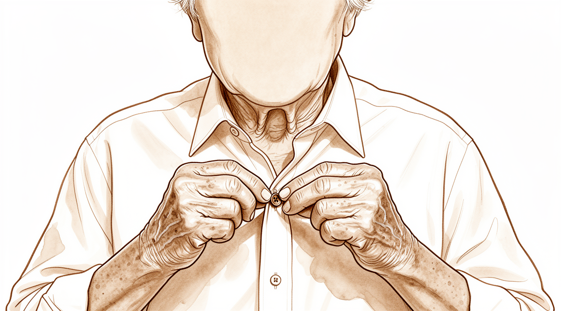
Bạn có thể cảm thấy đau và cứng khớp ngay tại đầu ngón tay, nơi khớp gặp móng tay. Đây là khớp liên đốt xa (DIP). Cơn đau thường bùng phát sau khi bạn sử dụng tay cho các hoạt động hàng ngày như với tay ra sau lưng để cài áo ngực hoặc nhét áo vào quần. Bạn có thể nhận thấy cơn đau nặng hơn khi vừa thức dậy vào buổi sáng và giảm dần khi bạn vận động tay.
Theo thời gian, khớp có thể thay đổi hình dạng. Đầu ngón tay có thể cong vào trong, tạo thành một đường cong được gọi là biến dạng cổ thiên nga. Điều này xảy ra do khớp phát triển co cứng duỗi, nghĩa là khớp bị mắc kẹt ở tư thế cong. Nếu bạn từng bị chấn thương trước đó như gãy ngón tay búa, bạn có thể thấy các dấu hiệu của viêm xương khớp do hao mòn. Tình trạng này tiến triển tương tự như quá trình lão hóa tự nhiên của khớp và dẫn đến giảm phạm vi vận động.
Cuộc sống hàng ngày có thể trở nên khó khăn khi khớp sưng hoặc bị khóa. Bạn có thể thấy khó cầm nắm đồ vật hoặc thực hiện các cử động tinh tế. Trong một số trường hợp, khớp cảm thấy không ổn định hoặc "lơ lửng", điều này có thể gây nhầm lẫn nếu không có biến dạng rõ ràng. Mặc dù cơn đau có thể gây khó chịu, điều quan trọng là phải biết rằng những thay đổi thấy rõ trên phim X-quang không phải lúc nào cũng tương ứng với mức độ đau mà bạn cảm nhận. Bác sĩ phẫu thuật của bạn sẽ xem xét các triệu chứng cụ thể của bạn để hướng dẫn việc chăm sóc sức khỏe.
Những gì thực sự đang xảy ra
Khớp liên đốt xa (khớp DIP) của bạn là khớp bản lề nhỏ nằm ở đầu ngón tay. Bên trong, sụn trơn hoạt động như một bộ giảm xóc, cho phép các xương trượt lên nhau mà không gây đau. Khi viêm xương khớp do hao mòn phát triển, lớp phủ này bị mòn đi. Các xương bắt đầu cọ xát trực tiếp vào nhau, gây ra cảm giác cọt kẹt và cứng khớp mà bạn cảm thấy. Tổn thương này cũng có thể làm lệch khớp, thường đẩy đầu ngón tay của bạn ngả ra sau.
Gân gập đầu ngón tay giống như một sợi dây chắc chắn gắn vào xương. Nếu sợi dây này bị rách hoặc khớp trở nên không ổn định, gân không thể kéo ngón tay một cách trơn tru. Đôi khi, khớp bị kẹt ở tư thế gập, được gọi là co rút gập. Theo thời gian, biến dạng này có thể trở nên nghiêm trọng hơn, khiến bạn khó duỗi thẳng ngón tay hoặc sử dụng nó cho các hoạt động hàng ngày.
Bác sĩ phẫu thuật của bạn xem xét mức độ tổn thương bề mặt khớp để quyết định hướng điều trị tốt nhất. Nếu xương quá nhỏ để bắt vít tiêu chuẩn, hoặc nếu khớp bị kẹt ở tư thế xấu, cần các kỹ thuật đặc biệt. Mục tiêu là ngăn chặn cơn đau và khôi phục chức năng, dù bằng cách hợp nhất các xương lại với nhau hoặc sử dụng một implant mềm để duy trì một số chuyển động. Các lựa chọn này nhằm mang lại sự giảm đau trong khi bảo vệ sức mạnh còn lại của ngón tay bạn.
Những gì chúng tôi có thể làm về vấn đề này
Bạn có thể bắt đầu bằng cách tự quản lý cơn đau và làm việc với một nhà vật lý trị liệu. Nẹp khớp liên đốt xa giúp giảm đau và cải thiện khả năng duỗi mà không gây cứng khớp hoặc hạn chế chuyển động của bạn. Tuy nhiên, việc giữ cố định khớp này sẽ làm giảm lực nắm tổng thể, với mức độ ảnh hưởng tăng dần từ ngón trỏ sang ngón út. Bạn nên dành thời gian cho các phương pháp không phẫu thuật này phát huy tác dụng trước khi xem xét các bước can thiệp xâm lấn hơn.
Nếu các biện pháp đơn giản không đủ hiệu quả, bác sĩ phẫu thuật của bạn có thể thảo luận về các lựa chọn y tế để quản lý các triệu chứng của bạn. Mặc dù bằng chứng nhấn mạnh các lựa chọn phẫu thuật cụ thể, nhưng cũng lưu ý rằng các phương pháp tiêm như collagenase có thể giúp cải thiện tình trạng co rút khớp, tuy nhiên bạn phải cân nhắc nguy cơ vấn đề tái phát. Đối với những người bị đau dữ dội ảnh hưởng đến cả khớp giữa và khớp cuối của cùng một ngón tay, việc điều trị đồng thời cho cả hai khớp thường được khuyến nghị. Bác sĩ phẫu thuật của bạn cũng sẽ xem xét sức khỏe cá nhân của bạn, vì các yếu tố như tiểu đường có thể làm tăng nguy cơ biến chứng sau điều trị.
Khi chăm sóc bảo tồn đạt đến giới hạn, phẫu thuật có thể được xem xét để ngăn chặn cơn đau hoặc khôi phục chức năng. Bác sĩ phẫu thuật của bạn có thể chọn một thủ thuật bảo tồn chuyển động để giữ cho khớp của bạn vẫn linh hoạt, hoặc một thủ thuật hợp nhất để cố định khớp tại chỗ nhằm đảm bảo sự ổn định. Trong một số trường hợp, một implant silicon được sử dụng để cung cấp giảm đau hiệu quả và phạm vi chuyển động từ 30–40 độ với tỷ lệ biến chứng tổng thể thấp là 5%. Phương pháp cụ thể phụ thuộc vào tình trạng khớp của bạn và mục tiêu của bạn trong cuộc sống hàng ngày.
Khi nào cần gặp bác sĩ
Hãy gặp bác sĩ đa khoa nếu bạn có cơn đau dai dẳng không cải thiện khi nghỉ ngơi, hoặc nếu ngón tay của bạn cảm thấy yếu và không vững. Hãy yêu cầu được bác sĩ chuyên khoa thăm khám nếu khớp của bạn bị kẹt, bị sập, hoặc nếu các triệu chứng ảnh hưởng đến giấc ngủ hoặc công việc của bạn. Bạn cũng nên tìm kiếm sự giúp đỡ nếu nhận thấy các triệu chứng xấu đi đột ngột. Cần lưu ý rằng một số tình trạng, chẳng hạn như chấn thương khớp liên đốt xa (DIP) có mảnh xương rời, có thể biểu hiện biến dạng rất ít ở giai đoạn đầu nhưng vẫn có thể dẫn đến viêm khớp sau này. Chẩn đoán sớm giúp phân biệt các vấn đề này với các bệnh lý khớp khác.
Evidence & references
Overview
- Percutaneous DIP joint arthrodesis is advantageous compared with open fusion techniques in select patients [1].
- Swan neck deformity progresses significantly over time due to increasing DIPJ flexion contracture [2].
- Simultaneous surgical intervention is recommended for severe painful osteoarthritis of both the PIP and DIP joints of the same digit [3].
- Denervation with cheilectomy presents a motion-preserving alternative to arthrodesis for symptomatic DIP joint osteoarthritis [4].
- Lateral approach and plate fixation for DIP joint arthrodesis yields results equivalent to traditional methods but with fewer major complications [5].
- The combination of DIP arthrodesis and PIP Swanson arthroplasty results in favorable outcomes regarding simultaneous bony union and flexibility [7].
- Silicone interpositional arthroplasty of the DIP joint is an acceptable alternative to arthrodesis, achieving excellent pain relief and a range of movement of 30–40 degrees [8].
- Silicone interpositional arthroplasty of the DIP joint has a low overall complication rate of 5% [8].
- The smile incision and reverse shotgun approach is a good surgical option for DIPJ arthrodesis when more volar part joint preparation and more volar implant insertion sites are necessary [9].
- The nonaxial multiple small screws (NMSS) technique is a feasible option for DIPJ and thumb IPJ arthrodesis, especially when a small finger is indicated and a significant flexion angle is required [11].
- Customized structural bone grafting addresses bone stock loss and medullary absence in failed DIPJ silicone arthroplasty, achieving reliable union rates and high patient satisfaction [13].
- There is no difference in biomechanical performance between K-wires and compression screws for DIPJ arthrodesis [20].
- Implant selection for DIPJ fusion should consider factors such as cost and complication profiles given the lack of difference in biomechanical performance between K-wires and compression screws [20].
Anatomy & Pathophysiology
- Palmar subluxation of a DIP joint without preexisting arthritic deformity is expected when more than one half of the dorsal articular surface is injured [6].
- Understanding of DIP joint morphology may lend insight into the biomechanics and disease progression within the DIP joints [10].
- A substantial number of distal phalanges are too small to accommodate commonly available headless compression screws, particularly in females and the small finger [31].
- Irreducibility was more commonly seen in dorsal than in volar dislocations of the DIP joint [33].
- Volar dislocations of the DIP joint carried a higher risk of instability immediately after reduction compared to dorsal dislocations [33].
- Biomechanically, dynamic tenodesis for the DIP joint using the remaining FDP tendon results in a flexion angle greater than 30 degrees [23].
- In a cadaveric model, tenodesis successfully restored coordinated interphalangeal joint flexion after a simulated zone I FDP laceration with improvements in DIP joint flexion and composite finger flexion [35].
- Lateral blocking with incremental joint angles allows a safer application of force for the healing tendon during palmar and lateral blocking exercises [26].
Classification
- Swan neck deformity in the DIP joint progresses significantly over time due to increasing DIPJ flexion contracture [2].
- Radiological osteoarthritis following a mallet finger fracture follows a similar course to the natural degenerative process in the DIP joint [12].
- Post-traumatic osteoarthritis of the DIP joint after mallet finger fractures is accompanied by a decrease in range of motion, though this does not clinically affect patient-reported outcome measures (PROMs) [12].
- The interrater reliability of the Kellgren & Lawrence classification system for post-traumatic osteoarthritis in the DIP joint after mallet finger fractures is considerably lower than initially assumed [34].
- The interrater reliability of the OARSI classification system for post-traumatic osteoarthritis in the DIP joint after mallet finger fractures is considerably lower than initially assumed [34].
- Current concepts regarding DIP joint osteoarthritis highlight the roles of cartilage, subchondral bone, and soft tissue structures in etiology, pathogenesis, and evaluation [19].
- Morphological understanding of DIP joint curvatures may provide insight into the biomechanics and disease progression within the DIP joints [10].
- Examination of type I and type II nerve endings provides new information on the sensory systems of the DIP joints and surrounding structures [32].
- Palmar subluxation of a DIP joint without preexisting arthritic deformity is expected when more than one half of the dorsal articular surface is injured [6].
Clinical Presentation
- Swan neck deformity in the DIPJ progresses significantly over time due to increasing DIPJ flexion contracture [2].
- Palmar subluxation of a DIP joint without preexisting arthritic deformity is expected when more than one half of the dorsal articular surface is injured [6].
- Floating DIP joint injuries can be misdiagnosed initially due to minimal deformity [17].
- Radiological osteoarthritis following a mallet finger fracture is similar to the natural degenerative process in the DIP joint [12].
- Radiological osteoarthritis after a mallet finger fracture is accompanied by a decrease in range of motion of the DIP joint [12].
- Radiological osteoarthritis after a mallet finger fracture does not clinically affect patient-reported outcome measures (PROMs) [12].
- Primary synovial chondromatosis of the DIPJ is an extremely rare entity that requires accurate diagnosis to distinguish from other arthropathies [27].
- Understanding the morphology of DIPJ curvatures may lend insight into the biomechanics and disease progression within the DIP joints [10].
- Osteoarthritis of the DIPJ involves roles of cartilage, subchondral bone, and soft tissue structures in its etiology, pathogenesis, and evaluation [19].
Investigations
- Percutaneous DIP joint arthrodesis is advantageous compared with open fusion techniques in select patients [1].
- Swan neck deformity progresses significantly over time due to increasing DIPJ flexion contracture [2].
- Simultaneous surgical intervention is recommended for severe painful osteoarthritis of both the PIP and DIP joints of the same digit [3].
- Denervation with cheilectomy presents a motion-preserving alternative to arthrodesis for symptomatic DIP joint osteoarthritis [4].
- Lateral approach and plate fixation for DIP joint arthrodesis yields results equivalent to traditional methods with fewer major complications [5].
- Palmar subluxation of a DIP joint without preexisting arthritic deformity is expected when more than one half of the dorsal articular surface is injured [6].
- The combination of DIP arthrodesis and PIP Swanson arthroplasty results in favorable outcomes regarding simultaneous bony union and flexibility [7].
- Silicone interpositional arthroplasty of the DIP joint is an acceptable alternative to arthrodesis, achieving excellent pain relief and a range of movement of 30–40 degrees with a low overall complication rate of 5% [8].
- The smile incision and reverse shotgun approach is a good surgical option for DIPJ arthrodesis when more volar part joint preparation and more volar implant insertion sites are necessary [9].
- Understanding the morphology of DIP joints may lend insight into the biomechanics and disease progression within the DIP joints [10].
- The nonaxial multiple small screws (NMSS) technique is a feasible option for DIPJ and thumb IPJ arthrodesis, especially when a small finger is indicated and a significant flexion angle is required [11].
- Radiological osteoarthritis after a mallet finger fracture is similar to the natural degenerative process in the DIP joint and is accompanied by a decrease in range of motion of the DIP joint [12].
- Radiological osteoarthritis after a mallet finger fracture does not clinically affect patient-reported outcome measures (PROMs) [12].
- Customized structural bone grafting addresses bone stock loss and medullary absence in failed DIPJ silicone arthroplasty, achieving reliable union rates and high patient satisfaction [13].
- Immobilization of the distal interphalangeal joint of any finger reduces the overall grip strength of the hand [16].
- The reduction in grip strength from DIP joint immobilization becomes progressively more pronounced from the index to the little fingers [16].
- Floating DIP joint injuries can be misdiagnosed initially due to minimal deformity [17].
- Open reduction and internal fixation is a viable treatment option for chronic floating DIP joint injuries, though osteoarthritis may develop [17].
- Arthrodesis of the distal interphalangeal joint often leads to complications [18].
- Current concepts regarding DIP joint osteoarthritis examine the etiology, pathogenesis, and evaluation of the condition, highlighting the roles of cartilage, subchondral bone, and soft tissue structures [19].
- A size mismatch existed between the anatomic dimensions of the DIP joint and commercially available headless compression screws [21].
- A distinct collagen septum exists between the extensor tendon and skin at the DIP joint [38].
Treatment
- Percutaneous DIP joint arthrodesis is advantageous compared with open fusion techniques in select patients [1].
- Simultaneous surgical intervention is recommended for severe painful osteoarthritis of both the PIP and DIP joints of the same digit [3].
- Denervation with cheilectomy presents a motion-preserving alternative to arthrodesis for symptomatic DIP joint osteoarthritis [4].
- Lateral approach and plate fixation for DIP joint arthrodesis yields results equivalent to traditional methods with fewer major complications [5].
- Simultaneous anterograde screw arthrodesis of the DIP joint and silastic PIP joint replacement results in favorable outcomes regarding bony union and flexibility [7].
- Silicone interpositional arthroplasty of the DIP joint is an acceptable alternative to arthrodesis, achieving excellent pain relief and a range of movement of 30–40 degrees [8].
- Silicone interpositional arthroplasty of the DIP joint has a low overall complication rate of 5% [8].
- The smile incision and reverse shotgun approach is a good surgical option for DIPJ arthrodesis when more volar part joint preparation and more volar implant insertion sites are necessary [9].
- The nonaxial multiple small screws (NMSS) technique is a feasible option for DIPJ and thumb IPJ arthrodesis, especially when a small finger is indicated and a significant flexion angle is required [11].
- Customized structural bone grafting addresses bone stock loss and medullary absence in failed DIPJ silicone arthroplasty, achieving reliable union rates and high patient satisfaction [13].
- Diabetes and surgeon experience are factors increasing the risk of postoperative complications in DIP/thumb IP joint arthrodeses [14].
- Splinting of the DIP joint reduces pain and improves extension at the joint without causing non-compliance, increased stiffness, or restriction of range of motion [15].
- Open reduction and internal fixation is a viable treatment option for chronic floating DIP joint injuries, though osteoarthritis may develop [17].
- Injection with collagenase Clostridium histolyticum is an option for the treatment of DIP joint contractures in Dupuytren disease, though the potential risk for recurrence should be carefully weighed [36].
- Open DIP joint cheilectomy is a safe and effective alternative to DIP joint arthrodesis in patients with symptomatic osteoarthritis who wish to preserve joint motion [37].
Complications
- Swan neck deformity in the DIPJ progresses significantly over time due to increasing DIPJ flexion contracture [2].
- Palmar subluxation of a DIP joint is expected when more than one half of the dorsal articular surface is injured, even without preexisting arthritic deformity [6].
- Radiological osteoarthritis following a mallet finger fracture follows a natural degenerative process and is accompanied by a decrease in DIPJ range of motion [12].
- Diabetes is identified as a factor increasing the risk of postoperative complications in DIP and thumb IP joint arthrodeses [14].
- Surgeon experience is identified as a factor increasing the risk of postoperative complications in DIP and thumb IP joint arthrodeses [14].
- Arthrodesis of the distal interphalangeal joint often leads to complications [18].
- Silicone interpositional arthroplasty of the DIP joint has a low overall complication rate of 5% [8].
- Failed Swanson's arthroplasty of the DIPJ can result in bone stock loss and medullary absence [13].
Recovery
- Percutaneous DIP joint arthrodesis is advantageous compared with open fusion techniques in select patients [1].
- Swan neck deformity progresses significantly over time due to increasing DIPJ flexion contracture [2].
- Simultaneous surgical intervention is recommended for severe painful osteoarthritis of both the PIP and DIP joints of the same digit [3].
- Denervation with cheilectomy presents a motion-preserving alternative to arthrodesis for symptomatic DIP joint osteoarthritis [4].
- Lateral approach and plate fixation for DIP joint arthrodesis yields results equivalent to traditional methods but with fewer major complications [5].
- Palmar subluxation of a DIP joint without preexisting arthritic deformity is expected when more than one half of the dorsal articular surface is injured [6].
- The combination of DIP arthrodesis and PIP Swanson arthroplasty results in favorable outcomes regarding simultaneous bony union and flexibility [7].
- Silicone interpositional arthroplasty of the DIP joint is an acceptable alternative to arthrodesis, achieving excellent pain relief and a range of movement of 30–40 degrees [8].
- Silicone interpositional arthroplasty of the DIP joint has a low overall complication rate of 5% [8].
- Radiological osteoarthritis after a mallet finger fracture is similar to the natural degenerative process in the DIP joint [12].
- Radiological osteoarthritis after a mallet finger fracture is accompanied by a decrease in range of motion of the DIP joint [12].
- The decrease in range of motion of the DIP joint following radiological osteoarthritis from a mallet finger fracture does not clinically affect PROMs [12].
- Customized structural bone grafting addresses bone stock loss and medullary absence in failed DIPJ silicone arthroplasty, achieving reliable union rates and high patient satisfaction [13].
- Diabetes is a factor increasing the risk of postoperative complications in DIP and thumb IP joint arthrodeses [14].
- Surgeon experience is a factor increasing the risk of postoperative complications in DIP and thumb IP joint arthrodeses [14].
- Splinting of the DIP joint reduces pain and improves extension at the joint [15].
- Splinting of the DIP joint does not give rise to non-compliance, increased stiffness, or restriction of range of motion [15].
Key Evidence
- [L4] In select patients, this percutaneous DIP joint arthrodesis is advantageous in comparison with open fusion techniques. [1] (10.1007/s11552-010-9265-9)
- [L5] The swan neck deformity in this individual progressed significantly with time because of increasing DIPJ flexion contracture. [2] (10.1016/j.jht.2009.11.005)
- [L3] The authors recommend simultaneous surgical intervention in case of severe painful OA of the PIP and DIP joints of the same digit. [3] (10.1177/17531934231191255)
- [L4] It presents a compelling motion-preserving alternative to arthrodesis for symptomatic DIP joint osteoarthritis. [4] (10.1016/j.jhsa.2026.01.027)
- [L4] The results obtained in this small series are equivalent to the traditional methods of DIP joint arthrodesis but with fewer major complications. [5] (10.1016/j.jhsa.2007.09.004)
- [L5] Palmar subluxation of a DIP joint without preexisting arthritic deformity is expected when more than one half of the dorsal articular surface is injured. [6] (10.1016/j.jhsa.2007.09.006)
- [L4] The combination of DIP arthrodesis and PIP Swanson arthroplasty resulted in a favourable outcome in terms of simultaneous bony union and flexibility. [7] (10.1177/17531934231215790)
- [L4] The study confirms that silicone interpositional arthroplasty of the DIP joint is an acceptable alternative to arthrodesis, achieving excellent pain relief and a range of movement of 30–40 degrees with a low overall complication rate of 5%. [8] (10.1177/1753193411422679)
- [L4] This technique may be a good surgical option for DIPJ arthrodesis when more volar part joint preparation and more volar implant insertion sites are necessary. [9] (10.1186/s12891-024-08016-6)
- [L5] Our understanding of morphology may lend insight into the biomechanics and disease progression within the DIP joints. [10] (10.1007/s11552-014-9605-2)
- [L4] Thus, the NMSS technique could be used as a feasible option in DIPJ and thumb IPJ arthrodesis, especially when a small finger is indicated and a significant flexion angle is required. [11] (10.1186/s12891-022-05473-9)
- [L4] Radiological OA after an MFF is similar to the natural degenerative process in the DIP joint and is accompanied by a decrease in range of motion of the DIP joint, which does not clinically affect PROMs. [12] (10.1016/j.jhsa.2023.03.027)
- [L4] A customized structural bone graft using the described technique addresses issues of bone stock loss and medullary absence in failed DIPJ silicone arthroplasty, achieving reliable union rates and high patient satisfaction. [13] (10.1177/17531934231151217)
- [L3] Diabetes and surgeon experience were identified as factors increasing the risk of postoperative complications in these DIP/thumb IP joint arthrodeses. [14] (10.1186/s12891-024-07361-w)
- [L2] It does not give rise to non-compliance, increased stiffness or restriction of range of motion. [15] (10.1016/j.jht.2013.08.004)
- [L4] Immobilization of the distal interphalangeal joint of any finger reduces the overall grip strength of the hand, with the effect becoming progressively more pronounced from the index to the little fingers. [16] (10.1177/1753193418765068)
- [Case_report] Floating DIP joint injuries can be misdiagnosed initially due to minimal deformity; open reduction and internal fixation is a viable treatment option for chronic cases, though osteoarthritis may develop. [17] (10.1016/j.jhsa.2010.05.025)
- [L3] Arthrodesis of the distal interphalangeal joint often leads to complications. [18] (10.1177/17531934221111641)
- [L5] This current concepts article examines the recent knowledge base regarding the etiology, pathogenesis, and evaluation of osteoarthritis of the distal interphalangeal joint, highlighting the roles of cartilage, subchondral bone, and soft tissue structures. [19] (10.1016/j.jhsa.2010.09.003)
- [L5] Given the lack of difference in biomechanical performance between K-wires and compression screws, consideration should be given to other factors such as cost and complication profiles when choosing an implant for DIPJ fusion. [20] (10.1177/1558944715627211)
- [L4] A size mismatch existed between the anatomic dimensions of the DIP joint and commercially available headless compression screws. [21] (10.1016/j.jhsa.2014.02.007)
- [L5] Biomechanically, dynamic tenodesis for the DIP joint using the remaining FDP tendon is a valuable procedure because it results in a flexion angle greater than 30 degrees. [23] (10.1016/j.jhsg.2020.08.007)
- [L5] This study supports the concept that lateral blocking with incremental joint angles allows a safer application of force for the healing tendon. [26] (10.1016/j.jht.2020.07.004)
- [Case_report] Primary synovial chondromatosis of the distal interphalangeal joint is an extremely rare entity that requires accurate diagnosis to distinguish from other arthropathies. [27] (10.1177/15589447211049520)
- [L4] A substantial number of distal phalanges are too small to accommodate commonly available headless compression screws, particularly in females and the small finger. [31] (10.1007/s11552-014-9679-x)
- [L5] Our examination of the distribution of type I and type II nerve endings provides new information on the sensory systems of the DIP joints and surrounding structures. [32] (10.1016/j.jhsa.2010.11.050)
- [L4] Irreducibility was more commonly seen in dorsal than in volar dislocations, while volar dislocations carried a higher risk of instability immediately after reduction. [33] (10.1177/1753193415616957)
- [L4] The interrater reliability of the Kellgren & Lawrence and OARSI classification systems for post-traumatic osteoarthritis in the distal interphalangeal joint after mallet finger fractures is considerably lower than initially assumed. [34] (10.1016/j.jhsa.2024.03.012)
- [L5] In this cadaveric model, this tenodesis successfully restored coordinated interphalangeal joint flexion after a simulated zone I FDP laceration with improvements in distal interphalangeal joint flexion and composite finger flexion. [35] (10.1016/j.jhsa.2013.10.009)
- [L4] Injection with CCH is an option for the treatment of DIP joint contractures in Dupuytren disease, though the potential risk for recurrence should be carefully weighed prior to its use. [36] (10.1016/j.jhsa.2018.07.004)
- [L4] Open DIP joint cheilectomy is a safe and effective alternative to DIP joint arthrodesis in patients with symptomatic osteoarthritis who wish to preserve joint motion. [37] (10.1016/j.jhsa.2017.07.006)
- [L5] We confirmed the existence of a distinct collagen septum between the extensor tendon and skin at the DIP joint using MRI and histology. [38] (10.1016/j.jhsa.2008.11.030)
References
[1] Treatment of Symptomatic Distal Interphalangeal Joint Arthritis with Percutaneous Arthrodesis: A Novel Technique in Select Patients. HAND. 2010. DOI: 10.1007/s11552-010-9265-9 [2] Swan Neck Deformity after Distal Interphalangeal Joint Flexion Contractures: A Biomechanical Analysis. Journal of Hand Therapy. 2010. DOI: 10.1016/j.jht.2009.11.005 [3] Does distal interphalangeal joint arthrodesis affect proximal interphalangeal joint arthroplasty outcomes in the same finger?. Journal of Hand Surgery (European Volume). 2023. DOI: 10.1177/17531934231191255 [4] Denervation with Cheilectomy of the Distal Interphalangeal Joint: Technique and Medium-Term Results. The Journal of Hand Surgery. 2026. DOI: 10.1016/j.jhsa.2026.01.027 [5] Alternative to the Distal Interphalangeal Joint Arthrodesis: Lateral Approach and Plate Fixation. The Journal of Hand Surgery. 2008. DOI: 10.1016/j.jhsa.2007.09.004 [6] A Biomechanical Study of Distal Interphalangeal Joint Subluxation After Mallet Fracture Injury. The Journal of Hand Surgery. 2008. DOI: 10.1016/j.jhsa.2007.09.006 [7] Simultaneous anterograde screw arthrodesis of distal interphalangeal joint and silastic proximal interphalangeal joint replacement for osteoarthritis. Journal of Hand Surgery (European Volume). 2023. DOI: 10.1177/17531934231215790 [8] Joint replacement in 131 painful osteoarthritic and post-traumatic distal interphalangeal joints. Journal of Hand Surgery (European Volume). 2011. DOI: 10.1177/1753193411422679 [9] Smile incision and reverse shotgun approach in distal interphalangeal joint arthrodesis. BMC Musculoskeletal Disorders. 2024. DOI: 10.1186/s12891-024-08016-6 [10] Curvatures of the DIP Joints of the Hand. HAND. 2014. DOI: 10.1007/s11552-014-9605-2 [11] Distal interphalangeal joint arthrodesis with nonaxial multiple small screws: a biomechanical analysis with axial headless compression screw and clinical result of 15 consecutive cases. BMC Musculoskeletal Disorders. 2022. DOI: 10.1186/s12891-022-05473-9 [12] Posttraumatic Osteoarthritis of the Distal Interphalangeal Joint: A Follow-Up Study of 12 Years After Nonsurgical Treatment of Mallet Finger Fractures. The Journal of Hand Surgery. 2023. DOI: 10.1016/j.jhsa.2023.03.027 [13] Salvage of failed Swanson’s arthroplasty of the distal interphalangeal joint. Journal of Hand Surgery (European Volume). 2023. DOI: 10.1177/17531934231151217 [14] Arthrodesis of distal interphalangeal and thumb interphalangeal joint: a retrospective cohort study of 149 cases. BMC Musculoskeletal Disorders. 2024. DOI: 10.1186/s12891-024-07361-w [15] Splinting of the Distal Interphalangeal Joint Reduces Pain and Improves Extension at the Joint; Results Front the Splint-OA Study. Journal of Hand Therapy. 2014. DOI: 10.1016/j.jht.2013.08.004 [16] Effect of immobilization of the distal interphalangeal joint of fingers on grip strength. Journal of Hand Surgery (European Volume). 2018. DOI: 10.1177/1753193418765068 [17] Floating Distal Interphalangeal Joint Injury: Case Report. The Journal of Hand Surgery. 2010. DOI: 10.1016/j.jhsa.2010.05.025 [18] Risk factors in distal interphalangeal joint arthrodesis in the hand: a retrospective study of 173 cases. Journal of Hand Surgery (European Volume). 2022. DOI: 10.1177/17531934221111641 [19] Osteoarthritis of the Distal Interphalangeal Joint. The Journal of Hand Surgery. 2010. DOI: 10.1016/j.jhsa.2010.09.003 [20] Biomechanical Analysis of Internal Fixation Methods for Distal Interphalangeal Joint Arthrodesis. HAND. 2016. DOI: 10.1177/1558944715627211 [21] Distal Interphalangeal Joint Bony Dimensions Related to Headless Compression Screw Sizes. The Journal of Hand Surgery. 2014. DOI: 10.1016/j.jhsa.2014.02.007 [23] The Effect of Flexor Digitorum Profundus Dynamic Tenodesis on the Distal Interphalangeal Joint: A Cadaver Study. Journal of Hand Surgery Global Online. 2020. DOI: 10.1016/j.jhsg.2020.08.007 [26] Tensile load on the flexor digitorum profundus tendon during palmar and lateral blocking exercises: Influence on blocking force and distal interphalangeal joint flexion angle. Journal of Hand Therapy. 2021. DOI: 10.1016/j.jht.2020.07.004 [27] Primary Distal Interphalangeal Joint Tenosynovial Chondromatosis of the Small Finger: A Case Report With Literature Review. HAND. 2022. DOI: 10.1177/15589447211049520 [31] Dimensional Analysis of the Distal Phalanx with Consideration of Distal Interphalangeal Joint Arthrodesis Using a Headless Compression Screw. HAND. 2014. DOI: 10.1007/s11552-014-9679-x [32] Distribution of Nerve Endings in Human Distal Interphalangeal Joint and Surrounding Structures. The Journal of Hand Surgery. 2011. DOI: 10.1016/j.jhsa.2010.11.050 [33] Differences between dorsal and volar dislocations of the distal interphalangeal joint of fingers: a report of 30 cases. Journal of Hand Surgery (European Volume). 2016. DOI: 10.1177/1753193415616957 [34] Rater Agreement of Post-Traumatic Osteoarthritis of the Distal Interphalangeal Joint 12 Years After a Mallet Finger Fracture. The Journal of Hand Surgery. 2024. DOI: 10.1016/j.jhsa.2024.03.012 [35] Tenodesis for Restoration of Distal Interphalangeal Joint Flexion in Unrepairable Flexor Digitorum Profundus Injuries. The Journal of Hand Surgery. 2014. DOI: 10.1016/j.jhsa.2013.10.009 [36] Collagenase Clostridium histolyticum for the Treatment of Distal Interphalangeal Joint Contractures in Dupuytren Disease. The Journal of Hand Surgery. 2019. DOI: 10.1016/j.jhsa.2018.07.004 [37] Cheilectomy for Treatment of Symptomatic Distal Interphalangeal Joint Osteoarthritis: A Review of 78 Patients. The Journal of Hand Surgery. 2017. DOI: 10.1016/j.jhsa.2017.07.006 [38] Dorsal Digital Septum of the Distal Interphalangeal Joint. The Journal of Hand Surgery. 2009. DOI: 10.1016/j.jhsa.2008.11.030


