

Cirurgia dos Dedos Folheto
Este protocolo orienta sua recuperação após a cirurgia no dedo realizada pelo Dr. Kieran Hirpara no Mater Private Hospital Rockhampton. Ele descreve o que esperar nas semanas seguintes à operação e apresenta o programa de exercícios que o ajudará a recuperar o movimento e a função do dedo e da mão. Leve esta página ou seu PDF à sua primeira sessão de fisioterapia ou terapia da mão, para que a reabilitação seja coordenada; seu terapeuta poderá ajustar o plano conforme o tipo de cirurgia e o andamento da sua recuperação.
Caso tenha alguma dúvida ou preocupação quanto à ferida cirúrgica, entre em contato com o consultório. Geralmente é útil tirar uma foto da ferida e enviá-la por e-mail para análise.
O que esperar
Os cuidados com a ferida são explicados no folheto de cuidados com feridas da clínica. Os exercícios abaixo são fundamentais para recuperar o movimento e a função do dedo e da mão.
Após a cicatrização da ferida, aplique calor na mão por 20 minutos antes de realizar esses exercícios. Depois de concluí-los, use gelo para reduzir qualquer inchaço ou inflamação.
Quando a ferida estiver totalmente curada, comece a massagem nas cicatrizes: faça círculos firmes sobre a incisão. Consulte o folheto de cuidados com feridas para obter mais informações sobre o manejo das cicatrizes.
Fique atento ao inchaço; caso tenha alguma preocupação, ligue para a clínica ou fale com um fisioterapeuta especializado em mão.
Três princípios sustentam a recuperação após a maioria das cirurgias nos dedos, e os exercícios abaixo colocam cada um deles em prática. O primeiro é controlar o inchaço: o inchaço persistente na mão torna os tecidos moles rígidos e limita o movimento das tendões e articulações; por isso, a elevação da mão, movimentos suaves e, quando necessário, massagem retrógrada e compressão são prioridades nas primeiras semanas [1]. O segundo é movimentação precoce e suave: os dedos enrijecem rapidamente; portanto, movê-los dentro dos limites indicados para a sua cirurgia específica (assim que a ferida e a operação permitirem) mantém as pequenas articulações flexíveis e ajuda os tendões a deslizarem, em vez de aderirem aos tecidos em cicatrização ao redor. O terceiro é deslizamento dos tendões. As diferentes posições dos dedos descritas no folheto (estendido, em gancho, “mesa” e punho fechado) não são escolhidas ao acaso: cada posição faz com que os tendões digitais profundos e superficiais se movam em graus distintos uns em relação aos outros e em relação à bainha tendinosa, o que garante seu livre deslizamento [2]. Os exercícios de bloqueio das articulações DIP e PIP visam esse mesmo deslizamento, articulação por articulação. O segredo é praticar pouco, mas com frequência: exercícios constantes, suaves e frequentes ao longo do dia trazem melhores resultados para a recuperação do que esforços intensos e esporádicos.
Precauções e limitações
É recomendado o uso funcional leve da mão para tarefas do dia a dia, como cuidados pessoais, alimentação, vestuário, escrita e digitação (a menos que seja indicado o contrário). Geralmente, será solicitado que você evite levantar pesos, fazer preensão, suportar carga ou expor a mão a impactos por até 6 semanas após a cirurgia, dependendo da lesão e do procedimento realizado. Mais orientações sobre precauções e limitações serão fornecidas na consulta pós-operatória.
Para o seu fisioterapeuta:
Manejo
- Cuidados com a ferida e a cicatriz conforme o manual de cuidados com feridas do consultório; inicie a massagem da cicatriz (círculos firmes sobre a incisão) assim que a ferida estiver totalmente cicatrizada.
- Após a cicatrização, aplique calor na mão por 20 minutos antes do programa de exercícios; use gelo após os exercícios para reduzir o inchaço e a inflamação.
- Monitore o inchaço; procure o consultório ou um terapeuta da mão caso surjam preocupações.
- O controle do edema é prioritário quando o inchaço for excessivo ou demorar a desaparecer: a elevação da mão e o movimento ativo são as medidas iniciais; a massagem retrógrada, a compressão e a mobilização manual do edema devem ser usadas como complemento à terapia padrão [1].
- Siga o programa de exercícios domiciliares conforme os cartões abaixo: flexão/extensão do punho; bloqueio das articulações interfalângicas distal (DIP) e proximal (PIP); deslizamentos tendíneos (Série A e Série B).
- As sequências de deslizamento tendíneo e bloqueio articular visam maximizar o movimento diferencial dos tendões flexores entre si e em relação à bainha tendínea; varie as posições (mão esticada, em gancho, na posição de “mesa” ou em punho fechado) em vez de repetir sempre a mesma posição [2].
Precauções
- O uso funcional leve da mão é recomendado para tarefas do dia a dia (cuidados pessoais, alimentação, vestuário, escrita, digitação), a menos que seja indicado o contrário.
- Evite levantar pesos, fazer preensão, suportar carga ou expor a mão a impactos por até 6 semanas após a cirurgia (dependendo da lesão e do procedimento realizado).
- As precauções e limitações específicas serão confirmadas na consulta pós-operatória.
- Este é um programa pós-operatório geral para os dedos; caso a cirurgia específica imponha limites de amplitude de movimento ou uso de talas (por exemplo, após reparo tendíneo), as instruções específicas dessa cirurgia têm prioridade.
Estes são os exercícios do seu manual, a serem realizados em casa conforme orientação do seu fisioterapeuta ou terapeuta da mão.
Seus exercícios
Kieran Hirpara 4.0
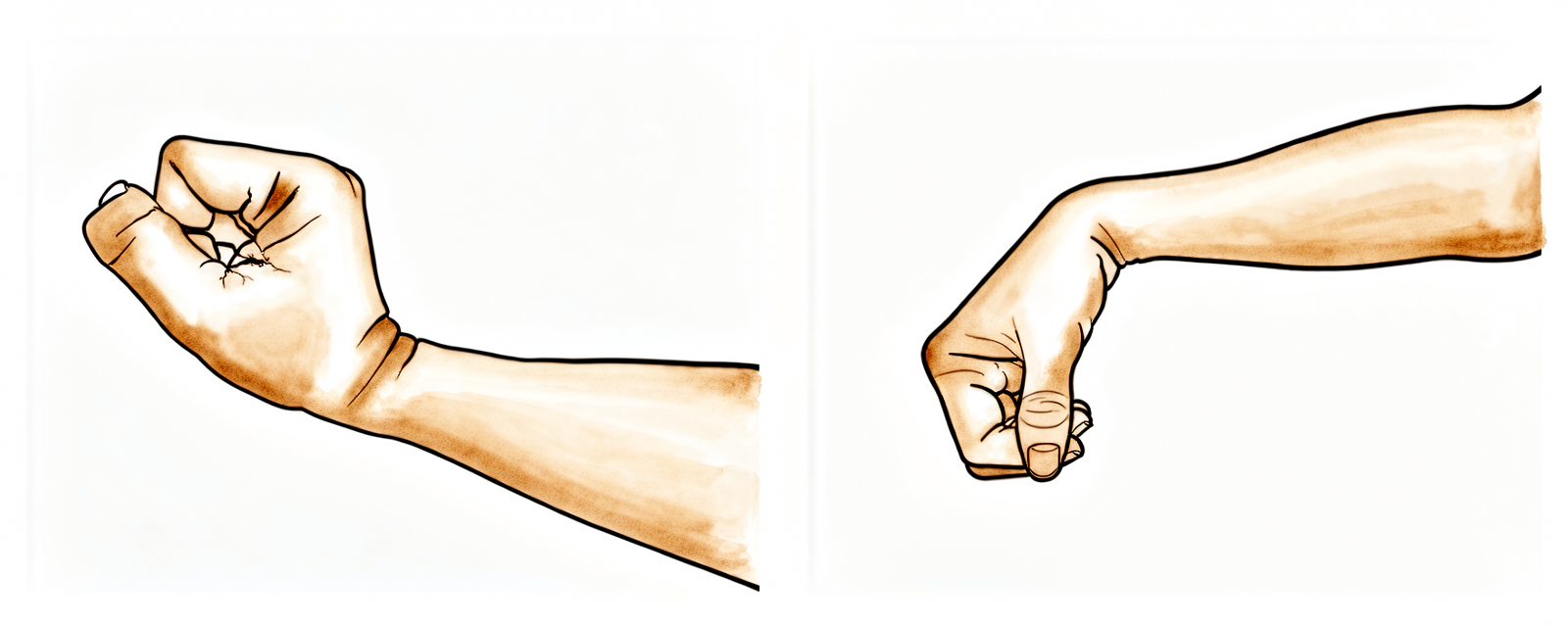
Flexão/extensão do punho
Apoie o cotovelo numa mesa e balance levemente o pulso para frente e para trás (ou apoie-o na borda de uma mesa ou poltrona, conforme ilustrado). Quando se sentir mais à vontade, segure a palma da mão com a outra mão e empurre o pulso para trás, de modo que os dedos apontem para o teto; depois faça o movimento inverso, fazendo com que apontem para o chão. Mantenha os dedos relaxados — eles se dobrarão ou esticarão por conta própria. Mantenha cada alongamento por 15 segundos; repita 5 vezes em cada direção.
10 repetições, 4–5 vezes por dia
Kieran Hirpara 4.0
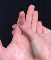
Bloqueio da articulação interfalângica distal
Comece com a palma da mão voltada para cima; use a outra mão para sustentar a mão afetada logo abaixo da articulação distal — a articulação interfalângica distal (AID). Flexione e estenda essa articulação, mantendo cada posição por 3–5 segundos. Apoie a articulação média apenas o suficiente para que ela não se dobre. É normal que os outros dedos se movam durante este exercício.
10 repetições, 4 vezes ao dia, diariamente.
Kieran Hirpara 4.0
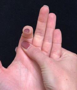
Bloqueio da articulação PIP
Comece com a palma da mão voltada para cima; apoie a mão afetada com a outra mão logo abaixo da segunda articulação — a articulação interfalângica proximal (AIP). Flexione e estenda o dedo na articulação do meio, mantendo cada posição por 3–5 segundos. Não há problema se os outros dedos também se moverem.
10 repetições, 4 vezes ao dia, diariamente.
Kieran Hirpara 4.0
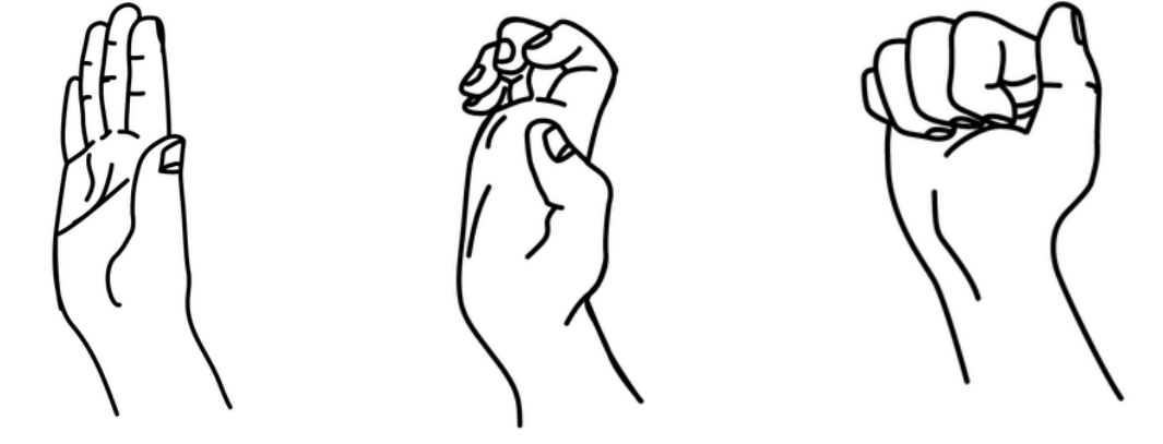
Deslizamentos tendinosos — Série A
Com a mão à sua frente e o pulso reto, estique completamente todos os dedos (1). Em seguida, dobre as pontas dos dedos para formar uma posição de “gancho”, mantendo os nós dos dedos voltados para cima (2). Por fim, faça um punho fechado, colocando o polegar por cima dos outros dedos (3).
5–10 repetições, 2–3 vezes ao dia, diariamente.
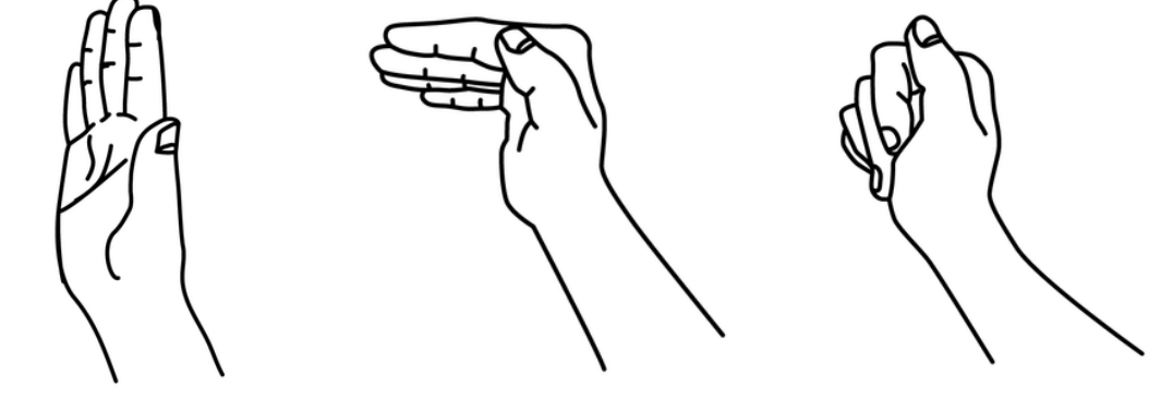
Kieran Hirpara 4.0
Deslizamentos tendinosos — Série B
Com a mão à sua frente e o pulso reto, estique completamente todos os dedos (1). Forme uma “mesa” com os dedos, dobrando-os na articulação da base (os nós dos dedos) e mantendo-os retos — certifique-se de que o pulso não se inclina para a frente (2). Em seguida, dobre os dedos na articulação média, tocando-os na palma da mão (3).
5–10 repetições, 2–3 vezes ao dia, diariamente.
Comece a realizar os exercícios abaixo somente sob orientação do Dr. Hirpara e do seu terapeuta ocupacional, respeitando sempre os limites e a amplitude de movimento que lhe foram indicados. Os exercícios de bloqueio são específicos: ao manter imóvel a articulação abaixo, você força o movimento a ocorrer na articulação que deseja recuperar, em vez daquela que já se move facilmente. A série de deslizamentos tendíneos mantém os tendões flexores deslizando suavemente dentro da bainha tendínea; já o alongamento do punho evita que ele fique rígido, enquanto o foco permanece nos dedos. É melhor fazer exercícios pequenos e frequentes do que sessões longas e esporádicas. Interrompa qualquer atividade que cause dor intensa.
Após seguir este protocolo
Este protocolo complementa as orientações gerais de recuperação fornecidas pelo consultório; consulte controle da dor pós-operatória, cuidados com a ferida e noções básicas sobre terapia da mão. Para cirurgias em que este programa costuma ser utilizado, veja liberação do dedo em gatilho e fasciectomia de Dupuytren.
Este programa de exercícios foi elaborado em parceria com Sarah Farrell, BOccThy, Terapeuta da Mão Credenciada.
Referências
[1] Miller LK, Jerosch-Herold C, Shepstone L. Eficácia das técnicas de controle do edema no edema subagudo da mão: uma revisão sistemática. J Hand Ther. 2017;30(4):432–446. https://pubmed.ncbi.nlm.nih.gov/28807598/ [2] Wehbé MA, Hunter JM. Deslizamento dos tendões flexores na mão. Parte II. Deslizamento diferencial. J Hand Surg Am. 1985;10(4):575–579. https://pubmed.ncbi.nlm.nih.gov/4020073/
Evidence & references
This is the clinical evidence summary written for health professionals. It is technical, and it lists the research this page was built from. You do not need to read it to understand your treatment or to make a decision about it.
Topic scope: This is a general post-operative finger rehabilitation program, not a single-procedure protocol. It applies as the default hand-therapy pathway after common finger operations where the repaired or released structure does not mandate its own protected range — most typically trigger finger (A1 pulley) release and Dupuytren's fasciectomy, and as a baseline mobility/oedema program after finger fracture fixation (proximal/middle phalanx ORIF or K-wire), PIP joint and volar-plate / collateral-ligament procedures, and minor soft-tissue work. The program rests on three levers the patient handout puts into practice — (1) oedema control, (2) early gentle protected movement, and (3) tendon gliding / joint-blocking to preserve differential excursion. It explicitly defers to the operation-specific protocol whenever the surgery carries a defined protected arc, motion limit or splinting regime (most importantly flexor or extensor tendon repair), which this general program does not attempt to reproduce.
Defining principle: fingers stiffen faster than almost any other joint complex in the body. The small interphalangeal joints, the gliding flexor/extensor tendons within their sheaths, and the dense soft-tissue envelope are all exquisitely sensitive to swelling and immobility — adhesions and joint contracture establish within days, not weeks. Rehabilitation is therefore a constant balancing act: protect the repaired structure for exactly the window it needs, and not one day longer, while restoring controlled glide and range early to outrun the stiffness. When in doubt, the default after finger surgery is controlled motion, not rest.
A. WHY EARLY CONTROLLED MOTION (THE CORE RATIONALE)
The unifying problem after any finger operation is the stiff finger: persistent oedema and immobility drive scar between the gliding planes, contract the joint capsule and collateral ligaments, and convert a mechanically sound repair into a functionally poor hand. The hand-surgery literature treats the stiff finger as a largely preventable complication of inadequate early rehabilitation rather than an inevitable consequence of surgery [The Stiff Finger; Stiff Digit, JAAOS].
- Immobilisation has a cost. Reduced range of motion after immobilisation arises from increased swelling, scarring between tendons and surrounding structures, and joint/ligament contracture — the exact mechanisms early motion is designed to defeat (BSSH early-mobilisation guidance).
- Adhesions establish early. The rationale for getting the patient moving — ideally with instructions given pre-operatively — and for a first therapy review at 5–7 days is to begin glide before adhesions become established (BSSH). The synthesis mirrors this: the home program starts as soon as the wound and operation allow, not at an arbitrary late milestone.
- Time to active exercise predicts the end result. In hand-fracture rehabilitation, earlier commencement of active exercise predicts greater total active range of motion at 6 weeks — a direct, measurable dose-response between early motion and outcome [Time to commencement of active exercise predicts TAM, Hand Therapy 2016].
B. EVIDENCE BY PROCEDURE GROUP
Finger fracture fixation (phalangeal ORIF / K-wire)
- The modern standard is stable fixation that permits early protected motion. Wide-awake surgery with early protected movement and pain-guided progression yields better finger ROM than rigid immobilisation (Saint John / pain-guided protocols; "better results with wide-awake surgery and early protected motion"). A systematic review and meta-analysis of mobilisation after ORIF of hand fractures supports earlier mobilisation over prolonged immobilisation for range without compromising union (ScienceDirect 2025 SR).
- Stable construct is the prerequisite. The whole early-motion strategy is contingent on the surgeon's judgement that the fixation will tolerate movement — which is why the synthesis hands the precaution set (load limits, the up-to-6-week no-lift window) back to the post-operative review. Surgeon to confirm per case.
- Minimally invasive fixation techniques are explicitly framed around preserving the soft-tissue envelope to reduce stiffness and allow early motion [Minimally Invasive Finger Fracture Management, Hand Clin].
PIP joint, volar-plate and collateral-ligament injuries
- These are stiffness-prone injuries where the management trade-off (stability vs early motion) is sharpest. The literature on PIP dislocations, fracture-dislocations and volar-plate injuries consistently favours early protected/active motion, often with buddy-strapping or a dorsal blocking approach, over static immobilisation, precisely because the PIP joint contracts so readily [PIP dislocations in athletes, Hand Clin; PIP fracture-dislocations, JBJS Rev; finger joint dislocations, Clin Sports Med].
- Buddy taping — depicted in the handout's hero image — is the canonical low-tech "protected early movement" tool here: it shares load with the neighbouring digit while permitting active glide.
Trigger finger (A1 pulley) release
- Release of the A1 pulley is a high-yield day procedure with reliably good patient-perceived recovery [Patient-Perceived Outcomes of Recovery After Trigger Digit Release, JHS 2023].
- Formal supervised therapy is usually NOT required for an uncomplicated release. A prospective randomised controlled trial found no significant difference in DASH, grip strength, ROM or pain between a structured post-operative occupational-therapy arm and a simple home-advice/ROM arm at final follow-up (RCT, PMC10671987). This validates the synthesis framing this as a home program with therapy escalation reserved for those who are slow to settle, stiff or swollen — not mandated for everyone.
Dupuytren's fasciectomy
- Therapy after fasciectomy centres on oedema and wound management, a home exercise program, and night extension splinting — a typical "brief" protocol runs 4 sessions (days 0-3, 2 wk, 4 wk, 8 wk) with a night extension orthosis to ~3 months (post-fasciectomy rehab trials).
- Routine night-splinting for all is contested. The SCoRD-type trials and subsequent work show static night splinting does not clearly improve ROM over hand therapy alone for unselected patients — splinting is best targeted at those losing extension, not applied universally [SCoRD protocol; Dutch Multidisciplinary Guideline on Dupuytren Disease].
- For established post-fasciectomy or post-fracture flexion stiffness, casting motion to mobilise stiffness (CMMS) is an evidence-supported salvage technique to regain digital flexion [Casting motion to mobilise stiffness, Hand Therapy 2010].
C. OEDEMA, SCAR AND STIFFNESS MANAGEMENT
- Oedema control is first-line and non-negotiable. Persistent hand oedema stiffens the soft tissues and degrades both ROM and function. The best systematic review of subacute hand oedema management concluded that active exercise enabling tendon gliding and muscular contraction acts as a pump to drive oedema away from the periphery, and supports elevation and active movement as first-line, with retrograde massage, compression and manual oedema mobilisation as adjuncts — there is no single superior modality, so the program layers them [Miller, Jerosch-Herold & Shepstone, J Hand Ther 2017]. This is reference [1] in the synthesis.
- Tendon gliding works through differential excursion. The straight / hook / tabletop / full-fist positions are not interchangeable repetitions: each moves the FDP relative to the FDS and relative to the sheath by a different amount, and it is this differential glide that keeps the tendons from scarring to one another and to the sheath [Wehbe & Hunter, J Hand Surg Am 1985 — reference [2] in the synthesis]. Joint-by-joint DIP and PIP blocking isolates the same glide at a single joint.
- Scar management. Once the wound is healed, scar massage and desensitisation reduce adherent scar over the incision — relevant to every open finger procedure and the surgical interval through which the tendons must glide.
- Heat before, ice after the exercise session is a standard hand-therapy adjunct to improve tissue extensibility for movement and settle the post-exercise inflammatory flare (consensus practice).
Phased timeline (maps to the synthesis sections)
| Phase | Window | Protect | Motion / glide | Oedema & scar | Notes |
|---|---|---|---|---|---|
| I — Settle & protect | Week 0-~2 | Protect per the specific operation (buddy tape / splint / load limits as set at review); light functional use for self-care, dressing, writing, typing | Begin gentle active motion within the operation's limits; tendon glides and DIP/PIP blocking as the wound and fixation allow | Elevation + active movement first-line for swelling; wound care per handout | First therapy review ideally 5-7 days to start glide before adhesions set (BSSH). No lifting/gripping/impact |
| II — Restore glide & range | Week ~2-6 | Wean protection as the structure consolidates; precautions confirmed at post-op review | Progress active ROM, full tendon-glide series, joint blocking; buddy strapping for PIP/collateral injuries | Once healed: commence scar massage (firm circles); heat before / ice after exercises; retrograde massage + compression if oedema persists | Most ROM is won in this window — frequent gentle practice beats occasional hard effort |
| III — Strengthen & return | Week ~6-12 | Protection generally off (operation-dependent) | Restore full ROM; introduce grip and functional strengthening | Continue scar work until mature; night extension splint to ~3 mo if losing extension (Dupuytren) | Return to lifting/gripping/impact from ~6 weeks per the operation; escalate persistent stiffness to hand therapy / CMMS |
Phase windows are typical and consensus-based; the operation-specific protocol and the surgeon's post-operative review override any timing here.
D. KEY CONTROVERSIES / EVIDENCE QUALITY
- Universal supervised therapy vs home program. For simple procedures (trigger finger release) an RCT shows no benefit of routine formal therapy over good home advice — supporting a targeted therapy model. For complex/stiffness-prone injuries (PIP, fracture-dislocation, fracture ORIF) early supervised hand therapy is far more clearly beneficial. The synthesis correctly pitches a home program with therapist escalation rather than mandating identical input for every operation. Moderate.
- Night-splinting after Dupuytren's fasciectomy. Routine static night splinting is not supported for unselected patients (SCoRD, Dutch guideline); reserve it for those demonstrably losing extension. Moderate (RCT/guideline).
- How early, and how much, to move a fixed fracture. Early protected motion is favoured, but it is strictly contingent on a stable construct — a judgement only the operating surgeon can make. The "early motion is better" evidence assumes adequate fixation. Moderate (SR), construct-dependent.
- The general protocol itself is a consensus scaffold. A single "finger surgery" rehab program necessarily generalises across heterogeneous operations; its three principles (oedema, early motion, glide) are very well supported, but the exact dosing/timing is expert-consensus, individualised by the treating therapist and surgeon.
E. EVIDENCE STRENGTH FLAGS (summary)
- STRONG: oedema control via elevation + active tendon-gliding exercise as first-line (SR, J Hand Ther 2017); tendon differential-excursion rationale for the varied glide positions (mechanistic, Wehbe & Hunter); early motion reduces stiffness/adhesions after finger surgery (consistent across the stiff-finger and BSSH literature).
- MODERATE (RCT / SR / guideline): early mobilisation > immobilisation after hand-fracture ORIF (SR + meta-analysis, 2025); time-to-active-exercise predicts 6-week TAM; no added benefit of routine formal therapy after simple trigger-finger release (RCT); selective (not universal) night splinting after Dupuytren's fasciectomy (SCoRD/Dutch guideline).
- WEAK / CONSENSUS: the precise phase windows and exercise dosing in this general program (expert hand-therapy consensus, individualised); heat-before/ice-after adjunct; the principle that operation-specific protocols override this general scaffold (sound clinical practice, not trial-derived).
CITATIONS
RAG corpus (180,000+ Orthopaedic articles)
- Time to commencement of active exercise predicts total active range of motion 6 weeks after hand-fracture fixation. Hand Therapy. 2016. DOI: 10.1177/1758998316679386
- Hardy MA. The Stiff Finger. Hand Clinics. 2010. DOI: 10.1016/j.hcl.2010.02.001
- Etiology, Evaluation, and Management Options for the Stiff Digit. JAAOS. DOI: 10.5435/jaaos-d-18-00310
- Phalangeal neck fractures of the proximal phalanx of the fingers in adults. Injury. 2010. DOI: 10.1016/j.injury.2010.06.017
- Minimally Invasive Finger Fracture Management. Hand Clinics. 2013. DOI: 10.1016/j.hcl.2013.08.014
- Management of Proximal Interphalangeal Joint Dislocations in Athletes. Hand Clinics. 2009. DOI: 10.1016/j.hcl.2009.05.008
- Treatment of Proximal Interphalangeal Joint Fracture-Dislocations. JBJS Reviews. DOI: 10.2106/jbjs.rvw.o.00019
- Management of Finger Joint Dislocation and Fracture-Dislocations in Athletes. Clinics in Sports Medicine. 2019. DOI: 10.1016/j.csm.2019.10.006
- Patient-Perceived Outcomes of Recovery After Trigger Digit Release. J Hand Surg Am. 2023. DOI: 10.1016/j.jhsa.2023.03.016
- Comparative Study of A1 Pulley Release and Ulnar Superficialis Slip Resection (trigger digit). J Hand Surg Am. 2022. DOI: 10.1016/j.jhsa.2022.04.021
- Use of casting motion to mobilize stiffness (CMMS) to regain digital flexion. Hand Therapy. 2010. DOI: 10.1258/ht.2010.010008
- Dutch Multidisciplinary Guideline on Dupuytren Disease. J Hand Surg Glob Online. 2022. DOI: 10.1016/j.jhsg.2022.11.008
- Factors affecting functional recovery after surgery and hand therapy in Dupuytren's patients. J Hand Ther. 2014. DOI: 10.1016/j.jht.2014.11.006
- Rehabilitation Regimens Following Surgical Repair of Extensor Tendon Injuries of the hand. DOI: 10.1007/s12593-012-0075-x
Hand-therapy / rehabilitation literature (URLs)
- Miller LK, Jerosch-Herold C, Shepstone L. Effectiveness of edema management techniques for subacute hand edema: a systematic review. J Hand Ther. 2017;30(4):432-446. https://pubmed.ncbi.nlm.nih.gov/28807598/
- Wehbe MA, Hunter JM. Flexor tendon gliding in the hand. Part II. Differential gliding. J Hand Surg Am.
- https://pubmed.ncbi.nlm.nih.gov/4020073/
- Systematic review and meta-analysis of mobilisation following ORIF of hand fractures. ScienceDirect.
- https://www.sciencedirect.com/science/article/pii/S1748681525003109
- Better results of finger fractures with wide-awake surgery and early protected motion. PMC. https://www.ncbi.nlm.nih.gov/pmc/articles/PMC4445035/
- The effectiveness of rehabilitation after open surgical release for trigger finger: a prospective, randomized, controlled study. PMC. https://www.ncbi.nlm.nih.gov/pmc/articles/PMC10671987/
- Splinting after contracture release for Dupuytren's contracture (SCoRD): RCT protocol. PMC. https://www.ncbi.nlm.nih.gov/pmc/articles/PMC2386788/
Published rehab protocols (patient-guidance / society — basis for the phase structure)
- The British Society for Surgery of the Hand (BSSH) — Guidelines. https://www.bssh.ac.uk/professionals/guidelines.aspx
- Pain-Guided Hand Therapy for early protected movement of finger fractures (The Saint John Protocol), ASSH. https://handsurgery.org/multimedia/files/preCourse/Pain%20Guided%20Hand%20Therapy%20for%20early%20protected%20movement%20finger%20fractures.pdf
- Rehabilitative Strategies Following Hand Fractures. Hand Clinics. https://www.hand.theclinics.com/article/S0749-0712(13)00066-8/fulltext
- University of Kentucky HealthCare — Hand Rehabilitation Protocols. https://ukhealthcare.uky.edu/sites/default/files/m21-0609_ortho_protocols-final.pdf





