Os Acromion
Patients › Shoulder
Patient-facing topic on os acromiale — failed fusion of an acromial ossification centre that may cause shoulder pain and contribute to subacromial impingement.
O que você está sentindo
Você pode sentir dor na parte superior do ombro. Esta condição afeta cerca de 2,13% dos pacientes com problemas no ombro que necessitam de ressonância magnética. A dor frequentemente decorre de uma lesão no manguito rotador, que é um grupo de músculos e tendões que movem seu braço. Essa lesão ocorre porque o fragmento ósseo adicional se move anormalmente e atrita contra seus outros tecidos.
Sua dor pode piorar após a realização de certas atividades. Ela também pode acordá-lo à noite ou doer quando você tenta dormir de lado. Tarefas diárias simples podem se tornar difíceis. Você pode ter dificuldade em alcançar as costas para fechar um sutiã ou guardar uma camisa. Esses movimentos exercem pressão na área onde os fragmentos ósseos não se unem completamente.
Se você já realizou cirurgia de substituição do ombro, pode notar sensibilidade logo acima desse fragmento ósseo. Isso ocorre em 1 em cada 4 pacientes após o procedimento. A boa notícia é que esse desconforto geralmente desaparece espontaneamente com o tempo para a maioria das pessoas. Seu cirurgião avaliará seus sintomas para decidir se esse fragmento ósseo adicional é a principal causa da sua dor.
O que está realmente acontecendo
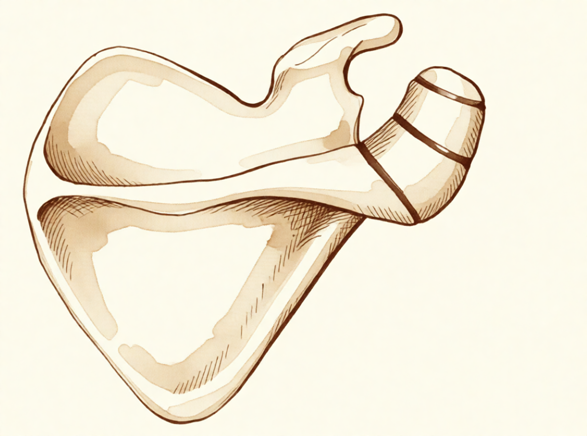
Seu ombro possui um pequeno fragmento ósseo chamado os acromiale que não se fundiu ao osso principal. Isso ocorre em aproximadamente 2,13% dos pacientes com problemas no ombro que necessitam de ressonância magnética. Pense neste ponto não fundido como uma junta solta entre duas partes do osso do seu ombro. Em vez de se mover como uma peça sólida única, esse fragmento solto pode esfregar e deslizar quando você move o braço.
Esse movimento anormal frequentemente irrita os tecidos moles próximos. O manguito rotador é um grupo de tendões que atuam como cordas para levantar e girar seu braço. Como o osso solto se move de maneira anormal, ele pode pinçar esses tendões. Esse pinçamento, ou impingement, frequentemente leva a uma ruptura do manguito rotador. Em atletas jovens, esse padrão específico de lesão é comum e requer avaliação cuidadosa para confirmar o diagnóstico.
Às vezes, esse osso solto pode sofrer lesão após uma queda ou trauma, embora isso seja raro. Se você tiver cirurgia por outros motivos, como uma artroplastia total reversa do ombro, pode sentir sensibilidade local nesse local em 1 em cada 4 pacientes. Essa dor geralmente desaparece espontaneamente ao longo do tempo. Ter esse fragmento ósseo adicional não parece prejudicar seus resultados gerais de cirurgias de substituição articular do ombro.
O que podemos fazer a respeito
A maioria das pessoas inicia com autocuidado e fisioterapia para controlar a dor. Seu fisioterapeuta orientará exercícios para fortalecer os músculos do ombro e melhorar a mobilidade. Essa abordagem visa reduzir o atrito na região onde se localiza o fragmento ósseo adicional. Você deve dar uma tentativa justa a este plano não cirúrgico antes de considerar outras etapas.
Se a dor persistir, seu cirurgião poderá discutir o uso de medicamentos ou injeções. Os anti-inflamatórios podem ajudar a reduzir o inchaço e a sensibilidade ao redor da articulação. Em alguns casos, uma injeção de cortisona é utilizada para acalmar a inflamação rapidamente. Embora as evidências não especifiquem durações exatas para esses tratamentos, muitos pacientes encontram alívio enquanto o medicamento faz efeito. Se você for um atleta competitivo, seu cirurgião avaliará os riscos cuidadosamente, pois a cirurgia geralmente não é recomendada para tenistas profissionais.
A cirurgia é normalmente considerada apenas se essas medidas conservadoras falharem no controle dos seus sintomas. O objetivo da operação é remover o fragmento ósseo instável ou fixá-lo no lugar para impedir o movimento doloroso. Essa decisão é tomada em conjunto com seu cirurgião após revisar seu caso específico e os resultados das imagens.
Quando procurar ajuda
Consulte o seu médico de família se tiver dor persistente no ombro que não melhora com o repouso. Solicite uma avaliação especializada se notar fraqueza, instabilidade, bloqueio do ombro ou se este ceder. Os sintomas que interferem com o sono ou com o trabalho também requerem atenção. Procure ajuda se experimentar uma piora súbita da dor. Esta condição está associada a lesões do manguito rotador, que podem causar impacto devido a um movimento anormal. Embora raro, a articulação pode ser lesada após trauma. Se for um jogador profissional de ténis, o tratamento cirúrgico geralmente não está indicado, mas um especialista pode confirmar o diagnóstico através de exame físico e exames de imagem.
Evidence & references
Overview
- In Thai patients with shoulder problems requiring MRI evaluation, the prevalence of os acromiale was 2.13% [1].
- Os acromiale is associated with rotator cuff injuries [5].
- A multicenter study aimed to determine the prevalence of and factors associated with os acromiale in the Japanese population [6].
- Operative management of a symptomatic os acromiale that has failed initial nonoperative treatment leads to decreased symptoms and improvement in clinical outcomes [2].
- Surgical options for symptomatic os acromiale include arthroscopic sub-total excision [16].
- Surgical options for symptomatic os acromiale include arthroscopic subacromial decompression of stable fragments [16].
- Surgical options for symptomatic os acromiale include open reduction and internal fixation of unstable fragments [16].
- Surgical treatment is usually not indicated for os acromiale in the professional tennis player [7].
- Ipsilateral os acromiale may be a relative contraindication to the clavicle hook plate [12].
- Postoperative local tenderness at the os acromiale can be expected in 1 out of 4 patients after reverse total shoulder arthroplasty [3].
- Postoperative local tenderness at the os acromiale resolves spontaneously over time in the majority of patients after reverse total shoulder arthroplasty [3].
- The presence of os acromiale does not appear to have a negative impact on clinical outcomes after reverse total shoulder arthroplasty [4].
- The outcome of reverse total shoulder arthroplasty does not seem to be negatively affected by the presence of an os acromiale [9].
Anatomy & Pathophysiology
- The prevalence of os acromiale in Thai patients with shoulder problems requiring MRI evaluation is 2.13% [1].
- Os acromiale is associated with rotator cuff injuries [5].
- A tear of the rotator cuff may often be associated with os acromiale, likely due to impingement from abnormal motion at the fibrous union site [8].
- Fused os acromiale, which has not been described previously, might be mistaken for a free ossicle in the clinical setting [10].
- Awareness of the os acromiale in the young athlete is crucial to confirm diagnosis through appropriate clinical examination and image studies [11].
- The synchondrosis of an os acromiale can be injured following trauma, though rarely [13].
- Postoperative local tenderness at the os acromiale can be expected in 1 out of 4 patients after reverse total shoulder arthroplasty [3].
- Postoperative local tenderness at the os acromiale resolves spontaneously over time in the majority of patients [3].
- The presence of os acromiale does not appear to have a negative impact on clinical outcomes after reverse total shoulder arthroplasty [4].
- The outcome of reverse total shoulder arthroplasty does not seem to be negatively affected by the presence of an os acromiale [9].
- Reverse shoulder arthroplasty improves range of motion, decreases pain, and increases patient satisfaction in patients with os acromiale and cuff tear arthropathy [24].
- Surgical treatment is usually not indicated for os acromiale in the professional tennis player [7].
Classification
- The prevalence of os acromiale in Thai patients with shoulder problems requiring MRI evaluation is 2.13% [1].
- Os acromiale is associated with rotator cuff injuries [5].
- A tear of the rotator cuff may often be associated with os acromiale, likely due to impingement from abnormal motion at the fibrous union site [8].
- Fused os acromiale, which has not been described previously, might be mistaken for a free ossicle in the clinical setting [10].
- Awareness of the os acromiale in the young athlete, appropriate clinical examination, and image studies are crucial to confirm diagnosis [11].
- The synchondrosis of an os acromiale can be injured following trauma, though rarely [13].
- Meta-os acromiale is the rarest subtype of os acromiale [15].
Clinical Presentation
- The prevalence of os acromiale in Thai patients with shoulder problems requiring MRI evaluation was 2.13% [1].
- Os acromiale is associated with rotator cuff injuries [5].
- A tear of the rotator cuff may often be associated with os acromiale, likely due to impingement from abnormal motion at the fibrous union site [8].
- The presence of os acromiale does not appear to have a negative impact on clinical outcomes after reverse total shoulder arthroplasty (rTSA) [4].
- The outcome of reverse total shoulder arthroplasty (RTSA) does not seem to be negatively affected by the presence of an os acromiale [9].
- Postoperative local tenderness at the os acromiale can be expected in 1 out of 4 patients following rTSA [3].
- Postoperative local tenderness at the os acromiale resolves spontaneously over time in the majority of patients [3].
- Surgical treatment is usually not indicated for os acromiale in professional tennis players [7].
- Awareness of the os acromiale, appropriate clinical examination, and image studies are crucial to confirm diagnosis in young athletes [11].
- Ipsilateral os acromiale may be a relative contraindication to the clavicle hook plate [12].
- The synchondrosis of an os acromiale can be injured following trauma, though rarely [13].
- Fused os acromiale, which has not been described previously, might be mistaken for a free ossicle in the clinical setting [10].
- Meta-os acromiale is the rarest subtype of os acromiale [15].
- Liberson reviewed 1800 shoulder girdles and identified an incidence of os acromiale of 1.4% [14].
- The lesion of os acromiale is bilateral in 62% of patients according to Liberson's review [14].
Investigations
- The prevalence of os acromiale in Thai patients with shoulder problems requiring MRI evaluation is 2.13% [1].
- Os acromiale is associated with rotator cuff injuries [5].
- A tear of the rotator cuff may often be associated with os acromiale, likely due to impingement from abnormal motion at the fibrous union site [8].
- Awareness of the os acromiale in the young athlete, appropriate clinical examination, and image studies are crucial to confirm diagnosis [11].
- The synchondrosis of an os acromiale can be injured following trauma, though rarely [13].
- Appropriate radiographic investigation for os acromiale injury includes axillary views [13].
- Fused os acromiale, which has not been described previously, might be mistaken for a free ossicle in the clinical setting [10].
- Liberson reviewed 1800 shoulder girdles and identified an incidence of os acromiale of 1.4% [14].
- The lesion of os acromiale is bilateral in 62% of patients according to Liberson's review [14].
Treatment
- Operative management of a symptomatic os acromiale that has failed initial nonoperative treatment leads to decreased symptoms and improvement in clinical outcomes [2].
- Surgical options for symptomatic os acromiale include arthroscopic sub-total excision, arthroscopic subacromial decompression of stable fragments, and open reduction and internal fixation of unstable fragments [16].
- Open reduction and internal fixation using cannulated screws or tension band wiring have superior outcomes in the literature in the treatment of symptomatic os acromiale [19].
- A new arthroscopic technique of fixation of os acromiale with absorbable screws provides promising clinical, cosmetic, and radiologic results with high patient satisfaction [17].
- A symptomatic os acromiale in a competitive female fastball pitcher was treated successfully with open reduction and internal fixation [21].
- Special consideration must be given to the type of tension-band construct used to achieve adequate compression and fixation for meta-os acromiale, the rarest subtype of os acromiale [15].
- Surgical treatment is usually not indicated for os acromiale in the professional tennis player [7].
- Ipsilateral os acromiale may be a relative contraindication to the clavicle hook plate [12].
- Postoperative local tenderness at the os acromiale can be expected in 1 out of 4 patients but resolves spontaneously over time in the majority of patients [3].
- The presence of os acromiale does not appear to have a negative impact on the clinical outcomes after reverse total shoulder arthroplasty (rTSA) [4].
- The outcome of reverse total shoulder arthroplasty (RTSA) does not seem to be negatively affected by the presence of an os acromiale [9].
Complications
- The prevalence of os acromiale in Thai patients with shoulder problems requiring MRI evaluation was 2.13% [1].
- Operative management of a symptomatic os acromiale that has failed initial nonoperative treatment leads to decreased symptoms and improvement in clinical outcomes [2].
- Postoperative local tenderness at the os acromiale can be expected in 1 out of 4 patients following reverse total shoulder arthroplasty [3].
- Postoperative local tenderness at the os acromiale resolves spontaneously over time in the majority of patients [3].
- The presence of os acromiale does not appear to have a negative impact on clinical outcomes after reverse total shoulder arthroplasty [4].
- Reverse total shoulder arthroplasty remains a safe and effective treatment option in the presence of os acromiale [4].
- Os acromiale is associated with rotator cuff injuries [5].
- A tear of the rotator cuff may often be associated with os acromiale, likely due to impingement from abnormal motion at the fibrous union site [8].
- Surgical treatment is usually not indicated for os acromiale in professional tennis players [7].
- Fused os acromiale, which has not been described previously, might be mistaken for a free ossicle in the clinical setting [10].
- The incidence of os acromiale identified by Liberson was 1.4% [14].
- The lesion of os acromiale was bilateral in 62% of patients in Liberson's review [14].
Recovery
- Operative management of a symptomatic os acromiale that has failed initial nonoperative treatment leads to decreased symptoms and improvement in clinical outcomes [2].
- Postoperative local tenderness at the os acromiale can be expected in 1 out of 4 patients but resolves spontaneously over time in the majority of patients [3].
- The presence of os acromiale does not appear to have a negative impact on the clinical outcomes after reverse total shoulder arthroplasty (rTSA) [4].
- Reverse total shoulder arthroplasty (rTSA) remains a safe and effective treatment option in patients with os acromiale [4].
- The outcome of reverse total shoulder arthroplasty (RTSA) does not seem to be negatively affected by the presence of an os acromiale [9].
- Surgical treatment is usually not indicated for os acromiale in professional tennis players [7].
Key Evidence
- [L3] In Thai patients with shoulder problems who required MRI evaluation, the prevalence of os acromiale was 2.13%. [1] (10.1177/23259671221078806)
- [L4] Operative management of a symptomatic os acromiale that has failed initial nonoperative treatment leads to decreased symptoms and improvement in clinical outcomes. [2] (10.1016/j.jse.2019.05.047)
- [L3] Postoperative local tenderness at the os acromiale can be expected in 1 out of 4 patients but resolves spontaneously over time in the majority of patients. [3] (10.1177/2325967120965131)
- [L4] The presence of os acromiale does not appear to have a negative impact on the clinical outcomes after surgery and rTSA remains a safe and effective treatment option. [4] (10.1016/j.xrrt.2025.01.002)
- [L3] The study supports previous findings that os acromiale is associated with rotator cuff injuries. [5] (10.1016/j.jseint.2025.05.015)
- [L3] This multicenter study aimed to determine the prevalence of and factors associated with os acromiale in the Japanese population. [6] (10.1016/j.jse.2025.01.008)
- [L4] Surgical treatment is usually not indicated for os acromiale in the professional tennis player. [7] (10.1177/2325967118773723)
- [L4] A tear of the rotator cuff may often be associated with os acromiale, likely due to impingement from abnormal motion at the fibrous union site. [8] (10.2106/00004623-198466080-00029)
- [L4] The outcome of RTSA does not seem to be negatively affected by the presence of an os acromiale. [9] (10.1016/j.jse.2017.02.012)
- [L4] An anatomical study showed that fused os acromiale, which has not been described previously, might be mistaken for a free ossicle in the clinical setting. [10] (10.2106/00004623-200003000-00010)
- [L4] Awareness of the os acromiale in the young athlete, appropriate clinical examination, and image studies are crucial to confirm diagnosis. [11] (10.1016/j.jseint.2020.02.008)
- [L4] Ipsilateral os acromiale may be a relative contraindication to the clavicle hook plate. [12] (10.1186/s12891-021-04841-1)
- [L4] This case highlights that the synchondrosis of an os acromiale can be injured following trauma, though rarely, and emphasizes the need for appropriate radiographic investigation including axillary views and a flexible surgical approach. [13] (10.1016/j.jse.2008.02.012)
- [L4] Meta–os acromiale is the rarest subtype of os acromiale, and special consideration must be given to the type of tension-band construct used to achieve adequate compression and fixation. [15] (10.1177/03635465211028238)
- [L5] Surgical options for symptomatic os acromiale include arthroscopic sub-total excision, arthroscopic subacromial decompression of stable fragments, and open reduction and internal fixation of unstable fragments. [16] (10.5435/jaaos-d-17-00011)
- [L4] This new arthroscopic technique of fixation of os acromiale with absorbable screws provides promising clinical, cosmetic, and radiologic results with high patient satisfaction. [17] (10.1016/j.jse.2011.12.011)
- [L4] Open reduction and internal fixation using cannulated screws, or tension band wiring have superior outcomes in the literature in the treatment of symptomatic os acromiale. [19] (10.1302/2058-5241.4.180100)
- [L4] A symptomatic os acromiale in a competitive female fastball pitcher was treated successfully with open reduction and internal fixation. [21] (10.1177/0363546506288305)
- [L4] Reverse shoulder arthroplasty improved ROM, decreased pain, and increased patient satisfaction in patients with os acromiale and cuff tear arthropathy. [24] (10.5397/cise.2019.00409)
References
[1] Prevalence of Os Acromiale in Thai Patients With Shoulder Problems: A Magnetic Resonance Imaging Study. Orthopaedic Journal of Sports Medicine. 2022. DOI: 10.1177/23259671221078806 [2] Os acromiale: systematic review of surgical outcomes. Journal of Shoulder and Elbow Surgery. 2020. DOI: 10.1016/j.jse.2019.05.047 [3] Os Acromiale in Reverse Total Shoulder Arthroplasty: A Cohort Study. Orthopaedic Journal of Sports Medicine. 2020. DOI: 10.1177/2325967120965131 [4] Clinical implications of reverse total shoulder arthroplasty with an os acromiale: a systematic review. JSES Reviews, Reports, and Techniques. 2025. DOI: 10.1016/j.xrrt.2025.01.002 [5] Prevalence and factors associated with os acromiale: a multicenter study. JSES International. 2025. DOI: 10.1016/j.jseint.2025.05.015 [6] The prevalence and associated factors of os acromiale: a multicenter study. Journal of Shoulder and Elbow Surgery. 2025. DOI: 10.1016/j.jse.2025.01.008 [7] Os Acromiale in Professional Tennis Players. Orthopaedic Journal of Sports Medicine. 2018. DOI: 10.1177/2325967118773723 [8] Rotator cuff tears associated with os acromiale.. The Journal of Bone & Joint Surgery. 1984. DOI: 10.2106/00004623-198466080-00029 [9] Reverse shoulder arthroplasty in patients with os acromiale. Journal of Shoulder and Elbow Surgery. 2017. DOI: 10.1016/j.jse.2017.02.012 [10] Os Acromiale: Frequency, Anatomy, and Clinical Implications. The Journal of Bone and Joint Surgery-American Volume. 2000. DOI: 10.2106/00004623-200003000-00010 [11] The unstable os acromiale: a cause of pain in the young athlete. JSES International. 2020. DOI: 10.1016/j.jseint.2020.02.008 [12] Os acromiale may be a contraindication of the clavicle hook plate: case reports and literature review. BMC Musculoskeletal Disorders. 2021. DOI: 10.1186/s12891-021-04841-1 [13] Fracture of an os acromiale with associated rupture of the coracoclavicular ligaments. Journal of Shoulder and Elbow Surgery. 2008. DOI: 10.1016/j.jse.2008.02.012 [14] Types of os acromiale according to Liberson. 2006. [15] Rare Symptomatic Meta–Os Acromiale in an Athlete. The American Journal of Sports Medicine. 2021. DOI: 10.1177/03635465211028238 [16] Symptomatic, Unstable Os Acromiale. Journal of the American Academy of Orthopaedic Surgeons. 2018. DOI: 10.5435/jaaos-d-17-00011 [17] Arthroscopically assisted internal fixation of the symptomatic unstable os acromiale with absorbable screws. Journal of Shoulder and Elbow Surgery. 2012. DOI: 10.1016/j.jse.2011.12.011 [19] Os acromiale: a review of its incidence, pathophysiology, and clinical management. EFORT Open Reviews. 2019. DOI: 10.1302/2058-5241.4.180100 [21] Surgical Stabilization of Os Acromiale in a Fast-Pitch Softball Pitcher. The American Journal of Sports Medicine. 2006. DOI: 10.1177/0363546506288305 [24] Reverse shoulder arthroplasty with os acromiale. Clinics in Shoulder and Elbow*. 2020. DOI: 10.5397/cise.2019.00409


