Doença de Kienböck
Patients › Wrist
Kienböck’s disease — progressive wrist pain from lunate avascular necrosis; diagnosis and treatment options.
O que você está sentindo
A doença de Kienböck é uma condição que afeta os pequenos ossos do seu punho. Não é muito comum. O problema geralmente piora com o tempo se não for tratado. Você pode notar inchaço ou rigidez no punho. A dor geralmente começa lentamente e aumenta gradualmente.
Você pode sentir sensibilidade ao pressionar a parte superior do punho. É aí que o osso escafoide (lunatum) se localiza. A dor pode dificultar tarefas simples. Você pode ter dificuldade para se impulsionar para cima ao sair de uma cadeira. Levantar objetos pode se tornar doloroso e causar fraqueza. Até mesmo girar uma maçaneta ou abrir um pote pode doer.
Seu punho pode parecer rígido pela manhã. Essa rigidez geralmente melhora à medida que você se move ao longo do dia. No entanto, a dor pode retornar após um período de uso das mãos. Atividades que sobrecarregam o punho, como fazer flexões ou carregar sacolas de compras, podem desencadear crises. Você também pode notar que sua força de preensão está mais fraca do que antes.
Em alguns casos, a dor pode ser aguda ou surda. Ela pode irradiar ao longo do antebraço. A dor noturna é possível, especialmente se você dormir de lado sobre esse braço. Isso pode perturbar seu descanso e deixá-lo cansado. Você pode ter dificuldade em alcançar as costas para fechar um sutiã ou alisar uma camisa. Esses movimentos esticam o punho e podem agravar o osso inflamado.
A gravidade dos seus sintomas depende de como os ossos estão formatados no seu punho. Essa forma afeta a quantidade de pressão exercida sobre o osso lunatum. A causa da doença provavelmente deve-se a vários fatores atuando em conjunto. Nem sempre está claro por que uma pessoa desenvolve a condição e outra não.
Se você ignorar os sintomas, a condição pode progredir para estágios mais avançados. Isso pode levar a alterações na forma dos ossos do punho. Seu cirurgião analisará radiografias para ver o quanto os ossos se deslocaram. No entanto, as radiografias padrão nem sempre mostram o quadro completo do colapso ósseo. É por isso que seu cirurgião pode solicitar exames de imagem mais detalhados para obter uma visão clara do que está acontecendo no interior do seu punho.
O que está realmente acontecendo
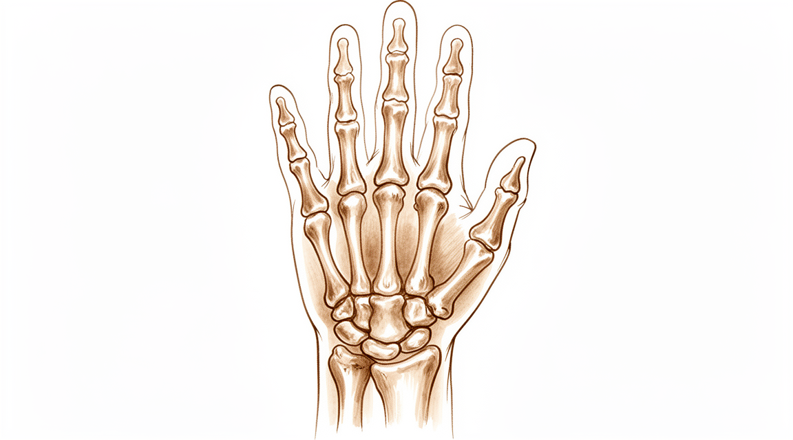
A doença de Kienböck é uma condição em que o suprimento sanguíneo para um pequeno osso do seu pulso, chamado lunato, é reduzido ou perdido. Sem sangue suficiente, esse osso começa a enfraquecer e pode, eventualmente, colapsar. Pense no lunato como um amortecedor central no seu pulso. Quando ele perde sua integridade estrutural, não consegue mais amortecer o impacto entre os ossos do seu antebraço e o restante da sua mão.
Esse colapso altera a forma como seu pulso se move. Normalmente, os ossos do seu pulso deslizam e rolam suavemente uns contra os outros. Quando o lunato está danificado, esse movimento torna-se irregular. Os ossos podem se deslocar de seu alinhamento normal, um problema conhecido como desalinhamento rotacional. Esse desalinhamento coloca estresse extra em outras partes da articulação do pulso, particularmente onde o osso rádio encontra o osso escafoide. Com o tempo, esse desgaste desigual pode levar à artrite nessas áreas específicas.
Seu cirurgião analisa essas mudanças para decidir o melhor caminho a seguir. Em pacientes mais jovens, o objetivo é frequentemente salvar o osso. Procedimentos como osteotomias radiais, onde o osso do antebraço é ajustado, podem melhorar os resultados e os achados radiográficos em adolescentes. Para outros, o encurtamento do capitato ou o enxerto ósseo vascularizado podem ser recomendados para restaurar o equilíbrio e o fluxo sanguíneo. Esses tratamentos visam manter o movimento do seu pulso o mais natural possível.
Em casos mais avançados, onde o osso colapsou significativamente, o foco muda para estabilizar a articulação. Cirurgias como a artrodese escafo-capitata, que funde dois ossos, podem proporcionar alívio da dor a longo prazo e melhorar a força de preensão. Embora esses procedimentos limitem algum movimento, eles criam uma plataforma estável que permite que você use sua mão de forma eficaz. Seu cirurgião escolherá a opção que melhor corresponde ao estágio da sua doença e às suas necessidades pessoais.
O que podemos fazer a respeito
Iniciamos com o tratamento não cirúrgico para gerenciar seus sintomas e proteger seu pulso. Esta abordagem foca na redução do estresse sobre os ossos da sua mão. Seu cirurgião pode recomendar repouso ou atividades específicas a evitar. A fisioterapia ajuda a manter a mobilidade e a força no seu pulso e mão. Esses métodos visam mantê-lo confortável enquanto monitoramos a condição.
Pesquisas mostram que os tratamentos não cirúrgicos podem alcançar resultados bons e excelentes em pacientes mais jovens cujos ossos ainda estão em crescimento. Para muitos, este caminho conservador é suficiente para gerenciar a dor e a função. No entanto, a doença de Kienböck é geralmente uma condição progressiva. Isso significa que pode piorar ao longo do tempo, potencialmente levando a alterações avançadas na articulação do pulso se não for controlada. Monitoramos esses sinais de perto durante seus exames regulares.
Se as medidas não cirúrgicas não proporcionarem alívio suficiente, discutimos opções de manejo médico. Medicamentos para dor e anti-inflamatórios ajudam a controlar o desconforto e o inchaço. Em alguns casos, podemos considerar injeções para reduzir a inflamação diretamente na articulação. Esses tratamentos oferecem alívio temporário e ajudam você a permanecer ativo. Eles não revertem as alterações ósseas subjacentes, mas podem melhorar sua qualidade de vida diária enquanto decidimos os próximos passos.
A cirurgia é considerada quando o tratamento conservador atinge seu limite ou se a doença progride apesar do tratamento. O objetivo da cirurgia é descarregar o osso afetado, restaurar o fluxo sanguíneo ou estabilizar a articulação. As opções incluem procedimentos para alterar a forma do osso rádio, enxertar novo osso com seu próprio suprimento sanguíneo ou fundir ossos específicos para aliviar a dor. A escolha depende do estágio da sua doença e das suas necessidades individuais.
Para pacientes adolescentes, as osteotomias radiais (alteração da forma do osso do antebraço) são eficazes para melhorar tanto os sintomas quanto os achados nos raios-X. Esses procedimentos podem proporcionar melhora por décadas em 75% dos pacientes. Em casos avançados onde o pulso colapsou, procedimentos de fusão, como a artrodese escafo-capitata, oferecem alívio significativo da dor e melhora funcional. Seu cirurgião explicará qual opção se adequa à sua situação específica.
Também utilizamos imagens, como raios-X e ressonância magnética, para acompanhar as alterações. Embora os raios-X simples às vezes possam não detectar sinais precoces de colapso, a imagem avançada nos ajuda a ver o estado real do seu osso lúneo. Isso garante que escolhamos o tratamento certo no momento certo. Seja você precisar de simples repouso ou de um procedimento cirúrgico, nosso objetivo é preservar a função do seu pulso e reduzir a dor.
O que esperar
A doença de Kienböck é uma condição em que o suprimento sanguíneo para um pequeno osso do pulso é reduzido, causando seu enfraquecimento e colapso. Esse processo é geralmente progressivo, o que significa que tende a piorar ao longo do tempo em vez de se estabilizar espontaneamente. Sem tratamento, a condição pode evoluir para alterações do Estágio IV, que envolvem artrite por desgaste significativo na articulação do pulso. A trajetória exata da doença não é totalmente conhecida, mas frequentemente leva a dor persistente e rigidez se não for manejada.
Com o tratamento, seu prognóstico melhora significativamente. Muitas opções cirúrgicas são projetadas para interromper essa progressão e aliviar a dor. Por exemplo, a osteotomia de encurtamento radial — um procedimento que ajusta o comprimento do osso do antebraço — proporciona melhora por décadas em 75% dos pacientes com doença sintomática. Esse tratamento é particularmente eficaz para o alívio duradouro da dor em pacientes com variação ulnar negativa, um alinhamento específico dos ossos do pulso. Outros procedimentos, como a artrodese escafo-capitata (união de dois ossos) ou a capsectomia proximal (remoção de uma fileira de pequenos ossos do pulso), oferecem benefícios duradouros e a longo prazo. Essas cirurgias tipicamente aliviam a dor, preservam a mobilidade funcional e mantêm uma força de preensão satisfatória por muitos anos.
Mesmo com o tratamento, a doença nem sempre progride rapidamente. A progressão radiográfica da doença de Kienböck ao longo de 1 ano ou mais é leve, em média, independentemente do tratamento escolhido. Isso significa que, embora a condição subjacente possa persistir, as alterações visíveis nos raios X frequentemente se estabilizam. Você pode esperar que seu cirurgião adapte o plano ao seu estágio e sintomas específicos. Seja você um adolescente ou um adulto, o objetivo é controlar a dor e manter o pulso funcional. Embora os resultados sejam geralmente positivos, é importante ter expectativas realistas. A doença é multifatorial, e os resultados individuais variam, mas os tratamentos modernos oferecem caminhos confiáveis para o alívio da dor e a melhora da função da mão.
Quando procurar ajuda
A doença de Kienböck é uma condição rara que afeta o punho. É geralmente considerada uma condição progressiva que pode levar a alterações articulares avançadas. O curso exato da doença não é totalmente conhecido. Deve consultar o seu médico de família se tiver dor persistente que não melhora com o repouso. Solicite uma avaliação por um especialista se notar fraqueza ou instabilidade no punho. Procure ajuda se a sua mão bloquear ou ceder. Contacte o seu cirurgião se os sintomas interferirem com o seu sono ou trabalho. O agravamento súbito da dor também é um motivo para procurar atendimento médico. Uma avaliação precoce ajuda a gerir eficazmente esta condição complexa.
Evidence & references
Overview
- Radial osteotomies are effective in improving short-term clinical outcomes and radiographic findings in teenage patients with Kienböck disease [1].
- Scaphocapitate arthrodesis demonstrates long-term clinical benefits for the treatment of collapsed Kienböck disease [2].
- Vascularized bone grafting for stage III Kienböck disease demonstrates favorable long-term results and is recommended as a surgical treatment [3].
- Good and excellent clinical and radiological outcomes can be achieved with both nonsurgical and surgical treatments in skeletally immature patients with Kienböck disease [5].
- Temporary scaphotrapezoidal joint fixation is recommended for the surgical treatment of adolescent Kienböck's disease [7].
- Radial shortening osteotomy provides decade-long improvement in 75% of patients and is a reasonable treatment for symptomatic Kienböck’s disease [8].
- Scaphocapitate arthrodesis is an effective procedure for the treatment of Kienböck disease, associated with satisfactory functional outcomes and significant improvement in pain scores and grip strength [13].
- Capitate shortening is a safe and effective approach for the treatment of early stages of Kienböck's disease and can be associated with a satisfying outcome [14].
- Advanced Kienböck's disease with carpal collapse is not a contraindication for carpal-sparing surgery radial shortening osteotomy [17].
- Lunate excision, capitate osteotomy, and intercarpal arthrodesis should be used with caution for advanced Kienböck's disease because it does not have good long-term results and is no longer widely used in Europe [25].
- The acceptance rate for negative outcomes studies regarding Kienböck's disease is higher than for other surgical disorders, indicating a relative decrease in positive outcome bias among published Kienböck's disease studies compared with other surgical disorders [41].
Anatomy & Pathophysiology
- Tendon ball arthroplasty and proximal carpal stabilization with tendon graft restore the integrity of the proximal carpal row in advanced Kienböck’s disease [28].
- Surgical treatments for scapholunate advanced collapse result in decreased wrist kinematic motion and functional performance compared with individuals with normal wrists [30].
- Lunate morphology affects the 3-dimensional kinematics of the carpus during wrist flexion and extension [31].
- Three- and 4-corner fusions produce motion that is smoother and more closely replicates the normal axis and functional motion of the wrist [32].
- Computed fiber elongations of the dorsal carpal ligaments vary linearly with wrist position [33].
- Four-dimensional computed tomography (4DCT) is a non-invasive and affordable method to assess and quantify wrist kinematics by incorporating the temporal dimension [34].
- During simple unresisted wrist motions, force in the scapholunate interosseous ligament does not exceed 20 N [35].
- Kinematic changes in scapholunate instability may predict the development of radioscaphoid arthritis and help identify a kinematically abnormal wrist [36].
- Rotational malalignment of the wrist has significant effects on carpal, distal radial, and distal radioulnar joint measurements [37].
- Scaphoid nonunions have a dramatic impact on carpal kinematics by partially uncoupling the proximal and distal carpal rows [42].
- Dynamic imaging may enable the derivation of a standardized protocol for mapping carpal motion that is clinically applicable and reproducible [44].
- Computer-aided CT analysis provides guidelines for measuring and quantifying carpal alignment three-dimensionally and establishes a database for normal values [45].
- More than half the motion of the carpus when the wrist was loaded in extension occurred at the midcarpal joint [47].
- Radioscapholunate fusion shows the most biomechanically similar behavior to the healthy wrist among three fusion types [51].
- Contact areas between the scaphoid and distal radius are maximized during full extension of the wrist, which helps stabilize the radiocarpal joint [52].
- A dorsally applied PLA plate restores carpal kinematics for 1,000 cycles of motion in unstable wrists where fixation is not compromised by carpal size or osteoporosis [53].
- The modification of the wrist center of rotation during flexion and extension indicates that stability is considered more important than mobility in clinical conditions [54].
- Correction of scapholunate dissociation may correlate with improved carpal dynamics and improved clinical outcomes [56].
- Postarthroscopic lunate excision alters normal carpal kinematics but maintains joint congruity in the short-term [57].
Classification
- Kienböck disease is generally considered a progressive condition that can end in Stage IV changes [6].
- Lunate morphology may affect the severity of Kienböck disease at the time of initial presentation [9].
- The patterns of carpal collapse differ between stage IIIb Kienböck disease and scapholunate dislocation in terms of radioscaphoid joint congruity [50].
- Traditional radiographic indices measured on plain radiographs have poor diagnostic performance in the detection of carpal collapse in Kienböck's disease [15].
Clinical Presentation
- Kienböck disease is a relatively infrequent carpal pathology [4].
- Kienböck disease is generally considered a progressive condition that can end in Stage IV changes [6].
- The natural history of Kienböck disease is not fully known [6].
- Lunate morphology may affect the severity of Kienböck disease at the time of initial presentation [9].
- The development of Kienböck disease is probably multifactorial [10].
- Traditional radiographic indices measured on plain radiographs have poor diagnostic performance in the detection of carpal collapse in Kienböck's disease [15].
Investigations
- Radial osteotomies are effective in improving short-term clinical outcomes and radiographic findings in teenage patients with Kienböck disease [1].
- Scaphocapitate arthrodesis demonstrates long-term clinical benefits for the treatment of collapsed Kienböck disease [2].
- Vascularized bone grafting for stage III Kienböck disease demonstrates favorable long-term results and is recommended as a surgical treatment [3].
- Good and excellent clinical and radiological outcomes can be achieved with both nonsurgical and surgical treatments in skeletally immature patients with Kienböck disease [5].
- Temporary scaphotrapezoidal joint fixation is recommended for the surgical treatment of adolescent Kienböck's disease [7].
- Radial shortening osteotomy provides decade-long improvement in 75% of patients and is a reasonable treatment for symptomatic Kienböck’s disease [8].
- Lunate morphology may affect the severity of Kienböck disease at the time of initial presentation [9].
- Traditional radiographic indices measured on plain radiographs have poor diagnostic performance in the detection of carpal collapse in Kienböck's disease [15].
- The Camembert osteotomy improved function in patients with early stage Kienböck disease, with MRI aspects improving in most cases and no patients experiencing lunate collapse [21].
- Radiographic progression of Kienböck over 1 year or more seems slight on average regardless of treatment [26].
- Computed tomography of the lunate in Kienböck disease is an important investigative tool [43].
- Lunates with advanced Kienböck's disease exhibit significantly denser, thicker, and more plate-like trabecular microstructure compared to normal lunates [58].
- Following medial femoral trochlea reconstruction of the proximal lunate for advanced Kienböck disease, cessation of radiocarpal collapse was observed [59].
Treatment
Non-Operative and General Considerations
- Good and excellent clinical and radiological outcomes can be achieved with both nonsurgical and surgical treatments in skeletally immature patients with Kienböck disease [5].
- The natural history of Kienböck's disease is generally considered a progressive condition that can end in Stage IV changes [6].
- Treatment strategies for Kienböck's disease focus on biomechanical unloading, vascularized bone grafts, or salvage procedures depending on the stage [6].
- There is limited, low-quality evidence that surgical treatment slows progression of Kienböck's disease [18].
- Many uncontrolled case series document slight improvement in motion and grip after surgical treatment without clear evidence that this is better than placebo or no intervention [18].
Radial Osteotomies and Shortening
- Radial osteotomies are effective in improving not only short-term clinical outcomes, but also radiographic findings in teenage patients with Kienböck disease [1].
- Radial shortening osteotomy provides decade-long improvement in 75% of patients and seems to be a reasonable treatment for symptomatic Kienböck’s disease [8].
- The medium- and long-term results of radial shortening osteotomy for Kienböck's disease in patients with negative ulnar variance are comparable to short-term results, providing long-lasting pain relief [16].
- Advanced Kienbock's disease with carpal collapse is not a contraindication for carpal-sparing surgery radial shortening osteotomy [17].
- Radius core decompression demonstrated favorable long-term results and could be considered as a surgical alternative for stage IIIA of Kienböck disease [61].
Vascularized Bone Grafts
- Vascularized bone grafting for stage III Kienböck disease demonstrated favorable long-term results and is recommended as a surgical treatment [3].
- Vascularized bone grafts are indicated for Kienböck's disease, while being contraindicated in advanced carpal collapse with degenerative changes [40].
Arthrodesis and Salvage Procedures
- The long-term clinical benefits of scaphocapitate arthrodesis for treatment of collapsed Kienböck disease are demonstrated [2].
- Scaphocapitate arthrodesis is an effective procedure for treatment of Kienböck disease associated with satisfactory functional outcomes and significant improvement in pain scores and grip strength [13].
- Scaphocapitate arthrodesis should be considered as a treatment option for wrist salvage in the patient with advanced Kienbock's disease given the significant postoperative reduction in associated pain symptoms at the time of follow-up [24].
- Scaphotrapeziotrapezoid arthrodesis with lunate excision for advanced Kienböck disease provided favorable clinical results in terms of pain relief and functional improvement [22].
- The procedure of lunate excision, capitate osteotomy, and intercarpal arthrodesis should be used with caution for advanced Kienböck's disease because it does not have good long-term results and is no longer widely used in Europe [25].
- Improved outcomes following proximal row carpectomy were associated with Kienbock's disease [55].
Capitate Shortening
- Capitate shortening is a safe and effective approach for treatment of the early stages of Kienböck's disease and can be associated with a satisfying outcome [14].
Arthroscopic Procedures
- Arthroscopic lunate core decompression appears to be an effective and safe surgery for treating Kienböck disease on the basis of mid-term follow-up [20].
Adolescent-Specific Interventions
- Temporary scaphotrapezoidal joint fixation is recommended for the surgical treatment of adolescent Kienböck's disease [7].
Complications
- Kienböck's disease is generally considered a progressive condition that can end in Stage IV changes [6].
- The natural history of Kienböck's disease is not fully known [6].
- Lunate morphology may affect the severity of Kienböck disease at the time of initial presentation [9].
- Negative ulnar variance has a longitudinal relationship with progressive Kienböck disease, though additional long-term study is needed to confirm this [19].
- The development of Kienböck disease is probably multifactorial [10].
- The observation of Kienböck disease and carpal coalition in one wrist is fortuitous [10].
Recovery
- Radial osteotomies improve short-term clinical outcomes in teenage patients with Kienböck disease [1].
- Radial osteotomies improve radiographic findings in teenage patients with Kienböck disease [1].
- Scaphocapitate arthrodesis provides long-term clinical benefits for collapsed Kienböck disease [2].
- Vascularized bone grafting demonstrates favorable long-term results for stage III Kienböck disease [3].
- Radial shortening osteotomy provides decade-long improvement in 75% of patients with symptomatic Kienböck’s disease [8].
- Radial shortening osteotomy provides long-lasting pain relief for patients with negative ulnar variance [16].
- Titanium lunate arthroplasty (TLA) shows promising longer-term results for stage III Kienböck disease [23].
- Scaphocapitate arthrodesis with lunate excision significantly alleviates pain in advanced Kienböck disease at a mean follow-up of 10.7 years [38].
- Scaphocapitate arthrodesis with lunate excision preserves functional mobility in advanced Kienböck disease at a mean follow-up of 10.7 years [38].
- Scaphocapitate arthrodesis with lunate excision maintains satisfactory grip strength in advanced Kienböck disease at a mean follow-up of 10.7 years [38].
- Proximal row carpectomy (PRC) is a durable long-term treatment option for radiocarpal degenerative arthritis and Kienböck's disease [46].
- Proximal row carpectomy allows for maintenance of wrist range of motion [46].
- Proximal row carpectomy improves grip strength [46].
- Proximal row carpectomy is a reliable and durable procedure for Lichtman stage IIIA or IIIB Kienböck's disease at an average follow-up of 10 years [49].
- Scaphocapitate arthrodesis (SCA) results in improved grip strength in patients with advanced stages of Kienböck disease in medium-term follow-up [48].
- Scaphocapitate arthrodesis (SCA) corrects carpal alignment in patients with advanced stages of Kienböck disease in medium-term follow-up [48].
- Radiographic progression of Kienböck disease over 1 year or more is slight on average regardless of treatment [26].
Key Evidence
- [L4] The current results indicate that radial osteotomies are effective in improving not only short-term clinical outcomes, but also radiographic findings in teenage patients with Kienböck disease. [1] (10.1097/01.blo.0000173254.46899.72)
- [L4] The long-term clinical benefits of scaphocapitate arthrodesis for treatment of collapsed Kienböck disease are demonstrated. [2] (10.1177/1753193413496177)
- [L3] Vascularized bone grafting for stage III Kienböck disease demonstrated favorable long-term results and is recommended as a surgical treatment. [3] (10.1016/j.jhsa.2013.02.010)
- [L5] This review article discusses the history, etiology, and course of Kienböck disease and reviews the literature on both the diagnosis and management of this relatively infrequent carpal pathology. [4] (10.5435/jaaos-d-20-00020)
- [L4] Good and excellent clinical and radiological outcomes can be achieved with both nonsurgical and surgical treatments in skeletally immature patients with Kienböck disease. [5] (10.1016/j.jhsa.2018.02.029)
- [L5] The natural history of Kienbock's disease is not fully known, though it is generally considered a progressive condition that can end in Stage IV changes; treatment strategies focus on biomechanical unloading, vascularized bone grafts, or salvage procedures depending on the stage. [6] (10.1016/j.hcl.2006.07.003)
- [L4] We therefore recommend this procedure for the surgical treatment of adolescent Kienböck's disease. [7] (10.1016/j.jhsa.2008.09.019)
- [L4] Radial shortening osteotomy provides decade-long improvement in 75% of patients and seems to be a reasonable treatment for symptomatic Kienböck’s disease. [8] (10.1177/1753193413512222)
- [L3] Lunate morphology may affect the severity of Kienböck disease at the time of initial presentation. [9] (10.1016/j.jhsa.2014.12.024)
- [Letter] The observation of Kienböck disease and carpal coalition in one wrist is fortuitous, and the development of Kienböck disease is probably multifactorial. [10] (10.1016/j.jhsa.2016.11.010)
- [L4] Scaphocapitate arthrodesis is an effective procedure for treatment of Kienböck disease associated with satisfactory functional outcomes and significant improvement in pain scores and grip strength. [13] (10.1016/j.jhsg.2023.03.014)
- [L2] Capitate shortening is a safe and effective approach for treatment of the early stages of Kienböck's disease and can be associated with a satisfying outcome. [14] (10.1177/15589447221081564)
- [L3] Traditional radiographic indices measured on plain radiographs have poor diagnostic performance in the detection of carpal collapse in Kienböck's disease. [15] (10.1177/17531934231153966)
- [L3] The medium- and long-term results of radial shortening osteotomy for Kienböck's disease in patients with negative ulnar variance are comparable to short-term results, providing long-lasting pain relief. [16] (10.1097/blo.0b013e318041d309)
- [L5] There is limited, low-quality evidence that surgical treatment slows progression of Kienböck's disease, and many uncontrolled case series document slight improvement in motion and grip after surgical treatment without clear evidence that this is better than placebo or no intervention. [18] (10.1016/j.jhsa.2009.10.013)
- [L2] Additional long-term study is needed to confirm the longitudinal relationship of negative ulnar variance with progressive Kienböck disease. [19] (10.1016/j.jhsa.2017.06.107)
- [L4] Arthroscopic lunate core decompression appears to be an effective and safe surgery for treating Kienböck disease on the basis of mid-term follow-up. [20] (10.1016/j.jhsa.2023.02.011)
- [L4] Scaphotrapeziotrapezoid arthrodesis with lunate excision for advanced Kienböck disease provided favorable clinical results in terms of pain relief and functional improvement. [22] (10.1016/j.jhsa.2012.08.031)
- [L4] The longer-term results of TLA for stage III Kienböck disease are promising. [23] (10.1016/j.jhsa.2018.02.009)
- [L4] Given the significant postoperative reduction in associated pain symptoms at the time of follow-up, scaphocapitate arthrodesis should be considered as a treatment option for wrist salvage in the patient with advanced Kienbock's disease. [24] (10.1007/s11552-014-9705-z)
- [L5] The procedure of lunate excision, capitate osteotomy, and intercarpal arthrodesis should be used with caution for advanced Kienböck's disease because it does not have good long-term results and is no longer widely used in Europe. [25] (10.1177/1753193418807360)
- [L4] Radiographic progression of Kienböck over 1 year or more seems slight on average regardless of treatment. [26] (10.1016/j.jhsa.2016.02.016)
- [L4] The technique demonstrated reduced wrist pain and improved wrist motion and grip strength while restoring the integrity of the proximal carpal row. [28] (10.1177/17531934241238939)
- [L2] Both surgical groups demonstrated decreased wrist kinematic motion and functional performance compared with individuals with normal wrists. [30] (10.1016/j.jhsa.2015.04.035)
- [L5] This study describes the effect of lunate morphology on 3-dimensional carpal kinematics during wrist flexion and extension. [31] (10.1016/j.jhsa.2014.09.019)
- [L3] Motion was smoother and more closely replicated the normal axis and functional motion of the wrist. [32] (10.1016/j.jhsa.2015.02.027)
- [L5] Despite complex carpal bone anatomy and kinematics, computed fiber elongations were found to vary linearly with wrist position. [33] (10.1016/j.jhsa.2012.04.025)
- [L5] Four-dimensional computed tomography (4DCT) is a promising, non-invasive, and affordable method to assess and quantify wrist kinematics, extending conventional CT by incorporating the temporal dimension. [34] (10.1177/17531934251326028)
- [L5] However, during simple unresisted wrist motions, the force did not exceed 20 N. [35] (10.1016/j.jhsa.2015.04.007)
- [L3] These kinematic changes may predict the development of radioscaphoid arthritis and help identify a kinematically abnormal wrist. [36] (10.1177/17531934241242676)
- [L4] Rotational malalignment of the wrist has significant effects on carpal, distal radial and distal radioulnar joint measurements. [37] (10.1177/1753193408090393)
- [L4] Scaphocapitate arthrodesis with lunate excision performed in an advanced stage of Kienböck disease significantly alleviates pain, while preserving functional mobility and satisfactory grip strength in the long term. [38] (10.1177/1753193417739247)
- [L4] This manuscript offers a current review of the techniques and outcomes of VBGs to the carpal bones, noting that VBGs are indicated for scaphoid nonunion with proximal pole AVN, Kienböck's disease, Preiser's disease, and capitate osteonecrosis, while being contraindicated in advanced carpal collapse with degenerative changes. [40] (10.1007/s11552-012-9479-0)
- [L2] The acceptance rate for negative outcomes studies regarding Kienböck's disease is higher than for other surgical disorders, indicating a relative decrease in positive outcome bias among published Kienböck's disease studies compared with other surgical disorders. [41] (10.1016/j.jhsa.2009.12.003)
- [L4] Scaphoid nonunions have a dramatic impact on carpal kinematics, partially uncoupling the proximal and distal carpal rows. [42] (10.1016/j.jhsa.2008.03.008)
- [L4] Computed tomography of the lunate in Kienböck disease is an important investigative tool. [43] (10.1016/j.jhsa.2018.05.008)
- [L4] With the increased focus on dynamic imaging for wrist motion, it may be possible to derive a standardized protocol for mapping the carpal motion that is clinically applicable and reproducible. [44] (10.1016/j.jhsg.2022.10.001)
- [L4] This study provides guidelines on how to measure and quantify carpal alignment three-dimensionally and establishes a database for normal values, which may be useful when analysing various wrist pathologies and kinematics. [45] (10.1177/17531934231160100)
- [L4] PRC is a durable long-term treatment option for radiocarpal degenerative arthritis and Kienböck's disease allowing for maintenance of wrist range of motion and improvement in grip strength. [46] (10.1016/s0363-5023(10)60087-1)
- [L4] More than half the motion of the carpus when the wrist was loaded in extension occurred at the midcarpal joint. [47] (10.1016/j.jhsa.2012.10.035)
- [L4] SCA resulted in improved grip strength with correction of carpal alignment in patients with advanced stages of Kienböck disease in medium-term follow-up. [48] (10.1016/j.jhsa.2014.12.013)
- [L4] At an average follow-up of 10 years, proximal row carpectomy is a reliable and durable procedure for patients with Lichtman stage IIIA or IIIB Kienböck's disease. [49] (10.1016/j.jhsa.2008.02.031)
- [L2] The patterns of carpal collapse differed between stage IIIb Kienböck disease and scapholunate dislocation in terms of radioscaphoid joint congruity. [50] (10.1016/j.jhsa.2014.10.035)
- [L5] The contact areas between the scaphoid and distal radius are maximized during full extension of the wrist, which helps stabilize the radiocarpal joint and potentially reduces the risk of injury to the carpus and the distal radius. [52] (10.1177/1753193413507810)
- [L5] The study shows that in the unstable wrist, following ligament sectioning, where fixation is not compromised by carpal size or osteoporosis, a dorsally applied PLA plate does restore carpal kinematics for 1,000 cycles of motion. [53] (10.1016/j.jhsa.2008.01.016)
- [L4] The study also characterized the modification of the wrist CoR during flexion and extension, noting that stability is considered more important than mobility in clinical conditions. [54] (10.1016/s0749-0712(03)00008-8)
- [L3] Improved outcomes were associated with age over 40, Kienbock's disease, concomitant neurectomy, non-labourer status, and surgery after 1990, while radiocapitate arthrosis did not correlate with clinical outcomes. [55] (10.1177/1753193415597096)
- [L5] This correction might correlate with improved carpal dynamics and improved clinical outcomes. [56] (10.1016/j.jhsa.2010.06.029)
- [L4] Although postarthroscopic lunate excision alters normal carpal kinematics, the joint congruity is maintained in the short-term. [57] (10.1016/j.jhsa.2025.01.024)
- [L4] Lunates with advanced Kienböck's disease exhibit significantly denser, thicker, and more plate-like trabecular microstructure compared to normal lunates. [58] (10.1177/1753193411422337)
- [L4] Following medial femoral trochlea reconstruction of the proximal lunate for advanced Kienböck disease, we observed a cessation of radiocarpal collapse. [59] (10.1016/j.jhsa.2019.12.008)
- [L4] In this limited series, the radius core decompression demonstrated favorable long-term results and could be considered as a surgical alternative for stage IIIA of Kienböck disease. [61] (10.1016/j.jhsa.2017.05.017)
References
[1] Radial Osteotomies for Teenage Patients with Kienb??ck Disease. Clinical Orthopaedics and Related Research. 2005. DOI: 10.1097/01.blo.0000173254.46899.72 [2] Scaphocapitate arthrodesis for treatment of late stage Kienböck disease. Journal of Hand Surgery (European Volume). 2013. DOI: 10.1177/1753193413496177 [3] Long-Term Results of Vascularized Bone Graft for Stage III Kienböck Disease. The Journal of Hand Surgery. 2013. DOI: 10.1016/j.jhsa.2013.02.010 [4] Osteonecrosis of the Lunate: Kienböck Disease. Journal of the American Academy of Orthopaedic Surgeons. 2020. DOI: 10.5435/jaaos-d-20-00020 [5] Kienböck Disease in the Skeletally Immature Patient. The Journal of Hand Surgery. 2018. DOI: 10.1016/j.jhsa.2018.02.029 [6] Kienböck's Disease: An Approach to Treatment. Hand Clinics. 2006. DOI: 10.1016/j.hcl.2006.07.003 [7] Temporary Scaphotrapezoidal Joint Fixation for Adolescent Kienböck's Disease. The Journal of Hand Surgery. 2009. DOI: 10.1016/j.jhsa.2008.09.019 [8] Long-term outcome (20 to 33 years) of radial shortening osteotomy for Kienböck’s lunatomalacia. Journal of Hand Surgery (European Volume). 2013. DOI: 10.1177/1753193413512222 [9] The Effect of Lunate Morphology in Kienböck Disease. The Journal of Hand Surgery. 2015. DOI: 10.1016/j.jhsa.2014.12.024 [10] Letter Regarding “Kienböck Disease and Carpal Coalitions: A Potential Correlation”. The Journal of Hand Surgery. 2017. DOI: 10.1016/j.jhsa.2016.11.010 [13] Clinical and Radiological Outcomes of Scaphocapitate Fusion in Kienböck Disease: A Systematic Review and Meta-Analysis. Journal of Hand Surgery Global Online. 2023. DOI: 10.1016/j.jhsg.2023.03.014 [14] Comparing the Radiologic and Functional Outcome of Radial Shortening Versus Capitate Shortening in Management of Kienböck’s Disease. HAND. 2022. DOI: 10.1177/15589447221081564 [15] Diagnostic performance of traditional radiographic indices in detection of carpal collapse in Kienböck’s disease. Journal of Hand Surgery (European Volume). 2023. DOI: 10.1177/17531934231153966 [16] Outcome of Kienböck's Disease 22 Years after Distal Radius Shortening Osteotomy. Clinical Orthopaedics & Related Research. 2007. DOI: 10.1097/blo.0b013e318041d309 [17] 10.1055-s-0039-1688947. n.d.. [18] Kienböck's Disease. The Journal of Hand Surgery. 2009. DOI: 10.1016/j.jhsa.2009.10.013 [19] Risk Factors of Lunate Collapse in Kienböck Disease. The Journal of Hand Surgery. 2017. DOI: 10.1016/j.jhsa.2017.06.107 [20] Arthroscopic Treatment of Kienböck Disease: Mid-Term Outcome of Arthroscopic Lunate Core Decompression. The Journal of Hand Surgery. 2024. DOI: 10.1016/j.jhsa.2023.02.011 [21] 10.1055-s-0039-1683931. n.d.. [22] Scaphotrapeziotrapezoid Arthrodesis and Lunate Excision for Advanced Kienböck Disease. The Journal of Hand Surgery. 2012. DOI: 10.1016/j.jhsa.2012.08.031 [23] Long-Term Clinical Outcome After Titanium Lunate Arthroplasty for Kienböck Disease. The Journal of Hand Surgery. 2018. DOI: 10.1016/j.jhsa.2018.02.009 [24] Limited Intercarpal Fusion as a Salvage Procedure for Advanced Kienbock Disease. HAND. 2014. DOI: 10.1007/s11552-014-9705-z [25] Lunate excision, capitate osteotomy, and intercarpal arthrodesis should be used with caution for advanced Kienböck’s disease. Journal of Hand Surgery (European Volume). 2018. DOI: 10.1177/1753193418807360 [26] Radiographic Progression of Kienböck Disease: Radial Shortening Versus No Surgery. The Journal of Hand Surgery. 2016. DOI: 10.1016/j.jhsa.2016.02.016 [28] Tendon ball arthroplasty and proximal carpal stabilization with tendon graft for advanced Kienböck’s disease. Journal of Hand Surgery (European Volume). 2024. DOI: 10.1177/17531934241238939 [30] Surgical Treatments for Scapholunate Advanced Collapse Wrist: Kinematics and Functional Performance. The Journal of Hand Surgery. 2015. DOI: 10.1016/j.jhsa.2015.04.035 [31] The Effect of Lunate Morphology on the 3-Dimensional Kinematics of the Carpus. The Journal of Hand Surgery. 2015. DOI: 10.1016/j.jhsa.2014.09.019 [32] Comparison of the Clinical and Functional Outcomes Following 3- and 4-Corner Fusions. The Journal of Hand Surgery. 2015. DOI: 10.1016/j.jhsa.2015.02.027 [33] Elongation of the Dorsal Carpal Ligaments: A Computational Study of In Vivo Carpal Kinematics. The Journal of Hand Surgery. 2012. DOI: 10.1016/j.jhsa.2012.04.025 [34] Dynamic wrist imaging: How it works and how to assess kinematic changes in wrists with scapholunate instability. Journal of Hand Surgery (European Volume). 2025. DOI: 10.1177/17531934251326028 [35] Force in the Scapholunate Interosseous Ligament During Active Wrist Motion. The Journal of Hand Surgery. 2015. DOI: 10.1016/j.jhsa.2015.04.007 [36] Radiocarpal and midcarpal kinematics in scapholunate instability: a four-dimensional CT study in vivo. Journal of Hand Surgery (European Volume). 2024. DOI: 10.1177/17531934241242676 [37] The Effect of Rotational Malalignment on X-rays of the Wrist. Journal of Hand Surgery (European Volume). 2009. DOI: 10.1177/1753193408090393 [38] Results of scaphocapitate arthrodesis with lunate excision in advanced Kienböck disease at 10.7-year mean follow-up. Journal of Hand Surgery (European Volume). 2017. DOI: 10.1177/1753193417739247 [40] Vascularized Bone Grafts for the Treatment of Carpal Bone Pathology. HAND. 2013. DOI: 10.1007/s11552-012-9479-0 [41] Publication Bias in Kienböck's Disease: Systematic Review. The Journal of Hand Surgery. 2010. DOI: 10.1016/j.jhsa.2009.12.003 [42] Interfragmentary Motion in Patients With Scaphoid Nonunion. The Journal of Hand Surgery. 2008. DOI: 10.1016/j.jhsa.2008.03.008 [43] Fixation of the Fractured Lunate in Kienböck Disease. The Journal of Hand Surgery. 2019. DOI: 10.1016/j.jhsa.2018.05.008 [44] Radiographic Evaluation of Carpal Mechanics and the Scapholunate Angle in a Clenched Fist with Dynamic Computed Tomography Imaging. Journal of Hand Surgery Global Online. 2023. DOI: 10.1016/j.jhsg.2022.10.001 [45] Three-dimensional carpal alignment: computer-aided CT analysis of carpal axes and normal ranges. Journal of Hand Surgery (European Volume). 2023. DOI: 10.1177/17531934231160100 [46] Minimum Twenty-year Follow-up of Proximal Row Carpectomy. The Journal of Hand Surgery. 2010. DOI: 10.1016/s0363-5023(10)60087-1 [47] In Vivo Kinematics of the Scaphoid, Lunate, Capitate, and Third Metacarpal in Extreme Wrist Flexion and Extension. The Journal of Hand Surgery. 2013. DOI: 10.1016/j.jhsa.2012.10.035 [48] Scaphocapitate Arthrodesis for Kienböck Disease. The Journal of Hand Surgery. 2015. DOI: 10.1016/j.jhsa.2014.12.013 [49] Proximal Row Carpectomy for Advanced Kienböck's Disease: Average 10-Year Follow-Up. The Journal of Hand Surgery. 2008. DOI: 10.1016/j.jhsa.2008.02.031 [50] In Vivo 3-Dimensional Analysis of Stage III Kienböck Disease: Pattern of Carpal Deformity and Radioscaphoid Joint Congruity. The Journal of Hand Surgery. 2015. DOI: 10.1016/j.jhsa.2014.10.035 [51] Load_transfer_through_the_radiocarpal_joint_and_the_effects_of_partial_wrist_art_1753193412441761. 1934. [52] Contact areas of the scaphoid and lunate with the distal radius in neutral and extension: correlation of falling strategies and distal radial anatomy. Journal of Hand Surgery (European Volume). 2013. DOI: 10.1177/1753193413507810 [53] Treatment of Scapholunate Dissociation With a Bioresorbable Polymer Plate: A Biomechanical Study. The Journal of Hand Surgery. 2008. DOI: 10.1016/j.jhsa.2008.01.016 [54] Electrogoniometric and radiologic evaluation of scapho-trapezo-trapezoid arthrodesis. Hand Clinics. 2003. DOI: 10.1016/s0749-0712(03)00008-8 [55] Factors associated with improved outcomes following proximal row carpectomy: a long-term outcome study of 144 patients. Journal of Hand Surgery (European Volume). 2015. DOI: 10.1177/1753193415597096 [56] Radiographic Evaluation of the Modified Brunelli Technique Versus the Blatt Capsulodesis for Scapholunate Dissociation in a Cadaver Model. The Journal of Hand Surgery. 2010. DOI: 10.1016/j.jhsa.2010.06.029 [57] Three-Dimensional In Vivo Kinematic Analysis of Kienböck Disease Treated with Arthroscopic Lunate Excision. The Journal of Hand Surgery. 2025. DOI: 10.1016/j.jhsa.2025.01.024 [58] Trabecular microstructure of the human lunate in Kienböck’s disease. Journal of Hand Surgery (European Volume). 2011. DOI: 10.1177/1753193411422337 [59] Preliminary Clinical, Radiographic, and Patient-Reported Outcomes of the Medial Femoral Trochlea Osteochondral Free Flap for Lunate Reconstruction in Advanced Kienböck Disease. The Journal of Hand Surgery. 2020. DOI: 10.1016/j.jhsa.2019.12.008 [61] Radius Core Decompression for Kienböck Disease Stage IIIA: Outcomes at 13 Years Follow-Up. The Journal of Hand Surgery. 2017. DOI: 10.1016/j.jhsa.2017.05.017


