Kienböck's Disease Info Evidence
Last reviewed
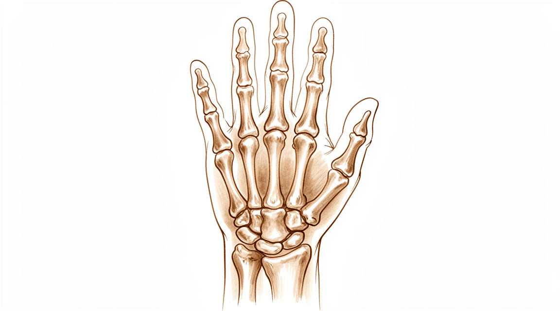
Kienböck’s disease — progressive wrist pain from lunate avascular necrosis; diagnosis and treatment options.
What you're feeling
Kienböck disease is a condition affecting the small bones in your wrist. It is not very common. The problem usually gets worse over time if left untreated. You may notice swelling or stiffness in your wrist. The pain often starts slowly and builds up.
You might feel tenderness when you press on the top of your wrist. This is where the lunate bone sits. The pain can make simple tasks difficult. You may struggle to push yourself up from a chair. Lifting objects can become painful and weak. Even turning a doorknob or opening a jar might hurt.
Your wrist may feel stiff in the morning. This stiffness often eases as you move around during the day. However, pain can return after you have been using your hands for a while. Activities that put weight on your wrist, like doing push-ups or carrying groceries, can trigger flare-ups. You might also notice that your grip strength is weaker than before.
In some cases, the pain can be sharp or dull. It may radiate up your forearm. Nighttime pain is possible, especially if you sleep on that side. This can disrupt your rest and leave you feeling tired. You might find it hard to reach behind your back to fasten a bra or tuck in a shirt. These movements stretch the wrist and can aggravate the inflamed bone.
The severity of your symptoms depends on how the bones are shaped in your wrist. This shape affects how much pressure is placed on the lunate. The cause of the disease is likely due to several factors working together. It is not always clear why one person develops it and another does not.
If you ignore the symptoms, the condition can progress to more advanced stages. This can lead to changes in the shape of your wrist bones. Your surgeon will look at X-rays to see how much the bones have shifted. However, standard X-rays do not always show the full picture of bone collapse. This is why your surgeon may order more detailed scans to get a clear view of what is happening inside your wrist.
What's actually happening
Kienböck’s disease is a condition where the blood supply to a small bone in your wrist, called the lunate, is reduced or lost. Without enough blood, this bone begins to weaken and can eventually collapse. Think of the lunate as a central shock absorber in your wrist. When it loses its structural integrity, it can no longer cushion the impact between your forearm bones and the rest of your hand.
This collapse changes how your wrist moves. Normally, the bones in your wrist slide and roll smoothly against each other. When the lunate is damaged, this motion becomes irregular. The bones may shift out of their normal alignment, a problem known as rotational malalignment. This misalignment puts extra stress on other parts of the wrist joint, particularly where the radius bone meets the scaphoid bone. Over time, this uneven wear can lead to arthritis in those specific areas.
Your surgeon looks at these changes to decide the best path forward. In younger patients, the goal is often to save the bone. Procedures like radial osteotomies, where the forearm bone is adjusted, can improve outcomes and radiographic findings in teenagers. For others, capitate shortening or vascularized bone grafting may be recommended to restore balance and blood flow. These treatments aim to keep your wrist moving as naturally as possible.
In more advanced cases where the bone has collapsed significantly, the focus shifts to stabilizing the joint. Surgeries like scaphocapitate arthrodesis, which fuses two bones together, can provide long-term relief from pain and improve grip strength. While these procedures limit some motion, they create a stable platform that allows you to use your hand effectively. Your surgeon will choose the option that best matches the stage of your disease and your personal needs.
What we can do about it
We start with non-surgical care to manage your symptoms and protect your wrist. This approach focuses on reducing stress on the bones in your hand. Your surgeon may recommend rest or specific activities to avoid. Physiotherapy helps maintain movement and strength in your wrist and hand. These methods aim to keep you comfortable while we monitor the condition.
Research shows that nonsurgical treatments can achieve good and excellent outcomes in younger patients whose bones are still growing. For many, this conservative path is sufficient to manage pain and function. However, Kienböck’s disease is generally a progressive condition. This means it can worsen over time, potentially leading to advanced changes in the wrist joint if left unchecked. We watch for these signs closely during your regular check-ups.
If non-surgical measures do not provide enough relief, we discuss medical management options. Pain medication and anti-inflammatories help control discomfort and swelling. In some cases, we may consider injections to reduce inflammation directly in the joint. These treatments offer temporary relief and help you stay active. They do not reverse the underlying bone changes but can improve your daily quality of life while we decide on the next steps.
Surgery is considered when conservative care reaches its limit or if the disease progresses despite treatment. The goal of surgery is to unload the affected bone, restore blood flow, or stabilize the joint. Options include procedures to change the shape of the radius bone, graft new bone with its own blood supply, or fuse specific bones together to relieve pain. The choice depends on the stage of your disease and your individual needs.
For teenage patients, radial osteotomies (changing the shape of the forearm bone) are effective in improving both symptoms and X-ray findings. These procedures can provide decade-long improvement in 75% of patients. In advanced cases where the wrist has collapsed, fusion procedures like scaphocapitate arthrodesis offer significant pain relief and functional improvement. Your surgeon will explain which option fits your specific situation.
We also use imaging like X-rays and MRI to track changes. While plain X-rays can sometimes miss early signs of collapse, advanced imaging helps us see the true state of your lunate bone. This ensures we choose the right treatment at the right time. Whether you need simple rest or a surgical procedure, our aim is to preserve your wrist function and reduce pain.
What to expect
Kienböck’s disease is a condition where the blood supply to a small wrist bone is reduced, causing it to weaken and collapse. This process is generally progressive, meaning it tends to worsen over time rather than settle on its own. Without treatment, the condition can advance to Stage IV changes, which involve significant wear-and-tear arthritis in the wrist joint. The exact path of the disease is not fully known, but it often leads to persistent pain and stiffness if left unmanaged.
With treatment, your outlook improves significantly. Many surgical options are designed to stop this progression and relieve pain. For example, radial shortening osteotomy—a procedure that adjusts the length of your forearm bone—provides decade-long improvement in 75% of patients with symptomatic disease. This treatment is particularly effective for long-lasting pain relief in patients with negative ulnar variance, a specific wrist bone alignment. Other procedures, such as scaphocapitate arthrodesis (joining two bones together) or proximal row carpectomy (removing a row of small wrist bones), offer durable, long-term benefits. These surgeries typically alleviate pain, preserve functional mobility, and maintain satisfactory grip strength for many years.
Even with treatment, the disease does not always progress rapidly. Radiographic progression of Kienböck disease over 1 year or more is slight on average, regardless of the treatment chosen. This means that while the underlying condition may persist, the visible changes on X-rays often stabilize. You can expect your surgeon to tailor the plan to your specific stage and symptoms. Whether you are a teenager or an adult, the goal is to manage pain and keep your wrist functional. While outcomes are generally positive, it is important to have realistic expectations. The disease is multifactorial, and individual results vary, but modern treatments offer reliable paths to pain relief and improved hand function.
When to see someone
Kienböck’s disease is a rare condition affecting the wrist. It is generally considered a progressive issue that can lead to advanced joint changes. The exact path it takes is not fully known. You should see your GP if you have persistent pain that does not improve with rest. Ask for a specialist review if you notice weakness or instability in your wrist. Seek help if your hand locks or gives way. Contact your surgeon if symptoms interfere with your sleep or work. Sudden worsening of pain is also a reason to seek care. Early evaluation helps manage this complex condition effectively.
Evidence & references
title: "Kienböck's Disease" slug: kienbocks-disease region: wrist audience: patient mesh_terms: ["Osteonecrosis", "Wrist Joint", "Lunate Bone", "Carpal Bones", "Radius", "Scaphoid Bone", "Arthrodesis", "Capitate Bone"] article_count: 547 model_used: Qwen3.6-35B-A3B-Q8_0.gguf generated_at: '2026-06-13T11:24:54+00:00' key_articles: - title: "Radial Osteotomies for Teenage Patients with Kienb??ck Disease" ref_num: 1 evidence_tier: paper evidence_level: 4 doi: 10.1097/01.blo.0000173254.46899.72 year: 2005 - title: "Scaphocapitate arthrodesis for treatment of late stage Kienböck disease" ref_num: 2 evidence_tier: paper evidence_level: 4 doi: 10.1177/1753193413496177 year: 2013 - title: "Long-Term Results of Vascularized Bone Graft for Stage III Kienböck Disease" ref_num: 3 evidence_tier: paper evidence_level: 3 doi: 10.1016/j.jhsa.2013.02.010 year: 2013 - title: "Osteonecrosis of the Lunate: Kienböck Disease" ref_num: 4 evidence_tier: paper evidence_level: 5 doi: 10.5435/jaaos-d-20-00020 year: 2020 - title: "Kienböck Disease in the Skeletally Immature Patient" ref_num: 5 evidence_tier: paper evidence_level: 4 doi: 10.1016/j.jhsa.2018.02.029 year: 2018 - title: "Kienböck's Disease: An Approach to Treatment" ref_num: 6 evidence_tier: paper evidence_level: 5 doi: 10.1016/j.hcl.2006.07.003 year: 2006 - title: "Temporary Scaphotrapezoidal Joint Fixation for Adolescent Kienböck's Disease" ref_num: 7 evidence_tier: paper evidence_level: 4 doi: 10.1016/j.jhsa.2008.09.019 year: 2009 - title: "Long-term outcome (20 to 33 years) of radial shortening osteotomy for Kienböck’s lunatomalacia" ref_num: 8 evidence_tier: paper evidence_level: 4 doi: 10.1177/1753193413512222 year: 2013 - title: "The Effect of Lunate Morphology in Kienböck Disease" ref_num: 9 evidence_tier: paper evidence_level: 3 doi: 10.1016/j.jhsa.2014.12.024 year: 2015 - title: "Letter Regarding “Kienböck Disease and Carpal Coalitions: A Potential Correlation”" ref_num: 10 evidence_tier: letter evidence_level: 4 doi: 10.1016/j.jhsa.2016.11.010 year: 2017 - title: "Clinical and Radiological Outcomes of Scaphocapitate Fusion in Kienböck Disease: A Systematic Review and Meta-Analysis" ref_num: 13 evidence_tier: paper evidence_level: 4 doi: 10.1016/j.jhsg.2023.03.014 year: 2023 - title: "Comparing the Radiologic and Functional Outcome of Radial Shortening Versus Capitate Shortening in Management of Kienböck’s Disease" ref_num: 14 evidence_tier: paper evidence_level: 2 doi: 10.1177/15589447221081564 year: 2022 - title: "Diagnostic performance of traditional radiographic indices in detection of carpal collapse in Kienböck’s disease" ref_num: 15 evidence_tier: paper evidence_level: 3 doi: 10.1177/17531934231153966 year: 2023 - title: "Outcome of Kienböck's Disease 22 Years after Distal Radius Shortening Osteotomy" ref_num: 16 evidence_tier: paper evidence_level: 3 doi: 10.1097/blo.0b013e318041d309 year: 2007 - title: "Kienböck's Disease" ref_num: 18 evidence_tier: paper evidence_level: 5 doi: 10.1016/j.jhsa.2009.10.013 year: 2009 - title: "Risk Factors of Lunate Collapse in Kienböck Disease" ref_num: 19 evidence_tier: paper evidence_level: 2 doi: 10.1016/j.jhsa.2017.06.107 year: 2017 - title: "Arthroscopic Treatment of Kienböck Disease: Mid-Term Outcome of Arthroscopic Lunate Core Decompression" ref_num: 20 evidence_tier: paper evidence_level: 4 doi: 10.1016/j.jhsa.2023.02.011 year: 2024 - title: "Scaphotrapeziotrapezoid Arthrodesis and Lunate Excision for Advanced Kienböck Disease" ref_num: 22 evidence_tier: paper evidence_level: 4 doi: 10.1016/j.jhsa.2012.08.031 year: 2012 - title: "Long-Term Clinical Outcome After Titanium Lunate Arthroplasty for Kienböck Disease" ref_num: 23 evidence_tier: paper evidence_level: 4 doi: 10.1016/j.jhsa.2018.02.009 year: 2018 - title: "Limited Intercarpal Fusion as a Salvage Procedure for Advanced Kienbock Disease" ref_num: 24 evidence_tier: paper evidence_level: 4 doi: 10.1007/s11552-014-9705-z year: 2014 - title: "Lunate excision, capitate osteotomy, and intercarpal arthrodesis should be used with caution for advanced Kienböck’s disease" ref_num: 25 evidence_tier: paper evidence_level: 5 doi: 10.1177/1753193418807360 year: 2018 - title: "Radiographic Progression of Kienböck Disease: Radial Shortening Versus No Surgery" ref_num: 26 evidence_tier: paper evidence_level: 4 doi: 10.1016/j.jhsa.2016.02.016 year: 2016 - title: "Tendon ball arthroplasty and proximal carpal stabilization with tendon graft for advanced Kienböck’s disease" ref_num: 28 evidence_tier: paper evidence_level: 4 doi: 10.1177/17531934241238939 year: 2024 - title: "Surgical Treatments for Scapholunate Advanced Collapse Wrist: Kinematics and Functional Performance" ref_num: 30 evidence_tier: paper evidence_level: 2 doi: 10.1016/j.jhsa.2015.04.035 year: 2015 - title: "The Effect of Lunate Morphology on the 3-Dimensional Kinematics of the Carpus" ref_num: 31 evidence_tier: paper evidence_level: 5 doi: 10.1016/j.jhsa.2014.09.019 year: 2015 - title: "Comparison of the Clinical and Functional Outcomes Following 3- and 4-Corner Fusions" ref_num: 32 evidence_tier: paper evidence_level: 3 doi: 10.1016/j.jhsa.2015.02.027 year: 2015 - title: "Elongation of the Dorsal Carpal Ligaments: A Computational Study of In Vivo Carpal Kinematics" ref_num: 33 evidence_tier: paper evidence_level: 5 doi: 10.1016/j.jhsa.2012.04.025 year: 2012 - title: "Dynamic wrist imaging: How it works and how to assess kinematic changes in wrists with scapholunate instability" ref_num: 34 evidence_tier: paper evidence_level: 5 doi: 10.1177/17531934251326028 year: 2025 - title: "Force in the Scapholunate Interosseous Ligament During Active Wrist Motion" ref_num: 35 evidence_tier: paper evidence_level: 5 doi: 10.1016/j.jhsa.2015.04.007 year: 2015 - title: "Radiocarpal and midcarpal kinematics in scapholunate instability: a four-dimensional CT study in vivo" ref_num: 36 evidence_tier: paper evidence_level: 3 doi: 10.1177/17531934241242676 year: 2024 - title: "The Effect of Rotational Malalignment on X-rays of the Wrist" ref_num: 37 evidence_tier: paper evidence_level: 4 doi: 10.1177/1753193408090393 year: 2009 - title: "Results of scaphocapitate arthrodesis with lunate excision in advanced Kienböck disease at 10.7-year mean follow-up" ref_num: 38 evidence_tier: paper evidence_level: 4 doi: 10.1177/1753193417739247 year: 2017 - title: "Vascularized Bone Grafts for the Treatment of Carpal Bone Pathology" ref_num: 40 evidence_tier: paper evidence_level: 4 doi: 10.1007/s11552-012-9479-0 year: 2013 - title: "Publication Bias in Kienböck's Disease: Systematic Review" ref_num: 41 evidence_tier: paper evidence_level: 2 doi: 10.1016/j.jhsa.2009.12.003 year: 2010 - title: "Interfragmentary Motion in Patients With Scaphoid Nonunion" ref_num: 42 evidence_tier: paper evidence_level: 4 doi: 10.1016/j.jhsa.2008.03.008 year: 2008 - title: "Fixation of the Fractured Lunate in Kienböck Disease" ref_num: 43 evidence_tier: paper evidence_level: 4 doi: 10.1016/j.jhsa.2018.05.008 year: 2019 - title: "Radiographic Evaluation of Carpal Mechanics and the Scapholunate Angle in a Clenched Fist with Dynamic Computed Tomography Imaging" ref_num: 44 evidence_tier: paper evidence_level: 4 doi: 10.1016/j.jhsg.2022.10.001 year: 2023 - title: "Three-dimensional carpal alignment: computer-aided CT analysis of carpal axes and normal ranges" ref_num: 45 evidence_tier: paper evidence_level: 4 doi: 10.1177/17531934231160100 year: 2023 - title: "Minimum Twenty-year Follow-up of Proximal Row Carpectomy" ref_num: 46 evidence_tier: paper evidence_level: 4 doi: 10.1016/s0363-5023(10)60087-1 year: 2010 - title: "In Vivo Kinematics of the Scaphoid, Lunate, Capitate, and Third Metacarpal in Extreme Wrist Flexion and Extension" ref_num: 47 evidence_tier: paper evidence_level: 4 doi: 10.1016/j.jhsa.2012.10.035 year: 2013 - title: "Scaphocapitate Arthrodesis for Kienböck Disease" ref_num: 48 evidence_tier: paper evidence_level: 4 doi: 10.1016/j.jhsa.2014.12.013 year: 2015 - title: "Proximal Row Carpectomy for Advanced Kienböck's Disease: Average 10-Year Follow-Up" ref_num: 49 evidence_tier: paper evidence_level: 4 doi: 10.1016/j.jhsa.2008.02.031 year: 2008 - title: "In Vivo 3-Dimensional Analysis of Stage III Kienböck Disease: Pattern of Carpal Deformity and Radioscaphoid Joint Congruity" ref_num: 50 evidence_tier: paper evidence_level: 2 doi: 10.1016/j.jhsa.2014.10.035 year: 2015 - title: "Contact areas of the scaphoid and lunate with the distal radius in neutral and extension: correlation of falling strategies and distal radial anatomy" ref_num: 52 evidence_tier: paper evidence_level: 5 doi: 10.1177/1753193413507810 year: 2013 - title: "Treatment of Scapholunate Dissociation With a Bioresorbable Polymer Plate: A Biomechanical Study" ref_num: 53 evidence_tier: paper evidence_level: 5 doi: 10.1016/j.jhsa.2008.01.016 year: 2008 - title: "Electrogoniometric and radiologic evaluation of scapho-trapezo-trapezoid arthrodesis" ref_num: 54 evidence_tier: paper evidence_level: 4 doi: 10.1016/s0749-0712(03)00008-8 year: 2003 - title: "Factors associated with improved outcomes following proximal row carpectomy: a long-term outcome study of 144 patients" ref_num: 55 evidence_tier: paper evidence_level: 3 doi: 10.1177/1753193415597096 year: 2015 - title: "Radiographic Evaluation of the Modified Brunelli Technique Versus the Blatt Capsulodesis for Scapholunate Dissociation in a Cadaver Model" ref_num: 56 evidence_tier: paper evidence_level: 5 doi: 10.1016/j.jhsa.2010.06.029 year: 2010 - title: "Three-Dimensional In Vivo Kinematic Analysis of Kienböck Disease Treated with Arthroscopic Lunate Excision" ref_num: 57 evidence_tier: paper evidence_level: 4 doi: 10.1016/j.jhsa.2025.01.024 year: 2025 - title: "Trabecular microstructure of the human lunate in Kienböck’s disease" ref_num: 58 evidence_tier: paper evidence_level: 4 doi: 10.1177/1753193411422337 year: 2011 - title: "Preliminary Clinical, Radiographic, and Patient-Reported Outcomes of the Medial Femoral Trochlea Osteochondral Free Flap for Lunate Reconstruction in Advanced Kienböck Disease" ref_num: 59 evidence_tier: paper evidence_level: 4 doi: 10.1016/j.jhsa.2019.12.008 year: 2020 - title: "Radius Core Decompression for Kienböck Disease Stage IIIA: Outcomes at 13 Years Follow-Up" ref_num: 61 evidence_tier: paper evidence_level: 4 doi: 10.1016/j.jhsa.2017.05.017 year: 2017 synthesis_version: "v2" verifier_status: skipped
Overview
- Radial osteotomies are effective in improving short-term clinical outcomes and radiographic findings in teenage patients with Kienböck disease [1].
- Scaphocapitate arthrodesis demonstrates long-term clinical benefits for the treatment of collapsed Kienböck disease [2].
- Vascularized bone grafting for stage III Kienböck disease demonstrates favorable long-term results and is recommended as a surgical treatment [3].
- Good and excellent clinical and radiological outcomes can be achieved with both nonsurgical and surgical treatments in skeletally immature patients with Kienböck disease [5].
- Temporary scaphotrapezoidal joint fixation is recommended for the surgical treatment of adolescent Kienböck's disease [7].
- Radial shortening osteotomy provides decade-long improvement in 75% of patients and is a reasonable treatment for symptomatic Kienböck’s disease [8].
- Scaphocapitate arthrodesis is an effective procedure for the treatment of Kienböck disease, associated with satisfactory functional outcomes and significant improvement in pain scores and grip strength [13].
- Capitate shortening is a safe and effective approach for the treatment of early stages of Kienböck's disease and can be associated with a satisfying outcome [14].
- Advanced Kienböck's disease with carpal collapse is not a contraindication for carpal-sparing surgery radial shortening osteotomy [17].
- Lunate excision, capitate osteotomy, and intercarpal arthrodesis should be used with caution for advanced Kienböck's disease because it does not have good long-term results and is no longer widely used in Europe [25].
- The acceptance rate for negative outcomes studies regarding Kienböck's disease is higher than for other surgical disorders, indicating a relative decrease in positive outcome bias among published Kienböck's disease studies compared with other surgical disorders [41].
Anatomy & Pathophysiology
- Tendon ball arthroplasty and proximal carpal stabilization with tendon graft restore the integrity of the proximal carpal row in advanced Kienböck’s disease [28].
- Surgical treatments for scapholunate advanced collapse result in decreased wrist kinematic motion and functional performance compared with individuals with normal wrists [30].
- Lunate morphology affects the 3-dimensional kinematics of the carpus during wrist flexion and extension [31].
- Three- and 4-corner fusions produce motion that is smoother and more closely replicates the normal axis and functional motion of the wrist [32].
- Computed fiber elongations of the dorsal carpal ligaments vary linearly with wrist position [33].
- Four-dimensional computed tomography (4DCT) is a non-invasive and affordable method to assess and quantify wrist kinematics by incorporating the temporal dimension [34].
- During simple unresisted wrist motions, force in the scapholunate interosseous ligament does not exceed 20 N [35].
- Kinematic changes in scapholunate instability may predict the development of radioscaphoid arthritis and help identify a kinematically abnormal wrist [36].
- Rotational malalignment of the wrist has significant effects on carpal, distal radial, and distal radioulnar joint measurements [37].
- Scaphoid nonunions have a dramatic impact on carpal kinematics by partially uncoupling the proximal and distal carpal rows [42].
- Dynamic imaging may enable the derivation of a standardized protocol for mapping carpal motion that is clinically applicable and reproducible [44].
- Computer-aided CT analysis provides guidelines for measuring and quantifying carpal alignment three-dimensionally and establishes a database for normal values [45].
- More than half the motion of the carpus when the wrist was loaded in extension occurred at the midcarpal joint [47].
- Radioscapholunate fusion shows the most biomechanically similar behavior to the healthy wrist among three fusion types [51].
- Contact areas between the scaphoid and distal radius are maximized during full extension of the wrist, which helps stabilize the radiocarpal joint [52].
- A dorsally applied PLA plate restores carpal kinematics for 1,000 cycles of motion in unstable wrists where fixation is not compromised by carpal size or osteoporosis [53].
- The modification of the wrist center of rotation during flexion and extension indicates that stability is considered more important than mobility in clinical conditions [54].
- Correction of scapholunate dissociation may correlate with improved carpal dynamics and improved clinical outcomes [56].
- Postarthroscopic lunate excision alters normal carpal kinematics but maintains joint congruity in the short-term [57].
Classification
- Kienböck disease is generally considered a progressive condition that can end in Stage IV changes [6].
- Lunate morphology may affect the severity of Kienböck disease at the time of initial presentation [9].
- The patterns of carpal collapse differ between stage IIIb Kienböck disease and scapholunate dislocation in terms of radioscaphoid joint congruity [50].
- Traditional radiographic indices measured on plain radiographs have poor diagnostic performance in the detection of carpal collapse in Kienböck's disease [15].
Clinical Presentation
- Kienböck disease is a relatively infrequent carpal pathology [4].
- Kienböck disease is generally considered a progressive condition that can end in Stage IV changes [6].
- The natural history of Kienböck disease is not fully known [6].
- Lunate morphology may affect the severity of Kienböck disease at the time of initial presentation [9].
- The development of Kienböck disease is probably multifactorial [10].
- Traditional radiographic indices measured on plain radiographs have poor diagnostic performance in the detection of carpal collapse in Kienböck's disease [15].
Investigations
- Radial osteotomies are effective in improving short-term clinical outcomes and radiographic findings in teenage patients with Kienböck disease [1].
- Scaphocapitate arthrodesis demonstrates long-term clinical benefits for the treatment of collapsed Kienböck disease [2].
- Vascularized bone grafting for stage III Kienböck disease demonstrates favorable long-term results and is recommended as a surgical treatment [3].
- Good and excellent clinical and radiological outcomes can be achieved with both nonsurgical and surgical treatments in skeletally immature patients with Kienböck disease [5].
- Temporary scaphotrapezoidal joint fixation is recommended for the surgical treatment of adolescent Kienböck's disease [7].
- Radial shortening osteotomy provides decade-long improvement in 75% of patients and is a reasonable treatment for symptomatic Kienböck’s disease [8].
- Lunate morphology may affect the severity of Kienböck disease at the time of initial presentation [9].
- Traditional radiographic indices measured on plain radiographs have poor diagnostic performance in the detection of carpal collapse in Kienböck's disease [15].
- The Camembert osteotomy improved function in patients with early stage Kienböck disease, with MRI aspects improving in most cases and no patients experiencing lunate collapse [21].
- Radiographic progression of Kienböck over 1 year or more seems slight on average regardless of treatment [26].
- Computed tomography of the lunate in Kienböck disease is an important investigative tool [43].
- Lunates with advanced Kienböck's disease exhibit significantly denser, thicker, and more plate-like trabecular microstructure compared to normal lunates [58].
- Following medial femoral trochlea reconstruction of the proximal lunate for advanced Kienböck disease, cessation of radiocarpal collapse was observed [59].
Treatment
Non-Operative and General Considerations
- Good and excellent clinical and radiological outcomes can be achieved with both nonsurgical and surgical treatments in skeletally immature patients with Kienböck disease [5].
- The natural history of Kienböck's disease is generally considered a progressive condition that can end in Stage IV changes [6].
- Treatment strategies for Kienböck's disease focus on biomechanical unloading, vascularized bone grafts, or salvage procedures depending on the stage [6].
- There is limited, low-quality evidence that surgical treatment slows progression of Kienböck's disease [18].
- Many uncontrolled case series document slight improvement in motion and grip after surgical treatment without clear evidence that this is better than placebo or no intervention [18].
Radial Osteotomies and Shortening
- Radial osteotomies are effective in improving not only short-term clinical outcomes, but also radiographic findings in teenage patients with Kienböck disease [1].
- Radial shortening osteotomy provides decade-long improvement in 75% of patients and seems to be a reasonable treatment for symptomatic Kienböck’s disease [8].
- The medium- and long-term results of radial shortening osteotomy for Kienböck's disease in patients with negative ulnar variance are comparable to short-term results, providing long-lasting pain relief [16].
- Advanced Kienbock's disease with carpal collapse is not a contraindication for carpal-sparing surgery radial shortening osteotomy [17].
- Radius core decompression demonstrated favorable long-term results and could be considered as a surgical alternative for stage IIIA of Kienböck disease [61].
Vascularized Bone Grafts
- Vascularized bone grafting for stage III Kienböck disease demonstrated favorable long-term results and is recommended as a surgical treatment [3].
- Vascularized bone grafts are indicated for Kienböck's disease, while being contraindicated in advanced carpal collapse with degenerative changes [40].
Arthrodesis and Salvage Procedures
- The long-term clinical benefits of scaphocapitate arthrodesis for treatment of collapsed Kienböck disease are demonstrated [2].
- Scaphocapitate arthrodesis is an effective procedure for treatment of Kienböck disease associated with satisfactory functional outcomes and significant improvement in pain scores and grip strength [13].
- Scaphocapitate arthrodesis should be considered as a treatment option for wrist salvage in the patient with advanced Kienbock's disease given the significant postoperative reduction in associated pain symptoms at the time of follow-up [24].
- Scaphotrapeziotrapezoid arthrodesis with lunate excision for advanced Kienböck disease provided favorable clinical results in terms of pain relief and functional improvement [22].
- The procedure of lunate excision, capitate osteotomy, and intercarpal arthrodesis should be used with caution for advanced Kienböck's disease because it does not have good long-term results and is no longer widely used in Europe [25].
- Improved outcomes following proximal row carpectomy were associated with Kienbock's disease [55].
Capitate Shortening
- Capitate shortening is a safe and effective approach for treatment of the early stages of Kienböck's disease and can be associated with a satisfying outcome [14].
Arthroscopic Procedures
- Arthroscopic lunate core decompression appears to be an effective and safe surgery for treating Kienböck disease on the basis of mid-term follow-up [20].
Adolescent-Specific Interventions
- Temporary scaphotrapezoidal joint fixation is recommended for the surgical treatment of adolescent Kienböck's disease [7].
Complications
- Kienböck's disease is generally considered a progressive condition that can end in Stage IV changes [6].
- The natural history of Kienböck's disease is not fully known [6].
- Lunate morphology may affect the severity of Kienböck disease at the time of initial presentation [9].
- Negative ulnar variance has a longitudinal relationship with progressive Kienböck disease, though additional long-term study is needed to confirm this [19].
- The development of Kienböck disease is probably multifactorial [10].
- The observation of Kienböck disease and carpal coalition in one wrist is fortuitous [10].
Recovery
- Radial osteotomies improve short-term clinical outcomes in teenage patients with Kienböck disease [1].
- Radial osteotomies improve radiographic findings in teenage patients with Kienböck disease [1].
- Scaphocapitate arthrodesis provides long-term clinical benefits for collapsed Kienböck disease [2].
- Vascularized bone grafting demonstrates favorable long-term results for stage III Kienböck disease [3].
- Radial shortening osteotomy provides decade-long improvement in 75% of patients with symptomatic Kienböck’s disease [8].
- Radial shortening osteotomy provides long-lasting pain relief for patients with negative ulnar variance [16].
- Titanium lunate arthroplasty (TLA) shows promising longer-term results for stage III Kienböck disease [23].
- Scaphocapitate arthrodesis with lunate excision significantly alleviates pain in advanced Kienböck disease at a mean follow-up of 10.7 years [38].
- Scaphocapitate arthrodesis with lunate excision preserves functional mobility in advanced Kienböck disease at a mean follow-up of 10.7 years [38].
- Scaphocapitate arthrodesis with lunate excision maintains satisfactory grip strength in advanced Kienböck disease at a mean follow-up of 10.7 years [38].
- Proximal row carpectomy (PRC) is a durable long-term treatment option for radiocarpal degenerative arthritis and Kienböck's disease [46].
- Proximal row carpectomy allows for maintenance of wrist range of motion [46].
- Proximal row carpectomy improves grip strength [46].
- Proximal row carpectomy is a reliable and durable procedure for Lichtman stage IIIA or IIIB Kienböck's disease at an average follow-up of 10 years [49].
- Scaphocapitate arthrodesis (SCA) results in improved grip strength in patients with advanced stages of Kienböck disease in medium-term follow-up [48].
- Scaphocapitate arthrodesis (SCA) corrects carpal alignment in patients with advanced stages of Kienböck disease in medium-term follow-up [48].
- Radiographic progression of Kienböck disease over 1 year or more is slight on average regardless of treatment [26].
Key Evidence
- [L4] The current results indicate that radial osteotomies are effective in improving not only short-term clinical outcomes, but also radiographic findings in teenage patients with Kienböck disease. (10.1097/01.blo.0000173254.46899.72)
- [L4] The long-term clinical benefits of scaphocapitate arthrodesis for treatment of collapsed Kienböck disease are demonstrated. (10.1177/1753193413496177)
- [L3] Vascularized bone grafting for stage III Kienböck disease demonstrated favorable long-term results and is recommended as a surgical treatment. (10.1016/j.jhsa.2013.02.010)
- [L5] This review article discusses the history, etiology, and course of Kienböck disease and reviews the literature on both the diagnosis and management of this relatively infrequent carpal pathology. (10.5435/jaaos-d-20-00020)
- [L4] Good and excellent clinical and radiological outcomes can be achieved with both nonsurgical and surgical treatments in skeletally immature patients with Kienböck disease. (10.1016/j.jhsa.2018.02.029)
- [L5] The natural history of Kienbock's disease is not fully known, though it is generally considered a progressive condition that can end in Stage IV changes; treatment strategies focus on biomechanical unloading, vascularized bone grafts, or salvage procedures depending on the stage. (10.1016/j.hcl.2006.07.003)
- [L4] We therefore recommend this procedure for the surgical treatment of adolescent Kienböck's disease. (10.1016/j.jhsa.2008.09.019)
- [L4] Radial shortening osteotomy provides decade-long improvement in 75% of patients and seems to be a reasonable treatment for symptomatic Kienböck’s disease. (10.1177/1753193413512222)
- [L3] Lunate morphology may affect the severity of Kienböck disease at the time of initial presentation. (10.1016/j.jhsa.2014.12.024)
- [Letter] The observation of Kienböck disease and carpal coalition in one wrist is fortuitous, and the development of Kienböck disease is probably multifactorial. (10.1016/j.jhsa.2016.11.010)
- [L4] Scaphocapitate arthrodesis is an effective procedure for treatment of Kienböck disease associated with satisfactory functional outcomes and significant improvement in pain scores and grip strength. (10.1016/j.jhsg.2023.03.014)
- [L2] Capitate shortening is a safe and effective approach for treatment of the early stages of Kienböck's disease and can be associated with a satisfying outcome. (10.1177/15589447221081564)
- [L3] Traditional radiographic indices measured on plain radiographs have poor diagnostic performance in the detection of carpal collapse in Kienböck's disease. (10.1177/17531934231153966)
- [L3] The medium- and long-term results of radial shortening osteotomy for Kienböck's disease in patients with negative ulnar variance are comparable to short-term results, providing long-lasting pain relief. (10.1097/blo.0b013e318041d309)
- [L5] There is limited, low-quality evidence that surgical treatment slows progression of Kienböck's disease, and many uncontrolled case series document slight improvement in motion and grip after surgical treatment without clear evidence that this is better than placebo or no intervention. (10.1016/j.jhsa.2009.10.013)
- [L2] Additional long-term study is needed to confirm the longitudinal relationship of negative ulnar variance with progressive Kienböck disease. (10.1016/j.jhsa.2017.06.107)
- [L4] Arthroscopic lunate core decompression appears to be an effective and safe surgery for treating Kienböck disease on the basis of mid-term follow-up. (10.1016/j.jhsa.2023.02.011)
- [L4] Scaphotrapeziotrapezoid arthrodesis with lunate excision for advanced Kienböck disease provided favorable clinical results in terms of pain relief and functional improvement. (10.1016/j.jhsa.2012.08.031)
- [L4] The longer-term results of TLA for stage III Kienböck disease are promising. (10.1016/j.jhsa.2018.02.009)
- [L4] Given the significant postoperative reduction in associated pain symptoms at the time of follow-up, scaphocapitate arthrodesis should be considered as a treatment option for wrist salvage in the patient with advanced Kienbock's disease. (10.1007/s11552-014-9705-z)
- [L5] The procedure of lunate excision, capitate osteotomy, and intercarpal arthrodesis should be used with caution for advanced Kienböck's disease because it does not have good long-term results and is no longer widely used in Europe. (10.1177/1753193418807360)
- [L4] Radiographic progression of Kienböck over 1 year or more seems slight on average regardless of treatment. (10.1016/j.jhsa.2016.02.016)
- [L4] The technique demonstrated reduced wrist pain and improved wrist motion and grip strength while restoring the integrity of the proximal carpal row. (10.1177/17531934241238939)
- [L2] Both surgical groups demonstrated decreased wrist kinematic motion and functional performance compared with individuals with normal wrists. (10.1016/j.jhsa.2015.04.035)
- [L5] This study describes the effect of lunate morphology on 3-dimensional carpal kinematics during wrist flexion and extension. (10.1016/j.jhsa.2014.09.019)
- [L3] Motion was smoother and more closely replicated the normal axis and functional motion of the wrist. (10.1016/j.jhsa.2015.02.027)
- [L5] Despite complex carpal bone anatomy and kinematics, computed fiber elongations were found to vary linearly with wrist position. (10.1016/j.jhsa.2012.04.025)
- [L5] Four-dimensional computed tomography (4DCT) is a promising, non-invasive, and affordable method to assess and quantify wrist kinematics, extending conventional CT by incorporating the temporal dimension. (10.1177/17531934251326028)
- [L5] However, during simple unresisted wrist motions, the force did not exceed 20 N. (10.1016/j.jhsa.2015.04.007)
- [L3] These kinematic changes may predict the development of radioscaphoid arthritis and help identify a kinematically abnormal wrist. (10.1177/17531934241242676)
- [L4] Rotational malalignment of the wrist has significant effects on carpal, distal radial and distal radioulnar joint measurements. (10.1177/1753193408090393)
- [L4] Scaphocapitate arthrodesis with lunate excision performed in an advanced stage of Kienböck disease significantly alleviates pain, while preserving functional mobility and satisfactory grip strength in the long term. (10.1177/1753193417739247)
- [L4] This manuscript offers a current review of the techniques and outcomes of VBGs to the carpal bones, noting that VBGs are indicated for scaphoid nonunion with proximal pole AVN, Kienböck's disease, Preiser's disease, and capitate osteonecrosis, while being contraindicated in advanced carpal collapse with degenerative changes. (10.1007/s11552-012-9479-0)
- [L2] The acceptance rate for negative outcomes studies regarding Kienböck's disease is higher than for other surgical disorders, indicating a relative decrease in positive outcome bias among published Kienböck's disease studies compared with other surgical disorders. (10.1016/j.jhsa.2009.12.003)
- [L4] Scaphoid nonunions have a dramatic impact on carpal kinematics, partially uncoupling the proximal and distal carpal rows. (10.1016/j.jhsa.2008.03.008)
- [L4] Computed tomography of the lunate in Kienböck disease is an important investigative tool. (10.1016/j.jhsa.2018.05.008)
- [L4] With the increased focus on dynamic imaging for wrist motion, it may be possible to derive a standardized protocol for mapping the carpal motion that is clinically applicable and reproducible. (10.1016/j.jhsg.2022.10.001)
- [L4] This study provides guidelines on how to measure and quantify carpal alignment three-dimensionally and establishes a database for normal values, which may be useful when analysing various wrist pathologies and kinematics. (10.1177/17531934231160100)
- [L4] PRC is a durable long-term treatment option for radiocarpal degenerative arthritis and Kienböck's disease allowing for maintenance of wrist range of motion and improvement in grip strength. (10.1016/s0363-5023(10)60087-1)
- [L4] More than half the motion of the carpus when the wrist was loaded in extension occurred at the midcarpal joint. (10.1016/j.jhsa.2012.10.035)
- [L4] SCA resulted in improved grip strength with correction of carpal alignment in patients with advanced stages of Kienböck disease in medium-term follow-up. (10.1016/j.jhsa.2014.12.013)
- [L4] At an average follow-up of 10 years, proximal row carpectomy is a reliable and durable procedure for patients with Lichtman stage IIIA or IIIB Kienböck's disease. (10.1016/j.jhsa.2008.02.031)
- [L2] The patterns of carpal collapse differed between stage IIIb Kienböck disease and scapholunate dislocation in terms of radioscaphoid joint congruity. (10.1016/j.jhsa.2014.10.035)
- [L5] The contact areas between the scaphoid and distal radius are maximized during full extension of the wrist, which helps stabilize the radiocarpal joint and potentially reduces the risk of injury to the carpus and the distal radius. (10.1177/1753193413507810)
- [L5] The study shows that in the unstable wrist, following ligament sectioning, where fixation is not compromised by carpal size or osteoporosis, a dorsally applied PLA plate does restore carpal kinematics for 1,000 cycles of motion. (10.1016/j.jhsa.2008.01.016)
- [L4] The study also characterized the modification of the wrist CoR during flexion and extension, noting that stability is considered more important than mobility in clinical conditions. (10.1016/s0749-0712(03)00008-8)
- [L3] Improved outcomes were associated with age over 40, Kienbock's disease, concomitant neurectomy, non-labourer status, and surgery after 1990, while radiocapitate arthrosis did not correlate with clinical outcomes. (10.1177/1753193415597096)
- [L5] This correction might correlate with improved carpal dynamics and improved clinical outcomes. (10.1016/j.jhsa.2010.06.029)
- [L4] Although postarthroscopic lunate excision alters normal carpal kinematics, the joint congruity is maintained in the short-term. (10.1016/j.jhsa.2025.01.024)
- [L4] Lunates with advanced Kienböck's disease exhibit significantly denser, thicker, and more plate-like trabecular microstructure compared to normal lunates. (10.1177/1753193411422337)
- [L4] Following medial femoral trochlea reconstruction of the proximal lunate for advanced Kienböck disease, we observed a cessation of radiocarpal collapse. (10.1016/j.jhsa.2019.12.008)
- [L4] In this limited series, the radius core decompression demonstrated favorable long-term results and could be considered as a surgical alternative for stage IIIA of Kienböck disease. (10.1016/j.jhsa.2017.05.017)
References
[1] Radial Osteotomies for Teenage Patients with Kienb??ck Disease. Clinical Orthopaedics and Related Research. 2005. DOI: 10.1097/01.blo.0000173254.46899.72 [2] Scaphocapitate arthrodesis for treatment of late stage Kienböck disease. Journal of Hand Surgery (European Volume). 2013. DOI: 10.1177/1753193413496177 [3] Long-Term Results of Vascularized Bone Graft for Stage III Kienböck Disease. The Journal of Hand Surgery. 2013. DOI: 10.1016/j.jhsa.2013.02.010 [4] Osteonecrosis of the Lunate: Kienböck Disease. Journal of the American Academy of Orthopaedic Surgeons. 2020. DOI: 10.5435/jaaos-d-20-00020 [5] Kienböck Disease in the Skeletally Immature Patient. The Journal of Hand Surgery. 2018. DOI: 10.1016/j.jhsa.2018.02.029 [6] Kienböck's Disease: An Approach to Treatment. Hand Clinics. 2006. DOI: 10.1016/j.hcl.2006.07.003 [7] Temporary Scaphotrapezoidal Joint Fixation for Adolescent Kienböck's Disease. The Journal of Hand Surgery. 2009. DOI: 10.1016/j.jhsa.2008.09.019 [8] Long-term outcome (20 to 33 years) of radial shortening osteotomy for Kienböck’s lunatomalacia. Journal of Hand Surgery (European Volume). 2013. DOI: 10.1177/1753193413512222 [9] The Effect of Lunate Morphology in Kienböck Disease. The Journal of Hand Surgery. 2015. DOI: 10.1016/j.jhsa.2014.12.024 [10] Letter Regarding “Kienböck Disease and Carpal Coalitions: A Potential Correlation”. The Journal of Hand Surgery. 2017. DOI: 10.1016/j.jhsa.2016.11.010 [13] Clinical and Radiological Outcomes of Scaphocapitate Fusion in Kienböck Disease: A Systematic Review and Meta-Analysis. Journal of Hand Surgery Global Online. 2023. DOI: 10.1016/j.jhsg.2023.03.014 [14] Comparing the Radiologic and Functional Outcome of Radial Shortening Versus Capitate Shortening in Management of Kienböck’s Disease. HAND. 2022. DOI: 10.1177/15589447221081564 [15] Diagnostic performance of traditional radiographic indices in detection of carpal collapse in Kienböck’s disease. Journal of Hand Surgery (European Volume). 2023. DOI: 10.1177/17531934231153966 [16] Outcome of Kienböck's Disease 22 Years after Distal Radius Shortening Osteotomy. Clinical Orthopaedics & Related Research. 2007. DOI: 10.1097/blo.0b013e318041d309 [17] 10.1055-s-0039-1688947. n.d.. [18] Kienböck's Disease. The Journal of Hand Surgery. 2009. DOI: 10.1016/j.jhsa.2009.10.013 [19] Risk Factors of Lunate Collapse in Kienböck Disease. The Journal of Hand Surgery. 2017. DOI: 10.1016/j.jhsa.2017.06.107 [20] Arthroscopic Treatment of Kienböck Disease: Mid-Term Outcome of Arthroscopic Lunate Core Decompression. The Journal of Hand Surgery. 2024. DOI: 10.1016/j.jhsa.2023.02.011 [21] 10.1055-s-0039-1683931. n.d.. [22] Scaphotrapeziotrapezoid Arthrodesis and Lunate Excision for Advanced Kienböck Disease. The Journal of Hand Surgery. 2012. DOI: 10.1016/j.jhsa.2012.08.031 [23] Long-Term Clinical Outcome After Titanium Lunate Arthroplasty for Kienböck Disease. The Journal of Hand Surgery. 2018. DOI: 10.1016/j.jhsa.2018.02.009 [24] Limited Intercarpal Fusion as a Salvage Procedure for Advanced Kienbock Disease. HAND. 2014. DOI: 10.1007/s11552-014-9705-z [25] Lunate excision, capitate osteotomy, and intercarpal arthrodesis should be used with caution for advanced Kienböck’s disease. Journal of Hand Surgery (European Volume). 2018. DOI: 10.1177/1753193418807360 [26] Radiographic Progression of Kienböck Disease: Radial Shortening Versus No Surgery. The Journal of Hand Surgery. 2016. DOI: 10.1016/j.jhsa.2016.02.016 [28] Tendon ball arthroplasty and proximal carpal stabilization with tendon graft for advanced Kienböck’s disease. Journal of Hand Surgery (European Volume). 2024. DOI: 10.1177/17531934241238939 [30] Surgical Treatments for Scapholunate Advanced Collapse Wrist: Kinematics and Functional Performance. The Journal of Hand Surgery. 2015. DOI: 10.1016/j.jhsa.2015.04.035 [31] The Effect of Lunate Morphology on the 3-Dimensional Kinematics of the Carpus. The Journal of Hand Surgery. 2015. DOI: 10.1016/j.jhsa.2014.09.019 [32] Comparison of the Clinical and Functional Outcomes Following 3- and 4-Corner Fusions. The Journal of Hand Surgery. 2015. DOI: 10.1016/j.jhsa.2015.02.027 [33] Elongation of the Dorsal Carpal Ligaments: A Computational Study of In Vivo Carpal Kinematics. The Journal of Hand Surgery. 2012. DOI: 10.1016/j.jhsa.2012.04.025 [34] Dynamic wrist imaging: How it works and how to assess kinematic changes in wrists with scapholunate instability. Journal of Hand Surgery (European Volume). 2025. DOI: 10.1177/17531934251326028 [35] Force in the Scapholunate Interosseous Ligament During Active Wrist Motion. The Journal of Hand Surgery. 2015. DOI: 10.1016/j.jhsa.2015.04.007 [36] Radiocarpal and midcarpal kinematics in scapholunate instability: a four-dimensional CT study in vivo. Journal of Hand Surgery (European Volume). 2024. DOI: 10.1177/17531934241242676 [37] The Effect of Rotational Malalignment on X-rays of the Wrist. Journal of Hand Surgery (European Volume). 2009. DOI: 10.1177/1753193408090393 [38] Results of scaphocapitate arthrodesis with lunate excision in advanced Kienböck disease at 10.7-year mean follow-up. Journal of Hand Surgery (European Volume). 2017. DOI: 10.1177/1753193417739247 [40] Vascularized Bone Grafts for the Treatment of Carpal Bone Pathology. HAND. 2013. DOI: 10.1007/s11552-012-9479-0 [41] Publication Bias in Kienböck's Disease: Systematic Review. The Journal of Hand Surgery. 2010. DOI: 10.1016/j.jhsa.2009.12.003 [42] Interfragmentary Motion in Patients With Scaphoid Nonunion. The Journal of Hand Surgery. 2008. DOI: 10.1016/j.jhsa.2008.03.008 [43] Fixation of the Fractured Lunate in Kienböck Disease. The Journal of Hand Surgery. 2019. DOI: 10.1016/j.jhsa.2018.05.008 [44] Radiographic Evaluation of Carpal Mechanics and the Scapholunate Angle in a Clenched Fist with Dynamic Computed Tomography Imaging. Journal of Hand Surgery Global Online. 2023. DOI: 10.1016/j.jhsg.2022.10.001 [45] Three-dimensional carpal alignment: computer-aided CT analysis of carpal axes and normal ranges. Journal of Hand Surgery (European Volume). 2023. DOI: 10.1177/17531934231160100 [46] Minimum Twenty-year Follow-up of Proximal Row Carpectomy. The Journal of Hand Surgery. 2010. DOI: 10.1016/s0363-5023(10)60087-1 [47] In Vivo Kinematics of the Scaphoid, Lunate, Capitate, and Third Metacarpal in Extreme Wrist Flexion and Extension. The Journal of Hand Surgery. 2013. DOI: 10.1016/j.jhsa.2012.10.035 [48] Scaphocapitate Arthrodesis for Kienböck Disease. The Journal of Hand Surgery. 2015. DOI: 10.1016/j.jhsa.2014.12.013 [49] Proximal Row Carpectomy for Advanced Kienböck's Disease: Average 10-Year Follow-Up. The Journal of Hand Surgery. 2008. DOI: 10.1016/j.jhsa.2008.02.031 [50] In Vivo 3-Dimensional Analysis of Stage III Kienböck Disease: Pattern of Carpal Deformity and Radioscaphoid Joint Congruity. The Journal of Hand Surgery. 2015. DOI: 10.1016/j.jhsa.2014.10.035 [51] Load_transfer_through_the_radiocarpal_joint_and_the_effects_of_partial_wrist_art_1753193412441761. 1934. [52] Contact areas of the scaphoid and lunate with the distal radius in neutral and extension: correlation of falling strategies and distal radial anatomy. Journal of Hand Surgery (European Volume). 2013. DOI: 10.1177/1753193413507810 [53] Treatment of Scapholunate Dissociation With a Bioresorbable Polymer Plate: A Biomechanical Study. The Journal of Hand Surgery. 2008. DOI: 10.1016/j.jhsa.2008.01.016 [54] Electrogoniometric and radiologic evaluation of scapho-trapezo-trapezoid arthrodesis. Hand Clinics. 2003. DOI: 10.1016/s0749-0712(03)00008-8 [55] Factors associated with improved outcomes following proximal row carpectomy: a long-term outcome study of 144 patients. Journal of Hand Surgery (European Volume). 2015. DOI: 10.1177/1753193415597096 [56] Radiographic Evaluation of the Modified Brunelli Technique Versus the Blatt Capsulodesis for Scapholunate Dissociation in a Cadaver Model. The Journal of Hand Surgery. 2010. DOI: 10.1016/j.jhsa.2010.06.029 [57] Three-Dimensional In Vivo Kinematic Analysis of Kienböck Disease Treated with Arthroscopic Lunate Excision. The Journal of Hand Surgery. 2025. DOI: 10.1016/j.jhsa.2025.01.024 [58] Trabecular microstructure of the human lunate in Kienböck’s disease. Journal of Hand Surgery (European Volume). 2011. DOI: 10.1177/1753193411422337 [59] Preliminary Clinical, Radiographic, and Patient-Reported Outcomes of the Medial Femoral Trochlea Osteochondral Free Flap for Lunate Reconstruction in Advanced Kienböck Disease. The Journal of Hand Surgery. 2020. DOI: 10.1016/j.jhsa.2019.12.008 [61] Radius Core Decompression for Kienböck Disease Stage IIIA: Outcomes at 13 Years Follow-Up. The Journal of Hand Surgery. 2017. DOI: 10.1016/j.jhsa.2017.05.017


