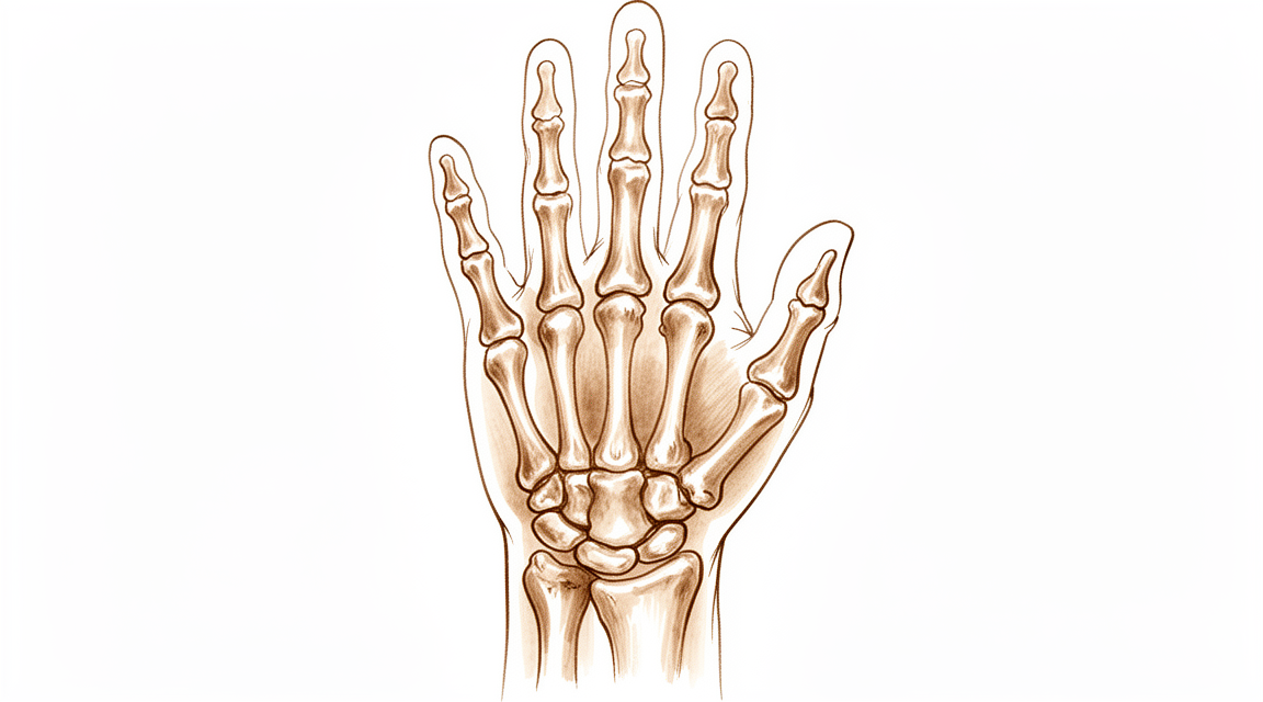
Kienböck病
Patients › Wrist
Kienböck’s disease — progressive wrist pain from lunate avascular necrosis; diagnosis and treatment options.
您的感受
Kienböck病(月骨无菌性坏死)是一种影响腕部小骨的疾病。它并不常见。如果不进行治疗,病情通常会随时间推移而恶化。您可能会注意到腕部出现肿胀或僵硬。疼痛通常缓慢开始并逐渐加重。
按压腕部背侧时,您可能会感到压痛。这正是月骨所在的位置。这种疼痛会使简单的日常活动变得困难。您可能难以从椅子上撑起身体。提举物体时可能会感到疼痛且无力。甚至转动门把手或打开罐子也可能引起疼痛。
早晨您的腕部可能会感到僵硬。这种僵硬通常在白天活动时逐渐缓解。然而,在使用双手一段时间后,疼痛可能会再次出现。使腕部承重的活动,如做俯卧撑或提购物袋,可能会诱发症状加重。您还可能会注意到握力较以前减弱。
在某些情况下,疼痛可能是尖锐的或钝痛的。疼痛可能会放射至前臂。夜间疼痛是可能的,尤其是如果您侧卧睡在该侧时。这可能会干扰您的休息,使您感到疲劳。您可能会发现很难将手伸到背后扣内衣或塞衬衫。这些动作会拉伸腕部,从而加重受累骨头的炎症。
您症状的严重程度取决于腕部骨骼的形状。这种形状会影响施加在月骨上的压力大小。该病的病因可能是多种因素共同作用的结果。为什么有些人会患病而其他人不会,原因并不总是很清楚。
如果您忽视症状,病情可能会进展到更严重的阶段。这可能导致腕部骨骼形状的改变。您的外科医生会查看X光片,以了解骨骼移位的情况。然而,标准X光片并不总能完全显示骨塌陷的全貌。这就是为什么您的外科医生可能会安排更详细的扫描,以便清楚地了解腕部内部发生的情况。
实际发生了什么
Kienböck病(月骨无菌性坏死)是一种腕部小骨——月骨——的血液供应减少或丧失的疾病。由于供血不足,该骨开始变弱,最终可能导致塌陷。可以将月骨视为腕部的中央减震器。当它失去结构完整性时,便无法再缓冲前臂骨骼与手部其余部分之间的冲击力。
这种塌陷会改变腕部的运动方式。正常情况下,腕骨之间会平滑地滑动和滚动。当发生月骨损伤时,这种运动变得不规则。骨骼可能会偏离其正常排列,这种情况称为旋转性排列不良。这种排列不齐会对腕关节的其他部位施加额外压力,特别是桡骨与舟骨相接的区域。随着时间的推移,这种不均匀的磨损可能导致这些特定区域发生关节炎。
外科医生会根据这些变化决定最佳治疗方案。在年轻患者中,目标通常是保留骨骼。例如,桡骨截骨术(调整前臂骨骼)可以改善青少年的临床结果和影像学表现。对于其他患者,可能会建议进行头状骨缩短术或带血管骨移植术,以恢复平衡和血液供应。这些治疗旨在尽可能保持腕部的自然活动。
在骨骼严重塌陷的更晚期病例中,重点转向稳定关节。诸如舟头关节融合术(将两块骨骼融合在一起)等手术可以提供长期的疼痛缓解并改善握力。虽然这些手术会限制部分活动,但它们能创建一个稳定的平台,使您能够有效地使用手部。外科医生将根据您疾病的分期和个人需求选择最合适的方案。
我们能采取的措施
我们从非手术治疗开始,以管理您的症状并保护手腕。这种方法侧重于减少手部骨骼所承受的压力。您的外科医生可能会建议休息或避免进行某些特定活动。物理治疗有助于维持手腕和手部的活动度与力量。这些方法旨在让您在病情监测期间保持舒适。
研究表明,对于骨骼仍在生长的年轻患者,非手术治疗可实现良好和优秀的疗效。对许多人而言,这种保守治疗途径足以控制疼痛并维持功能。然而,Kienböck病(月骨无菌性坏死)通常是一种进展性疾病。这意味着如果不加以控制,病情可能会随时间推移而恶化,最终导致腕关节出现晚期改变。在您的定期复查中,我们会密切监测这些迹象。
如果非手术治疗措施无法提供足够的缓解,我们将讨论药物治疗方案。止痛药和抗炎药有助于控制不适和肿胀。在某些情况下,我们可能会考虑进行关节内注射,以直接减轻关节内的炎症。这些治疗可提供暂时性缓解,并帮助您保持活动能力。它们不能逆转潜在的骨骼改变,但在我们决定下一步治疗方案时,可以改善您的日常生活质量。
当保守治疗达到极限或疾病在治疗期间仍进展时,会考虑手术治疗。手术的目的是减轻受影响骨骼的压力、恢复血液供应或稳定关节。可选方案包括改变桡骨形状的手术、移植带有自身血液供应的新骨,或融合特定骨骼以缓解疼痛。具体选择取决于您的疾病分期和个体需求。
对于青少年患者,桡骨截骨术(改变前臂骨的形状)在改善症状和X线表现方面效果显著。这些手术可为75%的患者提供长达十年的改善。在腕关节已塌陷的晚期病例中,舟头关节融合术等融合手术可提供显著的疼痛缓解和功能改善。您的外科医生将解释哪种方案最适合您的具体情况。
我们还使用X线和MRI等影像学检查来追踪病情变化。虽然普通X线有时可能漏诊早期的塌陷迹象,但先进的影像学检查有助于我们准确了解月骨的真实状态。这确保我们在正确的时机选择正确的治疗方案。无论您需要简单的休息还是手术治疗,我们的目标都是保留您的手腕功能并减轻疼痛。
预期情况
Kienböck病是一种由于腕部一小块骨骼的血液供应减少,导致其变弱并塌陷的疾病。这一过程通常是进行性的,意味着病情倾向于随时间推移而恶化,而非自行缓解。若不进行治疗,病情可能进展至IV期改变,涉及腕关节的显著磨损性关节炎。该疾病的确切发展路径尚未完全明确,但若不加管理,通常会导致持续性疼痛和僵硬。
经过治疗,您的预后将显著改善。许多手术方案旨在阻止病情进展并缓解疼痛。例如,桡骨缩短截骨术——一种调整前臂骨长度的手术——可为75%的症状性患者提供长达十年的改善。该治疗对于尺骨负变异(一种特定的腕骨排列)患者长期缓解疼痛尤为有效。其他手术,如舟头关节融合术(将两块骨头连接在一起)或近排腕骨切除术(移除一行小腕骨),可提供持久、长期的益处。这些手术通常能缓解疼痛,保持功能性活动度,并在多年内维持令人满意的握力。
即使接受治疗,疾病也不总是迅速进展。无论选择何种治疗,Kienböck病在1年或更长时间内的影像学进展平均而言较为轻微。这意味着虽然潜在疾病可能持续存在,但X光片上的可见变化通常会趋于稳定。您可以预期您的外科医生会根据您的具体分期和症状量身定制治疗方案。无论您是青少年还是成年人,目标都是管理疼痛并保持腕关节的功能性。虽然总体结果通常较为积极,但保持现实的期望非常重要。该疾病是多因素的,个体结果存在差异,但现代治疗提供了可靠的途径以实现疼痛缓解和改善手部功能。
何时就医
Kienböck病是一种影响腕部的罕见疾病。它通常被认为是一种进展性问题,可导致严重的关节改变。其确切的发展途径尚未完全明确。如果您有持续不缓解的疼痛,即使休息后也无改善,应就诊于全科医生(GP)。如果您注意到腕部无力或不稳,请要求专科医生评估。如果您的手出现卡顿或无力感,请及时就医。如果症状干扰您的睡眠或工作,请联系您的外科医生。疼痛突然加重也是就医的原因。早期评估有助于有效管理这一复杂疾病。
Evidence & references
Overview
- Radial osteotomies are effective in improving short-term clinical outcomes and radiographic findings in teenage patients with Kienböck disease [1].
- Scaphocapitate arthrodesis demonstrates long-term clinical benefits for the treatment of collapsed Kienböck disease [2].
- Vascularized bone grafting for stage III Kienböck disease demonstrates favorable long-term results and is recommended as a surgical treatment [3].
- Good and excellent clinical and radiological outcomes can be achieved with both nonsurgical and surgical treatments in skeletally immature patients with Kienböck disease [5].
- Temporary scaphotrapezoidal joint fixation is recommended for the surgical treatment of adolescent Kienböck's disease [7].
- Radial shortening osteotomy provides decade-long improvement in 75% of patients and is a reasonable treatment for symptomatic Kienböck’s disease [8].
- Scaphocapitate arthrodesis is an effective procedure for the treatment of Kienböck disease, associated with satisfactory functional outcomes and significant improvement in pain scores and grip strength [13].
- Capitate shortening is a safe and effective approach for the treatment of early stages of Kienböck's disease and can be associated with a satisfying outcome [14].
- Advanced Kienböck's disease with carpal collapse is not a contraindication for carpal-sparing surgery radial shortening osteotomy [17].
- Lunate excision, capitate osteotomy, and intercarpal arthrodesis should be used with caution for advanced Kienböck's disease because it does not have good long-term results and is no longer widely used in Europe [25].
- The acceptance rate for negative outcomes studies regarding Kienböck's disease is higher than for other surgical disorders, indicating a relative decrease in positive outcome bias among published Kienböck's disease studies compared with other surgical disorders [41].
Anatomy & Pathophysiology
- Tendon ball arthroplasty and proximal carpal stabilization with tendon graft restore the integrity of the proximal carpal row in advanced Kienböck’s disease [28].
- Surgical treatments for scapholunate advanced collapse result in decreased wrist kinematic motion and functional performance compared with individuals with normal wrists [30].
- Lunate morphology affects the 3-dimensional kinematics of the carpus during wrist flexion and extension [31].
- Three- and 4-corner fusions produce motion that is smoother and more closely replicates the normal axis and functional motion of the wrist [32].
- Computed fiber elongations of the dorsal carpal ligaments vary linearly with wrist position [33].
- Four-dimensional computed tomography (4DCT) is a non-invasive and affordable method to assess and quantify wrist kinematics by incorporating the temporal dimension [34].
- During simple unresisted wrist motions, force in the scapholunate interosseous ligament does not exceed 20 N [35].
- Kinematic changes in scapholunate instability may predict the development of radioscaphoid arthritis and help identify a kinematically abnormal wrist [36].
- Rotational malalignment of the wrist has significant effects on carpal, distal radial, and distal radioulnar joint measurements [37].
- Scaphoid nonunions have a dramatic impact on carpal kinematics by partially uncoupling the proximal and distal carpal rows [42].
- Dynamic imaging may enable the derivation of a standardized protocol for mapping carpal motion that is clinically applicable and reproducible [44].
- Computer-aided CT analysis provides guidelines for measuring and quantifying carpal alignment three-dimensionally and establishes a database for normal values [45].
- More than half the motion of the carpus when the wrist was loaded in extension occurred at the midcarpal joint [47].
- Radioscapholunate fusion shows the most biomechanically similar behavior to the healthy wrist among three fusion types [51].
- Contact areas between the scaphoid and distal radius are maximized during full extension of the wrist, which helps stabilize the radiocarpal joint [52].
- A dorsally applied PLA plate restores carpal kinematics for 1,000 cycles of motion in unstable wrists where fixation is not compromised by carpal size or osteoporosis [53].
- The modification of the wrist center of rotation during flexion and extension indicates that stability is considered more important than mobility in clinical conditions [54].
- Correction of scapholunate dissociation may correlate with improved carpal dynamics and improved clinical outcomes [56].
- Postarthroscopic lunate excision alters normal carpal kinematics but maintains joint congruity in the short-term [57].
Classification
- Kienböck disease is generally considered a progressive condition that can end in Stage IV changes [6].
- Lunate morphology may affect the severity of Kienböck disease at the time of initial presentation [9].
- The patterns of carpal collapse differ between stage IIIb Kienböck disease and scapholunate dislocation in terms of radioscaphoid joint congruity [50].
- Traditional radiographic indices measured on plain radiographs have poor diagnostic performance in the detection of carpal collapse in Kienböck's disease [15].
Clinical Presentation
- Kienböck disease is a relatively infrequent carpal pathology [4].
- Kienböck disease is generally considered a progressive condition that can end in Stage IV changes [6].
- The natural history of Kienböck disease is not fully known [6].
- Lunate morphology may affect the severity of Kienböck disease at the time of initial presentation [9].
- The development of Kienböck disease is probably multifactorial [10].
- Traditional radiographic indices measured on plain radiographs have poor diagnostic performance in the detection of carpal collapse in Kienböck's disease [15].
Investigations
- Radial osteotomies are effective in improving short-term clinical outcomes and radiographic findings in teenage patients with Kienböck disease [1].
- Scaphocapitate arthrodesis demonstrates long-term clinical benefits for the treatment of collapsed Kienböck disease [2].
- Vascularized bone grafting for stage III Kienböck disease demonstrates favorable long-term results and is recommended as a surgical treatment [3].
- Good and excellent clinical and radiological outcomes can be achieved with both nonsurgical and surgical treatments in skeletally immature patients with Kienböck disease [5].
- Temporary scaphotrapezoidal joint fixation is recommended for the surgical treatment of adolescent Kienböck's disease [7].
- Radial shortening osteotomy provides decade-long improvement in 75% of patients and is a reasonable treatment for symptomatic Kienböck’s disease [8].
- Lunate morphology may affect the severity of Kienböck disease at the time of initial presentation [9].
- Traditional radiographic indices measured on plain radiographs have poor diagnostic performance in the detection of carpal collapse in Kienböck's disease [15].
- The Camembert osteotomy improved function in patients with early stage Kienböck disease, with MRI aspects improving in most cases and no patients experiencing lunate collapse [21].
- Radiographic progression of Kienböck over 1 year or more seems slight on average regardless of treatment [26].
- Computed tomography of the lunate in Kienböck disease is an important investigative tool [43].
- Lunates with advanced Kienböck's disease exhibit significantly denser, thicker, and more plate-like trabecular microstructure compared to normal lunates [58].
- Following medial femoral trochlea reconstruction of the proximal lunate for advanced Kienböck disease, cessation of radiocarpal collapse was observed [59].
Treatment
Non-Operative and General Considerations
- Good and excellent clinical and radiological outcomes can be achieved with both nonsurgical and surgical treatments in skeletally immature patients with Kienböck disease [5].
- The natural history of Kienböck's disease is generally considered a progressive condition that can end in Stage IV changes [6].
- Treatment strategies for Kienböck's disease focus on biomechanical unloading, vascularized bone grafts, or salvage procedures depending on the stage [6].
- There is limited, low-quality evidence that surgical treatment slows progression of Kienböck's disease [18].
- Many uncontrolled case series document slight improvement in motion and grip after surgical treatment without clear evidence that this is better than placebo or no intervention [18].
Radial Osteotomies and Shortening
- Radial osteotomies are effective in improving not only short-term clinical outcomes, but also radiographic findings in teenage patients with Kienböck disease [1].
- Radial shortening osteotomy provides decade-long improvement in 75% of patients and seems to be a reasonable treatment for symptomatic Kienböck’s disease [8].
- The medium- and long-term results of radial shortening osteotomy for Kienböck's disease in patients with negative ulnar variance are comparable to short-term results, providing long-lasting pain relief [16].
- Advanced Kienbock's disease with carpal collapse is not a contraindication for carpal-sparing surgery radial shortening osteotomy [17].
- Radius core decompression demonstrated favorable long-term results and could be considered as a surgical alternative for stage IIIA of Kienböck disease [61].
Vascularized Bone Grafts
- Vascularized bone grafting for stage III Kienböck disease demonstrated favorable long-term results and is recommended as a surgical treatment [3].
- Vascularized bone grafts are indicated for Kienböck's disease, while being contraindicated in advanced carpal collapse with degenerative changes [40].
Arthrodesis and Salvage Procedures
- The long-term clinical benefits of scaphocapitate arthrodesis for treatment of collapsed Kienböck disease are demonstrated [2].
- Scaphocapitate arthrodesis is an effective procedure for treatment of Kienböck disease associated with satisfactory functional outcomes and significant improvement in pain scores and grip strength [13].
- Scaphocapitate arthrodesis should be considered as a treatment option for wrist salvage in the patient with advanced Kienbock's disease given the significant postoperative reduction in associated pain symptoms at the time of follow-up [24].
- Scaphotrapeziotrapezoid arthrodesis with lunate excision for advanced Kienböck disease provided favorable clinical results in terms of pain relief and functional improvement [22].
- The procedure of lunate excision, capitate osteotomy, and intercarpal arthrodesis should be used with caution for advanced Kienböck's disease because it does not have good long-term results and is no longer widely used in Europe [25].
- Improved outcomes following proximal row carpectomy were associated with Kienbock's disease [55].
Capitate Shortening
- Capitate shortening is a safe and effective approach for treatment of the early stages of Kienböck's disease and can be associated with a satisfying outcome [14].
Arthroscopic Procedures
- Arthroscopic lunate core decompression appears to be an effective and safe surgery for treating Kienböck disease on the basis of mid-term follow-up [20].
Adolescent-Specific Interventions
- Temporary scaphotrapezoidal joint fixation is recommended for the surgical treatment of adolescent Kienböck's disease [7].
Complications
- Kienböck's disease is generally considered a progressive condition that can end in Stage IV changes [6].
- The natural history of Kienböck's disease is not fully known [6].
- Lunate morphology may affect the severity of Kienböck disease at the time of initial presentation [9].
- Negative ulnar variance has a longitudinal relationship with progressive Kienböck disease, though additional long-term study is needed to confirm this [19].
- The development of Kienböck disease is probably multifactorial [10].
- The observation of Kienböck disease and carpal coalition in one wrist is fortuitous [10].
Recovery
- Radial osteotomies improve short-term clinical outcomes in teenage patients with Kienböck disease [1].
- Radial osteotomies improve radiographic findings in teenage patients with Kienböck disease [1].
- Scaphocapitate arthrodesis provides long-term clinical benefits for collapsed Kienböck disease [2].
- Vascularized bone grafting demonstrates favorable long-term results for stage III Kienböck disease [3].
- Radial shortening osteotomy provides decade-long improvement in 75% of patients with symptomatic Kienböck’s disease [8].
- Radial shortening osteotomy provides long-lasting pain relief for patients with negative ulnar variance [16].
- Titanium lunate arthroplasty (TLA) shows promising longer-term results for stage III Kienböck disease [23].
- Scaphocapitate arthrodesis with lunate excision significantly alleviates pain in advanced Kienböck disease at a mean follow-up of 10.7 years [38].
- Scaphocapitate arthrodesis with lunate excision preserves functional mobility in advanced Kienböck disease at a mean follow-up of 10.7 years [38].
- Scaphocapitate arthrodesis with lunate excision maintains satisfactory grip strength in advanced Kienböck disease at a mean follow-up of 10.7 years [38].
- Proximal row carpectomy (PRC) is a durable long-term treatment option for radiocarpal degenerative arthritis and Kienböck's disease [46].
- Proximal row carpectomy allows for maintenance of wrist range of motion [46].
- Proximal row carpectomy improves grip strength [46].
- Proximal row carpectomy is a reliable and durable procedure for Lichtman stage IIIA or IIIB Kienböck's disease at an average follow-up of 10 years [49].
- Scaphocapitate arthrodesis (SCA) results in improved grip strength in patients with advanced stages of Kienböck disease in medium-term follow-up [48].
- Scaphocapitate arthrodesis (SCA) corrects carpal alignment in patients with advanced stages of Kienböck disease in medium-term follow-up [48].
- Radiographic progression of Kienböck disease over 1 year or more is slight on average regardless of treatment [26].
Key Evidence
- [L4] The current results indicate that radial osteotomies are effective in improving not only short-term clinical outcomes, but also radiographic findings in teenage patients with Kienböck disease. [1] (10.1097/01.blo.0000173254.46899.72)
- [L4] The long-term clinical benefits of scaphocapitate arthrodesis for treatment of collapsed Kienböck disease are demonstrated. [2] (10.1177/1753193413496177)
- [L3] Vascularized bone grafting for stage III Kienböck disease demonstrated favorable long-term results and is recommended as a surgical treatment. [3] (10.1016/j.jhsa.2013.02.010)
- [L5] This review article discusses the history, etiology, and course of Kienböck disease and reviews the literature on both the diagnosis and management of this relatively infrequent carpal pathology. [4] (10.5435/jaaos-d-20-00020)
- [L4] Good and excellent clinical and radiological outcomes can be achieved with both nonsurgical and surgical treatments in skeletally immature patients with Kienböck disease. [5] (10.1016/j.jhsa.2018.02.029)
- [L5] The natural history of Kienbock's disease is not fully known, though it is generally considered a progressive condition that can end in Stage IV changes; treatment strategies focus on biomechanical unloading, vascularized bone grafts, or salvage procedures depending on the stage. [6] (10.1016/j.hcl.2006.07.003)
- [L4] We therefore recommend this procedure for the surgical treatment of adolescent Kienböck's disease. [7] (10.1016/j.jhsa.2008.09.019)
- [L4] Radial shortening osteotomy provides decade-long improvement in 75% of patients and seems to be a reasonable treatment for symptomatic Kienböck’s disease. [8] (10.1177/1753193413512222)
- [L3] Lunate morphology may affect the severity of Kienböck disease at the time of initial presentation. [9] (10.1016/j.jhsa.2014.12.024)
- [Letter] The observation of Kienböck disease and carpal coalition in one wrist is fortuitous, and the development of Kienböck disease is probably multifactorial. [10] (10.1016/j.jhsa.2016.11.010)
- [L4] Scaphocapitate arthrodesis is an effective procedure for treatment of Kienböck disease associated with satisfactory functional outcomes and significant improvement in pain scores and grip strength. [13] (10.1016/j.jhsg.2023.03.014)
- [L2] Capitate shortening is a safe and effective approach for treatment of the early stages of Kienböck's disease and can be associated with a satisfying outcome. [14] (10.1177/15589447221081564)
- [L3] Traditional radiographic indices measured on plain radiographs have poor diagnostic performance in the detection of carpal collapse in Kienböck's disease. [15] (10.1177/17531934231153966)
- [L3] The medium- and long-term results of radial shortening osteotomy for Kienböck's disease in patients with negative ulnar variance are comparable to short-term results, providing long-lasting pain relief. [16] (10.1097/blo.0b013e318041d309)
- [L5] There is limited, low-quality evidence that surgical treatment slows progression of Kienböck's disease, and many uncontrolled case series document slight improvement in motion and grip after surgical treatment without clear evidence that this is better than placebo or no intervention. [18] (10.1016/j.jhsa.2009.10.013)
- [L2] Additional long-term study is needed to confirm the longitudinal relationship of negative ulnar variance with progressive Kienböck disease. [19] (10.1016/j.jhsa.2017.06.107)
- [L4] Arthroscopic lunate core decompression appears to be an effective and safe surgery for treating Kienböck disease on the basis of mid-term follow-up. [20] (10.1016/j.jhsa.2023.02.011)
- [L4] Scaphotrapeziotrapezoid arthrodesis with lunate excision for advanced Kienböck disease provided favorable clinical results in terms of pain relief and functional improvement. [22] (10.1016/j.jhsa.2012.08.031)
- [L4] The longer-term results of TLA for stage III Kienböck disease are promising. [23] (10.1016/j.jhsa.2018.02.009)
- [L4] Given the significant postoperative reduction in associated pain symptoms at the time of follow-up, scaphocapitate arthrodesis should be considered as a treatment option for wrist salvage in the patient with advanced Kienbock's disease. [24] (10.1007/s11552-014-9705-z)
- [L5] The procedure of lunate excision, capitate osteotomy, and intercarpal arthrodesis should be used with caution for advanced Kienböck's disease because it does not have good long-term results and is no longer widely used in Europe. [25] (10.1177/1753193418807360)
- [L4] Radiographic progression of Kienböck over 1 year or more seems slight on average regardless of treatment. [26] (10.1016/j.jhsa.2016.02.016)
- [L4] The technique demonstrated reduced wrist pain and improved wrist motion and grip strength while restoring the integrity of the proximal carpal row. [28] (10.1177/17531934241238939)
- [L2] Both surgical groups demonstrated decreased wrist kinematic motion and functional performance compared with individuals with normal wrists. [30] (10.1016/j.jhsa.2015.04.035)
- [L5] This study describes the effect of lunate morphology on 3-dimensional carpal kinematics during wrist flexion and extension. [31] (10.1016/j.jhsa.2014.09.019)
- [L3] Motion was smoother and more closely replicated the normal axis and functional motion of the wrist. [32] (10.1016/j.jhsa.2015.02.027)
- [L5] Despite complex carpal bone anatomy and kinematics, computed fiber elongations were found to vary linearly with wrist position. [33] (10.1016/j.jhsa.2012.04.025)
- [L5] Four-dimensional computed tomography (4DCT) is a promising, non-invasive, and affordable method to assess and quantify wrist kinematics, extending conventional CT by incorporating the temporal dimension. [34] (10.1177/17531934251326028)
- [L5] However, during simple unresisted wrist motions, the force did not exceed 20 N. [35] (10.1016/j.jhsa.2015.04.007)
- [L3] These kinematic changes may predict the development of radioscaphoid arthritis and help identify a kinematically abnormal wrist. [36] (10.1177/17531934241242676)
- [L4] Rotational malalignment of the wrist has significant effects on carpal, distal radial and distal radioulnar joint measurements. [37] (10.1177/1753193408090393)
- [L4] Scaphocapitate arthrodesis with lunate excision performed in an advanced stage of Kienböck disease significantly alleviates pain, while preserving functional mobility and satisfactory grip strength in the long term. [38] (10.1177/1753193417739247)
- [L4] This manuscript offers a current review of the techniques and outcomes of VBGs to the carpal bones, noting that VBGs are indicated for scaphoid nonunion with proximal pole AVN, Kienböck's disease, Preiser's disease, and capitate osteonecrosis, while being contraindicated in advanced carpal collapse with degenerative changes. [40] (10.1007/s11552-012-9479-0)
- [L2] The acceptance rate for negative outcomes studies regarding Kienböck's disease is higher than for other surgical disorders, indicating a relative decrease in positive outcome bias among published Kienböck's disease studies compared with other surgical disorders. [41] (10.1016/j.jhsa.2009.12.003)
- [L4] Scaphoid nonunions have a dramatic impact on carpal kinematics, partially uncoupling the proximal and distal carpal rows. [42] (10.1016/j.jhsa.2008.03.008)
- [L4] Computed tomography of the lunate in Kienböck disease is an important investigative tool. [43] (10.1016/j.jhsa.2018.05.008)
- [L4] With the increased focus on dynamic imaging for wrist motion, it may be possible to derive a standardized protocol for mapping the carpal motion that is clinically applicable and reproducible. [44] (10.1016/j.jhsg.2022.10.001)
- [L4] This study provides guidelines on how to measure and quantify carpal alignment three-dimensionally and establishes a database for normal values, which may be useful when analysing various wrist pathologies and kinematics. [45] (10.1177/17531934231160100)
- [L4] PRC is a durable long-term treatment option for radiocarpal degenerative arthritis and Kienböck's disease allowing for maintenance of wrist range of motion and improvement in grip strength. [46] (10.1016/s0363-5023(10)60087-1)
- [L4] More than half the motion of the carpus when the wrist was loaded in extension occurred at the midcarpal joint. [47] (10.1016/j.jhsa.2012.10.035)
- [L4] SCA resulted in improved grip strength with correction of carpal alignment in patients with advanced stages of Kienböck disease in medium-term follow-up. [48] (10.1016/j.jhsa.2014.12.013)
- [L4] At an average follow-up of 10 years, proximal row carpectomy is a reliable and durable procedure for patients with Lichtman stage IIIA or IIIB Kienböck's disease. [49] (10.1016/j.jhsa.2008.02.031)
- [L2] The patterns of carpal collapse differed between stage IIIb Kienböck disease and scapholunate dislocation in terms of radioscaphoid joint congruity. [50] (10.1016/j.jhsa.2014.10.035)
- [L5] The contact areas between the scaphoid and distal radius are maximized during full extension of the wrist, which helps stabilize the radiocarpal joint and potentially reduces the risk of injury to the carpus and the distal radius. [52] (10.1177/1753193413507810)
- [L5] The study shows that in the unstable wrist, following ligament sectioning, where fixation is not compromised by carpal size or osteoporosis, a dorsally applied PLA plate does restore carpal kinematics for 1,000 cycles of motion. [53] (10.1016/j.jhsa.2008.01.016)
- [L4] The study also characterized the modification of the wrist CoR during flexion and extension, noting that stability is considered more important than mobility in clinical conditions. [54] (10.1016/s0749-0712(03)00008-8)
- [L3] Improved outcomes were associated with age over 40, Kienbock's disease, concomitant neurectomy, non-labourer status, and surgery after 1990, while radiocapitate arthrosis did not correlate with clinical outcomes. [55] (10.1177/1753193415597096)
- [L5] This correction might correlate with improved carpal dynamics and improved clinical outcomes. [56] (10.1016/j.jhsa.2010.06.029)
- [L4] Although postarthroscopic lunate excision alters normal carpal kinematics, the joint congruity is maintained in the short-term. [57] (10.1016/j.jhsa.2025.01.024)
- [L4] Lunates with advanced Kienböck's disease exhibit significantly denser, thicker, and more plate-like trabecular microstructure compared to normal lunates. [58] (10.1177/1753193411422337)
- [L4] Following medial femoral trochlea reconstruction of the proximal lunate for advanced Kienböck disease, we observed a cessation of radiocarpal collapse. [59] (10.1016/j.jhsa.2019.12.008)
- [L4] In this limited series, the radius core decompression demonstrated favorable long-term results and could be considered as a surgical alternative for stage IIIA of Kienböck disease. [61] (10.1016/j.jhsa.2017.05.017)
References
[1] Radial Osteotomies for Teenage Patients with Kienb??ck Disease. Clinical Orthopaedics and Related Research. 2005. DOI: 10.1097/01.blo.0000173254.46899.72 [2] Scaphocapitate arthrodesis for treatment of late stage Kienböck disease. Journal of Hand Surgery (European Volume). 2013. DOI: 10.1177/1753193413496177 [3] Long-Term Results of Vascularized Bone Graft for Stage III Kienböck Disease. The Journal of Hand Surgery. 2013. DOI: 10.1016/j.jhsa.2013.02.010 [4] Osteonecrosis of the Lunate: Kienböck Disease. Journal of the American Academy of Orthopaedic Surgeons. 2020. DOI: 10.5435/jaaos-d-20-00020 [5] Kienböck Disease in the Skeletally Immature Patient. The Journal of Hand Surgery. 2018. DOI: 10.1016/j.jhsa.2018.02.029 [6] Kienböck's Disease: An Approach to Treatment. Hand Clinics. 2006. DOI: 10.1016/j.hcl.2006.07.003 [7] Temporary Scaphotrapezoidal Joint Fixation for Adolescent Kienböck's Disease. The Journal of Hand Surgery. 2009. DOI: 10.1016/j.jhsa.2008.09.019 [8] Long-term outcome (20 to 33 years) of radial shortening osteotomy for Kienböck’s lunatomalacia. Journal of Hand Surgery (European Volume). 2013. DOI: 10.1177/1753193413512222 [9] The Effect of Lunate Morphology in Kienböck Disease. The Journal of Hand Surgery. 2015. DOI: 10.1016/j.jhsa.2014.12.024 [10] Letter Regarding “Kienböck Disease and Carpal Coalitions: A Potential Correlation”. The Journal of Hand Surgery. 2017. DOI: 10.1016/j.jhsa.2016.11.010 [13] Clinical and Radiological Outcomes of Scaphocapitate Fusion in Kienböck Disease: A Systematic Review and Meta-Analysis. Journal of Hand Surgery Global Online. 2023. DOI: 10.1016/j.jhsg.2023.03.014 [14] Comparing the Radiologic and Functional Outcome of Radial Shortening Versus Capitate Shortening in Management of Kienböck’s Disease. HAND. 2022. DOI: 10.1177/15589447221081564 [15] Diagnostic performance of traditional radiographic indices in detection of carpal collapse in Kienböck’s disease. Journal of Hand Surgery (European Volume). 2023. DOI: 10.1177/17531934231153966 [16] Outcome of Kienböck's Disease 22 Years after Distal Radius Shortening Osteotomy. Clinical Orthopaedics & Related Research. 2007. DOI: 10.1097/blo.0b013e318041d309 [17] 10.1055-s-0039-1688947. n.d.. [18] Kienböck's Disease. The Journal of Hand Surgery. 2009. DOI: 10.1016/j.jhsa.2009.10.013 [19] Risk Factors of Lunate Collapse in Kienböck Disease. The Journal of Hand Surgery. 2017. DOI: 10.1016/j.jhsa.2017.06.107 [20] Arthroscopic Treatment of Kienböck Disease: Mid-Term Outcome of Arthroscopic Lunate Core Decompression. The Journal of Hand Surgery. 2024. DOI: 10.1016/j.jhsa.2023.02.011 [21] 10.1055-s-0039-1683931. n.d.. [22] Scaphotrapeziotrapezoid Arthrodesis and Lunate Excision for Advanced Kienböck Disease. The Journal of Hand Surgery. 2012. DOI: 10.1016/j.jhsa.2012.08.031 [23] Long-Term Clinical Outcome After Titanium Lunate Arthroplasty for Kienböck Disease. The Journal of Hand Surgery. 2018. DOI: 10.1016/j.jhsa.2018.02.009 [24] Limited Intercarpal Fusion as a Salvage Procedure for Advanced Kienbock Disease. HAND. 2014. DOI: 10.1007/s11552-014-9705-z [25] Lunate excision, capitate osteotomy, and intercarpal arthrodesis should be used with caution for advanced Kienböck’s disease. Journal of Hand Surgery (European Volume). 2018. DOI: 10.1177/1753193418807360 [26] Radiographic Progression of Kienböck Disease: Radial Shortening Versus No Surgery. The Journal of Hand Surgery. 2016. DOI: 10.1016/j.jhsa.2016.02.016 [28] Tendon ball arthroplasty and proximal carpal stabilization with tendon graft for advanced Kienböck’s disease. Journal of Hand Surgery (European Volume). 2024. DOI: 10.1177/17531934241238939 [30] Surgical Treatments for Scapholunate Advanced Collapse Wrist: Kinematics and Functional Performance. The Journal of Hand Surgery. 2015. DOI: 10.1016/j.jhsa.2015.04.035 [31] The Effect of Lunate Morphology on the 3-Dimensional Kinematics of the Carpus. The Journal of Hand Surgery. 2015. DOI: 10.1016/j.jhsa.2014.09.019 [32] Comparison of the Clinical and Functional Outcomes Following 3- and 4-Corner Fusions. The Journal of Hand Surgery. 2015. DOI: 10.1016/j.jhsa.2015.02.027 [33] Elongation of the Dorsal Carpal Ligaments: A Computational Study of In Vivo Carpal Kinematics. The Journal of Hand Surgery. 2012. DOI: 10.1016/j.jhsa.2012.04.025 [34] Dynamic wrist imaging: How it works and how to assess kinematic changes in wrists with scapholunate instability. Journal of Hand Surgery (European Volume). 2025. DOI: 10.1177/17531934251326028 [35] Force in the Scapholunate Interosseous Ligament During Active Wrist Motion. The Journal of Hand Surgery. 2015. DOI: 10.1016/j.jhsa.2015.04.007 [36] Radiocarpal and midcarpal kinematics in scapholunate instability: a four-dimensional CT study in vivo. Journal of Hand Surgery (European Volume). 2024. DOI: 10.1177/17531934241242676 [37] The Effect of Rotational Malalignment on X-rays of the Wrist. Journal of Hand Surgery (European Volume). 2009. DOI: 10.1177/1753193408090393 [38] Results of scaphocapitate arthrodesis with lunate excision in advanced Kienböck disease at 10.7-year mean follow-up. Journal of Hand Surgery (European Volume). 2017. DOI: 10.1177/1753193417739247 [40] Vascularized Bone Grafts for the Treatment of Carpal Bone Pathology. HAND. 2013. DOI: 10.1007/s11552-012-9479-0 [41] Publication Bias in Kienböck's Disease: Systematic Review. The Journal of Hand Surgery. 2010. DOI: 10.1016/j.jhsa.2009.12.003 [42] Interfragmentary Motion in Patients With Scaphoid Nonunion. The Journal of Hand Surgery. 2008. DOI: 10.1016/j.jhsa.2008.03.008 [43] Fixation of the Fractured Lunate in Kienböck Disease. The Journal of Hand Surgery. 2019. DOI: 10.1016/j.jhsa.2018.05.008 [44] Radiographic Evaluation of Carpal Mechanics and the Scapholunate Angle in a Clenched Fist with Dynamic Computed Tomography Imaging. Journal of Hand Surgery Global Online. 2023. DOI: 10.1016/j.jhsg.2022.10.001 [45] Three-dimensional carpal alignment: computer-aided CT analysis of carpal axes and normal ranges. Journal of Hand Surgery (European Volume). 2023. DOI: 10.1177/17531934231160100 [46] Minimum Twenty-year Follow-up of Proximal Row Carpectomy. The Journal of Hand Surgery. 2010. DOI: 10.1016/s0363-5023(10)60087-1 [47] In Vivo Kinematics of the Scaphoid, Lunate, Capitate, and Third Metacarpal in Extreme Wrist Flexion and Extension. The Journal of Hand Surgery. 2013. DOI: 10.1016/j.jhsa.2012.10.035 [48] Scaphocapitate Arthrodesis for Kienböck Disease. The Journal of Hand Surgery. 2015. DOI: 10.1016/j.jhsa.2014.12.013 [49] Proximal Row Carpectomy for Advanced Kienböck's Disease: Average 10-Year Follow-Up. The Journal of Hand Surgery. 2008. DOI: 10.1016/j.jhsa.2008.02.031 [50] In Vivo 3-Dimensional Analysis of Stage III Kienböck Disease: Pattern of Carpal Deformity and Radioscaphoid Joint Congruity. The Journal of Hand Surgery. 2015. DOI: 10.1016/j.jhsa.2014.10.035 [51] Load_transfer_through_the_radiocarpal_joint_and_the_effects_of_partial_wrist_art_1753193412441761. 1934. [52] Contact areas of the scaphoid and lunate with the distal radius in neutral and extension: correlation of falling strategies and distal radial anatomy. Journal of Hand Surgery (European Volume). 2013. DOI: 10.1177/1753193413507810 [53] Treatment of Scapholunate Dissociation With a Bioresorbable Polymer Plate: A Biomechanical Study. The Journal of Hand Surgery. 2008. DOI: 10.1016/j.jhsa.2008.01.016 [54] Electrogoniometric and radiologic evaluation of scapho-trapezo-trapezoid arthrodesis. Hand Clinics. 2003. DOI: 10.1016/s0749-0712(03)00008-8 [55] Factors associated with improved outcomes following proximal row carpectomy: a long-term outcome study of 144 patients. Journal of Hand Surgery (European Volume). 2015. DOI: 10.1177/1753193415597096 [56] Radiographic Evaluation of the Modified Brunelli Technique Versus the Blatt Capsulodesis for Scapholunate Dissociation in a Cadaver Model. The Journal of Hand Surgery. 2010. DOI: 10.1016/j.jhsa.2010.06.029 [57] Three-Dimensional In Vivo Kinematic Analysis of Kienböck Disease Treated with Arthroscopic Lunate Excision. The Journal of Hand Surgery. 2025. DOI: 10.1016/j.jhsa.2025.01.024 [58] Trabecular microstructure of the human lunate in Kienböck’s disease. Journal of Hand Surgery (European Volume). 2011. DOI: 10.1177/1753193411422337 [59] Preliminary Clinical, Radiographic, and Patient-Reported Outcomes of the Medial Femoral Trochlea Osteochondral Free Flap for Lunate Reconstruction in Advanced Kienböck Disease. The Journal of Hand Surgery. 2020. DOI: 10.1016/j.jhsa.2019.12.008 [61] Radius Core Decompression for Kienböck Disease Stage IIIA: Outcomes at 13 Years Follow-Up. The Journal of Hand Surgery. 2017. DOI: 10.1016/j.jhsa.2017.05.017


