மீடியல் எபிகொண்டிலிடிஸ் (கோல்ஃப் வீரரின் முழங்கை)
Patients › Rehabilitation
கோல்ப் வீரரின் முழங்காலுக்கு (ஃப்ளெக்சர்-ப்ரோனேட்டர் டெண்டினோசிஸ்) சுமை அடிப்படையிலான மறுவாழ்வு, அல்னார்-நரம்பு திரையிடல், விசித்திரமான தலைகீழ்-டைலர்-திருப்பு சுமை, மற்றும் அறுவை சிகிச்சை தேவைப்படும் சிறுபான்மையினருக்கு ஒரு தனி பிந்தைய அறுவை சிகிச்சை பாதை.
இந்த பக்கம் உங்கள் மீட்பு வழிகாட்டுகிறது நடுத்தர epicondylitis (பொதுவாக கால்பந்து வீரரின் முழங்கைபெரும்பாலான மக்கள் அறுவை சிகிச்சை இல்லாமல் முழுமையாக குணமடைகிறார்கள், மேலும் சிகிச்சையின் மூலக்கல்லானது ஓய்வெடுப்பதை விட நிலையான, சுமை அடிப்படையிலான உடற்பயிற்சி திட்டமாகும். இது உங்கள் வீட்டு உடற்பயிற்சி திட்டத்துடன் தொடங்குகிறது, அதைத் தொடர்ந்து கட்டமைக்கப்பட்ட மருத்துவ நெறிமுறை எழுதப்பட்டுள்ளது உங்கள் உடற்பயிற்சி நிபுணர் அல்லது கை சிகிச்சை நிபுணருக்கு; இந்த பக்கத்தை அல்லது அதன் PDF ஐ உங்கள் முதல் சிகிச்சை வருகைக்கு எடுத்துச் செல்லுங்கள், இதனால் உங்கள் மறுவாழ்வு ஒருங்கிணைந்ததாக இருக்கும். உங்கள் சிகிச்சையாளர் உங்கள் மீட்பு எவ்வாறு முன்னேறுகிறது என்பதைப் பொறுத்து திட்டத்தை சரிசெய்யலாம்.
உங்கள் சிறிய மற்றும் மோதிர விரல்களில் ஊசிகள் மற்றும் ஊசிகள், மயக்கம் அல்லது பலவீனம் ஏற்பட்டால், அறைகள் அல்லது உங்கள் சிகிச்சையாளருக்கு தெரியப்படுத்துங்கள்; உல்னார் நரம்பு உள் முழங்கையின் பின்னால் சரியாக இயங்குகிறது மற்றும் சில நேரங்களில் தனி கவனம் தேவைப்படுகிறது.
எதிர்பார்ப்பது என்ன
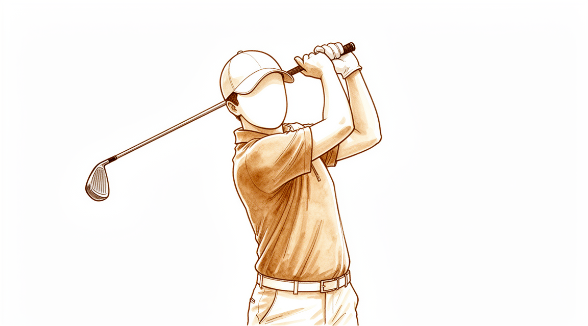
கோல்ஃபர்'ஸ் முழங்கை என்பது முழங்கையின் உள் பக்கத்தில் உள்ள இடுப்புகளின் உடைந்துபோகும் (மோசமடையும்) பிரச்சினையாகும்: நெகிழ்வு-பிரானேட்டர் இடுப்புகள், இது மணிக்கட்டை வளைத்து, கையை கீழே திருப்புகிறது, அங்கு அவை நடுத்தர எபிகோண்டைல் என்று அழைக்கப்படும் எலும்பு முட்டுடன் இணைகின்றன. பழைய பெயர் "எபிகோண்டைலைடிஸ்" இருந்தபோதிலும், இது உண்மையில் ஒரு அழற்சி அல்ல; அதிக சுமை காரணமாக இடுப்பு பலவீனமாகி, ஒழுங்கற்றதாகிவிட்டது. அதனால்தான் நவீன சிகிச்சை ஓய்வு மற்றும் அழற்சி எதிர்ப்பு மருந்துகள் அல்ல, ஆனால் ஒரு படிப்படியான திட்டம் மெதுவாக இடுப்பை மீண்டும் முழு வலிமைக்கு ஏற்றும்.
குணமடைவதற்கு பொறுமை தேவைப்படுகிறது. கோல்ஃப்பரின் முழங்கை பொதுவாக சுய-கட்டுப்படுத்தும், ஆனால் அது முழுமையாக குடியேற 6 முதல் 18 மாதங்கள் ஆகலாம். நல்ல செய்தி என்னவென்றால், பெரும்பாலான மக்கள் ஒரு நல்ல பழமைவாத திட்டத்துடன் சிறப்பாக வருகிறார்கள், ஒருபோதும் அறுவை சிகிச்சை தேவையில்லை. குறைந்தது ஆறு மாத தரமான சிகிச்சை தோல்வியடைந்த பிறகு மட்டுமே அறுவை சிகிச்சை கருதப்படுகிறது.
உட்புற முழங்கை வெளிப்புற (டென்னிஸ்) முழங்கையிலிருந்து வேறுபடுகிறது கால்நடை நரம்பு கால்பந்து வீரரின் முழங்கை நோயாளிகளில் பாதிக்கும் மேற்பட்டவர்கள் இந்த நரம்பில் சில எரிச்சலைக் கொண்டிருக்கிறார்கள், எனவே உங்கள் சிகிச்சையாளர் ஒவ்வொரு வருகையின் போதும் அதைச் சரிபார்த்து, குறிப்பிட்ட நரம்பு-குதிக்கும் பயிற்சிகளைச் சேர்க்கலாம்.
முன்னெச்சரிக்கைகள் மற்றும் வரம்புகள்
செய்:
- சாதாரண அன்றாட பணிகளுக்கு வசதியான வரம்புகளுக்குள் கையை தொடர்ந்து பயன்படுத்தவும்.
- [அடிக்குறிப்புகள்]
- கடுமையான உடற்பயிற்சியின் போது முதுகெலும்பு தசை மீது ஒரு எதிர்மறை வலிமை கொண்ட பிரேஸை அணியுங்கள்.
- [பக்கம் 3-ன் படம்]
செய்யாதது:
- முழங்கையை முழுவதுமாக ஓய்வெடுக்கவோ அல்லது அதை ஒரு அச்சுப்பொறியில் வைக்கவோ வேண்டாம்; நரம்பு குணமடைய மென்மையான சுமை தேவை.
- உங்கள் வலிமை மீட்கப்படும் வரை, வால்ஜஸ் சுமைகளை அதிகரிக்கும் செயல்பாடுகளை ஆரம்பத்தில் தவிர்க்கவும்ஃ கோல்ஃப், வீசுதல் (குறிப்பாக ஹோக்கிங் மற்றும் முடுக்கம் கட்டங்கள்), நீச்சல் மற்றும் ராக்கெட் விளையாட்டுகள்.
- எந்தவொரு உடற்பயிற்சியையும் கூர்மையான வலியாகத் தள்ளாதீர்கள், மேலும் நரம்புகளை ஊசிகள் மற்றும் ஊசிகளாக அல்லது மயக்கமாகத் தள்ளாதீர்கள்.
- உங்கள் அல்னார் நரம்பு அறிகுறிகள் (சிறிய மற்றும் மோதிர விரல்களில் நமைச்சல் அல்லது மயக்கம்) மோசமடைந்தால், உங்கள் சுமைகளை முன்னேற்றுவதற்கு முன்பு நிதானமாக திரும்பி பரிசோதனை செய்யுங்கள்.
உங்கள் பயிற்சிகள்
இவை உங்கள் கையேட்டில் உள்ள பயிற்சிகள். டாக்டர் ஹிர்பாரா மற்றும் உங்கள் சிகிச்சையாளரின் வழிகாட்டுதலின்படி அவற்றைத் தொடங்குங்கள். ஆரம்ப வாரங்களில் வலி, மென்மையான இயக்கம் மற்றும் ஐசோமெட்ரிக் பிடிப்புகள் ஆகியவற்றிற்கு முக்கியத்துவம் அளிக்கப்படுகிறது; நீங்கள் மேம்படுத்தும்போது விசித்திரமான தலைகீழ் டைலர் திருப்பம் மற்றும் பிடியை வலுப்படுத்துதல் சேர்க்கப்படுகின்றன. அல்னார்-நரம்பு சறுக்கல் சேர்க்கப்பட்டுள்ளது, ஏனெனில் நரம்பு பெரும்பாலும் உள் முழங்கையில் ஈடுபடுகிறது; அதை மென்மையாக வைத்திருங்கள்.
உங்கள் மருத்துவ நெறிமுறை
இந்த பக்கத்தின் மீதமுள்ள பகுதி மருத்துவ மறுவாழ்வு நெறிமுறை ஆகும். இந்த பிரிவு உங்கள் பிசியோதெரபிஸ்ட் அல்லது கை சிகிச்சையாளருக்கு வழங்கப்பட வேண்டும். இது வெறுமனே நேர அடிப்படையில் அல்லாமல் அளவுகோல்-கேட் ஆகும்: கட்டங்களுக்கிடையேயான முன்னேற்றம் பட்டியலிடப்பட்ட இலக்குகளை பூர்த்தி செய்வதைப் பொறுத்தது, வெறுமனே காலண்டரில் அல்ல. ஒவ்வொரு வருகைக்கும் (டினெல் அடையாளம், சப்ளக்ஸேஷன்) அல்னார் நரம்பு திரையிடப்படுகிறது, ஏனெனில் சுமார் 5060% மருத்துவ வழக்குகள் ஒத்த அல்னார் நரம்பு அறிகுறிகளைக் கொண்டுள்ளன, அவை கன்சர்வேடிவ் பராமரிப்பு தோல்வியடைய முக்கிய காரணம்.
கீழே இரண்டு பாதைகள் உள்ளன: செயல்படாத திட்டம் (முதல் வரிசை, பெரும்பான்மை) மற்றும் அறுவை சிகிச்சைக்குப் பின் சிகிச்சையில் தோல்வி அடைந்த பிறகு அறுவை சிகிச்சைக்குச் செல்லும் சிறுபான்மையினருக்கு.
செயல்படாத பாதை
கட்டம் I: கடுமையான / வலி கட்டுப்பாடு (02 வாரங்கள்)
குறிக்கோள்கள்: வலியைக் குறைக்கும்; முழு சுமை இல்லாத இயக்க வரம்பை மீட்டெடுக்கும்.
- ஒப்பீட்டு ஓய்வு மற்றும் செயல்பாடு மாற்றம்: வலியைக் கட்டுப்பாடாகப் பயன்படுத்துதல்; இயங்காமல் இருப்பதைத் தவிர்ப்பது. கோல்ஃப், வீசுதல், நீச்சல், ராக்கெட் விளையாட்டுகள், பளு தூக்குதல் மற்றும் மீண்டும் மீண்டும் பிடித்தல் ஆகியவற்றை மாற்றியமைத்தல்.
- விருப்பமான எதிர்வினை வலிமை வலுவூட்டி பொதுவான பிளெக்ஸர் வெகுஜனத்தின் மீது; கடுமையான வலி இருந்தால் ஒரு மணிக்கட்டு அடுக்கு பயன்படுத்தப்படலாம்.
- வலி கட்டுப்பாட்டு துணைஃ பனி, மென்மையான திசு வேலை / IASTM, மென்மையான வலி இல்லாத செயலில் உள்ள இயக்க வரம்பு (AROM), நரம்பு சறுக்கல்கள்.
- அல்னார் நரம்பை திரையிடுதல் (டினெல், சப்ளக்ஸேஷன்).
- முன்னேற்றத்திற்கான அளவுகோல்கள்: முழுமையாக ஏற்றப்படாத AROM வலி இல்லாமல்; சுயாதீனமான வீட்டு நிரல்.
கட்டம் II: சப்-அக்யூட் / ஆரம்பகட்ட சுமை (24 வாரங்கள்)
குறிக்கோள்கள்: flexor-pronator சுமை தொடங்க; அருகில் உள்ள சங்கிலி உரையாற்ற.
- ஐசோமெட்ரிக் மணிக்கட்டு-ஃப்ளெக்சர் மற்றும் ப்ரோனேட்டர் சுமை (ஒளி).
- 90° முழங்கை வளைவில் மணிக்கட்டு வளைக்கும் கருவிகளின் படிப்படியான நீட்டிப்பு.
- அருகிலுள்ள இயக்கச் சங்கிலி: ஸ்கேப்புலர் ஸ்டேபிலைசர்ஸ் (செரடஸ் அன்டீரியர், மிட் / லோயர் டிராபெசியஸ்) மற்றும் ரோட்டேட்டர் கஃப், நடுத்தர முழங்கை ஓவர்லோட் வால்ஜஸ்-உந்துதல் கொண்ட எறிபவர்களில் முக்கியமானவை.
- முன்னேற்றத்திற்கான அளவுகோல்கள்: முழு ROM பராமரிக்கப்படுகிறது; 90 ° நீட்டிப்பு தாங்குகிறது; ~ 70% எதிர் பக்க வலிமை.
கட்டம் III: வலுவூட்டல் / திரும்புதல் (46+ வாரங்கள்)
குறிக்கோள்கள்: சுமை சகிப்புத்தன்மையை மீட்டெடுக்க மற்றும் செயல்பாடு மற்றும் விளையாட்டு திரும்ப.
- எக்ஸென்ட்ரிக்-கான்சென்ட்ரிக் சுமை மணிக்கட்டு வளைவு மற்றும் முதுகெலும்பு வளைவு: டைலர் திருப்பத்தின் நடுத்தர அனலாக் "திருப்பமான டைலர் திருப்பம்" (ஒரு ஃப்ளெக்ஸ்பாரில் எக்ஸென்ட்ரிக் மணிக்கட்டு வளைவு) ஒருங்கிணைந்த எக்ஸென்ட்ரிக்-கான்சென்ட்ரிக் சுமை விரும்பப்படுகிறது; ஆரம்பகால வலி நிவாரணத்திற்கு ஐசோமெட்ரிக்ஸ் பயனுள்ளதாக இருக்கும்.
- இயக்கத்துடன் அணிதிரட்டுதல்; முழங்கை நீட்டிக்கப்பட்ட நிலைக்கு முன்னேற்றம்.
- பிடியை வலுப்படுத்துதல், பின்னர் விளையாட்டு சார்ந்த சுமை; வீசுபவர்களுக்கு, ஒரு இடைவெளி வீசுதல் திட்டம்; பிளையோமெட்ரிக் கடைசியாக.
- முழங்கை அறிகுறிகளற்றதாக மாறும்போது எதிர் வலிமை வளைவை நிறுத்துங்கள்; உபகரணங்கள் மற்றும் நுட்பத்தை உரையாற்றுங்கள்.
- விளையாட்டுக்கு திரும்புவதற்கான அளவுகோல்கள்: ~ 90% எதிர் பக்க வலிமை, வலி இலவச செயல்பாடு, சுய மேலாண்மை.
அறுவை சிகிச்சைக்கு பிந்தைய பாதை (ஃப்ளெக்சர்-ப்ரோனேட்டர் டிப்ரிட்மென்ட் ± பழுதுபார்ப்பு ± அல்னார் நரம்பு நடைமுறை)
அறுவை சிகிச்சை ≥6 மாதங்கள் கன்சர்வேடிவ் பராமரிப்பில் தோல்வியுற்ற சிறுபான்மைக்கு ஒதுக்கப்பட்டுள்ளது. திறந்த Nirschl- வகை அறுவை சிகிச்சை நோயியல் flexor-pronator தோற்றத்தை நீக்குகிறது மற்றும் பொதுவாக அதை சரிசெய்கிறது / மீண்டும் இணைக்கிறது; இடுப்பு நரம்பு மதிப்பீடு செய்யப்பட்டு பாதுகாக்கப்படுகிறது, decompression அல்லது முந்தைய இடமாற்றம் வழக்குகள் ஒரு விகிதத்தில் ஒரே நேரத்தில் செய்யப்படுகிறது.
கட்டம் 1: பாதுகாத்தல் (02 வாரங்கள்)
- பின்புற நீளமான கை அடுக்கு (முழங்கை + மணிக்கட்டு) 1014 நாட்கள்; சமுதாய பயன்பாட்டிற்கான ஸ்லிங்.
- உயர்வு மற்றும் வீக்கம் கட்டுப்பாடு; விரல் / இடுப்பு-கிளைட் AROM; செயலில் தோள்பட்டை ROM; மென்மையான கர்ப்பப்பை வாய் AROM.
- முன்னெச்சரிக்கைகள்: உயர்த்தவோ, தள்ளவோ, இழுக்கவோ அல்லது வலுக்கட்டாயமாகப் பிடிக்கவோ கூடாதுஃ பழுதுபார்ப்பைப் பாதுகாக்கவும்.
கட்டம் 2: ROM மீட்பு (26 வாரங்கள்)
- ~ 2 வார வருகையின் போதுஃ தையல் அகற்றுதல்; நடுநிலை மணிக்கட்டு ஆர்த்தோசிஸ் முழுநேர (சுகாதாரத்திற்காக); வீக்கத்திற்காக முழங்காலில் Tubigrip.
- தொடங்கு AROM முழங்கை வளைவு/நீட்டிப்பு (24 wk), பின்னர் 4-வழி மணிக்கட்டு AROM + முன்கை சுழற்சி மற்றும் விரல் / கட்டைவிரல் AROM (46 wk).
- 46 வாரங்களில் அறிமுகப்படுத்தப்பட்ட உல்னார் நரம்பு ஸ்லைடுகள் (மத்திய-குறிப்பிட்ட சேர்த்தல்).
- தோள்பட்டை உறுதிப்படுத்தல் (ஈர்ப்பு எதிர்ப்பு). 6 வாரங்கள் வரை எதிர்ப்பு வலுவடையவில்லை.
கட்டம் 3: பலப்படுத்துதல் (612 வாரங்கள்)
- (இரவுப் பயன்பாடு ஆரம்பத்தில் தொடரலாம்).
- படிப்படியான எதிர்ப்பு வலுவூட்டல் மார்பு மற்றும் முதுகெலும்பு. எந்த எதிர்ப்பு supination/pronation ஆரம்பத்தில்; supination/ நடுநிலை, உடன் ஒன்பதாவது வாரத்தில் இருந்து லேசான எடையை உயர்த்துதல்.
கட்டம் 4: செயல்பாடு / விளையாட்டுக்கு திரும்புதல் (1216+ வாரங்கள்)
- முன்னேற்றம் அதிகரிப்பு அனைத்து முதுகெலும்பு நிலைகளும் அனுமதிக்கப்பட்டபடி; ~1216 வாரங்களில் முழுமையான செயல்பாட்டுக்கு திரும்புதல்விளையாட்டு வீரர்களுக்கான விளையாட்டு-குறிப்பிட்ட / இடைவெளி வீசுதல் திட்டம். முழுமையான மீட்பு பொதுவாக 36 மாதங்கள் ஆகும்.
உல்னார் நரம்பு முன்னெச்சரிக்கைகள்: ஒரு முந்தைய மாற்றம் செய்யப்பட்டது என்றால், முழங்கை வளைவை முன்கூட்டியே குறைக்கவும் தொடர்ச்சியான அல்லது மோசமான அல்னார் அறிகுறிகள் அறுவை சிகிச்சை முன்னெடுப்பதற்கு முன் அறுவை சிகிச்சை பரிசீலனை தேவை.
வேலை மற்றும் செயற்பாட்டிற்கு திரும்புதல்
நீங்கள் எவ்வளவு விரைவாக திரும்புகிறீர்கள் என்பது நீங்கள் எந்த பாதையில் இருக்கிறீர்கள் மற்றும் உங்கள் வேலை மற்றும் விளையாட்டின் தேவைகளைப் பொறுத்தது.
செயல்படாத. கோல்ஃப், வீசுதல் விளையாட்டுகள், நீச்சல் மற்றும் ராக்கெட் விளையாட்டுகள் வலுவூட்டும் கட்டத்தில் மீண்டும் எளிதாக்கப்படுகின்றன, உங்கள் வலிமை கிட்டத்தட்ட 90% மற்ற பக்கமாகவும், செயல்பாடு வலியற்றதாகவும் இருக்கும். கோல்ஃப்பரின் முழங்கை சுய-வரையறுக்கப்பட்டிருப்பதால், முழுமையான தீர்மானம் 6 முதல் 18 மாதங்கள் வரை ஆகலாம், இருப்பினும் அன்றாட செயல்பாடு மிக விரைவாக மேம்படுகிறது.
அறுவை சிகிச்சைக்குப் பின். லேசான, கட்டுப்படுத்தப்பட்ட பயன்பாடு ஆரம்பத்தில் தொடங்குகிறது, ஆனால் பழுதுபார்ப்பைப் பாதுகாக்க கனமான தூக்குதல் மற்றும் பிடிப்பு ஆகியவை நிறுத்தப்படுகின்றன. பெரும்பாலான மக்கள் சுமார் 12 முதல் 16 வாரங்களுக்குள் முழு செயல்பாட்டிற்குத் திரும்புகிறார்கள், முழுமையான மீட்பு பொதுவாக 3 முதல் 6 மாதங்கள் வரை ஆகும். எறிதல் விளையாட்டு வீரர்கள் போட்டிக்குத் திரும்புவதற்கு முன்பு படிப்படியான இடைவெளி எறிதல் திட்டத்தைப் பின்பற்றுகிறார்கள்.
வாகனம் ஓட்டுதல்: நீங்கள் ஒரு ஸ்பிளின்ட் அல்லது ஸ்லிங்கில் இருக்கும்போது வாகனம் ஓட்டுவதைத் தவிர்க்கவும், அல்லது முழங்கை பாதுகாப்பாக காரை கட்டுப்படுத்த முடியாத அளவுக்கு வலிக்கிறது. நீங்கள் ஸ்பிளின்ட்டை விட்டு வெளியேறியதும், உங்கள் பரிசோதனையில் உறுதிப்படுத்தப்பட்டபடி கையை வசதியாக நகர்த்த முடிந்தவுடன் மீண்டும் தொடரவும்.
உங்கள் நெறிமுறை பிறகு
இந்த நெறிமுறை நடைமுறையின் பொது மீட்பு ஆலோசனையுடன் இணைந்து செயல்படுகிறது; அறுவை சிகிச்சைக்குப் பிந்தைய வலியை நிர்வகித்தல், காயம் பராமரிப்பு மற்றும் கை சிகிச்சை அடிப்படைகள். கோல்ப் வீரரின் முழங்கை அதன் வெளிப்புற-முழங்கை சகா, டென்னிஸ் முழங்கைடன் அதன் சுமை அடிப்படையிலான அணுகுமுறையை பகிர்ந்து கொள்கிறது; நீங்கள் சமமானதை விரும்பினால் உங்கள் சிகிச்சையாளரிடம் கேளுங்கள் பக்கவாட்டு எபிகொண்டிலிட்டிஸ் உங்கள் முழங்கை எவ்வாறு முன்னேறுகிறது என்பதைப் பொறுத்து உங்கள் பிசியோதெரபிஸ்ட் அல்லது கை சிகிச்சையாளரால் உங்கள் தொடர்ச்சியான மீட்பு தனித்தனியாக வழிநடத்தப்படுகிறது.
Evidence & references
Medial Epicondylitis (Golfer's Elbow) — Flexor-Pronator Tendinosis: Conservative Loading & Post-operative Rehabilitation
Topic scope: (A) the loading-based non-operative rehabilitation of medial epicondylitis — a degenerative tendinopathy of the flexor-pronator origin (chiefly flexor carpi radialis and pronator teres) at the medial epicondyle — with mandatory ulnar-nerve screening; and (B) post-operative rehabilitation after open flexor-pronator debridement (± repair, ± concurrent ulnar nerve decompression/transposition), reserved for the minority failing ≥6 months of quality conservative care.
Defining principle: medial epicondylitis is not an inflammatory condition but a degenerative tendinosis, so the treatment is graded tendon loading, not rest. The protocol mirrors lateral elbow tendinopathy but with two practice-defining differences Dr Hirpara emphasises: (1) the loaded group is the wrist flexors/pronators (hence the eccentric "reverse Tyler twist" rather than the lateral Tyler twist), and (2) the ulnar nerve lies immediately behind the medial epicondyle, so concomitant ulnar neuritis (~50–60% of cases) is screened at every visit and is the leading reason conservative care fails. Surgery is a last resort after ≥6 months.
Medial epicondylitis is far less studied than its lateral counterpart — it is ~5–10× less common (prevalence ~0.4% vs 1.3%; ~10–20% of all epicondylitis). Most evidence is extrapolated from lateral elbow tendinopathy and from older operative case series; dedicated medial RCTs are sparse. Phase timelines below come from institutional Standard-of-Care protocols (Mass General Brigham combined medial/lateral; UVA medial debridement; Campbell's / Nirschl) plus operative series.
A. NON-OPERATIVE REHABILITATION (phased)
First-line; the majority resolve without surgery. Largely the SAME phased structure as the lateral elbow (Mass General Brigham publishes ONE combined medial/lateral protocol), with the loading target shifted to the flexor-pronator mass. Expected resolution 6–18 months (self-limited).
Phase I — Acute / pain control (~0–2 weeks). Relative rest + activity modification using pain as the limiter (avoid immobilisation). Aggravators to modify: golf, throwing (esp. late-cocking / acceleration valgus load), swimming, bowling, racquet sports, weightlifting, repetitive gripping. Optional counterforce brace over the common flexor mass; wrist splint if acutely painful. Pain-control adjuncts: ice, soft-tissue / IASTM, gentle pain-free AROM, dry needling, nerve glides. Screen the ulnar nerve (Tinel, subluxation). Criterion to progress: full unloaded AROM without pain; independent home program.
Phase II — Sub-acute / early loading (~2–4 weeks). Isometric wrist-flexor and pronator loading (minimal load). Progressive stretching of the wrist flexors at 90° elbow flexion. Proximal kinetic chain: scapular stabilisers and rotator cuff — critical in throwers, where medial elbow overload is valgus-driven. Criteria to progress: full ROM maintained; tolerates the 90° stretch; ~70% contralateral strength.
Phase III — Late / strengthening & return (~4–6+ weeks). Eccentric and concentric loading of wrist flexion and forearm pronation — the medial analogue of the Tyler twist is a "reverse Tyler twist" (eccentric wrist flexion on the FlexBar). Combined eccentric-concentric loading is favoured; isometrics for early analgesia. Mobilisation-with-movement; progress stretching to the elbow-extended position. Grip strengthening, then sport-specific loading; for throwers, an interval throwing program; plyometrics last. Wean counterforce brace as asymptomatic; equipment/technique modification. Return-to-sport criteria: ~90% contralateral strength, pain-free function, self-management.
B. POST-OPERATIVE REHABILITATION (flexor-pronator debridement ± repair, ± ulnar nerve procedure)
Surgery is for the minority failing ≥6 months of conservative care. The open Nirschl-type operation debrides the pathologic flexor-pronator origin (incision posterior to the medial epicondyle to spare the medial antebrachial cutaneous nerve), with repair/reattachment commonly by suture anchor. The ulnar nerve must be assessed and protected: ulnar neuritis is addressed concurrently (decompression or anterior transposition) in roughly 20–50% of operative series. The phase timeline blends the UVA "Golfer's Elbow Debridement (with tendon repair)" protocol and the Verma / Midwest-Orthopaedics-at-Rush medial/lateral debridement protocol.
Phase 1 — Protect / immobilise (Weeks 0–2). Posterior long-arm splint (elbow + wrist) for 10–14 days; sling for community use. Elevation; oedema control; finger/tendon-glide AROM; unaffected-joint motion; active shoulder ROM; gentle cervical AROM. Precautions: NO lifting / pushing / pulling / forceful gripping; protect the repair.
Phase 2 — ROM restoration (Weeks 2–6). At the 2-wk visit: suture removal; transition to a wrist orthosis in neutral full-time (off for hygiene); Tubigrip at the elbow for swelling. Begin AROM elbow flexion/extension (2–4 wk), then 4-way wrist AROM + forearm rotation, finger/thumb AROM (4–6 wk). Ulnar nerve glides introduced ~weeks 4–6 (the explicit medial-specific addition). Scapular stabilisation (gravity-resisted). No resistance strengthening until after 6 weeks.
Phase 3 — Strengthening (Weeks 6–12). Wean the orthosis as tolerated (consider night use early). Progressive resistive strengthening of wrist and forearm; per Verma, no resisted supination/pronation early, lifting begun in supination/neutral, with light pronated lifting from ~week 9.
Phase 4 — Return to activity / sport (Weeks 12–16+). Progress lifting in all forearm positions as tolerated; full return to activity by ~12–16 weeks; sport-specific / interval throwing program for athletes. Full recovery commonly 3–6 months.
Ulnar nerve precautions: if an anterior transposition was performed, limit end-range elbow flexion early and progress nerve excursion gradually; persistent or worsening ulnar symptoms warrant surgeon review before advancing loading.
C. PHASED TIMELINE SUMMARY
| Pathway | Phase | Window | Immobilisation | Loading / key actions | Criteria / milestone |
|---|---|---|---|---|---|
| Non-op | I — Pain control | 0–2 wk | None (avoid casting); optional counterforce brace | Activity modification; pain-free AROM; nerve glides; ulnar screen | Full unloaded AROM, pain-free |
| Non-op | II — Early loading | 2–4 wk | None | Isometric flexor/pronator load; 90° wrist-flexor stretch; scapular/cuff | ~70% contralateral strength |
| Non-op | III — Strengthen / return | 4–6+ wk | Wean brace | Reverse Tyler twist (eccentric); grip; sport-specific; throwers' interval program | ~90% strength, pain-free → RTS |
| Post-op | 1 — Protect | 0–2 wk | Posterior long-arm splint 10–14 d + sling | Finger glides, shoulder ROM; oedema control | No resistance; repair protected |
| Post-op | 2 — ROM restore | 2–6 wk | Neutral wrist orthosis | Elbow AROM → 4-way wrist + forearm rotation; ulnar glides wk 4–6 | No resistance until >6 wk |
| Post-op | 3 — Strengthen | 6–12 wk | Wean orthosis | Progressive resistance; supinated/neutral lifting → light pronated ~wk 9 | Restored strength in safe positions |
| Post-op | 4 — Return | 12–16+ wk | None | Lifting all forearm positions; interval throwing | Full return ~12–16 wk; recovery 3–6 mo |
D. KEY CONTROVERSIES / EVIDENCE QUALITY
- Sparse high-level evidence. Almost no medial-specific RCTs; recommendations are extrapolated from lateral elbow and from retrospective operative series (Kurvers & Verhaar 1995 remains a cornerstone). Strength of evidence is materially weaker than for lateral epicondylitis.
- Ulnar nerve is the dominant modifier. Concomitant ulnar neuropathy (reported 23–60%) worsens prognosis and is the leading reason conservative care fails; whether and how to address it surgically (decompression vs transposition vs medial epicondylectomy) is debated. Outcomes are reliably worse when ulnar symptoms coexist and are untreated.
- PRP may rival surgery for type-1 disease. Bohlen et al (OJSM 2020) found 2 leukocyte-rich PRP injections matched surgery for recalcitrant type-1 medial epicondylitis (29/33 success each) with faster recovery (pain-free ~56 vs ~108 days; full ROM ~42 vs ~96 days) — the surgical delay partly attributed to post-op bracing. Small evidence base.
- Corticosteroid: short-term only. As with the lateral elbow, steroid gives transient relief without durable benefit and risks recurrence; repeated injections show diminishing returns.
- Eccentric vs concentric. Same unsettled debate as the lateral elbow; combined eccentric-concentric flexor-pronator loading is the pragmatic standard, but direct medial trial data are minimal.
- Surgical technique. Open Nirschl debridement with repair is reliable in case series; arthroscopic medial debridement is emerging (claimed ulnar-nerve protection) but is technically demanding and under-evidenced. Debridement alone vs with repair remains unsettled.
E. EVIDENCE STRENGTH FLAGS (summary)
- MODERATE (non-operative rehab): the phased loading program — extrapolated largely from lateral elbow tendinopathy and combined medial/lateral institutional protocols; combined eccentric-concentric flexor-pronator loading is the pragmatic standard.
- LOW–MODERATE (post-operative rehab): phase timelines from institutional debridement protocols (UVA; Verma/Rush) and operative case series; no defining post-op rehab RCT.
- MODERATE (PRP for type-1 disease): single comparative study (Bohlen OJSM 2020) matching surgery with faster recovery; small sample.
- CONSENSUS / EXPERT: ulnar-nerve screening at every visit, ulnar-glide timing (wk 4–6 post-op), and the forearm-position lifting progression — drawn from surgeon-guidance protocols and operative practice rather than trial data.
CITATIONS
RAG corpus (180,000+ Orthopaedic articles)
- Kurvers H, Verhaar J. The results of operative treatment of medial epicondylitis. J Bone Joint Surg Am. 1995. (ulnar neuritis coexistence 23–50%)
- Bohlen HL, et al. Platelet-rich plasma is an equal alternative to surgery in the treatment of type 1 medial epicondylitis. Orthop J Sports Med. 2020. DOI: 10.1177/2325967120908952
- Platelet-rich plasma versus Tenex in the treatment of medial and lateral epicondylitis. J Shoulder Elbow Surg. 2019.
- Ellenbecker TS, Nirschl R, Renstrom P. Current concepts in examination and treatment of elbow tendon injury. Sports Health. 2012.
- Rehabilitation of the thrower's elbow. Clin Sports Med. 2004.
- Nirschl surgical technique for concomitant lateral and medial elbow tendinosis. Am J Sports Med. 2011.
- Imaging of the elbow in the overhead throwing athlete. Am J Sports Med. 2003. (ulnar neuritis in ~60% of throwers with medial epicondylitis)
- Outcome of partial medial epicondylectomy for cubital tunnel syndrome. Clin Orthop Relat Res. 2006.
- Coonrad RW, Hooper WR. Tennis elbow: its course, natural history, conservative and surgical management (includes medial). J Bone Joint Surg Am. 1973.
- Green's Operative Hand Surgery. 2021. (medial vs lateral prevalence; combined treatment chapter; Nirschl technique)
- Campbell's Operative Orthopaedics. 2020. (Box 46.3 Rehabilitation Protocol for Epicondylitis [Wilk/Arrigo/Andrews]; Nirschl medial technique, posterior incision sparing the MABC nerve)
Published protocols (URLs)
- University of Virginia Orthopaedics — Medial Epicondyle (Golfer's Elbow) Debridement (with tendon repair), Rehabilitation Guidelines. https://med.virginia.edu/orthopaedic-surgery/wp-content/uploads/sites/242/2024/09/Medial-Epicondyle-Golfers-Elbow-Debridement-with-tendon-repair.pdf
- Midwest Orthopaedics at Rush (Nikhil Verma, MD) — Post-Operative Rehabilitation Guidelines for Medial/Lateral Epicondyle Debridement. https://www.sportssurgerychicago.com/patient-resources/rehab-manuals/mediallateral-epicondyle-debridement/
- Mass General Brigham Sports Medicine — Rehabilitation Protocol for Medial/Lateral Epicondylalgia (non-operative), rev. April 2021. https://www.massgeneral.org/assets/MGH/pdf/orthopaedics/sports-medicine/physical-therapy/rehabilitation-protocol-for-medial-lateral-epicondylitis.pdf


