ரேடியல் தலை மாற்று
Patients › Rehabilitation
முழங்காலில் உள்ள ரேடியல் தலையை ஒரு உலோக உள்வைப்புடன் மாற்றியமைத்த பின்னர் பாதுகாக்கப்பட்ட மீட்பு திட்டம், முழங்கை மற்றும் முன்கை இயக்கம் ஆகியவற்றைச் சுற்றி விறைப்புத்தன்மையைத் தடுக்க, முன்கை எந்தவொரு சரிசெய்யப்பட்ட இழைகளையும் பாதுகாக்க நிலைநிறுத்தப்பட்டு, வசதிக்காக முழங்கை ஒரு எளிய ஸ்லிங்கில் ஓய்வெடுத்தது.
இந்த நெறிமுறை உங்கள் மீட்பு வழிகாட்டுகிறது ரேடியல் தலை மாற்றம் இது உங்கள் வீட்டு உடற்பயிற்சி திட்டத்துடன் தொடங்குகிறது, அதைத் தொடர்ந்து உங்கள் கை சிகிச்சையாளருக்காக எழுதப்பட்ட கட்டமைக்கப்பட்ட மருத்துவ நெறிமுறை; இந்த பக்கத்தை அல்லது அதன் PDF ஐ உங்கள் முதல் சிகிச்சை வருகைக்கு கொண்டு வாருங்கள், இதனால் உங்கள் மறுவாழ்வு ஒருங்கிணைந்ததாக இருக்கும். உங்கள் சிகிச்சையாளர் உங்கள் மீட்பு எவ்வாறு முன்னேறுகிறது என்பதையும், உங்கள் அறுவை சிகிச்சையின் போது சரியாக என்ன சரிசெய்யப்பட்டது என்பதையும் பொறுத்து திட்டத்தை சரிசெய்யலாம்.
அறுவை சிகிச்சைக்குப் பிறகு உங்கள் காயத்தைப் பற்றி ஏதேனும் கவலைகள் இருந்தால், அறைகளைத் தொடர்பு கொள்ளுங்கள். காயத்தின் புகைப்படத்தை எடுத்து அதை மதிப்பாய்வு செய்ய மின்னஞ்சல் அனுப்புவது பெரும்பாலும் உதவியாக இருக்கும்.
எதிர்பார்ப்பது என்ன
ரேடியல் தலை என்பது இரண்டு முதுகெலும்புகளில் ஒன்றின் வட்டமான மேல் பகுதியாகும், அங்கு அது முழங்கை சந்திக்கிறது. இது சரிசெய்ய முடியாத அளவுக்கு பல துண்டுகளாக உடைக்கப்படும்போது, அது சிறியதாக மாற்றப்படுகிறது உலோக உள்வைப்பு இது ஒரு நிலையான, ஒத்த முழங்கை மற்றும் ஒரு மென்மையான முதுகெலும்பு சுழற்சி அச்சு மீட்டெடுக்கிறது. இது பெரும்பாலும் ஒரு சிக்கலான காயத்தை சரிசெய்யும் ஒரு பகுதியாக செய்யப்படுகிறது, ஒரு எலும்பு முறிவு-விலகல் சில நேரங்களில் ஒரு "பயங்கரமான முக்கோணம்", அங்கு ரேடியல் தலை, கொரோனோய்டின் ஒரு பகுதி, மற்றும் முழங்கை பக்கத்தில் உள்ள இழைகள் அனைத்தும் சேர்ந்து காயமடைகின்றன.
உள்வைப்பு ஸ்திரத்தன்மையை மீட்டெடுப்பதால், உங்கள் மறுவாழ்வின் முன்னுரிமை விறைப்புத்தன்மையை தடுக்க ஆரம்ப பாதுகாக்கப்பட்ட இயக்கம் ஆகும்; முழங்கைகள் இந்த வகையான காயத்திற்குப் பிறகு விறைப்புத்தன்மைக்கு மிகவும் ஆளாகின்றன, அதற்கு எதிரான சிறந்த பாதுகாப்பு ஆரம்பத்தில் நகர்வதைத் தொடங்குவதாகும். உங்கள் முழங்கை ஒரு வசதிக்காக எளிய ஸ்லிங் உடற்பயிற்சிகளுக்கு இடையில் (இல்லை கீறல் கொண்ட ப்ரேஸ்), மற்றும் உங்கள் உடற்பயிற்சிகள் மற்றும் கழுவுவதற்கு ஸ்லிங் அகற்றப்படுகிறது.
இரண்டு விஷயங்கள் நீங்கள் எவ்வளவு விரைவாக மற்றும் எவ்வளவு தூரம் நகர்கிறீர்கள் என்பதை தீர்மானிக்கின்றன:
- சரிசெய்யப்பட்ட எந்த இழைகளும் பாதுகாக்கப்பட வேண்டும். முழங்கையின் வெளிப்புறத்தில் உள்ள இழை (பக்க பக்கவாட்டு இழை) சரிசெய்யப்பட்டால், முன்கை பிடித்து உடற்பயிற்சி செய்யப்படுகிறது (அவள்) தம் கரங்களால் (குனிந்து) சாய்ந்தவள். ஆரம்பத்தில்; உட்புறத்தில் உள்ள இழை (மத்திய பக்கவாட்டு இழை) சரிசெய்யப்பட்டால், அது நடைபெறுகிறது கரத்தால் மேலே (சூப்பினேட்டட்)உங்கள் சிகிச்சையாளர் உங்களுக்கு எது பொருந்தும் என்று சொல்வார்.
- முழங்கை பக்கவாட்டு அழுத்தத்திலிருந்து (வாரஸ்) பாதுகாக்கப்பட வேண்டும், ஆரம்பத்தில், முழு நேராக இருந்து முழங்கை உறுதியற்றதாக இருந்தால். அதனால்தான், ஒரே நேரத்தில் அல்லாமல், படிப்படியாக இயக்கம் திறக்கப்படுகிறது.
இயக்கம் சீராக முன்னேறுகிறது, பொதுவாக ஆறு வாரங்களுக்குப் பிறகு வலுவடைந்து, மூன்று மாதங்களுக்குள் முழு செயல்பாட்டிற்கு திரும்புகிறது. உள்வைப்பு மற்றும் குணமடைதல் பல மாதங்களுக்கு தொடர்ந்து குடியேறுகிறது, அதனால்தான் கனமான சுமை படிப்படியாக மீண்டும் கட்டமைக்கப்படுகிறது.
முன்னெச்சரிக்கைகள் மற்றும் வரம்புகள்
- வசதிக்காக வழிகாட்டுதலின்படி எளிய ஸ்லிங்கை அணியுங்கள்; இது ஒரு ஹிஞ்ச் செய்யப்பட்ட ப்ரேஸ் அல்ல, உடற்பயிற்சி மற்றும் கழுவுதலுக்கு அகற்றப்படுகிறது.
- ஆரம்ப பயிற்சிகளின் போது உங்கள் சிகிச்சையாளர் உங்களுக்குக் கொடுக்கும் நிலையில் முதுகெலும்பை வைத்திருங்கள் (வெளிப்புற இழை சரிசெய்யப்பட்டால், பனை-கீழே, உள் ஒன்று சரிசெய்யப்பட்டால், பனை-மேலே, நடுநிலை என்றால் இரண்டும்), இது பழுதுபார்ப்பைப் பாதுகாக்கிறது.
- செய் இல்லை முழங்கால் வழியாக பக்கவாட்டு (வாருஸ்) அழுத்தத்தை செலுத்துங்கள்; முழங்காலில் சாய்வதைத் தவிர்க்கவும் அல்லது ஆரம்பத்தில் உங்கள் உடல் முழுவதும் ஆதரவற்ற கை தொங்கவிடவும்.
- செய் இல்லை முழங்கை நிலையற்றதாக இருப்பதாகக் கூறப்பட்டால், முழங்கையை முழுமையாக நேராகச் செய்யுங்கள்; உங்கள் அனுமதிக்கப்பட்ட வரம்பிற்குள் மட்டுமே நேராகச் செய்யுங்கள்.
- செய் இல்லை (பொதுவாக ஆறு வாரங்களுக்குள்) சுத்தம் செய்யப்படும் வரை அறுவை சிகிச்சை செய்யப்பட்ட கை வழியாக எடையை உயர்த்தவும், தள்ளவும், இழுக்கவும் அல்லது தாங்கவும்; ஆரம்பத்தில் கையை லேசாக பயன்படுத்தவும்.
- உங்கள் தோள்பட்டை, மணிக்கட்டு மற்றும் விரல்கள் தொடக்கத்தில் இருந்து நகரும் வைத்து, மற்றும் இல்லை உங்கள் கை ஸ்லிங்கில் இருக்கும்போது அல்லது சக்கரத்தை பாதுகாப்பாக கட்டுப்படுத்த முடியாதபோது வாகனம் ஓட்டுங்கள்.
காயம், வீக்கம் மற்றும் வடுக்கள் மேலாண்மை, நடைமுறையில் பார்க்க காயம் பராமரிப்பு வழிகாட்டல்.
உங்கள் பயிற்சிகள்
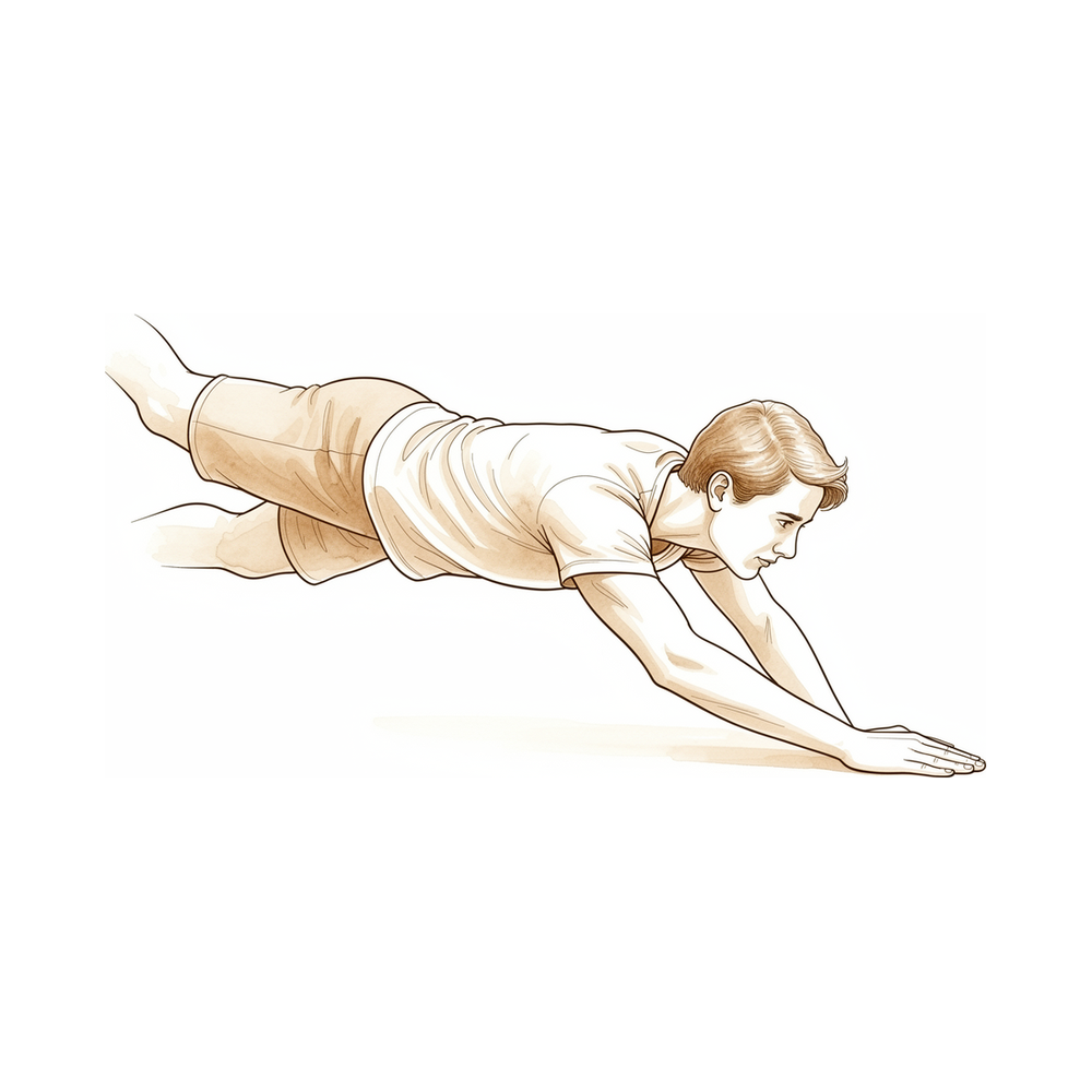
இவை உங்கள் கையேட்டில் உள்ள பயிற்சிகள். டாக்டர் ஹிர்பரா மற்றும் உங்கள் கை சிகிச்சையாளரின் வழிகாட்டுதலின்படி மட்டுமே அவற்றைத் தொடங்குங்கள், உங்களுக்கு வழங்கப்பட்ட எந்தவொரு வரம்பிலும் முன்கை நிலைகளிலும் தங்கியிருங்கள். ஆரம்ப பயிற்சிகள் எந்தவொரு பழுதுபார்ப்பையும் இல்லாமல் கடினத்தன்மையைத் தடுக்க முழங்கை மற்றும் முன்கைகளை நகர்த்துகின்றன: செயலில் உதவியுடன் முழங்கை வளைத்தல் மற்றும் நேராக்குதல், உங்கள் அனுமதிக்கப்பட்ட திசையில் முன்கை சுழற்சி, மற்றும் தோள்பட்டை மற்றும் கையை இலவசமாக வைத்திருத்தல். வலுவூட்டல் மற்றும் வடுக்களை பராமரிப்பு பிற்கால கட்டங்களுக்கு சொந்தமானது, மேலும் நீங்கள் குறிப்பாக அழிக்கப்படும் வரை தொடங்கக்கூடாது. கூர்மையான வலி அல்லது முழங்கை வழிவகுக்கும் என்ற உணர்வை ஏற்படுத்தும் எதையும் நிறுத்துங்கள்.
உங்கள் மருத்துவ நெறிமுறை
இந்த பக்கத்தின் மீதமுள்ள பகுதி ரேடியல் தலை மாற்று (ரேடியல் ஹெட் ஆர்த்தோபிளாஸ்டிரி) க்குப் பிறகு மறுவாழ்வுக்கான மருத்துவ நெறிமுறை ஆகும், இது பெரும்பாலும் மறுசீரமைக்க முடியாத கம்யூட்டட் ரேடியல் ஹெட் எலும்பு முறிவு, பெரும்பாலும் ஒரு பயங்கரமான முக்கோண எலும்பு முறிவு-வெளியேறலின் ஒரு பகுதியாக செய்யப்படுகிறது. இந்த பிரிவு கை சிகிச்சையாளருக்கு வழங்கப்பட வேண்டும், மேலும் ஒவ்வொரு கட்டமும் என்ன நடக்கிறது என்பதற்கான எளிய ஆங்கில விளக்கத்துடன் திறக்கப்படுகிறது. உள்வைப்பு ஒரு நிலையான, ஒத்திசைவான ரேடியோ கேபிடெலர் விசைப்பலகலை மீட்டமைக்கிறது, எனவே வழிகாட்டும் கொள்கை இந்த முழங்கைகள் பலவீனமாக இருப்பதைத் தடுக்க ஆரம்ப இயக்கம் பாதுகாக்கப்படுகிறது, வில் மற்றும் முன் கை சுழற்சி எந்தவொரு இணை-இணைப்பு மற்றும் கொரோனாய்டு பழுதுபார்ப்புகளின் ஒருமைத்தன் மூலம் வாயிலிடப்படுகிறது.
சிகிச்சைக்கு முன்னர், நோயாளியின் செயல்பாட்டு அறிக்கை மற்றும் பரிசோதனையின் கீழ் மயக்க நிலைத்தன்மையின் மதிப்பீட்டைச் சரிபார்த்து, சிகிச்சையளிக்கும் அறுவை சிகிச்சையாளருடன் தொடர்பு கொள்ளுங்கள்ஃ எந்த பக்கவாட்டு இழைகள் மற்றும்/அல்லது கொரோனோயிட் சரி செய்யப்பட்டது, நிலையான வளைவு இன்ட்ரா-ஆபரேட்டிவ் முறையில் நிரூபிக்கப்பட்டது, மற்றும் பாதுகாப்பு முதுகெலும்பு சுழற்சி. டாக்டர் ஹிர்பாரா வசதிக்காக ஒரு எளிய ஸ்லிங்கில் முழங்கை ஓய்வெடுக்கிறார் (ஹிஞ்சன்ஸ் பேரேட் இல்லை) மற்றும் நிலைத்தன்மை அனுமதிக்கும் போது விரைவான, ஆரம்ப இயக்க அணுகுமுறையை விரும்புகிறார். முதுகெலும்பு நிலை விதிஃ எல்சிஎல் பழுது → உடற்பயிற்சி/ஓய்வு நிலையில்; எம்சிஎல் பழுது → சுபினேஷன்; இரண்டும் → நடுநிலை நடு நிலைவார்ஸ் அழுத்தத்தைத் தவிர்க்கவும், முழங்கை நிலையற்றதாக இருந்தால், முனைய நீட்டிப்பு ஆரம்பத்தில்.
கட்டம் I ஆரம்பகால பாதுகாக்கப்பட்ட இயக்கம் (0 முதல் 2 வாரங்கள் வரை)
முதல் இரண்டு வாரங்கள் காயத்தின் ஸ்திரத்தன்மை (பெரும்பாலும் முதல் வாரத்திற்குள்) கடினத்தன்மையை விட முன்னேற அனுமதிக்கும்போது மென்மையான பாதுகாக்கப்பட்ட இயக்கத்தைத் தொடங்குகின்றன. ஆறுதலுக்காக ஒரு எளிய ஸ்லிங்கில் கை ஓய்வெடுக்கிறது, உடற்பயிற்சிகள் மற்றும் சுகாதாரத்திற்காக. முழங்கை அதன் பாதுகாப்பான வில் வழியாக நகர்கிறது, முதுகெலும்பு எந்த இழைக்கான பாதுகாப்பு சுழற்சியில் பராமரிக்கப்படுகிறது.
உங்கள் கை சிகிச்சையாளருக்கு:
கல்வி மற்றும் முன்னெச்சரிக்கைகள் - அசையாத நிலையில் வசதிக்காக எளிய ஸ்லிங் (ஹிஞ்ச் செய்யப்பட்ட பிரேஸ் இல்லை); உடற்பயிற்சிகள் மற்றும் கழுவுதல் - சுறுசுறுப்பான-உதவி/சுறுசுறுப்பான முழங்கை வளைவுநீட்டிப்பு அறுவை சிகிச்சைக்குள் நிரூபிக்கப்பட்ட நிலையான வில்; முழங்கை நிலையற்ற இருந்தால் முனை நீட்டிப்பு தவிர்க்க - பாதுகாப்பு நிலையில் முதுகெலும்பு சுழற்சி: எல்சிஎல் சரிசெய்யப்பட்டால் ப்ரோனேஷன், எம்சிஎல் சரிசெய்யப்பட்டால் சப்னைஷன், இருவரும் சரிசெய்யப்பட்டால் நடுநிலை நடுத்தர வரம்பு - வார்ஸ் அழுத்தம் இல்லை எந்த நேரத்திலும்; வார்ஸ் நடுநிலையற்ற மற்றும் கூட்டு coapt ஈர்ப்பு பயன்படுத்த இடதுபுறம் உடற்பயிற்சிகளை செய்ய - எடை தாங்கி அல்லது இயக்கப்படும் கை மூலம் தள்ளுதல் இல்லை
நிர்வாகம் - காயம்: இயக்கம் தொடங்குவதற்கு முன்னர் காயம் நிலையானது என்பதை உறுதிப்படுத்த - வீக்கம்: உயர்வு, மென்மையான கை பம்ப், தேவைக்கேற்ப பனி - உடற்பயிற்சிகள்: AAROM/செயல்பட்ட முழங்கை வளைவு நிலையான வளைவில் நீட்டிப்பு; முழங்கை 90° இல் பாதுகாக்கப்பட்ட திசையில் முன்னோக்கி / மேல்நோக்கி; முழு செயலில் தோள்பட்டை, மணிக்கட்டு, கை மற்றும் பிடியில் ROM
முன்னேற்றத்திற்கான அளவுகோல்கள் - காயம் அமைந்து; பாதுகாக்கப்பட்ட வில் உள்ள வசதியான கட்டுப்படுத்தப்பட்ட இயக்கம்
கட்டம் II வில் மற்றும் முதுகெலும்பு சுழற்சியின் முன்னேற்றம் (2 முதல் 6 வாரங்கள் வரை)
சுமார் இரண்டு முதல் ஆறு வாரங்கள் வரை பாதுகாக்கப்பட்ட வளைவு படிப்படியாக முழு நீட்டிப்பு நோக்கி விரிவடைகிறது மற்றும் முன்கை சுழற்சி இரண்டு திசைகளிலும் திறக்கப்படுகிறது, முழுமையான முனையம் / சுப்பினேஷன் குறிக்கோளுடன் சுமார் எட்டு வாரங்கள். வலுவூட்டல் மற்றும் சுமை இன்னும் நிறுத்தப்பட்டுள்ளது.
உங்கள் கை சிகிச்சையாளருக்கு:
மதிப்பீடுகள் - செயலில் மற்றும் செயலற்ற முழங்கை வளைவு நீட்டிப்பு மற்றும் முதுகெலும்பு சுழற்சி; வலி மற்றும் வீக்கம்; காயம் / ஸ்கார் ஆய்வு; நிலைத்தன்மை அறிகுறிகள்
கல்வி மற்றும் முன்னெச்சரிக்கைகள் - முன்னேற்றம் முழு நீட்டிப்பு ஸ்திரத்தன்மை அனுமதிக்கிறது (எந்தவொரு ஆரம்ப நீட்டிப்பு தொகுதி படிப்படியாக வெளியிடப்படுகிறது) - முன்னேற்றம் இரு திசைகளிலும் முதுகெலும்பு சுழற்சி முழு நோக்கி, இன்னும் இந்த கட்டத்தில் ஆரம்பத்தில் சரிசெய்யப்பட்ட இழை நினைவில் - வார்ஸ் அழுத்தம் மற்றும் கை வழியாக எந்த சுமை தவிர்க்க தொடர்ந்து
நிர்வாகம் - உடற்பயிற்சிகள்ஃ முழங்கை வளைவு நீட்டிப்பு வளைவை முழுமையாக விரிவுபடுத்துதல்; முழு ROM க்கு முன்னேற்றம் (இலக்கு 8 வாரங்களுக்குள் முழுமையானது); காயம் குணமடைந்தவுடன் வடுக்கள் சிகிச்சையைத் தொடங்குங்கள்; தோள் / மணிக்கட்டு / கை ROM ஐத் தொடரவும் - மீதமுள்ள நிலையற்ற தன்மை ஒரு கவலையாக இருக்கும்போது ஒரு மேல்நிலை (சிற்றலை) இயக்கம் திட்டம் பயனுள்ளதாக இருக்கும்
முன்னேற்றத்திற்கான அளவுகோல்கள் - முழுமையான வலியற்ற ROM ஐ நெருங்குகிறது; நிலையற்ற தன்மை அறிகுறிகள் இல்லை; வலி ≤3/10
கட்டம் III வலுவூட்டல் மற்றும் திரும்புதல் (6 முதல் 12 வாரங்கள் மற்றும் அதற்கு அப்பால்)
இயக்கம் மீட்டெடுக்கப்பட்டு, பழுதுபார்ப்புகள் பாதுகாப்பானதாகக் கருதப்பட்டவுடன் (பொதுவாக ஆறு வாரங்களுக்குள்), வலுவூட்டல் தொடங்குகிறது மற்றும் படிப்படியாக கட்டமைக்கப்படுகிறது (முதலில் பிடியுங்கள், பின்னர் எதிர்க்கப்பட்ட முழங்கை மற்றும் முன்கை வேலை), அடுத்த வாரங்களில் முன்னேறுகிறது. கனமான செயல்பாட்டிற்கு திரும்புவது அளவுகோல் அடிப்படையிலானது, பொதுவாக மூன்று மாதங்களுக்குள்.
உங்கள் கை சிகிச்சையாளருக்கு:
மதிப்பீடுகள் - முழங்கை மற்றும் முதுகெலும்பு வலிமை மற்ற பக்கத்திற்கு எதிராக; சுமைக்கு வலி / வீக்கம் பதில்; செயல்பாட்டு மற்றும் வேலை-/விளையாட்டு-குறிப்பிட்ட சோதனைகள்
கல்வி மற்றும் முன்னெச்சரிக்கைகள் - தொடங்கு மென்மையான எதிர்ப்பு வலுவூட்டல் (கைப்பிடிப்பு → எதிர்ப்பு முழங்கை வளைவு நீட்டிப்பு மற்றும் pro/sup) சுமார் ஆறு வாரங்களில் இருந்து; கட்டமைக்க சுமை படிப்படியாக - அனுமதிக்கப்பட்டபடி செயல்பாட்டு மற்றும் வேலை-குறிப்பிட்ட சுமைக்கு முன்னேற்றம்; திடீர் கனமான அல்லது தாக்க சுமைகளை முன்கூட்டியே தவிர்க்கவும்
நிர்வாகம் - உடற்பயிற்சிகள்: படிப்படியான எதிர்ப்புடன் முழங்கை/முதுகெலும்பு வலுவூட்டல் (பேண்ட் → லேசான எடைகள்); பிடிப்பு வலுவூட்டல்; படிப்படியான செயல்பாட்டு சுமை; மீதமுள்ள இயக்கம் தொடர்பான வேலைகளைத் தொடரவும் - தொடர்ச்சியான அல்லது மோசமான வலி, இயந்திர அறிகுறிகள் அல்லது இயக்கம் இழப்பு (சாத்தியமான இம்ப்ளான்ட் அதிகப்படியான நிரப்புதல் / தளர்த்தல் அல்லது மூலக்கூறு உடைதல்) ஆகியவற்றைக் கண்காணிக்கவும், சிகிச்சையளிக்கும் மருத்துவரை மீண்டும் பரிந்துரைக்கவும் - இயக்கம் செயல்பாட்டு மற்றும் வலிமை கிட்டத்தட்ட சமச்சீரற்ற உள்ளது ஒருமுறை வெளியேற்றத்தை கருத்தில்
முழுமையான செயல்பாட்டிற்கு திரும்புவதற்கான அளவுகோல்கள் - செயல்பாட்டு வலி இல்லாத ROM; கிட்டத்தட்ட சமச்சீரற்ற வலிமை; சுமை கீழ் நம்பிக்கையான, நிலையான முழங்கை
வேலை மற்றும் செயற்பாட்டிற்கு திரும்புதல்
எளிய தினசரி கை பயன்பாடு (உண்ணல், எழுதுதல், எளிய சுய-பராமரிப்பு) தொடக்கத்திலிருந்தே ஊக்குவிக்கப்படுகிறது, அது வசதியாக இருக்கும் வரை, அது முழங்கை வழியாக தள்ளுதல், தூக்குதல் அல்லது எடையை சுமப்பது ஆகியவற்றை உள்ளடக்காது. உங்கள் கை ஸ்லிங்கில் இருக்கும்போது அல்லது சக்கரத்தை பாதுகாப்பாகக் கட்டுப்படுத்த முடியாதபோது நீங்கள் ஓட்டக்கூடாது என்பதால், ஆரம்ப வாரங்களில் போக்குவரத்துக்கான உதவியைத் திட்டமிடுங்கள்; நீங்கள் ஸ்லிங்கிலிருந்து வெளியேறி, காரை கட்டுப்படுத்த முடிந்தவுடன் ஓட்டுதல் தொடர்கிறது, உங்கள் மதிப்பாய்வில் உறுதிப்படுத்தப்பட்டுள்ளது.
வலுவூட்டல் பொதுவாக ஆறு வாரங்களிலிருந்து தொடங்குகிறது மற்றும் படிப்படியாக கட்டமைக்கப்படுகிறது. கனமான வேலை, தூக்குதல் மற்றும் விளையாட்டுக்கு திரும்புவது பொதுவாக மூன்று மாதங்கள் ஆகும், மேலும் முழு வலி இல்லாத இயக்கம் மற்றும் ஒரு நிலையான முழங்காலுடன் போதுமான, சமச்சீர் வலிமையை மீட்டெடுப்பதை அடிப்படையாகக் கொண்டது, இது டாக்டர் ஹிர்பாரா மற்றும் உங்கள் கை சிகிச்சையாளரால் தீர்மானிக்கப்படுகிறது. கனமான கையேடு வேலை மற்றும் தொடர்பு விளையாட்டு ஆகியவை ஒரே அளவுகோல் அடிப்படையிலான முன்னேற்றத்தைப் பின்பற்றுகின்றன.
உங்கள் நெறிமுறை பிறகு
இந்த நெறிமுறை நடைமுறையின் பொது மீட்பு ஆலோசனையுடன் இணைந்து செயல்படுகிறது; அறுவை சிகிச்சைக்குப் பிந்தைய வலியை நிர்வகித்தல், காயம் பராமரிப்பு மற்றும் வடு மேலாண்மைமேலே உள்ள படிப்படியான திட்டம், வெளியிடப்பட்ட மறுவாழ்வு வழிகாட்டுதல்களை பிரதிபலிக்கிறது. ரேடியல் ஹெட் ஆர்த்ரோபிளாஸ்டிசி மற்றும் பயங்கரமான-மூன்றெழுத்து புனரமைப்பிற்குப் பிறகு, உங்கள் தொடர்ச்சியான மீட்பு டாக்டர் ஹிர்பரா மற்றும் உங்கள் கை சிகிச்சையாளரால் தனித்தனியாக வழிகாட்டப்படுகிறது. உங்கள் முழங்கை எவ்வாறு முன்னேறுகிறது மற்றும் சரியாக என்ன சரி செய்யப்பட்டது என்பதைப் பொறுத்து.
Evidence & references
Radial Head Replacement — Procedure Outcomes & Post-operative Rehabilitation (Radial Head Arthroplasty for Unreconstructable Fracture / Terrible Triad)
Topic scope: post-operative rehabilitation after radial head arthroplasty (RHA) — replacement of an unreconstructable comminuted radial head with a metallic implant — performed either in isolation or, more commonly, as one component of reconstructing a fracture-dislocation (the "terrible triad": radial head + coronoid + lateral collateral ligament ± medial collateral ligament). The radial head is a key secondary stabiliser of the elbow against valgus and axial (posterolateral rotatory) load, so the implant exists to restore a stable, congruent radiocapitellar articulation and forearm axis — not merely to fill a defect.
Defining principle of the rehab here: the implant restores stability, so the dominant clinical enemy is stiffness, to which these elbows are strongly predisposed. The rehab is therefore an early protected-motion pathway — start moving within days to a week — explicitly gated by the integrity of the collateral-ligament and coronoid repairs done at the same operation. The two deliberate restraints are (1) the forearm rotation position that offloads the repaired ligament (pronation protects a repaired LCL; supination protects a repaired MCL; neutral mid-range when both), and (2) avoidance of varus stress and, where the elbow was unstable, early terminal extension. A simple sling is worn for comfort — not a hinged brace. The single biggest branch point is how much residual instability was demonstrated on examination under anaesthesia, which determines how fast the arc and forearm rotation are released.
A. PROCEDURE OUTCOMES (radial head arthroplasty; repair-vs-replace context)
Metallic RHA is a reliable reconstruction for the unreconstructable radial head, and — critically for rehab — it restores enough stability to permit early motion even in the setting of associated dislocation, provided the ligaments and coronoid are addressed.
- RHA restores elbow stability and kinematics when the native head is unreconstructable, but ligament repair is required to fully restore stability. Cadaveric work shows radial head excision alters kinematics and stability, arthroplasty restores them in the ligament-intact elbow, and in the ligament-disrupted elbow arthroplasty plus LCL repair is needed to correct varus–valgus laxity [Beingessner et al., J Bone Joint Surg Am 2004, DOI 10.2106/00004623-200408000-00018]. Strong (mechanistic/biomechanical).
- RHA gives functional, durable ROM in unstable elbow injuries equivalent to stable injuries. A 15-year single-surgeon series (68 patients) found patients with unstable radial head fractures plus dislocation achieved flexion and rotational arcs similar to stable injuries, with no difference in complication rate or implant survivorship — though supination loss was ~10° greater in the unstable group [Lott et al., J Shoulder Elbow Surg 2018, DOI 10.1016/j.jse.2017.10.011]. Moderate (Level II cohort).
- Long-term monopolar implant survival is good, with stiffness/sizing the main failure modes. A 15-year follow-up of the Acumed anatomical (press-fit, monopolar) implant for Mason III–IV fractures confirms durable function and survival, with the principal complications being joint stiffness, malpositioning and improper sizing [Tarallo et al., J Shoulder Elbow Surg 2026, DOI 10.1016/j.jse.2025.05.038]. Moderate (long-term cohort).
- Implant failure/revision risk is real, especially with associated instability. In a young active (military) cohort, RHA carried higher implant-failure rates than ORIF (20% vs 2.9%), and dislocation, coronoid fracture and concomitant ligament repair each predicted complications — underscoring that the injury complex, not just the implant, drives outcome [Kusnezov et al., HAND 2017, DOI 10.1177/1558944717715136]. Moderate.
- Terrible-triad reconstruction aims explicitly to restore stability sufficient for early motion. Comprehensive reviews frame the entire surgical sequence (LCL repair, radial head fix/replace, ± coronoid, ± MCL/fixator) as a means to permit early ROM and pre-empt stiffness, posttraumatic arthrosis and instability [Fahs et al., J Am Acad Orthop Surg 2024, DOI 10.5435/jaaos-d-24-00310]. Moderate–strong (narrative review).
B. REHABILITATION / THERAPY EVIDENCE
The rehab evidence base is built on biomechanics + surgical-series protocols rather than RCTs: there is strong agreement on early protected motion and on forearm-position-based ligament protection, but the exact arc and timing are individualised to intra-operative stability.
- Early motion is the consensus priority to prevent stiffness. Across operative series and textbook protocols, formal active and active-assisted ROM is begun within the first week once wound stability is confirmed, with splinting between sessions usually discontinued by 2–3 weeks and strengthening from ~6 weeks [Monica & Mudgal, Hand Clin 2010, DOI 10.1016/j.hcl.2010.04.008; Duckworth et al., Clin Orthop Relat Res 2014, DOI 10.1007/s11999-014-3516-y]. Moderate (consensus/series).
- Motion is gated by stability, with varus stress avoided at all times. Where instability is a concern, an overhead (supine) rehabilitation protocol begun ~10–14 days post-op achieves early motion while gravity coapts the joint and neutralises varus; "a stiff stable elbow is preferred over a loose incongruous one" [Rockwood and Green's Fractures in Adults, 2019]. Moderate (textbook consensus).
- Forearm rotation is positioned to protect the repaired ligament. Published RHA protocols position and exercise the forearm in pronation when the LCL was repaired, supination when the MCL was repaired, and neutral mid-range when both were repaired, progressing to full rotation as the repair consolidates [single-centre RHA protocol & narrative review, ResearchGate 2018; UVA / Christ Hospital RHA PT protocols — see URLs]. Weak–moderate (protocol consensus).
- A coronoid fracture treated without fixation does not preclude early motion in selected triads. Where the LCL and radial head are addressed and intra-operative fluoroscopic stability is confirmed, type I–II coronoid fractures can be left unfixed and still rehabilitated with early motion to good ROM and DASH scores [Papatheodorou et al., Clin Orthop Relat Res 2014, DOI 10.1007/s11999-014-3471-7]. Moderate (Level IV series).
- Restoring radiocapitellar contact (by replacement) is what permits the early-motion pathway in the unstable elbow; conservative or excision pathways are reserved for stable patterns and depend on the same early-mobilisation principle [Charalambous et al., J Shoulder Elbow Surg 2011, DOI 10.1016/j.jse.2011.02.013]. Moderate.
Recovery trajectory (expected, evidence-anchored)
| Phase | Window | Restraint | Therapy focus | Strength / load | Notes |
|---|---|---|---|---|---|
| I — Early protected motion | Week 0–2 (often start <1 wk) | Simple sling for comfort (no hinged brace); stable-arc only; forearm in ligament-protective rotation; no varus stress | Active/active-assisted elbow flexion–extension within the intra-operative stable arc; forearm pro/sup in the protected direction; full shoulder/wrist/hand ROM; supine overhead programme if unstable | None | Wound stability confirmed before motion; "stiff-stable > loose-incongruous" |
| II — Arc & rotation progression | Week 2–6 | Release extension block / forearm rotation gradually as stability allows | Progress elbow arc to full extension; open forearm rotation both directions; scar management once healed | None | Aim full pronation/supination by ~8 weeks; supination is the slowest to recover (~10° residual loss common) |
| III — Strengthening & return | Week 6–12+ | Restrictions lifted as repairs consolidate | Grip → resisted elbow/forearm strengthening; graded functional and work-specific loading | Begin ~6 wk, build gradually | Return to heavier work/sport criterion-based ~3 months; watch for overstuffing/loosening/capitellar wear |
(Phase windows mirror the precautions in the patient protocol; they are typical, stability-gated guides, not trial-derived deadlines.)
C. KEY CONTROVERSIES / EVIDENCE QUALITY
- Repair (ORIF) vs replace (RHA) the radial head. For reconstructable heads, ORIF is generally preferred and no prosthesis equals the native head biomechanically; for unreconstructable comminution (Mason III–IV) or in the unstable/dislocated elbow, RHA is the more reliable option because fixation constructs fail under the higher stresses [Kusnezov et al. 2017; Charalambous et al. 2011; Leigh & Ball, J Shoulder Elbow Surg 2012, DOI 10.1016/j.jse.2012.03.005]. Moderate; selection-dependent.
- Terrible-triad early motion vs protected immobilisation. Modern practice favours restoring enough stability (LCL ± radial head ± coronoid ± MCL/fixator) to permit early motion and avoid stiffness; the supine/overhead protocol exists precisely to reconcile early motion with residual instability. The trade-off ("stiff-stable preferred over loose-incongruous") is consensus, not RCT-settled [Rockwood and Green 2019; Fahs et al. 2024]. Moderate (consensus).
- Monopolar vs bipolar implants. Both are used; bipolar designs were intended to self-align and tolerate sizing imperfection, while monopolar anatomical implants show good long-term survival. No clear superiority is established, and overstuffing/sizing error harms either design more than the bearing type does [Tarallo et al. 2026; Doornberg et al., J Bone Joint Surg 2007, DOI 10.2106/jbjs.e.01340]. Weak (no head-to-head superiority).
- Implant-related complications. Overstuffing the radiocapitellar joint, malsizing and stem loosening cause capitellar erosion/osteopenia, pain and stiffness; capitellar erosion is reported from metal-on-cartilage articulation, and accurate head height/diameter is the key technical guard [Van Riet et al., J Bone Joint Surg 2004, DOI 10.2106/00004623-200405000-00028; Monica & Mudgal 2010]. Rehab cannot fix a malsized implant — persistent loading pain/stiffness warrants surgical review. Moderate.
- Supination is the laggard. Across series, forearm supination is the motion most likely to remain mildly deficient (≈10° loss), partly from scarring and partly from MCL-protective early positioning; patients should be counselled accordingly [Lott et al. 2018]. Moderate natural-history.
D. EVIDENCE STRENGTH FLAGS (summary)
- STRONG (biomechanical / mechanistic): RHA restores elbow stability and kinematics only in concert with collateral-ligament repair (varus–valgus laxity corrected by RHA + LCL repair, not RHA alone).
- MODERATE: functional ROM after RHA in unstable injuries equivalent to stable injuries with good implant survivorship (Level II–IV cohorts); long-term monopolar implant survival with stiffness/sizing as main failure modes; early-motion-to-prevent-stiffness as the governing rehab principle; supine/overhead protocol for the unstable elbow; supination as the slowest-recovering arc.
- WEAK / CONSENSUS: the specific forearm-position-by-repaired-ligament rehab rule (pronation for LCL, supination for MCL, neutral for both) and the exact phase timings (protocol-derived, stability-gated, not RCT-validated); monopolar-vs-bipolar bearing choice (no proven superiority).
CITATIONS
RAG corpus (180,000+ Orthopaedic articles)
- The Effect of Radial Head Excision and Arthroplasty on Elbow Kinematics and Stability. J Bone Joint Surg Am. 2004. DOI: 10.2106/00004623-200408000-00018
- Radial Head Arthroplasty. Hand Clin. 2010. DOI: 10.1016/j.hcl.2010.04.008
- Results after radial head arthroplasty in unstable fractures. J Shoulder Elbow Surg. 2018. DOI: 10.1016/j.jse.2017.10.011
- Long-term survival of Acumed anatomical radial head implant for Mason type III-IV fractures: a 15-year follow-up. J Shoulder Elbow Surg. 2026. DOI: 10.1016/j.jse.2025.05.038
- Operative Management of Unstable Radial Head Fractures in a Young Active Population. HAND. 2017. DOI: 10.1177/1558944717715136
- Management of Elbow Terrible Triad Injuries: A Comprehensive Review and Update. J Am Acad Orthop Surg. 2024. DOI: 10.5435/jaaos-d-24-00310
- Terrible Triad Injuries of the Elbow: Does the Coronoid Always Need to Be Fixed? Clin Orthop Relat Res. 2014. DOI: 10.1007/s11999-014-3471-7
- Radial Head Replacement for Acute Complex Fractures: What Are the Rate and Risk Factors for Revision or Removal? Clin Orthop Relat Res. 2014. DOI: 10.1007/s11999-014-3516-y
- Radial head reconstruction versus replacement in the treatment of terrible triad injuries of the elbow. J Shoulder Elbow Surg. 2012. DOI: 10.1016/j.jse.2012.03.005
- Comminuted radial head fractures: aspects of current management. J Shoulder Elbow Surg. 2011. DOI: 10.1016/j.jse.2011.02.013
- Radial Head Arthroplasty with a Modular Metal Spacer to Treat Acute Traumatic Elbow Instability. J Bone Joint Surg Am. 2007. DOI: 10.2106/jbjs.e.01340
- Capitellar Erosion Caused by a Metal Radial Head Prosthesis. J Bone Joint Surg Am. 2004. DOI: 10.2106/00004623-200405000-00028
- Comparative study of radial head resection and prosthetic replacement in surgical release of stiff elbows. Int Orthop. 2014. DOI: 10.1007/s00264-014-2594-5
- Rockwood and Green's Fractures in Adults (terrible-triad surgical pitfalls; overhead/early-motion protocol; "stiff-stable preferred"). Wolters Kluwer, 2019.
Radial head replacement rehabilitation literature (URLs)
- Rehabilitation protocol after radial head arthroplasty — a single-centre experience and narrative review of the literature. ResearchGate (2018). https://www.researchgate.net/publication/326168570
- University of Virginia, Department of Orthopaedic Surgery — Radial Head Replacement Rehabilitation Guidelines (forearm-position-by-ligament; arc progression). https://med.virginia.edu/orthopaedic-surgery/wp-content/uploads/sites/242/2024/09/Radial-head-replacement.pdf
- The Christ Hospital — Radial Head Replacement Physical Therapy Protocol (Rao). https://www.thechristhospital.com/landingpages/Documents/Rao%20PT%20Protocols/Operative/Elbow/Rao%20Radial%20Head%20Replacement%20r1.pdf
- Cheshire Arm Clinic — Physiotherapy Protocol for Radial Head Replacement. https://cheshirearmclinic.co.uk/wp-content/uploads/2021/09/Radial-Head-Replacement.pdf
- Denver Shoulder — Rehabilitation Protocol: Radial Head Replacement. https://www.denvershouldersurgeon.com/pdf/radial-head-replacement-protocol.pdf


