桡骨头置换术
Patients › Rehabilitation
肘关节桡骨头金属置换术后的保护性康复计划,以早期受控的肘关节和前臂活动为核心,以预防关节僵硬;前臂置于保护修复韧带的位置,肘关节使用简易吊带悬吊以提供舒适。
本方案旨在指导您在 Mater Private Hospital Rockhampton 接受 Kieran Hirpara 医生进行的桡骨头置换术(即使用小型金属植入物替换粉碎的桡骨头)后的康复过程。方案首先介绍您的家庭锻炼计划,随后附上为您的手部治疗师制定的结构化临床方案;请在首次治疗时携带此页面或其 PDF 版本,以确保康复过程协调一致。您的治疗师可能会根据您的康复进展以及手术中具体修复的情况对计划进行调整。
如果您在术后对伤口有任何疑虑,请与诊所联系。拍摄伤口照片并通过电子邮件发送以供审查通常很有帮助。
预期情况
桡骨头是前臂两根骨头之一靠近肘部的圆形顶端。当桡骨头碎裂成过多碎片而无法修复时,会植入一个小型金属假体,以恢复稳定的、匹配的肘关节以及平滑的前臂旋转轴。这通常作为修复更复杂损伤的一部分进行,这种损伤有时被称为“恐怖三联征”的骨折脱位,即桡骨头、冠状突的一部分以及肘部侧方的韧带同时受损。
由于假体恢复了稳定性,康复的首要任务是早期受保护的活动,以防止关节僵硬;肘关节在此类损伤后极易发生僵硬,而预防僵硬的最佳方法是尽早开始活动。在锻炼之间,肘部使用简易吊带以提供舒适(而非铰链式支具),锻炼和清洗时需取下吊带。
有两个因素决定了您开始活动的时间和幅度:
- 任何被修复的韧带都需要受到保护。 如果肘部外侧的韧带(外侧副韧带)被修复,前臂在早期会被固定并保持在掌心向下(旋前)的位置进行锻炼;如果内侧的韧带(内侧副韧带)被修复,则保持在掌心向上(旋后)的位置;如果两者都被修复,则保持在中间中立位。您的治疗师会告知您具体适用于哪种情况。
- 肘部必须避免侧向(内翻)应力,且在早期避免完全伸直,以防肘关节不稳定。这就是为什么活动范围是分阶段逐步打开,而不是一次性全部开放。
活动范围会稳步增加,强化训练通常从大约六周开始,完全恢复活动大约在三个月时进行。假体和愈合过程会在数月内持续稳定,因此较重的负荷是逐步增加的。
注意事项与限制
- 按医嘱佩戴简易吊带以提供舒适感;该吊带并非铰链式支具,进行锻炼和清洗时需取下。
- 在早期锻炼期间,保持前臂处于治疗师指导的位置(外侧韧带修复者掌心向下,内侧韧带修复者掌心向上,双侧修复者保持中立位),以保护修复部位。
- 严禁对肘部施加侧向(内翻)应力;早期避免倚靠肘部或让手臂在身体侧方无支撑地垂挂。
- 若被告知肘关节不稳定,早期严禁强行完全伸直;仅在允许的活动范围内进行伸直。
- 在获得许可前(通常为术后六周),严禁通过患肢提、推、拉物体或承重;早期手部活动应轻微。
- 从开始即保持肩、腕及手指活动;当手臂佩戴吊带或无法安全控制方向盘时,严禁驾驶。
关于伤口、肿胀及瘢痕管理,请参阅诊所的伤口护理指南。
您的锻炼
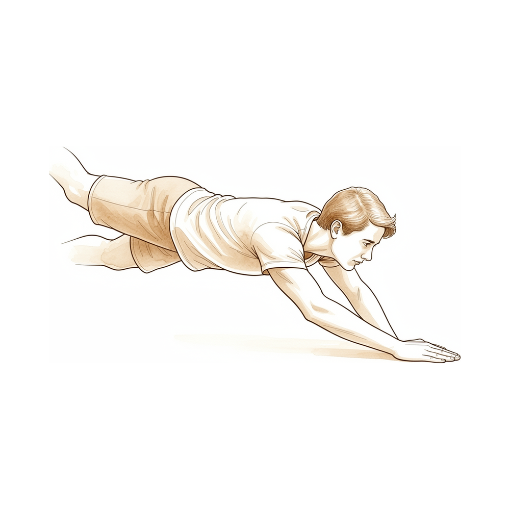
这些是您讲义中的锻炼项目。仅在 Hirpara 医生和您的手部治疗师的指导下开始,并严格在您被允许的关节活动范围和前臂位置内进行。早期锻炼旨在保持肘部和前臂的活动,以防止僵硬,同时避免对任何修复部位造成牵拉:包括主动辅助性的肘部屈伸、在您允许方向上的轻柔前臂旋转,以及保持肩部和手部自由活动。强化训练和疤痕护理属于后期阶段,在您获得明确许可之前不得开始。如果任何动作引起锐痛或肘部有不稳感,请立即停止。
您的临床方案
本页面其余部分为桡骨头置换术(桡骨头成形术)后的分阶段康复临床方案,该手术最常因无法重建的粉碎性桡骨头骨折而实施,通常作为“恐怖三联征”骨折脱位的一部分。本节内容将提供给手部治疗师,每个阶段均以通俗易懂的语言解释当前的治疗目标。植入物恢复了稳定且匹配的桡尺近端关节(桡尺关节),因此指导原则是在允许的情况下尽早进行受保护的活动,以预防此类肘关节易出现的僵硬,活动范围及前臂旋转受限于侧副韧带和冠状突修复的完整性。
治疗前,请查阅患者的手术记录及麻醉下稳定性评估结果,并与主治外科医生沟通以下事项:哪些侧副韧带和/或冠状突进行了修复、术中显示的稳定活动弧,以及需要保护的前臂旋转角度。Hirpara 医生为求舒适将肘关节置于简单吊带中(不使用铰链式支具),并在稳定性允许的情况下倾向于加速的早期活动策略。前臂位置原则:LCL(外侧副韧带)修复 → 旋前位锻炼/休息;MCL(内侧副韧带)修复 → 旋后位;两者均修复 → 中立位;避免内翻应力,若肘关节不稳定,则早期避免终末伸直。
第一阶段——早期受控活动(第0至2周)
在伤口稳定性允许的情况下(通常在第一周内),尽早开始轻柔的受控活动,以预防僵硬。手臂佩戴简易吊带以提供舒适,仅在锻炼和清洁时取下。肘部在保护性前臂旋转位置下,于安全活动范围内活动,具体旋转位置取决于所修复的韧带。
致手部治疗师:
教育与注意事项 - 使用简易吊带固定以提供舒适(不使用铰链式支具);仅在锻炼和清洗时取下 - 在术中证实的稳定活动范围内开始肘部主动辅助/主动屈伸;若肘关节不稳定,避免终末伸直 - 保护性体位下的前臂旋转:若修复外侧副韧带(LCL),则保持旋前;若修复内侧副韧带(MCL),则保持旋后;若两者均修复,则保持中立位中间范围 - 任何情况下均禁止内翻应力;在不稳定情况下进行过头锻炼时应取仰卧位,以消除内翻应力并利用重力使关节面贴合 - 禁止通过患侧手臂负重或推撑
管理 - 伤口:按医嘱进行外科敷料处理;开始活动前确认伤口稳定性 - 水肿:抬高患肢,轻柔的手部泵血运动,必要时冰敷 - 锻炼:在稳定范围内进行肘部主动辅助/主动屈伸;肘部屈曲90°位,在保护方向下进行前臂旋前/旋后;肩、腕、手及握力的全范围主动活动
进阶标准 - 伤口愈合稳定;在保护范围内进行受控活动时感到舒适
第二阶段——逐步扩大活动弧及前臂旋转(第2至6周)
从大约第2周至第6周,受保护的活动弧逐渐向完全伸直位扩大,前臂旋转在两个方向上逐步开放,目标是在大约第8周时实现完全旋前/旋后。此时仍避免进行力量训练和负重。
供您的手部治疗师参考:
评估 - 主动及被动肘关节屈伸及前臂旋转;疼痛及肿胀情况;伤口/瘢痕复查;稳定性症状
教育与注意事项 - 在稳定性允许的情况下,逐步达到完全伸直(如有早期伸直受限,应逐步解除) - 逐步向完全位进展双向前臂旋转,在此阶段早期仍需谨慎对待修复的韧带 - 继续避免内翻应力及任何经上肢的负重
管理 - 练习:将肘关节屈伸活动弧扩大至完全位;逐步进展旋前/旋后至完全活动范围(目标是在约8周时达到完全位);伤口愈合后开始瘢痕管理;继续肩、腕、手部的活动度练习 - 若存在残留的不稳定性顾虑,仰卧位(过顶)活动度训练方案仍然有益
进阶标准 - 接近无痛的完全活动范围;无稳定性症状;疼痛评分≤3/10
第三阶段——强化与回归(第6周至12周及以后)
一旦恢复活动度且修复被认定为牢固(通常在6周左右),即可开始强化训练,并逐步增加负荷(先进行握力训练,随后进行抗阻肘部和前臂训练),并在随后的几周中逐步推进。回归更重活动的标准是达到特定指标,通常在3个月左右。
供您的手部治疗师参考:
评估 - 与对侧相比的肘部和前臂力量;负荷下的疼痛/肿胀反应;根据需要进行功能和特定工作/运动测试
教育与注意事项 - 从大约6周开始进行轻柔的抗阻强化训练(握力 → 抗阻肘屈曲-伸展和旋前/旋后);逐步增加负荷 - 根据耐受情况进展至功能和特定工作负荷;早期避免突然的重负荷或冲击性负荷
管理 - 练习:渐进性抗阻肘部/前臂强化训练(弹力带 → 轻重量);握力强化训练;分级功能负荷;继续任何残留的活动度训练 - 监测并报告持续或加重的疼痛、机械症状或活动度丧失(可能的植入物过度填充/松动或肱骨小头磨损),如果恢复停滞或结果不佳,则转诊回主治医生 - 一旦活动度达到功能水平且力量接近对称,可考虑出院
回归全面活动的标准 - 无痛的功能性活动度;接近对称的力量;在负荷下肘部自信且稳定
重返工作与活动
从术后初期开始,鼓励在舒适范围内进行日常轻度手部活动(如进食、书写、轻度自理),前提是活动不涉及通过肘部进行推、提或承重。由于在手臂佩戴吊带期间或无法安全控制方向盘时不得驾驶,请在早期数周安排他人协助交通;待拆除吊带并经复查确认能够安全操控车辆后,方可恢复驾驶。
强化训练通常从术后约六周开始,并逐步增加强度。重返较重体力工作、提举重物及参与体育运动的时间通常在术后三个月左右,其依据是肘关节恢复无痛且充分、对称的力量,并由 Hirpara 医生及您的手部治疗师根据临床评估而非仅凭术后日历来判断。较重的体力劳动及对抗性运动也遵循相同的基于标准的渐进原则。
术后方案
本方案与诊所的一般康复建议并行;请参阅术后疼痛管理、伤口护理和疤痕管理。上述分阶段计划反映了桡骨头置换术和“恐怖三联征”重建术后的康复指南,您的持续康复将由Hirpara医生和您的手部治疗师根据肘关节的进展情况及具体修复内容,进行个体化指导。
Evidence & references
Radial Head Replacement — Procedure Outcomes & Post-operative Rehabilitation (Radial Head Arthroplasty for Unreconstructable Fracture / Terrible Triad)
Topic scope: post-operative rehabilitation after radial head arthroplasty (RHA) — replacement of an unreconstructable comminuted radial head with a metallic implant — performed either in isolation or, more commonly, as one component of reconstructing a fracture-dislocation (the "terrible triad": radial head + coronoid + lateral collateral ligament ± medial collateral ligament). The radial head is a key secondary stabiliser of the elbow against valgus and axial (posterolateral rotatory) load, so the implant exists to restore a stable, congruent radiocapitellar articulation and forearm axis — not merely to fill a defect.
Defining principle of the rehab here: the implant restores stability, so the dominant clinical enemy is stiffness, to which these elbows are strongly predisposed. The rehab is therefore an early protected-motion pathway — start moving within days to a week — explicitly gated by the integrity of the collateral-ligament and coronoid repairs done at the same operation. The two deliberate restraints are (1) the forearm rotation position that offloads the repaired ligament (pronation protects a repaired LCL; supination protects a repaired MCL; neutral mid-range when both), and (2) avoidance of varus stress and, where the elbow was unstable, early terminal extension. A simple sling is worn for comfort — not a hinged brace. The single biggest branch point is how much residual instability was demonstrated on examination under anaesthesia, which determines how fast the arc and forearm rotation are released.
A. PROCEDURE OUTCOMES (radial head arthroplasty; repair-vs-replace context)
Metallic RHA is a reliable reconstruction for the unreconstructable radial head, and — critically for rehab — it restores enough stability to permit early motion even in the setting of associated dislocation, provided the ligaments and coronoid are addressed.
- RHA restores elbow stability and kinematics when the native head is unreconstructable, but ligament repair is required to fully restore stability. Cadaveric work shows radial head excision alters kinematics and stability, arthroplasty restores them in the ligament-intact elbow, and in the ligament-disrupted elbow arthroplasty plus LCL repair is needed to correct varus–valgus laxity [Beingessner et al., J Bone Joint Surg Am 2004, DOI 10.2106/00004623-200408000-00018]. Strong (mechanistic/biomechanical).
- RHA gives functional, durable ROM in unstable elbow injuries equivalent to stable injuries. A 15-year single-surgeon series (68 patients) found patients with unstable radial head fractures plus dislocation achieved flexion and rotational arcs similar to stable injuries, with no difference in complication rate or implant survivorship — though supination loss was ~10° greater in the unstable group [Lott et al., J Shoulder Elbow Surg 2018, DOI 10.1016/j.jse.2017.10.011]. Moderate (Level II cohort).
- Long-term monopolar implant survival is good, with stiffness/sizing the main failure modes. A 15-year follow-up of the Acumed anatomical (press-fit, monopolar) implant for Mason III–IV fractures confirms durable function and survival, with the principal complications being joint stiffness, malpositioning and improper sizing [Tarallo et al., J Shoulder Elbow Surg 2026, DOI 10.1016/j.jse.2025.05.038]. Moderate (long-term cohort).
- Implant failure/revision risk is real, especially with associated instability. In a young active (military) cohort, RHA carried higher implant-failure rates than ORIF (20% vs 2.9%), and dislocation, coronoid fracture and concomitant ligament repair each predicted complications — underscoring that the injury complex, not just the implant, drives outcome [Kusnezov et al., HAND 2017, DOI 10.1177/1558944717715136]. Moderate.
- Terrible-triad reconstruction aims explicitly to restore stability sufficient for early motion. Comprehensive reviews frame the entire surgical sequence (LCL repair, radial head fix/replace, ± coronoid, ± MCL/fixator) as a means to permit early ROM and pre-empt stiffness, posttraumatic arthrosis and instability [Fahs et al., J Am Acad Orthop Surg 2024, DOI 10.5435/jaaos-d-24-00310]. Moderate–strong (narrative review).
B. REHABILITATION / THERAPY EVIDENCE
The rehab evidence base is built on biomechanics + surgical-series protocols rather than RCTs: there is strong agreement on early protected motion and on forearm-position-based ligament protection, but the exact arc and timing are individualised to intra-operative stability.
- Early motion is the consensus priority to prevent stiffness. Across operative series and textbook protocols, formal active and active-assisted ROM is begun within the first week once wound stability is confirmed, with splinting between sessions usually discontinued by 2–3 weeks and strengthening from ~6 weeks [Monica & Mudgal, Hand Clin 2010, DOI 10.1016/j.hcl.2010.04.008; Duckworth et al., Clin Orthop Relat Res 2014, DOI 10.1007/s11999-014-3516-y]. Moderate (consensus/series).
- Motion is gated by stability, with varus stress avoided at all times. Where instability is a concern, an overhead (supine) rehabilitation protocol begun ~10–14 days post-op achieves early motion while gravity coapts the joint and neutralises varus; "a stiff stable elbow is preferred over a loose incongruous one" [Rockwood and Green's Fractures in Adults, 2019]. Moderate (textbook consensus).
- Forearm rotation is positioned to protect the repaired ligament. Published RHA protocols position and exercise the forearm in pronation when the LCL was repaired, supination when the MCL was repaired, and neutral mid-range when both were repaired, progressing to full rotation as the repair consolidates [single-centre RHA protocol & narrative review, ResearchGate 2018; UVA / Christ Hospital RHA PT protocols — see URLs]. Weak–moderate (protocol consensus).
- A coronoid fracture treated without fixation does not preclude early motion in selected triads. Where the LCL and radial head are addressed and intra-operative fluoroscopic stability is confirmed, type I–II coronoid fractures can be left unfixed and still rehabilitated with early motion to good ROM and DASH scores [Papatheodorou et al., Clin Orthop Relat Res 2014, DOI 10.1007/s11999-014-3471-7]. Moderate (Level IV series).
- Restoring radiocapitellar contact (by replacement) is what permits the early-motion pathway in the unstable elbow; conservative or excision pathways are reserved for stable patterns and depend on the same early-mobilisation principle [Charalambous et al., J Shoulder Elbow Surg 2011, DOI 10.1016/j.jse.2011.02.013]. Moderate.
Recovery trajectory (expected, evidence-anchored)
| Phase | Window | Restraint | Therapy focus | Strength / load | Notes |
|---|---|---|---|---|---|
| I — Early protected motion | Week 0–2 (often start <1 wk) | Simple sling for comfort (no hinged brace); stable-arc only; forearm in ligament-protective rotation; no varus stress | Active/active-assisted elbow flexion–extension within the intra-operative stable arc; forearm pro/sup in the protected direction; full shoulder/wrist/hand ROM; supine overhead programme if unstable | None | Wound stability confirmed before motion; "stiff-stable > loose-incongruous" |
| II — Arc & rotation progression | Week 2–6 | Release extension block / forearm rotation gradually as stability allows | Progress elbow arc to full extension; open forearm rotation both directions; scar management once healed | None | Aim full pronation/supination by ~8 weeks; supination is the slowest to recover (~10° residual loss common) |
| III — Strengthening & return | Week 6–12+ | Restrictions lifted as repairs consolidate | Grip → resisted elbow/forearm strengthening; graded functional and work-specific loading | Begin ~6 wk, build gradually | Return to heavier work/sport criterion-based ~3 months; watch for overstuffing/loosening/capitellar wear |
(Phase windows mirror the precautions in the patient protocol; they are typical, stability-gated guides, not trial-derived deadlines.)
C. KEY CONTROVERSIES / EVIDENCE QUALITY
- Repair (ORIF) vs replace (RHA) the radial head. For reconstructable heads, ORIF is generally preferred and no prosthesis equals the native head biomechanically; for unreconstructable comminution (Mason III–IV) or in the unstable/dislocated elbow, RHA is the more reliable option because fixation constructs fail under the higher stresses [Kusnezov et al. 2017; Charalambous et al. 2011; Leigh & Ball, J Shoulder Elbow Surg 2012, DOI 10.1016/j.jse.2012.03.005]. Moderate; selection-dependent.
- Terrible-triad early motion vs protected immobilisation. Modern practice favours restoring enough stability (LCL ± radial head ± coronoid ± MCL/fixator) to permit early motion and avoid stiffness; the supine/overhead protocol exists precisely to reconcile early motion with residual instability. The trade-off ("stiff-stable preferred over loose-incongruous") is consensus, not RCT-settled [Rockwood and Green 2019; Fahs et al. 2024]. Moderate (consensus).
- Monopolar vs bipolar implants. Both are used; bipolar designs were intended to self-align and tolerate sizing imperfection, while monopolar anatomical implants show good long-term survival. No clear superiority is established, and overstuffing/sizing error harms either design more than the bearing type does [Tarallo et al. 2026; Doornberg et al., J Bone Joint Surg 2007, DOI 10.2106/jbjs.e.01340]. Weak (no head-to-head superiority).
- Implant-related complications. Overstuffing the radiocapitellar joint, malsizing and stem loosening cause capitellar erosion/osteopenia, pain and stiffness; capitellar erosion is reported from metal-on-cartilage articulation, and accurate head height/diameter is the key technical guard [Van Riet et al., J Bone Joint Surg 2004, DOI 10.2106/00004623-200405000-00028; Monica & Mudgal 2010]. Rehab cannot fix a malsized implant — persistent loading pain/stiffness warrants surgical review. Moderate.
- Supination is the laggard. Across series, forearm supination is the motion most likely to remain mildly deficient (≈10° loss), partly from scarring and partly from MCL-protective early positioning; patients should be counselled accordingly [Lott et al. 2018]. Moderate natural-history.
D. EVIDENCE STRENGTH FLAGS (summary)
- STRONG (biomechanical / mechanistic): RHA restores elbow stability and kinematics only in concert with collateral-ligament repair (varus–valgus laxity corrected by RHA + LCL repair, not RHA alone).
- MODERATE: functional ROM after RHA in unstable injuries equivalent to stable injuries with good implant survivorship (Level II–IV cohorts); long-term monopolar implant survival with stiffness/sizing as main failure modes; early-motion-to-prevent-stiffness as the governing rehab principle; supine/overhead protocol for the unstable elbow; supination as the slowest-recovering arc.
- WEAK / CONSENSUS: the specific forearm-position-by-repaired-ligament rehab rule (pronation for LCL, supination for MCL, neutral for both) and the exact phase timings (protocol-derived, stability-gated, not RCT-validated); monopolar-vs-bipolar bearing choice (no proven superiority).
CITATIONS
RAG corpus (180,000+ Orthopaedic articles)
- The Effect of Radial Head Excision and Arthroplasty on Elbow Kinematics and Stability. J Bone Joint Surg Am. 2004. DOI: 10.2106/00004623-200408000-00018
- Radial Head Arthroplasty. Hand Clin. 2010. DOI: 10.1016/j.hcl.2010.04.008
- Results after radial head arthroplasty in unstable fractures. J Shoulder Elbow Surg. 2018. DOI: 10.1016/j.jse.2017.10.011
- Long-term survival of Acumed anatomical radial head implant for Mason type III-IV fractures: a 15-year follow-up. J Shoulder Elbow Surg. 2026. DOI: 10.1016/j.jse.2025.05.038
- Operative Management of Unstable Radial Head Fractures in a Young Active Population. HAND. 2017. DOI: 10.1177/1558944717715136
- Management of Elbow Terrible Triad Injuries: A Comprehensive Review and Update. J Am Acad Orthop Surg. 2024. DOI: 10.5435/jaaos-d-24-00310
- Terrible Triad Injuries of the Elbow: Does the Coronoid Always Need to Be Fixed? Clin Orthop Relat Res. 2014. DOI: 10.1007/s11999-014-3471-7
- Radial Head Replacement for Acute Complex Fractures: What Are the Rate and Risk Factors for Revision or Removal? Clin Orthop Relat Res. 2014. DOI: 10.1007/s11999-014-3516-y
- Radial head reconstruction versus replacement in the treatment of terrible triad injuries of the elbow. J Shoulder Elbow Surg. 2012. DOI: 10.1016/j.jse.2012.03.005
- Comminuted radial head fractures: aspects of current management. J Shoulder Elbow Surg. 2011. DOI: 10.1016/j.jse.2011.02.013
- Radial Head Arthroplasty with a Modular Metal Spacer to Treat Acute Traumatic Elbow Instability. J Bone Joint Surg Am. 2007. DOI: 10.2106/jbjs.e.01340
- Capitellar Erosion Caused by a Metal Radial Head Prosthesis. J Bone Joint Surg Am. 2004. DOI: 10.2106/00004623-200405000-00028
- Comparative study of radial head resection and prosthetic replacement in surgical release of stiff elbows. Int Orthop. 2014. DOI: 10.1007/s00264-014-2594-5
- Rockwood and Green's Fractures in Adults (terrible-triad surgical pitfalls; overhead/early-motion protocol; "stiff-stable preferred"). Wolters Kluwer, 2019.
Radial head replacement rehabilitation literature (URLs)
- Rehabilitation protocol after radial head arthroplasty — a single-centre experience and narrative review of the literature. ResearchGate (2018). https://www.researchgate.net/publication/326168570
- University of Virginia, Department of Orthopaedic Surgery — Radial Head Replacement Rehabilitation Guidelines (forearm-position-by-ligament; arc progression). https://med.virginia.edu/orthopaedic-surgery/wp-content/uploads/sites/242/2024/09/Radial-head-replacement.pdf
- The Christ Hospital — Radial Head Replacement Physical Therapy Protocol (Rao). https://www.thechristhospital.com/landingpages/Documents/Rao%20PT%20Protocols/Operative/Elbow/Rao%20Radial%20Head%20Replacement%20r1.pdf
- Cheshire Arm Clinic — Physiotherapy Protocol for Radial Head Replacement. https://cheshirearmclinic.co.uk/wp-content/uploads/2021/09/Radial-Head-Replacement.pdf
- Denver Shoulder — Rehabilitation Protocol: Radial Head Replacement. https://www.denvershouldersurgeon.com/pdf/radial-head-replacement-protocol.pdf


