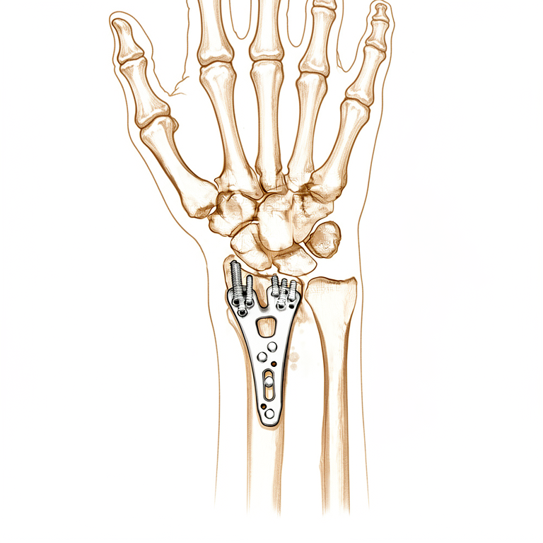
டிஸ்டல் ரேடியஸ் ORIF
Patients › Wrist
Open reduction and volar plate fixation of the distal radius — operation, recovery, rehabilitation.
இந்த அறுவை சிகிச்சை ஏன் பரிந்துரைக்கப்பட்டது
உங்கள் அறுவை சிகிச்சையாளர் ஒரு உலோகத் தகடு மற்றும் திருகுகளைப் பயன்படுத்தி உங்கள் மணிக்கட்டில் உடைந்த எலும்பை சரிசெய்ய திறந்த குறைப்பு மற்றும் உள் உறுதிப்படுத்தல் (ORIF என்றும் அழைக்கப்படுகிறது) பரிந்துரைத்துள்ளார். இந்த அறுவை சிகிச்சை பொதுவாக எலும்பு இடத்திலிருந்து நகர்ந்த அல்லது பல துண்டுகளாக உடைந்த சிக்கலான எலும்புகளைக் கொண்ட பெரியவர்களுக்கு வழங்கப்படுகிறது. அறுவை சிகிச்சை போன்ற அறுவை சிகிச்சை அல்லாத விருப்பங்கள் முதலில் முயற்சிக்கப்பட்டாலும், அந்த முறைகள் போதுமான ஸ்திரத்தன்மையையோ அல்லது குணப்படுத்துதலையோ வழங்க முடியாதபோது அறுவை சிகிச்சை பரிந்துரைக்கப்படுகிறது.
இந்த அறுவை சிகிச்சையின் முக்கிய நோக்கம் உங்கள் மணிக்கட்டு மூட்டு இயல்பான வடிவத்தை மீட்டெடுப்பதும், உங்கள் கையை விரைவாக நகர்த்த அனுமதிப்பதும் ஆகும். சான்றுகள் மற்ற முறைகளுடன் ஒப்பிடும்போது ஒரு தட்டு பயன்படுத்துவது விரைவான மற்றும் நீடித்த மீட்புக்கான சிறந்த முடிவுகளை வழங்குகிறது என்பதைக் காட்டுகிறது. இது எலும்பு சரியாக குணமடையாதது தொடர்பான சிக்கல்களின் அபாயத்தையும் கணிசமாகக் குறைக்கிறது.
அறுவை சிகிச்சைக்கு முன்
உங்கள் அறுவை சிகிச்சைக்கு முன் ஆறு மணி நேரம் நோன்பு நோற்கவும், உங்கள் அறுவை சிகிச்சையாளர் அறிவுறுத்தியபடி இரத்தத்தை மெல்லிய மருந்துகளை நிறுத்தவும். யாராவது உங்களை வீட்டிற்கு அழைத்துச் செல்ல ஏற்பாடு செய்யுங்கள், உங்கள் தற்போதைய அனைத்து மருந்துகளின் பட்டியலையும் கொண்டு வாருங்கள். உங்கள் எலும்பு மற்றும் மூட்டு ஆரோக்கியத்தை சரிபார்க்க, உங்களுக்கு எக்ஸ்-கதிர்கள் மற்றும் ஒருவேளை எம்.ஆர்.ஐ அல்லது இரத்த பரிசோதனை தேவைப்படலாம். உங்கள் அறுவை சிகிச்சையாளர் எலும்பை சீரமைக்க ஒரு நிலையான வெட்டு மூலம் ஒரு திறந்த அறுவை சிகிச்சையை மேற்கொள்வார், அதை ஒரு தட்டுடன் பாதுகாப்பார். சில நேரங்களில், சீரமைப்பு சரியானது என்பதை உறுதிப்படுத்த மூட்டுக்குள் ஒரு சிறிய கேமரா பயன்படுத்தப்படுகிறது. வசதியான ஆடைகளை அணிந்து, உங்கள் மயக்க பரிசோதனைக்கு தயாராக வந்து சேருங்கள்.
அன்று
நீங்கள் மருத்துவமனைக்கு வந்து உங்கள் அறுவை சிகிச்சையாளர் மற்றும் மயக்க மருத்துவரை சந்திப்பீர்கள். இந்த அறுவை சிகிச்சை பொது மயக்க மருந்தின் கீழ் செய்யப்படுகிறது. அறுவை சிகிச்சைக்கு நீங்கள் முழுமையாக தூங்குவீர்கள். சில நோயாளிகளுக்கு அறுவை சிகிச்சைக்குப் பிந்தைய வலி நிவாரணத்திற்காக ஒரு பிராந்திய நரம்புத் தடுப்பு இருக்கலாம்; உங்கள் தனிப்பட்ட சூழ்நிலைகளின் அடிப்படையில் மயக்க மருத்துவர் நாள் தீர்மானிக்கிறார்.
அறுவை சிகிச்சைக்குப் பிறகு நீங்கள் அறுவை சிகிச்சை அறைக்குச் செல்வீர்கள், அங்கு உங்கள் அறுவை சிகிச்சையாளர் உங்கள் எலும்பை அடைய உங்கள் சருமத்தில் ஒரு வெட்டு வைப்பார். உடைந்த துண்டுகளை இடத்தில் வைத்திருக்க அவர்கள் ஒரு உலோகத் தகடு மற்றும் திருகுகளைப் பயன்படுத்துகிறார்கள். அறுவை சிகிச்சைக்குப் பிறகு, நீங்கள் குணமடைந்து எழுந்திருப்பீர்கள், அதே நேரத்தில் செவிலியர்கள் உங்களைப் பார்த்து உங்கள் வலியை நிர்வகிப்பார்கள்.
அறுவை சிகிச்சையின் உள்ளடக்கம்
உடைந்த எலும்பை அடைய உங்கள் அறுவை சிகிச்சையாளர் மணிக்கட்டில் ஒரு வெட்டு செய்வார். இது ஒரு திறந்த அணுகுமுறையாகும், அதாவது சிறிய சாவி துளை கேமராக்களைப் பயன்படுத்துவதற்குப் பதிலாக பகுதி முழுமையாக வெளிப்படும். இந்த திறப்பு மூலம், உங்கள் அறுவை சிகிச்சை நிதானமாக உடைந்த எலும்பு துண்டுகளை சரியான நிலைக்கு நகர்த்துகிறது.
எலும்பு சீரமைக்கப்பட்டவுடன், உங்கள் அறுவை சிகிச்சை நிபுணர் ஒரு உலோக தகடு மற்றும் திருகுகளைப் பயன்படுத்தி அதைப் பாதுகாக்கிறார். இந்த முறை ஆரம்ப மற்றும் நீடித்த மீட்புக்கான சிறந்த முடிவுகளை வழங்குகிறது மற்றும் குணப்படுத்தும் சிக்கல்களைக் குறைக்க உதவுகிறது. குணமடையும் போது எல்லாவற்றையும் சீராக வைத்திருக்க ரேடியஸ் எலும்பின் பக்கத்தில் தகடு வைக்கப்படுகிறது. சில சந்தர்ப்பங்களில், உங்கள் அறுவை சிகிச்சை நிபுணர் ஒரு சிறப்பு திருகு நுட்பத்தைப் பயன்படுத்தலாம்.
எலும்பு சரிசெய்யப்பட்ட பிறகு, உங்கள் அறுவை சிகிச்சை நிபுணர் தையல்களால் வெட்டு மூடுகிறார். வன்பொருள் எரிச்சலை ஏற்படுத்தினால், பின்னர் உங்களுக்கு இரண்டாவது சிறிய அறுவை சிகிச்சை தேவைப்படலாம், ஏனெனில் இந்த பிளேட்டிங்கிற்கு உட்படும் நோயாளிகளுக்கு வன்பொருள் அகற்றுதல் தேவைப்பட 28% வாய்ப்பு உள்ளது. இந்த அறுவை சிகிச்சைக்குப் பிறகு உங்கள் கை மற்றும் மணிக்கட்டில் ஆரம்பகால இயக்கம் ஊக்குவிக்கப்படுகிறது, நீங்கள் காத்திருந்தால் விட விரைவாக செயல்பாட்டை மீண்டும் பெற உதவுகிறது.
அறுவை சிகிச்சைக்குப் பிறகு
உங்கள் வலி பொது மருத்துவத்துடன் நிர்வகிக்கப்படும் ஒரு மீட்பு வார்டில் நீங்கள் எழுந்திருப்பீர்கள். உங்கள் கை வெட்டுடன் ஒரு பிணைப்புடன் ஒரு ஸ்லிங் அல்லது ப்ரேஸில் இருக்கும். உங்கள் அறுவை சிகிச்சையாளர் எலும்பை சரிசெய்ய ஒரு திறந்த வெட்டு செய்தார், சில நேரங்களில் ஒரு சிறிய கேமராவைப் பயன்படுத்தி மூட்டு சரிபார்க்கவும். உங்கள் செயல்பாட்டிற்கு உதவ உங்கள் விரல்கள் மற்றும் மணிக்கட்டுகளை ஆரம்பத்தில் நகர்த்தத் தொடங்கலாம். இந்த அறுவை சிகிச்சைக்குப் பிறகு பெரும்பாலான நோயாளிகள் ஒரு இரவு மருத்துவமனையில் தங்கியிருக்கிறார்கள், இருப்பினும் சிலர் அதே நாளில் வீட்டிற்குச் செல்ல முடியும். முதல் 24 மணிநேரங்களுக்கு யாராவது உங்களுடன் இருக்க வேண்டும்.
மீட்பு
அறுவை சிகிச்சையிலிருந்து உங்கள் உடல் குணமடையும்போது இது இயல்பானது. உங்கள் அறுவை சிகிச்சையாளர் ஒரு தட்டு மற்றும் திருகுகளைப் பயன்படுத்தி உங்கள் மணிக்கட்டில் ஒரு சிறிய வெட்டு வழியாக எலும்பை வைத்திருக்கக்கூடும். நீங்கள் ஓய்வெடுக்கும் போது அந்த பகுதியை பாதுகாக்க நீங்கள் ஒரு பிரேஸ் அல்லது ஒரு பிளாஸ்ட் வைத்திருக்கலாம்.
உங்கள் கை இயல்பு நிலைக்கு திரும்புவதற்கு ஆரம்ப இயக்கம் முக்கியமானது. உங்கள் பிசியோதெரபிஸ்ட் கடினத்தன்மையைக் குறைக்க மென்மையான பயிற்சிகள் மூலம் உங்களுக்கு வழிகாட்டுவார். நீங்கள் சுற்றி நடக்கும்போது ஒரு ஸ்லிங் அல்லது பிராஸ் அணிவீர்கள், ஆனால் உங்களால் முடிந்தவுடன் உங்கள் விரல்கள் மற்றும் முழங்கைகளை நகர்த்தத் தொடங்க வேண்டும். இந்த சிறிய இயக்கங்கள் உங்கள் கையை நீண்ட நேரம் காத்திருப்பதை விட விரைவாக உணர உதவுகின்றன.
உங்கள் அறுவை சிகிச்சை நிபுணர் உங்களுக்கு அனுமதி அளித்தவுடன் நீங்கள் லேசான வீட்டு நடவடிக்கைகளுக்குத் திரும்பலாம். உங்கள் காலவரிசை மற்றவர்களிடமிருந்து மாறுபடலாம், எனவே உங்கள் அறுவை சிகிச்சை நிபுணர் மற்றும் உடலியல் குழுவின் குறிப்பிட்ட ஆலோசனையைப் பின்பற்றவும். உங்கள் செயல்பாட்டு அளவை எப்போது பாதுகாப்பாக அதிகரிக்க வேண்டும் என்பதை அவர்கள் சரியாக அறிய உதவுவார்கள்.
என்ன தவறு நடக்கலாம்
பெரும்பாலான நோயாளிகள் நன்றாக இருக்கிறார்கள், ஆனால் எப்போதாவது பிரச்சினைகள் ஏற்படலாம். உங்கள் அறுவை சிகிச்சை நிபுணரும் குழுவும் எந்தவொரு பிரச்சினையையும் ஆரம்பத்தில் கண்டறிய உங்களை உன்னிப்பாகக் கண்காணிப்பார்கள்.
சில சமயங்களில் உங்கள் எலும்பை அதன் இடத்தில் வைத்திருக்கப் பயன்படுத்தப்படும் உலோகக் கருவிகள் அசௌகரியமாகிவிடும் அல்லது எரிச்சலை ஏற்படுத்தும். நீங்கள் வலியை அல்லது தட்டு மீது அழுத்தத்தை உணர்ந்தால், அதை அகற்ற உங்களுக்கு இரண்டாவது சிறிய செயல்முறை தேவைப்படலாம். இது நான்கு நோயாளிகளில் ஒருவருக்கு நிகழ்கிறது.
உங்கள் மணிக்கட்டு நெகிழ்வாகவோ அல்லது பலவீனமாகவோ உணர்ந்தால், உடனடியாக உங்கள் அறுவை சிகிச்சை நிபுணரிடம் சொல்லுங்கள், இதனால் அவர்கள் உங்கள் சிகிச்சையை வழிநடத்த முடியும்.
சில சந்தர்ப்பங்களில், காலப்போக்கில் எலும்பு சீரமைப்பு சற்று மாறக்கூடும். திருகுகள் எலும்பில் போதுமான ஆழத்தில் வைக்கப்படவில்லை என்றால் இது ஏற்படலாம். நீங்கள் ஒரு புதிய கிளிக் ஒலியை உணர்ந்தால் அல்லது உங்கள் கை வித்தியாசமாக இருப்பதைக் கவனித்தால், அடுத்த சோதனையில் அதைக் கொண்டு வாருங்கள்.
ஒட்டுமொத்தமாக, கடுமையான பிரச்சினைகள் அரிதானவை. இந்த அறுவை சிகிச்சைக்கு உட்படுத்தப்பட்ட பெரும்பாலான மக்கள் ஆரம்பகால இயக்கம் மற்றும் கவனமான பின்தொடர்தல் மூலம் நன்றாக மீண்டு வருகிறார்கள். இந்த பக்கத்தில் உள்ள சிக்கல்களின் அட்டவணை வழக்கமான விகிதங்களை பட்டியலிடுகிறது.
எப்போது எங்களை அழைக்க வேண்டும்
உங்களுக்கு காய்ச்சல், அதிகரிக்கும் சிவப்பு, அல்லது உங்கள் வெட்டுக்களில் இருந்து வடிகால் இருந்தால் எங்களை அழைக்கவும். நீங்கள் திடீரென்று கடுமையான வலியை உணர்ந்தால், உங்கள் கன்றுக்குட்டி வீங்கியிருப்பதைக் கவனித்தால் அல்லது சுவாசிப்பதில் சிரமம் இருந்தால் அவசரநிலைக்குச் செல்லுங்கள். உங்கள் விரல்களில் உணர்வை இழந்துவிட்டால் அல்லது உங்கள் கையை நகர்த்த முடியாவிட்டால் உடனடியாக அழைக்கவும். உங்கள் மீட்பைப் பாதுகாக்க இந்த அறிகுறிகளுக்கு அவசர மதிப்பீடு தேவை.
Evidence & references
Overview
- Early mobilization for patients with distal radius fractures treated with ORIF may have a beneficial effect on function at earlier stages compared to late mobilization [1].
- Mean differences in DASH scores at 6 weeks for early mobilization in distal radius fractures treated with ORIF surpass the minimal clinically important difference [1].
- Substantial variations in surgical direct costs for distal radius ORIF exist [2].
- Implant choice is the predominant driver of cost variation for distal radius ORIF [2].
- The LOS length can be calculated for the Lift-Off Screw technique in a distal radius fracture model [3].
- The Lift-Off Screw technique can potentially be used with any distal radius periarticular locking plate with locking options in the shaft [3].
- Open reduction and internal fixation with a plate offers the best results for adult patients with a distal radius fracture in terms of early and sustained functional recovery [5].
- Open reduction and internal fixation with a plate offers the best results for adult patients with a distal radius fracture in terms of reduction in fracture healing complications [5].
- Radial column plating of the distal radius is a safe treatment modality [6].
- Radial column plating of the distal radius is a valuable adjunct in the setting of complex distal radius fractures [6].
- There is a 28% chance that hardware removal may be required for patients undergoing radial column plating of the distal radius [6].
- Placing the distal row of screws greater than 3mm from subchondral bone in comminuted intra-articular distal radius fractures was associated with increased odds of worsening ulnar variance [8].
- The treatment of displaced intra-articular distal radius fractures with a dorsally versus a volarly placed interlocking plate system demonstrated similar clinical results [9].
Anatomy & Pathophysiology
- Distal radius fractures are the most common fractures of the upper extremity in the United States, occurring in over 300,000 cases per year [27].
- Distal radius fractures exhibit a bimodal distribution with young patients sustaining high-energy trauma and elderly patients sustaining low-energy falls [27].
- Distal radius fractures are the most common upper extremity osteoporotic fracture in elderly patients [27].
- The distal radius articular surface is biconcave with distinct scaphoid and lunate facets [27].
- The distal radioulnar joint (DRUJ) is the articulation between the radius and ulna at the sigmoid notch [27].
- Lister tubercle is a small dorsal prominence on the distal radius that serves as a landmark for the dorsal approach to the wrist [27].
- Lister tubercle is a cause of attritional rupture of the extensor pollicis longus (EPL) after a distal radius fracture [27].
- The metaphysis of the distal radius has a thin cortex and is vulnerable to bending forces [27].
- The brachioradialis insertion on the radial styloid acts as a deforming force in distal radius fractures [27].
- In a normal wrist, the distal radius bears 80% of the axial load in neutral ulnar variance [27].
- The carpus encompasses two rows of eight bones that serve as a bridge between the forearm and the hand [16].
- The proximal carpal row from radial to ulnar includes the scaphoid, lunate, and triquetrum [16].
- The proximal carpal row is referred to as the key intercalated segment between the forearm and the distal row of the carpus [16].
- There are no direct tendon attachments to the proximal carpal row [16].
- The movement of the proximal carpal row results from the shape of the bones, their interaction with other bones, and ligament attachments [16].
- The pisiform bone is a sesamoid bone enclosed within the sheath of the flexor carpi ulnaris tendon and should not theoretically be considered within the proximal carpal row [16].
- The distal carpal row from radial to ulnar includes the trapezium, trapezoid, capitate, and hamate [16].
- The distal carpal row articulates with the proximal carpal row and distally with the five metacarpals of the hand [16].
- The trapezium articulates with the first metacarpal, the trapezoid with the second, the capitate with the third, and the hamate with the fourth and fifth metacarpals [16].
- The capitate and trapezoid are tightly connected to the metacarpals [16].
- There is 30 to 40 degrees of flexion–extension and rotation at the metacarpotrapezial joint [16].
- Motion at the distal carpal row is controlled by the extrinsic wrist flexors and extensors [16].
- The ligaments of the wrist are predominantly contained within the joint capsule [16].
- The inherent stability of the carpal rows is predominantly due to the support of extrinsic and intrinsic ligaments that reinforce the capsule of the carpus [16].
- Apart from the scaphocapitate ligament, carpal ligaments are not described consistently across anatomical studies [16].
- The extrinsic palmar radiocarpal ligaments include the transverse carpal, radioscaphocapitate (RSC), radioscapholunate (RSL), radial collateral, long radiolunate (RLT), and short radiolunate ligaments [23].
- The extrinsic ulnocarpal ligaments include the ulnotriquetral (dorsal and palmar), ulnolunate, and ulnocapitate ligaments [23].
- The strong oblique extrinsic palmar radial ligaments prevent the carpus from translating medially on the angulated slope of the distal radius through two V-shaped ligamentous bands [23].
- One V-shaped band is proximal (long radiolunate, radioscapholunate, ulnolunate, ulnotriquetral) and connects the forearm to the proximal carpal row [23].
- One V-shaped band is distal (radioscaphocapitate, ulnocapitate) and connects the forearm to the distal carpal row [23].
- Between the radial and ulnar palmar ligaments there is a V-shaped interligamentous sulcus over the capitolunate articulation known as the space of Poirier [23].
- Maximal space in the space of Poirier is seen when the wrist is dorsiflexed [23].
- The space of Poirier almost disappears in palmar flexion [23].
- The lunate displaces into the carpal canal through the space of Poirier during dorsal dislocations [23].
- The arcuate ligament is found in the central third of the palmar joint capsule and is thought to be formed from the interdigitation of transverse fibers of the radioscaphocapitate, ulnocapitate, triquetrocapitate, and volar scaphotriquetral ligaments [23].
- The arcuate ligament forms a support sling for the midcarpal region, in particular the head of the capitate [23].
- The extrinsic dorsal carpal ligaments include the dorsal radiocarpal (DRC) ligament and the dorsal intercarpal ligament [23].
- The dorsal intercarpal ligament forms a V-shaped configuration [23].
- The ulnodorsal capsule of the wrist is reinforced by the ulnolunate and ulnotriquetral ligaments and the floors of the fifth and sixth extensor compartments [23].
- Some studies suggest an essential role of the dorsal carpal ligaments in scapholunate stability [23].
- The intrinsic ligaments connect the individual carpal bones to one another [23].
- The intrinsic ligaments are intra-articular short fibers that connect and hold the carpal bones of both the proximal or distal rows to each other [23].
- There is a contiguous merging of the interosseous ligaments with the joint articular cartilage [23].
- The intrinsic ligaments include the palmar midcarpal ligaments (scaphotrapeziotrapezoid, scaphocapitate, triquetrocapiate, triquetrohamate) [23].
- The intrinsic ligaments include the proximal interosseous ligaments (scapholunate, lunotriquetral) [23].
- The intrinsic ligaments include the distal interosseous ligaments (trapeziotrapezoid, trapeziocapitate, capitohamate) [23].
- The pisotriquetral ligament bridges the pisotriquetral joint [23].
- The pisohamate ligament is an extension of the flexor carpi ulnaris [23].
- On the radial side of the wrist, the V-shaped scaphotrapezium–trapezoid ligament provides stability to the scaphoid–trapezium–trapezoid articulation as well as the scaphoid itself [23].
- Recent studies suggest the existence of two distinct ligaments on the radial side: the scaphotrapezoid and scaphotrapezium ligaments [23].
- The scaphocapitate is a large robust ligament that provides midcarpal stability [23].
- The scaphoid bone is located in the proximal carpal row on the radial aspect of the wrist [21].
- The scaphoid is a small, irregular S-shaped tubular bone [21].
- The scaphoid lies entirely within the wrist joint and is located at a 45-degree plane to the longitudinal and horizontal axis of the wrist [21].
- The scaphoid articulates with the trapezium/trapezoid (distal surface), radius (proximal/lateral surface), capitate (medial surface), and lunate (medial surface) [21].
- The proximal articular surface of the scaphoid is convex and articulates with the radius [21].
- The capitate head articulates with a sulcus on the scaphoid located across the radial articular surface [21].
- The scaphoid gently pronates and flexes distally such that the distal pole sits ulnarly angulated relative to the proximal pole [21].
- At the distal articular surface, two distinct articular facets for the trapezium and trapezoid are present forming the STT joint [21].
- Over 80% of the scaphoid surface is covered with articular cartilage [21].
- The scaphoid has a reduced capacity for periosteal healing and an increased tendency for delayed union and nonunion [21].
- The scaphoid is ridged across its nonarticular dorsoradial surface, along which the critical dorsal ridge vessels traverse [21].
- The ridge is the insertion point for both the dorsal component of the scapholunate and intercarpal ligaments [21].
- The ligamentous attachments of the scaphoid are predominantly found on the nonarticular dorsoradial surface [21].
- The radioscapocapitate ligament does not attach to the bone itself but crosses the waist, acting as a sling across it allowing it to rotate [21].
- There are no tendon attachments to the scaphoid [21].
- The scaphoid acts as a midcarpal joint "bridge" linking and synchronizing the motions of the proximal and distal carpal rows [21].
- Motion of the scaphoid includes rotation proximally and gliding distally [21].
- The potential for nonunion of the scaphoid is often ascribed to the meagre, largely retrograde blood supply [21].
- The blood supply to the scaphoid is provided by two vascular pedicles originating from the scaphoid branches of the radial artery [21].
- The dorsal branch enters via the small foramina along the spiral groove and dorsal ridge of the scaphoid and supplies 70% to 80% of the scaphoid proximally, including the proximal pole [21].
- The volar branch enters via the scaphoid tubercle and supplies the remaining 20% to 30% of distal scaphoid [21].
- The waist of the scaphoid has been shown to have minimal or no perforating vasculature [21].
- No vessels perforate the proximal dorsal cartilaginous area or through the scapholunate ligament [21].
- Proximal fractures are inexorably associated with at least temporary disruption of the interosseous blood supply to the proximal pole [21].
- The metacarpals form a unique cascade similar to that of a Roman arch [20].
- The index and small fingers form the borders of the metacarpal cascade while the middle finger forms the keystone [20].
- The metacarpals are inherently stable secondary to the origins of the intrinsic muscles of the hand as well as the stout attachments of the deep transverse intermetacarpal ligaments [20].
- The bony architecture of the metacarpal is made up of a distal and proximal metaphysis [20].
- The metaphyseal portion of the metacarpal has a thinner cortex and does not provide rigid purchase for cortical fixation [20].
- The diaphyseal portion of the metacarpal provides better purchase with its tubular structure and thick cortices [20].
- The middle and ring fingers have the most inherent stability secondary to their central location [20].
- The border digits are more prone to shortening, rotation, and angulation [20].
- The CMC joints increase in stability and decrease in dorsal-volar mobility when moving from the small to index finger metacarpal [20].
- The small and ring fingers are far more tolerant to deformity than are the index and middle finger metacarpals [20].
- In the sagittal plane, the metacarpal head has a cam-shaped articular surface with a larger volar-dorsal diameter than a proximal-distal diameter [20].
- In the transverse plane, the metacarpal head resembles a trapezoid [20].
- The collateral ligaments of the metacarpal head attach on the dorsal aspect of the metacarpal head within the recess and are thus elongated in flexion [20].
- The combination of the trapezoidal shape of the head, origin and insertion of the collateral ligaments, and cam-shaped morphology contribute to inherent stability during MCP flexion [20].
- The metacarpals of the hand and thumb are subcutaneous in position and are thus susceptible to penetrating and crush injuries [20].
- The metacarpals have a curvature to their diaphyseal segment and often flex during the making of a fist [20].
- The metacarpal necks are susceptible to fracture during punching [20].
- The fifth metacarpal has the most anterior–posterior arc at the CMC joint [20].
- Metacarpal fractures are third in frequency only to phalangeal fractures and distal radius fractures in the upper limb [17].
- Approximately 70% of metacarpal fractures occur during the second and fifth decades of life [17].
- The vast majority of metacarpal fractures are inherently stable and can be treated via closed means with early range of motion [17].
- The flexor carpi radialis (FCR) approach provides excellent exposure for volar plating [26].
- Incising the FCR sheath on its radial aspect may help to avoid injury to the palmar cutaneous branch of the median nerve [26].
- A brachioradialis tenotomy removes a deforming force during the FCR approach [26].
- Repair of the pronator quadratus did not result in better outcomes at 12 months in a randomized trial of 72 patients [26].
- The extended FCR approach aims to facilitate adequate distal exposure including the volar ulnar corner [26].
- By releasing the superficial and deep leaves of the FCR sheath to the level of the trapezium, the carpal tunnel contents are more easily retracted ulnarly [26].
- Pronation of the ulnar shaft through the fracture exposes the dorsal metaphysis in subacute fractures [26].
- Biasing the skin incision more midline allows development of the interval between the digital flexors and ulnar neurovascular bundle [26].
- Retracting the carpal tunnel contents radially provides excellent visualization of the lunate facet fragment [26].
- The approach for the volar ulnar facet fragment is extensile with an open carpal tunnel release [26].
- A radial column approach requires protection of the superficial branch of the radial nerve [26].
- The deep interval for a radial column approach is through the first dorsal compartment or the interval between the first and second compartments [26].
- The dorsal approach is the traditional approach for plating distal radius fractures [26].
- Enthusiasm for dorsal plating waned as a result of extensor tendon complications [26].
- A dorsal approach may be necessary to reduce and stabilize the dorsal ulnar facet fragment [26].
- Elevation of the fourth dorsal compartment on its ulnar aspect exposes the dorsal ulnar corner [26].
- Unlike the volar approach, the dorsal approach affords the option of visualizing the articular surface through a dorsal capsulotomy [26].
- The most common tendon to rupture following application of a volar plate is the flexor pollicis longus [34].
- Rupture of the flexor pollicis longus is due to volar extension of the plate beyond the so-called watershed line [34].
- Dorsal tendons such as the extensor pollicis longus and extensor digitorum communis can fray and rupture from prominent screw tips following volar insertion [34].
- Dorsal plates or constructs are now preferred for dorsal shearing fractures and complex articular fractures [34].
- Distraction (or bridge) plate fixation is increasingly utilized for complex articular fracture, those with complex metaphyseal or diaphyseal fragmentation in particular [34].
- Application of the bridge/distraction plate should not be a substitute for accurate ORIF [34].
- Fractures of the radial styloid may be associated with SL ligament injuries because the intra-articular fracture line extends into the joint at that level [34].
- In the setting of isolated radial styloid fractures, intercarpal ligament injuries must be suspected [34].
- Intra-articular displacement (or diastasis) greater than 2 mm is an indication for surgery for fractures of the radial styloid [34].
- Compression screw fixation with partially threaded 3.5- or 4.0-mm cancellous screws can effectively compress radial styloid fragments and maintain the reduction [34].
- Alternative fixation options for radial styloid fractures include K-wires and fragment-specific pin plate and screw fixation [34].
- The distal radioulnar joint is assessed following stabilization of the radius [34].
- Slightly greater laxity than the opposite uninjured wrist is to be expected after distal radius fracture treatment [34].
- Only frank dislocation with forearm rotation merits surgery to stabilize the distal radioulnar joint [34].
- The presence of a displaced fracture at the base of the ulnar styloid is not in itself an indication for surgical fixation [34].
- Clinical stability of the distal radioulnar joint must be elucidated and compared with
Classification
- Early mobilization for patients with distal radius fractures treated with ORIF may have a beneficial effect on function at earlier stages compared to late mobilization [1].
- Mean differences in DASH scores at 6 weeks for early mobilization in distal radius fractures treated with ORIF surpass the minimal clinically important difference [1].
- Substantial variations in surgical direct costs for distal radius ORIF exist [2].
- Implant choice is the predominant driver of cost variation for distal radius ORIF [2].
- The LOS length can be calculated for the lift-off screw technique in a distal radius fracture model [3].
- The lift-off screw technique can potentially be used with any distal radius periarticular locking plate with locking options in the shaft [3].
- Open reduction and internal fixation with a plate offers the best results for adult patients with a distal radius fracture in terms of early and sustained functional recovery [5].
- Open reduction and internal fixation with a plate offers the best results for adult patients with a distal radius fracture in terms of a reduction in fracture healing complications [5].
- Radial column plating of the distal radius is a safe treatment modality [6].
- Radial column plating of the distal radius is a valuable adjunct in the setting of complex distal radius fractures [6].
- There is a 28% chance that hardware removal may be required for patients undergoing radial column plating of the distal radius [6].
- Placing the distal row of screws greater than 3mm from subchondral bone in comminuted intra-articular distal radius fractures was associated with increased odds of worsening ulnar variance [8].
- The treatment of displaced intra-articular distal radius fractures with a dorsally versus a volarly placed interlocking plate system demonstrated similar clinical results [9].
Clinical Presentation
- Early mobilization for patients with distal radius fractures treated with ORIF may have a beneficial effect on function at earlier stages compared to late mobilization [1].
- Mean differences in DASH scores at 6 weeks for early mobilization in distal radius fractures treated with ORIF surpass the minimal clinically important difference [1].
- Substantial variations in surgical direct costs for distal radius ORIF exist, with implant choice being the predominant driver [2].
- Radial column plating of the distal radius is a safe treatment modality and a valuable adjunct in the setting of complex distal radius fractures [6].
- Patients undergoing radial column plating of the distal radius should be counseled that there is a 28% chance that hardware removal may be required [6].
- Placing the distal row of screws greater than 3mm from subchondral bone in comminuted intra-articular distal radius fractures was associated with increased odds of worsening ulnar variance [8].
- Treatment of displaced intra-articular distal radius fractures with a dorsally versus a volarly placed interlocking plate system demonstrated similar clinical results [9].
- An isolated radial shaft fracture is uncommon in adults because the forearm functions as a ring [35].
- Fractures within 7.5 cm of the radiocarpal joint are more likely to represent a Galeazzi fracture (radial shaft fracture with associated distal radioulnar joint injury) [35].
- Galeazzi fractures require surgical fixation with rare exceptions [35].
- Examination may reveal a prominent ulnar head in cases of dorsal distal radioulnar joint subluxation or dislocation [35].
- Radiographic parameters associated with injury to the distal radioulnar joint include fracture of the base of the ulnar styloid [35].
- Radiographic parameters associated with injury to the distal radioulnar joint include widening of the distal radioulnar joint on the AP wrist radiograph [35].
- Radiographic parameters associated with injury to the distal radioulnar joint include dislocation of the ulna relative to the radius on a true lateral wrist radiograph [35].
- Radiographic parameters associated with injury to the distal radioulnar joint include more than 5 mm of shortening of the radius relative to the ulna (ulnar positive variance) when compared with the contralateral wrist [35].
- Radial shaft fracture line obliquity greater than 30° is a statistically significant predictor of distal radioulnar joint instability [35].
- Closed management of displaced forearm axis fractures in adults results in poor outcomes [35].
- Surgery is indicated for all Galeazzi fractures to restore the structural integrity of the forearm ring [35].
- Complications following management of Galeazzi fractures of the radius include nonunion [35].
- Complications following management of Galeazzi fractures of the radius include malunion with limited forearm rotation [35].
- Complications following management of Galeazzi fractures of the radius include nerve or vessel injury [35].
- Complications following management of Galeazzi fractures of the radius include infection [35].
- Complications following management of Galeazzi fractures of the radius include complex regional pain syndrome [35].
- Complications following management of Galeazzi fractures of the radius include persistent distal radioulnar joint instability [35].
Investigations
- Early mobilization for distal radius fractures treated with ORIF may have a beneficial effect on function at earlier stages compared to late mobilization, with mean differences in DASH scores at 6 weeks surpassing the minimal clinically important difference [1].
- Substantial variations in surgical direct costs for distal radius ORIF exist, with implant choice being the predominant driver [2].
- The length of stay (LOS) can be calculated for a distal radius fracture model using the Lift-Off Screw technique, which can potentially be used with any distal radius periarticular locking plate with locking options in the shaft [3].
- Open reduction and internal fixation with a plate offers the best results for adult patients with a distal radius fracture in terms of early and sustained functional recovery and a reduction in fracture healing complications [5].
- Radial column plating of the distal radius is a safe treatment modality, but patients should be counseled that there is a 28% chance that hardware removal may be required [6].
- Placing the distal row of screws greater than 3mm from subchondral bone in comminuted intra-articular distal radius fractures was associated with increased odds of worsening ulnar variance [8].
- Treatment of displaced intra-articular distal radius fractures with a dorsally versus a volarly placed interlocking plate system demonstrated similar clinical results [9].
- An understanding of the anatomy and kinematics of the eight carpal bones is essential for the diagnosis and management of carpal injuries [16].
- The proximal carpal row includes the scaphoid, lunate, and triquetrum, serving as a key intercalated segment between the forearm and the distal row of the carpus [16].
- The distal carpal row includes the trapezium, trapezoid, capitate, and hamate, articulating with the proximal carpal row and distally with the five metacarpals [16].
- The inherent stability of the carpal rows is predominantly due to the support of extrinsic and intrinsic ligaments that reinforce the capsule of the carpus [16].
- Apart from the scaphocapitate ligament, carpal ligaments are not described consistently across anatomical studies [16].
- The incidence of metacarpal fractures is third in frequency only to phalangeal fractures and distal radius fractures in the upper limb [17].
- Approximately 70% of metacarpal fractures occur during the second and fifth decades of life [17].
- The vast majority of metacarpal fractures are inherently stable and can be treated via closed means with early range of motion [17].
- Assessment of metacarpal fractures requires evaluation of skin integrity, neurovascular status, ligamentous evaluation, and tendon function [17].
- Malrotation must be tested in metacarpal fracture assessment, with particular attention toward the border digits [17].
- Initial X-rays for suspected scaphoid fractures should include posteroanterior (PA) with wrist in ulnar deviation, lateral, semi-pronated oblique, and semi-supinated oblique views [24].
- An anteroposterior (AP) view with the fist clenched can be added if there is a suspicion of a scapholunate injury [24].
- In a normal lateral X-ray, the axes of the radius, lunate, capitate, and third metacarpal are co-linear [24].
- In a normal lateral X-ray, the scaphoid projects at an angle of about 45 degrees to the line of the radius [24].
- Dorsal intercalated segmental instability (DISI) is characterized by the lunate tilting dorsally and the scaphoid tilting somewhat volarwards [24].
- Volar intercalated segmental instability (VISI) is characterized by the lunate and scaphoid tilting somewhat volarwards [24].
- Radiocarpal instability in the acute phase presents with wrist swelling, deformity, and pain aggravated by wrist motion [25].
- Dorsal wrist swelling and tenderness are most noticeable at the radiocarpal level in acute radiocarpal instability [25].
- In patients with ulnar translation of the carpus, the radiographic appearance often shows the lunate positioned just distal to the ulna and a large space between the radial styloid and the scaphoid [25].
- CT may be required to better define associated bony injuries in radiocarpal instability [25].
- MRI can be used to determine the extent of ligamentous disruption in radiocarpal instability [25].
- Scaphoid fractures account for almost 75% of all carpal fractures [30].
- The usual mechanism for scaphoid fractures is forced hyperextension of the wrist [30].
- Scaphoid fractures occur in three anatomical locations: distal tubercle, waist, and proximal pole [30].
- The blood supply of the scaphoid arises from the dorsal distal pole, meaning the proximal pole has a poor blood supply [30].
- Precisely localized tenderness in the anatomical snuffbox is an important diagnostic sign for scaphoid fracture [30].
- Examination for scaphoid fracture should include pressure backwards over the scaphoid tubercle, palpation over the proximal pole, and telescoping of the thumb base [30].
- X-rays for suspected scaphoid fractures should include AP, lateral, and two oblique views [30].
- A CT scan is more sensitive for diagnosing a scaphoid fracture than X-rays [30].
- MRI is the definitive way to confirm or exclude a diagnosis of scaphoid fracture if the technique is available [30].
- If X-rays are normal but clinical features are suggestive of a scaphoid fracture, the patient should be immobilized and either re-X-rayed in 2 weeks or undergo an MRI scan [30].
- Secondary imaging modalities are predominantly used in the assessment of scaphoid fractures and the diagnosis of intercarpal ligament injury and any associated instability [31].
- Ultrasound scanning (USS) is used for suspected carpal fractures and ligament injuries [31].
- CT (2D/3D) is used for suspected carpal fractures, fracture displacement, malunion, nonunion, and bone loss [31].
- Dynamic CT is used by some for ligament injuries [31].
- Bone scintigraphy is used for suspected carpal fractures and avulsion injuries [31].
- Arthrography ± videofluoroscopy is used for ligament injuries [31].
- MRI is useful in detecting suspected fractures and avascular necrosis (AVN) of the carpus [31].
- Wrist arthroscopy can be used as an aid to the diagnosis of ligament injuries and fracture displacement [31].
- The lateral intrascaphoid angle is created by lines drawn perpendicular to the proximal and distal articular surfaces/poles of the scaphoid in a sagittal view [31].
- The normal lateral intrascaphoid angle is 30 degrees ±5 degrees [31].
- An angle greater than 35 degrees is used as a cut-off for scaphoid displacement [31].
- The AP intrascaphoid angle is created by lines drawn perpendicular to the proximal and distal articular surfaces in coronal views [31].
- The normal AP intrascaphoid angle is 40 degrees ± 5 degrees [31].
- The dorsal cortical angle is created by tangential lines drawn along the dorsal cortices of the proximal and distal scaphoid fragments in a sagittal view [31].
- The normal dorsal cortical angle is 140 degrees, with abnormal values greater than 160 degrees [31].
- The scaphoid height-to-length ratio is the ratio of the lines measuring the height and length of the scaphoid in a sagittal view [31].
- The normal scaphoid height-to-length ratio is 0.60, with abnormal values greater than 0.65 [31].
- Live/video fluoroscopic evaluation of the wrist can provide diagnostic clarity for dynamic instability with sensitivities reported between 86% and 95% [31].
- Sensitivities for live/video fluoroscopic evaluation are reported between 86% and 95%, with similar specificity between 80% and 97% for diagnosing scapholunate ligament injury [31].
- Ultrasound scanning provides an additional tool for the detection of carpal ligament injuries [31].
- Standard scaphoid radiographic views detect most carpal injuries [31].
- The DISI pattern is most commonly associated with displaced scaphoid fractures and scapholunate dissociation (SLD) [31].
- Perilunate dislocations can be missed on standard imaging [31].
- Assessment of Gilula's lines can aid in the diagnosis of perilunate dislocations [31].
- CT is useful in the diagnosis of suspected carpal fractures and assessment of union [31].
- MRI is useful in detecting suspected fractures and AVN of the carpus [31].
- Wrist arthroscopy can be used as an aid to the diagnosis of ligament injuries and fracture displacement [31].
- A cortical ring sign with the wrist in flexion, pronation, and ulnar deviation can help localize the ulnar aspect of the proximal pole of the scaphoid [32].
- A stab incision or mini-open incision can be performed to avoid injury to the soft tissue during percutaneous scaphoid fixation [32].
- The wire is advanced antegrade along the central axis of the scaphoid and then pulled distally from the palmar side until the proximal guidewire clears the radiocarpal joint [32].
- The wire is advanced retrograde until it exits the skin dorsally for percutaneous scaphoid fixation [32].
- Several millimeters should be subtracted from the measured screw length to avoid prominence at either end during scaphoid fixation [32].
- Biomechanical studies support the view that larger screws are stronger [32].
- Cannulated screws have been shown to improve central placement of the screw when compared with the Herbert screw [32].
- High-quality images are required for scaphoid fracture detection, and if initial X-rays are normal, the wrist should be treated and immobilized [24].
- 10–15% of scaphoid fractures are not visible on initial X-rays [24].
- Early MRI reduces uncertainty and streamlines care for suspected scaphoid fractures [24].
- If MRI is not available, repeated X-rays are needed 2 weeks later for suspected scaphoid fractures [24].
- If there is still doubt after a further 2 weeks, X-ray again for suspected scaphoid fractures [24].
- Unusual gaps between the scaphoid and the lunate suggest disruption of ligaments [24].
- With traumatic instability, the linked carpal segments collapse like the buckled carriages of a derailed train [24].
- In chronic causes for ulnar translation, rheumatoid arthritis and developmental deformities (e.g., Madelung deformity) are included [25].
- A decrease in the ulnocarpal index may provide the only clue to diagnosis in subtle ulnar shift cases [25].
- Scaphoid fractures are rare in children and in the elderly [30].
- Some scaphoid fractures, especially distal oblique and waist fractures, are unstable, which predisposes to non-union or malunion [30].
- If any of the clinical signs (snuffbox tenderness, pressure over tubercle, palpation of proximal pole, telescoping) are positive, the suspicion for a scaphoid fracture should be high [30].
- The crack in a scaphoid fracture is usually transverse through the narrowest part of the bone (the waist), but it may be more proximal or more distal [30].
- One must always look for signs of associated carpal displacement when evaluating scaphoid fractures [30].
- A CT scan is useful for showing the fracture configuration of a scaphoid fracture [30].
- The wrist should be held dorsiflexed and the thumb forwards in the 'glass-holding' position (the so-called scaphoid plaster) for suspected scaphoid fractures [30].
- The cast for suspected scaphoid fractures extends from the upper forearm to just short of the metacarpophalangeal joints of the fingers, but incorporates the proximal phalanx of the thumb [30].
- At least four X-rays should be taken of possible scaphoid fractures [30].
- Even if X-rays are normal initially, if there is any doubt, the patient should be put in plaster and either re-X-rayed in 2 weeks or an MRI scan should be obtained [30].
- The length of the screw for scaphoid fixation should be measured carefully, and several millimeters subtracted from the measured length to avoid prominence at either end [32].
- Predrilling is used depending on the type of screw during scaphoid fixation [32].
- If there is resistance during drilling for scaphoid fixation, it may indicate a bend in the wire that will lead to drill or wire breakage [32].
- Even if the screw is self-drilling, there is a risk of distraction of the fracture during scaphoid fixation [32].
- If distraction occurs during scaphoid fixation, the screw is removed, and the track drilled [32].
- If there is a bend in the wire during scaphoid fixation, the wire can usually be further advanced so that the drill passes over a nonbent part of the wire [32].
- After drilling for scaphoid fixation, the screw is advanced under image intensification [32].
- It is important to judge the screw length prior to fully seating the screw during scaphoid fixation [32].
- Central positioning of the screw without joint penetration at the radiocarpal joint or prominence at the scaphotrapezial joint should be confirmed with AP, lateral, supinated, and pronated views of the wrist after scaphoid fixation [32].
- There are a number of screws available which vary in size and pitch variation and can be partially, fully, or tip threaded [32].
- Some screws have ancillary techniques for achieving compression, such as special screwdrivers or mobile parts of the screw [32].
- A high index of suspicion is critical for scaphoid injuries as they can be missed in up to 25% of cases [31].
- Additional cost-effective tools to help in the diagnosis of scaphoid injuries can be performed with live/video fluoroscopic evaluation of the wrist [31].
- Few comparative studies exist regarding the utility of ultrasound scanning for carpal ligament injuries [31].
- The technique of ultrasound scanning for carpal ligament injuries is operator dependent [31].
- Ultrasound scanning has yet to be universally adopted for carpal ligament injuries because of operator dependence [31].
- Standard scaphoid radiographic views detect most carpal injuries [31].
- The DISI pattern is most commonly associated with displaced scaphoid fractures and SLD [31].
- Perilunate dislocations can be missed [31].
- Assessment of Gilula's lines can aid in the diagnosis of perilunate dislocations [31].
- CT is useful in the diagnosis of suspected carpal fractures and assessment of union [31].
- MRI is useful in detecting suspected fractures and AVN of the carpus [31].
- Wrist arthroscopy can be used as an aid to the diagnosis of ligament injuries and fracture displacement [31].
Treatment
- Early mobilization for patients with distal radius fractures treated with ORIF may have a beneficial effect on function at earlier stages compared to late mobilization [1].
- Mean differences in DASH scores at 6 weeks for early mobilization in distal radius fractures treated with ORIF surpass the minimal clinically important difference [1].
- Substantial variations in surgical direct costs for distal radius ORIF exist [2].
- Implant choice is the predominant driver of cost variation for distal radius ORIF [2].
- The LOS length can be calculated for the Lift-Off Screw technique in a distal radius fracture model [3].
- The Lift-Off Screw technique can potentially be used with any distal radius periarticular locking plate with locking options in the shaft [3].
- Open reduction and internal fixation with a plate offers the best results for adult patients with a distal radius fracture in terms of early and sustained functional recovery [5].
- Open reduction and internal fixation with a plate offers the best results for adult patients with a distal radius fracture in terms of reduction in fracture healing complications [5].
- Radial column plating of the distal radius is a safe treatment modality [6].
- Radial column plating of the distal radius is a valuable adjunct in the setting of complex distal radius fractures [6].
- There is a 28% chance that hardware removal may be required for patients undergoing radial column plating of the distal radius [6].
- Placing the distal row of screws greater than 3mm from subchondral bone in comminuted intra-articular distal radius fractures was associated with increased odds of worsening ulnar variance [8].
- The treatment of displaced intra-articular distal radius fractures with a dorsally versus a volarly placed interlocking plate system demonstrated similar clinical results [9].
- Open reduction and internal fixation is a recommended option for extra-articular distal humeral fractures in elderly patients aged 70 years or older in whom conservative treatment has failed [19].
- The patient in the case of proximal plate migration after internal fixation of a pediatric distal radius fracture required washout and hardware removal, with resolution of symptoms [40].
Complications
- Early mobilization for patients with distal radius fractures treated with ORIF may have a beneficial effect on function at earlier stages compared to late mobilization, with mean differences in DASH scores at 6 weeks surpassing the minimal clinically important difference [1].
- Substantial variations in surgical direct costs for distal radius ORIF exist, and implant choice is the predominant driver [2].
- The LOS length can be calculated, and the lift-off screw technique can potentially be used with any distal radius periarticular locking plate with locking options in the shaft [3].
- Open reduction and internal fixation with a plate offers the best results for adult patients with a distal radius fracture in terms of early and sustained functional recovery and a reduction in fracture healing complications [5].
- Radial column plating of the distal radius is a safe treatment modality, but patients should be counseled that there is a 28% chance that hardware removal may be required [6].
- In a two-center retrospective cohort of comminuted intra-articular distal radius fractures, placing the distal row of screws greater than 3mm from subchondral bone was associated with increased odds of worsening ulnar variance [8].
- The treatment of displaced intra-articular distal radius fractures with a dorsally versus a volarly placed interlocking plate system demonstrated similar clinical results [9].
- A low overall complication rate of 12.3% was found in a retrospective study of 822 patients treated with volar locking plate fixation of distal radius fractures, with 4.8% major and 7.5% minor complications [39].
Recovery
- Early mobilization for patients with distal radius fractures treated with ORIF may have a beneficial effect on function at earlier stages compared to late mobilization [1].
- Mean differences in DASH scores at 6 weeks for early mobilization in distal radius fractures treated with ORIF surpass the minimal clinically important difference [1].
- Substantial variations in surgical direct costs for distal radius ORIF exist [2].
- Implant choice is the predominant driver of cost variation for distal radius ORIF [2].
- Open reduction and internal fixation with a plate offers the best results for adult patients with a distal radius fracture in terms of early and sustained functional recovery [5].
- Open reduction and internal fixation with a plate offers the best results for adult patients with a distal radius fracture in terms of a reduction in fracture healing complications [5].
- Radial column plating of the distal radius is a safe treatment modality [6].
- Radial column plating of the distal radius is a valuable adjunct in the setting of complex distal radius fractures [6].
- There is a 28% chance that hardware removal may be required for patients undergoing radial column plating of the distal radius [6].
- Placing the distal row of screws greater than 3mm from subchondral bone in comminuted intra-articular distal radius fractures was associated with increased odds of worsening ulnar variance [8].
Key Evidence
- [L1] Functionally, at earlier stages, early mobilization for patients with distal radius fractures treated with ORIF may have a beneficial effect compared to late mobilization, with mean differences in DASH scores at 6 weeks surpassing the minimal clinically important difference. [1] (10.1186/s13018-021-02837-0)
- [L3] Substantial variations in surgical direct costs for distal radius ORIF exist, and implant choice is the predominant driver. [2] (10.1016/j.jhsa.2018.04.015)
- [L5] The LOS length can be calculated, and this technique can potentially be used with any distal radius periarticular locking plate with locking options in the shaft. [3] (10.1016/j.jhsa.2018.02.011)
- [L1] A network meta-analysis of randomized trials revealed that open reduction and internal fixation with a plate offers the best results for adult patients with a distal radius fracture, in terms of early and sustained functional recovery and a reduction in fracture healing complications. [5] (10.5435/jaaos-d-18-00424)
- [L4] Radial column plating of the distal radius is a safe treatment modality and a valuable adjunct in the setting of complex distal radius fractures, but patients should be counseled that there is a 28% chance that hardware removal may be required. [6] (10.1177/1558944718760861)
- [L4] In this two-center retrospective cohort of comminuted intra-articular distal radius fractures, placing the distal row of screws greater than 3mm from subchondral bone was associated with increased odds of worsening ulnar variance. [8] (10.1016/j.jhsa.2025.03.016)
- [L3] The treatment of displaced intra-articular distal radius fractures with a dorsally versus a volarly placed interlocking plate system demonstrated similar clinical results. [9] (10.1177/1558944716675129)
- [L4] Osteosynthesis by open reduction and internal fixation is a recommended option for extra-articular distal humeral fractures in elderly patients aged 70 years or older in whom conservative treatment has failed. [19] (10.1016/j.jse.2017.08.024)
- [L4] A low overall complication rate of 12.3% was found, with 4.8% major and 7.5% minor complications. [39] (10.1016/j.jhsa.2022.11.012)
- [L4] The patient required washout and hardware removal, with resolution of his symptoms. [40] (10.1177/15589447221130083)
References
[1] In adults, early mobilization may be beneficial for distal radius fractures treated with open reduction and internal fixation: a systematic review and meta-analysis. Journal of Orthopaedic Surgery and Research. 2021. DOI: 10.1186/s13018-021-02837-0 [2] Evaluation of Factors Driving Cost Variation for Distal Radius Fracture Open Reduction Internal Fixation. The Journal of Hand Surgery. 2018. DOI: 10.1016/j.jhsa.2018.04.015 [3] Lift-Off Screw Results in Accurate Sagittal Tilt Correction in a Distal Radius Fracture Model. The Journal of Hand Surgery. 2018. DOI: 10.1016/j.jhsa.2018.02.011 [5] Interventions for Distal Radius Fractures: A Network Meta-analysis of Randomized Trials. Journal of the American Academy of Orthopaedic Surgeons. 2019. DOI: 10.5435/jaaos-d-18-00424 [6] Complications of Radial Column Plating of the Distal Radius. HAND. 2018. DOI: 10.1177/1558944718760861 [8] The Association Between Distal Screw and Articular Subsidence in the Open Treatment of Intra-articular Distal Radius Fractures. The Journal of Hand Surgery. 2026. DOI: 10.1016/j.jhsa.2025.03.016 [9] Management of Intra-Articular Distal Radius Fractures: Volar or Dorsal Locking Plate—Which Has Fewer Complications?. HAND. 2016. DOI: 10.1177/1558944716675129 [16] Rockwood And Green S Fractures In Adults. 42: Fractures of the Distal Radius and Ulna > Pathoanatomy and Applied Anatomy Relating to Carpal Fractures and Dislocations. [17] Rockwood And Green S Fractures In Adults. 42: Fractures of the Distal Radius and Ulna > Metacarpal Fractures. [19] Open reduction and internal fixation for nonunion of extra-articular distal humeral fractures in patients 70 years and older. Journal of Shoulder and Elbow Surgery. 2018. DOI: 10.1016/j.jse.2017.08.024 [20] Rockwood And Green S Fractures In Adults. 42: Fractures of the Distal Radius and Ulna > Pathoanatomy and Applied Anatomy Relating to Metacarpal Fractures. [21] Rockwood And Green S Fractures In Adults. 42: Fractures of the Distal Radius and Ulna > Pathoanatomy and Applied Anatomy Related to Scaphoid Fractures. [23] Rockwood And Green S Fractures In Adults. 42: Fractures of the Distal Radius and Ulna > Extrinsic Ligaments. [24] Apley And Solomon S Concise System Of Orthopaedics And Trauma. FRACTURES OF THE DISTAL RADIUS IN CHILDREN > Imaging. [25] Rockwood And Green S Fractures In Adults. 42: Fractures of the Distal Radius and Ulna > Assessment of Radiocarpal Instability. [26] Orthopaedic Knowledge Update Trauma. Fractures of the Forearm and Distal Radius > Surgical Approaches. [27] Miller S Review Of Orthopaedics. SECTION 16 PATELLAR TRACKING IN TOTAL KNEE ARTHROPLASTY > DISTAL RADIUS FRACTURES. [30] Apley And Solomon S Concise System Of Orthopaedics And Trauma. FRACTURES OF THE DISTAL RADIUS IN CHILDREN > FRACTURE OF THE SCAPHOID. [31] Rockwood And Green S Fractures In Adults. 42: Fractures of the Distal Radius and Ulna > Secondary Imaging Methods. [32] Rockwood And Green S Fractures In Adults. 42: Fractures of the Distal Radius and Ulna > Dorsal Approach. [34] Aaos Comprehensive Orthopaedic Review 3. Wrist Fractures and Dislocations, Carpal Dissociation, and Distal Radius Fractures > III. Fractures of the Distal Radius. [35] Orthopaedic Knowledge Update Trauma. Fractures of the Forearm and Distal Radius > Forearm Fractures > Galeazzi Fractures. [39] Complications After Volar Locking Plate Fixation of Distal Radius Fractures: A Retrospective Study of 822 Patients. The Journal of Hand Surgery. 2024. DOI: 10.1016/j.jhsa.2022.11.012 [40] Proximal Plate Migration After Internal Fixation of a Pediatric Distal Radius Fracture. HAND. 2023. DOI: 10.1177/15589447221130083


