Giải phóng hố khuỷu tay
Patients › Elbow
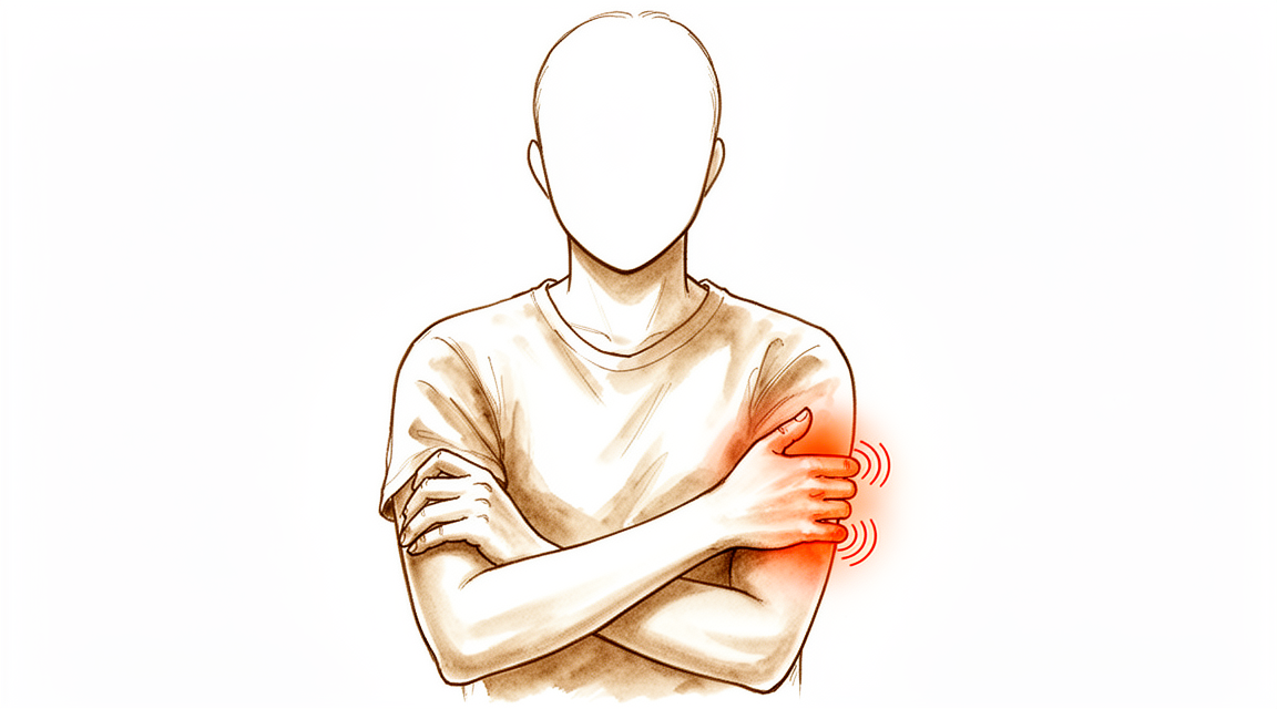
Cubital tunnel release relieves ulnar nerve compression—addressing numbness/weakness in the ring & little fingers.
Lý do phẫu thuật này được đề xuất
Bác sĩ phẫu thuật của bạn đã đề xuất ca phẫu thuật này vì bạn có khả năng cao mắc hội chứng ống khuỷu, một tình trạng trong đó dây thần kinh trụ bị chèn ép tại khuỷu tay. Tình trạng này gây tê ở ngón tay giữa và ngón út, yếu tay và đôi khi gây đau ở khuỷu tay. Mặc dù các phương pháp không phẫu thuật như thay đổi hoạt động và vật lý trị liệu thường được áp dụng trước, phẫu thuật được khuyến nghị khi những phương pháp này không mang lại đủ sự giảm nhẹ triệu chứng.
Thủ thuật này bao gồm việc rạch một vết mổ nhỏ để giải phóng các mô chèn ép xung quanh dây thần kinh trụ của bạn. Thủ thuật này thường được chỉ định cho những bệnh nhân có tình trạng tê liệt tiến triển, yếu cơ hoặc teo cơ bàn tay không cải thiện sau điều trị bảo tồn. Mục tiêu chính là ngăn ngừa tổn thương dây thần kinh tiến triển nặng hơn và giảm nhẹ các triệu chứng của bạn. Hầu hết bệnh nhân trải qua cải thiện đáng kể về giảm đau và chức năng bàn tay sau ca phẫu thuật này.
Trước khi phẫu thuật
Bạn sẽ cần nhịn ăn trong vài giờ trước khi phẫu thuật và ngừng một số loại thuốc theo chỉ định của bác sĩ phẫu thuật. Vui lòng sắp xếp để có người đưa bạn về nhà và mang theo danh sách tất cả các loại thuốc hiện tại bạn đang dùng. Bạn có thể cần làm xét nghiệm máu, chụp X-quang hoặc chụp cộng hưởng từ (MRI) để kiểm tra khuỷu tay và lập kế hoạch cho thủ thuật. Bác sĩ phẫu thuật của bạn sẽ thực hiện giải phóng hố khuỷu tay mở bằng một vết rạch thông thường duy nhất tại vị trí phẫu thuật. Phương pháp này cho phép tiếp cận trực tiếp đến dây thần kinh trụ để giảm áp lực. Hãy mặc quần áo thoải mái khi đến cuộc hẹn và sẵn sàng cho việc đánh giá gây mê ngắn.
Vào ngày phẫu thuật
Bạn sẽ đến bệnh viện và gặp bác sĩ gây mê để thảo luận về kế hoạch chăm sóc của bạn. Phẫu thuật này được thực hiện dưới gây mê toàn thân. Bạn sẽ hoàn toàn bất tỉnh trong suốt quá trình phẫu thuật. Một số bệnh nhân cũng có thể được phong bế thần kinh khu vực để giảm đau sau phẫu thuật; bác sĩ gây mê sẽ quyết định vào ngày phẫu thuật dựa trên tình trạng cá nhân của bạn.
Sau đó, bác sĩ phẫu thuật sẽ đưa bạn đến phòng mổ. Thủ thuật được thực hiện thông qua một vết rạch thông thường duy nhất tại vị trí phẫu thuật. Sau đó, bạn sẽ tỉnh dậy tại khu vực hồi sức, nơi đội ngũ y tế sẽ theo dõi mức độ thoải mái và quá trình hồi phục của bạn. Hầu hết bệnh nhân có tỷ lệ biến chứng ngắn hạn thấp là 5,6% với phương pháp này. Bạn sẽ được xuất viện khi tình trạng ổn định và cảm thấy thoải mái.
Thủ thuật phẫu thuật bao gồm
Bác sĩ phẫu thuật của bạn sẽ thực hiện một vết rạch duy nhất ở phía trước khuỷu tay để tiếp cận dây thần kinh trụ. Dây thần kinh này thường bị chèn ép bên trong một đường hầm mô. Trong quá trình phẫu thuật, bác sĩ phẫu thuật của bạn sẽ giải phóng dây thần kinh bị kẹt ra khỏi đường hầm chặt chẽ đang chèn ép lên nó.
Trong một số trường hợp, dây thần kinh có thể bị trượt khỏi vị trí khi bạn gập khuỷu tay. Nếu điều này xảy ra, bác sĩ phẫu thuật của bạn có thể di chuyển dây thần kinh đến một vị trí mới dưới cơ để giữ cho nó an toàn. Bước này được gọi là chuyển vị trí trước. Bác sĩ phẫu thuật đảm bảo dây thần kinh nằm thoải mái ở vị trí mới trước khi khâu vết rạch.
Bác sĩ phẫu thuật của bạn sẽ khâu vết mổ bằng chỉ. Bạn sẽ có một lớp băng gạc che phủ khu vực này sau khi thủ thuật hoàn tất. Trong khi một số kỹ thuật sử dụng các vết rạch nhỏ kiểu cửa sổ, bác sĩ phẫu thuật của bạn sử dụng phương pháp mở này để đảm bảo tiếp cận rõ ràng với dây thần kinh và các mô xung quanh.
Sau phẫu thuật
Bạn sẽ tỉnh lại tại khu vực hồi sức. Bác sĩ phẫu thuật của bạn sử dụng phương pháp tiếp cận mở với một vết rạch thông thường duy nhất ở khuỷu tay. Bạn sẽ được băng bó và đeo nẹp hoặc bó tay. Đau đớn được kiểm soát bằng thuốc giảm đau toàn thân. Đây là một ca phẫu thuật ngày, vì vậy bạn sẽ được xuất viện về nhà trong cùng ngày. Bạn phải có người ở lại chăm sóc bạn trong 24 giờ đầu tiên. Bạn có thể bắt đầu vận động khuỷu tay ngay sau khi phẫu thuật. Điều này giúp bạn sớm trở lại công việc. Vết thương sẽ được chăm sóc khi bạn hồi phục tại nhà.
Phục hồi
Bạn có thể sẽ cảm thấy một số cơn đau và sưng ở khuỷu tay và bàn tay trong vài ngày đầu. Đây là điều bình thường khi cơ thể bạn đang hồi phục. Bác sĩ phẫu thuật có thể khuyên bạn giữ cánh tay ở tư thế nâng cao để giúp giảm sưng. Bạn có thể nhận thấy các triệu chứng giảm bớt cả ở bàn tay và ở những vùng nằm ngoài đường đi của dây thần kinh trong quá trình hồi phục.
Bác sĩ phẫu thuật sẽ hướng dẫn bạn khi nào nên bắt đầu vận động khuỷu tay. Việc vận động khuỷu tay ngay sau phẫu thuật có thể giúp bạn trở lại làm việc sớm hơn. Bạn có thể sử dụng nạng treo tay hoặc nẹp để tăng sự thoải mái, nhưng chuyên viên vật lý trị liệu sẽ dạy bạn các bài tập nhẹ nhàng để khôi phục tầm vận động. Một khi tình trạng sưng giảm và bạn có thể nắm bắt mà không đau, bạn sẽ dần dần quay trở lại các hoạt động hàng ngày.
Quá trình hồi phục khác nhau ở mỗi người. Thời gian hồi phục của bạn có thể khác biệt dựa trên các triệu chứng trước phẫu thuật và cách cơ thể bạn hồi phục. Bác sĩ phẫu thuật và chuyên viên vật lý trị liệu sẽ hướng dẫn bạn qua từng bước để đảm bảo kết quả tốt nhất.
Những biến chứng có thể xảy ra
Hầu hết bệnh nhân đều hồi phục tốt, nhưng đôi khi có thể xảy ra các vấn đề. Bác sĩ phẫu thuật và đội ngũ y tế sẽ theo dõi sát sao bạn để phát hiện sớm bất kỳ vấn đề nào.
Đôi khi, cơn đau hoặc cảm giác tê bì bạn cảm thấy có thể không biến mất hoàn toàn. Trong những trường hợp hiếm gặp, các triệu chứng có thể xuất hiện ở những vùng nằm ngoài khu vực nơi dây thần kinh bị chèn ép. Nếu các triệu chứng của bạn không cải thiện hoặc trở nên nghiêm trọng hơn sau phẫu thuật, hãy báo ngay cho bác sĩ phẫu thuật của bạn.
Nhiễm trùng là không phổ biến, nhưng vẫn có thể xảy ra. Bạn có thể nhận thấy vùng da xung quanh vết mổ bị đỏ lan rộng, cảm giác nóng lên, hoặc cơn đau nhức nhối sâu không thuyên giảm khi dùng các loại thuốc giảm đau thông thường. Nếu bạn thấy những dấu hiệu này, hãy gọi ngay cho phòng khám.
Dây thần kinh trụ của bạn có thể bị lệch vị trí sau phẫu thuật. Bạn có thể cảm thấy có tiếng lách cách hoặc ma sát ở khuỷu tay, hoặc dây thần kinh có cảm giác như đang trượt dưới da. Hãy báo cảm giác này cho bác sĩ phẫu thuật để họ kiểm tra.
Bạn cũng có thể cảm thấy tê bì hoặc kiến bò ở mặt trong của cẳng tay. Điều này xảy ra nếu một dây thần kinh nhỏ dưới da gần vết mổ bị kích thích. Hãy cho bác sĩ phẫu thuật biết nếu cảm giác này kéo dài hoặc gây khó chịu cho bạn.
Nếu bạn cần phẫu thuật lần thứ hai vì lần đầu tiên không thành công, kết quả sẽ khó dự đoán hơn. Bạn có thể cảm thấy giảm bớt cơn đau, nhưng có thể không lấy lại hoàn toàn sức mạnh hoặc cảm giác nếu tổn thương trước đó quá nghiêm trọng. Nếu các triệu chứng tái phát, hãy báo cho bác sĩ phẫu thuật để họ thảo luận các lựa chọn điều trị với bạn.
Bảng biến chứng trên trang này liệt kê các tỷ lệ thường gặp nếu bạn muốn biết chi tiết.
Khi nào nên gọi cho chúng tôi
Hãy gọi cho chúng tôi nếu bạn bị sốt, đỏ tăng dần hoặc có dịch tiết từ vết mổ. Liên hệ ngay với bác sĩ phẫu thuật của bạn nếu bạn cảm thấy đau dữ dội đột ngột, tê mới xuất hiện hoặc không thể cử động tay. Hãy đến cơ sở y tế khẩn cấp nếu bạn nhận thấy sưng bắp chân hoặc khó thở. Những dấu hiệu này có thể cho thấy nhiễm trùng hoặc huyết khối. Ngay cả khi ca phẫu thuật của bạn diễn ra tốt đẹp, hãy báo cáo ngay bất kỳ suy yếu mới hoặc mất cảm giác nào.
Evidence & references
Anatomy & Pathophysiology
- Cubital tunnel syndrome is the second most common entrapment neuropathy of the upper extremity, occurring after carpal tunnel syndrome [1].
- The incidence of cubital tunnel syndrome is reported to be nearly 21 cases per 100,000 people per year [1].
- The prevalence of cubital tunnel syndrome in the United States population is between 1.8% and 5.9% [9].
- Pain is not a common symptom of cubital tunnel syndrome, though an aching pain localized to the elbow or proximal forearm may be reported [1].
- Patients typically present with worsening sensory numbness of the hand and digits in an ulnar distribution [1].
- Progression of cubital tunnel syndrome may involve motor weakness of the hypothenar musculature and clawing of the hand due to loss of intrinsic musculature [1].
- The most common site of ulnar nerve entrapment is about the elbow [8].
- The ulnar nerve's posterior location and superficial course make it particularly susceptible to irritation, compression, and traction, particularly with elbow motion [8].
- A cadaver study using three-dimensional modeling found that elbow flexion diminishes the volume of the cubital tunnel and elongates the nerve [8].
- Both compression and nerve tension can contribute to cubital tunnel syndrome [8].
- Ulnar nerve compression may stem from space-occupying lesions [8].
- Ulnar nerve compression may occur proximally at the ligament of Struthers (medial intermuscular septum) [8].
- Ulnar nerve compression may occur from fascial bands distally between the ulnar and humeral heads of the flexor carpi ulnaris [8].
- Ulnar nerve compression may occur about the roof of the cubital tunnel in patients with an anconeus epitrochlearis [8].
- The dorsal ulnar cutaneous nerve arises from the ulnar nerve approximately 6 cm proximal to the wrist [1].
- The dorsal ulnar cutaneous nerve innervates the ulnar aspect of the dorsum of the hand [1].
- Diminished sensation in the dorsal ulnar cutaneous nerve territory localizes the lesion proximal to this branch, likely within the cubital tunnel [1].
- Preserved dorsal sensation suggests the lesion may be localized to Guyon canal in the wrist [1].
- Subluxation or dislocation of the ulnar nerve during elbow flexion can be palpated and may be associated with increased symptoms [1].
- Ulnar nerve mobility may be associated with dislocation of the medial head of the triceps [1].
- Ultrasonography can demonstrate hypoechoic, enlarged nerve fascicles in ulnar neuropathy at the elbow [1].
- Ultrasonography sensitivity for diagnosing ulnar neuropathy at the elbow ranges from 46% to 100% [1].
- Ultrasonography specificity for diagnosing ulnar neuropathy at the elbow ranges from 43% to 97% [1].
- MRI can demonstrate enlarged, T2-hyperintense nerve lesions proximal to a point of compression with distal muscle atrophy in ulnar neuropathy at the elbow [1].
- MRI sensitivity for diagnosing ulnar neuropathy at the elbow is as high as 95% [1].
- MRI specificity for diagnosing ulnar neuropathy at the elbow is as high as 80% [1].
- The arcade of Struthers is located in the distal third of the arm and may tether the ulnar nerve [1].
- Structures that may tether the ulnar nerve distally after transposition include branches of the medial antebrachial cutaneous nerve [1].
- Structures that may tether the ulnar nerve distally after transposition include vascular branches from the ulnar artery [1].
- Structures that may tether the ulnar nerve distally after transposition include Osborne fascia [1].
- Structures that may tether the ulnar nerve distally after transposition include ulnar motor branches to the flexor carpi ulnaris [1].
- Structures that may tether the ulnar nerve distally after transposition include the distal intermuscular septum [1].
- Structures that may tether the ulnar nerve distally after transposition include the flexor-pronator muscle origin [1].
- Structures that may tether the ulnar nerve distally after transposition include the investing fascia of the flexor digitorum superficialis overlying the ulnar nerve [1].
- The elbow flexion test exacerbates dysesthesias in the small finger and ulnar side of the ring finger [8].
- Advanced findings of cubital tunnel syndrome include intrinsic muscle weakness of the hand, hypothenar wasting, and resulting functional impairment [8].
- Patients with cubital tunnel release often had a history of trauma to the anatomic site of the older cubital tunnel [9].
- Male gender is a risk factor for cubital tunnel syndrome [9].
- Patients with diabetes have both carpal tunnel and cubital tunnel procedures more often than non-diabetic populations [9].
- In situ decompression shows increased strain value in flexion [8].
- Anterior transposition demonstrates increased regional strain when the arm is in extension [8].
- The subluxated or "perched" ulnar nerve with elbow flexion after in situ release occurs in 21% to 34% of cases [9].
- The subluxated or "perched" ulnar nerve with elbow flexion after in situ release most often occurs in younger males [9].
- In a retrospective review of 67 in situ releases, 45% were unstable and required transposition [9].
Clinical Presentation
- Cubital tunnel syndrome is the second most common entrapment neuropathy of the upper extremity, occurring after carpal tunnel syndrome [1, 8].
- The incidence of cubital tunnel syndrome is reported to be nearly 21 cases per 100,000 people per year [1].
- The prevalence of cubital tunnel syndrome in the United States population is between 1.8% and 5.9% [9].
- Pain is not a common symptom of cubital tunnel syndrome, though an aching pain localized to the elbow or proximal forearm may be reported [1].
- Patients typically present with worsening sensory numbness of the hand and digits in an ulnar distribution [1].
- Symptoms may progress to motor weakness of the hypothenar musculature and clawing of the hand due to loss of intrinsic musculature [1].
- Compression of the ulnar nerve may occur anywhere along its course, but the most common site of entrapment is about the elbow [8].
- The ulnar nerve's posterior location and superficial course make it particularly susceptible to irritation, compression, and traction with elbow motion [8].
- Elbow flexion diminishes the volume of the cubital tunnel and elongates the nerve, suggesting that both compression and nerve tension contribute to cubital tunnel syndrome [8].
- Ulnar nerve compression may stem from space-occupying lesions, compression proximally at the ligament of Struthers (medial intermuscular septum), and fascial bands distally between the ulnar and humeral heads of the flexor carpi ulnaris [8].
- Compression may also occur about the roof of the cubital tunnel in patients with an anconeus epitrochlearis [8].
- Clinical manifestations include dysesthesias in the small finger and ulnar side of the ring finger exacerbated by prolonged elbow flexion [8].
- Later in the disease process, patients may complain of grip weakness and hand atrophy [8].
- More advanced findings include intrinsic muscle weakness of the hand, hypothenar wasting, and resulting functional impairment [8].
- Mild dysfunction is characterized by intermittent paresthesias and subjective weakness [9].
- Moderate dysfunction presents as intermittent paresthesias and measurable weakness [9].
- Severe dysfunction is characterized by persistent paresthesias and measurable weakness [9].
- Male gender is a risk factor for cubital tunnel syndrome [9].
- Patients with cubital tunnel release often have a history of trauma to the anatomic site of the older cubital tunnel [9].
- Patients with carpal tunnel release are older, more often female, have higher body mass indexes, and have concomitant hand tendinopathies compared to those with cubital tunnel release [9].
- The diagnosis is made by clinical history and physical examination, with adjunct electrophysiology and imaging as needed [1].
- A detailed ulnar nerve examination should include assessment of sensation in the distribution of the dorsal ulnar cutaneous nerve [1].
- The dorsal ulnar cutaneous nerve innervates the ulnar aspect of the dorsum of the hand and arises from the ulnar nerve approximately 6 cm proximal to the wrist [1].
- If sensation is diminished in the dorsal ulnar cutaneous nerve territory, the localization is proximal to this branch and likely within the cubital tunnel [1].
- With preserved dorsal sensation, the lesion may be localized to Guyon canal in the wrist [1].
- Electrophysiology is helpful to ensure proper diagnosis and targeted treatment [1].
- In some patients, subluxation or dislocation of the ulnar nerve during elbow flexion can be palpated and may be associated with increased symptoms [1].
- Ulnar nerve mobility may be associated with dislocation of the medial head of the triceps [1].
- Ultrasonography can demonstrate hypoechoic, enlarged nerve fascicles with a sensitivity of 46% to 100% and specificity of 43% to 97% [1].
- MRI can demonstrate enlarged, T2-hyperintense nerve lesions proximal to a point of compression with distal muscle atrophy, with sensitivity as high as 95% and specificity of 80% [1].
- MRI and ultrasonography are increasingly utilized to aid in the diagnosis of ulnar neuropathy at the elbow [1].
- The shoulder internal rotation test has been compared with the elbow flexion test in the diagnosis of cubital tunnel syndrome [3].
- Conservative management via activity modification, anti-inflammatory medications, and therapy can be attempted after diagnosis [1].
- Conservative treatment usually is attempted for 3 months before surgical treatment is considered [9].
- Patients with mild-to-moderate cubital tunnel syndrome are instructed to avoid prolonged elbow flexion for sleeping [9].
- Svernlöv reported improvement in 89.5% of patients with mild-to-moderate cubital tunnel syndrome treated conservatively with elbow extension splinting, patient education, and activity modification [9].
- The splint should not be fitted with the forearm held in pronation because this may aggravate symptoms [9].
- Towels or pillows secured around the elbow may limit elbow flexion adequately during sleep [9].
- Amyotrophic lateral sclerosis or Pancoast tumors can be accompanied by ulnar nerve symptoms and mimic cubital tunnel syndrome [7].
- If sensory loss far exceeds motor complaints, a sensory neuropathy should be considered [7].
- If motor complaints exceed sensory complaints, compression in the Guyon canal should be considered, especially if proximal extrinsic hand muscles are normal and there is no thenar atrophy [7].
- If there is additional thenar atrophy, radiculopathy or rarely compression in the thoracic outlet should be considered [7].
- If extrinsic motors are equally involved, brachial plexus neuritis can be considered or upper motor neuron disease if deep tendon reflexes are increased [7].
- Electrodiagnostic studies by an experienced examiner are critical in the differential diagnosis and localization of the level of compression [7].
- EMG/NCS may be ordered to confirm the extent of compression as well as sites, including identifying additional sites of compression such as above the elbow in the cervical spine [8].
- First-line treatment includes NSAIDs, night splints to avoid elbow flexion, elbow pads, and behavioral modification [8].
Investigations
- Cubital tunnel syndrome is the second most common entrapment neuropathy of the upper extremity, occurring behind carpal tunnel syndrome [1].
- The incidence of cubital tunnel syndrome is reported to be nearly 21 cases per 100,000 people per year [1].
- Pain is not a common symptom of cubital tunnel syndrome, though an aching pain localized to the elbow or proximal forearm may be reported [1].
- Typical presentation includes worsening sensory numbness of the hand and digits in an ulnar distribution [1].
- Progression of cubital tunnel syndrome may involve motor weakness of the hypothenar musculature and clawing of the hand due to loss of intrinsic musculature [1].
- Diagnosis is made by clinical history and physical examination, with adjunct electrophysiology and imaging as needed [1].
- A detailed ulnar nerve examination should assess sensation in the distribution of the dorsal ulnar cutaneous nerve [1].
- The dorsal ulnar cutaneous nerve innervates the ulnar aspect of the dorsum of the hand and arises from the ulnar nerve approximately 6 cm proximal to the wrist [1].
- Diminished sensation in the dorsal ulnar cutaneous nerve territory localizes the lesion proximal to that branch, likely within the cubital tunnel [1].
- Preserved dorsal sensation suggests the lesion may be localized to Guyon canal in the wrist [1].
- Electrophysiology is helpful to ensure proper diagnosis and targeted treatment [1].
- Subluxation or dislocation of the ulnar nerve during elbow flexion can be palpated and may be associated with increased symptoms [1].
- Ulnar nerve mobility may be associated with dislocation of the medial head of the triceps [1].
- Ultrasonography demonstrates hypoechoic, enlarged nerve fascicles in ulnar neuropathy at the elbow [1].
- Ultrasonography sensitivity for ulnar neuropathy at the elbow ranges from 46% to 100% [1].
- Ultrasonography specificity for ulnar neuropathy at the elbow ranges from 43% to 97% [1].
- MRI demonstrates enlarged, T2-hyperintense nerve lesions proximal to a point of compression with distal muscle atrophy in ulnar neuropathy at the elbow [1].
- MRI sensitivity for ulnar neuropathy at the elbow is as high as 95% [1].
- MRI specificity for ulnar neuropathy at the elbow is as high as 80% [1].
- MRI and ultrasonography are utilized as adjuncts in the preoperative setting and when evaluating recurrent or persistent symptoms [1].
- Conservative management for cubital tunnel syndrome includes activity modification, anti-inflammatory medications, and therapy [1].
- Many cases of cubital tunnel syndrome require surgical intervention despite conservative management [1].
- Commonly utilized surgical techniques for ulnar nerve surgery within the cubital tunnel include in situ decompression and anterior transposition [1].
- Surgical decompression techniques vary from unroofing the cubital tunnel to circumferential neurolysis from the distal third of the arm (arcade of Struthers) through the cubital tunnel to distal (Osborne fascia) [1].
- Anterior transposition of the ulnar nerve can be performed subcutaneously, intramuscularly, or in a submuscular fashion [1].
- Transposition involves more mobilization of the nerve and more extensive degrees of dissection of the soft tissues around the elbow compared to in situ decompression [1].
- Structures that may tether the ulnar nerve distally after transposition include branches of the medial antebrachial cutaneous nerve, vascular branches from the ulnar artery, Osborne fascia, ulnar motor branches to the flexor carpi ulnaris, the distal intermuscular septum, the flexor-pronator muscle origin, and the investing fascia of the flexor digitorum superficialis [1].
- A Cochrane review demonstrated no difference in symptom severity scores at follow-up (6 and 12 months) between in situ decompression and anterior transposition techniques [1].
- There was insufficient evidence to recommend a best treatment between in situ decompression and anterior transposition [1].
- In situ decompression has a reported success rate of 65.3% to 94.1% [1].
- Subcutaneous transposition has a reported success rate of 77.7% to 94% [1].
- Complications from in situ decompression and transposition include ulnar instability, infection, and medial antebrachial cutaneous nerve injury [1].
- The complication rate for in situ decompression is reported to be 3% [1].
- The complication rate for transposition is reported to be higher, up to 14%, likely due to more extensive dissection [1].
- Rates of secondary surgery are higher in patients undergoing anterior transposition compared to in situ release (11.1% vs 2.5%) [1].
- A recent trend favors in situ decompression over ulnar nerve transposition due to similar outcomes but higher complication and revision surgery rates with transposition [1].
- Endoscopic techniques for cubital tunnel release have outcomes similar to in situ decompression for symptom relief and return to work [1].
- Patients undergoing endoscopic techniques report higher scar satisfaction compared to other techniques [1].
- Anterior transposition should be considered in patients where ulnar nerve subluxation or dislocation is apparent and reproducible during elbow flexion on physical examination [1].
- In patients with ulnar neuropathy and no evidence of active subluxation, in situ decompression is feasible and cost-effective [1].
- In patients with recurrent symptoms after surgery, a significant proportion (77%) can experience either motor and/or sensory improvement after revision cubital tunnel decompression [1].
- 23% of patients achieve complete recovery from symptoms at final follow-up after revision cubital tunnel decompression [1].
- Complete recovery after revision surgery may be due to pain relief even when neurologic findings do not improve [1].
- For endoscopic cubital tunnel release, the patient is placed supine with the shoulder abducted and externally rotated and the arm on an arm table [4].
- A tourniquet is placed high on the brachium for endoscopic cubital tunnel release to avoid interfering with the surgical release [4].
- The arm is elevated off the table to facilitate instrumentation of the cubital tunnel during endoscopic release [4].
- A 2-cm incision is made through the skin over the cubital tunnel, just posterior to the medial epicondyle, for endoscopic release [4].
- During endoscopic exposure, dissection is carried down with scissors to the medial epicondyle while protecting superficial nerves [4].
- The deep fascia is not violated during the initial exposure of endoscopic cubital tunnel release [4].
- The ulnar nerve is identified by palpation directly posterior to the medial epicondyle during endoscopic release [4].
- An 8-cm skin incision along the course of the ulnar nerve centered over the posterior aspect of the medial epicondyle is used for open anterior transposition [4].
- The incision for open anterior transposition is carried to the deep fascia while carefully protecting the medial brachial and antebrachial cutaneous nerves [4].
- The medial epicondyle is exposed subperiosteally during open anterior transposition, incising the common flexor-pronator origin while protecting the ulnar collateral ligament [4].
- The ulnar nerve is identified and retracted posteriorly during exposure of the medial epicondyle in open anterior transposition, with care to protect the mesoneurium [4].
- The entire medial epicondyle and a portion of the supracondylar ridge are removed with an osteotome or rongeur during open anterior transposition to release the insertion of the medial intermuscular septum [4].
- The medial intermuscular septum is exposed and excised proximally to the insertion of the coracobrachialis muscle during open anterior transposition, releasing the arcade of Struthers [4].
- A bone rasp is used during open anterior transposition to ensure no bony ridges remain in the area of the osteotomy [4].
- The periosteum is reattached to the common flexor-pronator tendon during open anterior transposition to separate the raw cancellous surface from the ulnar nerve [4].
- The ulnar nerve is allowed to seek its own position adjacent to the medial humeral condyle after open anterior transposition [4].
- The tourniquet is deflated before routine closure of subcutaneous tissues and skin in open anterior transposition to allow adequate hemostasis [4].
- Postoperative care for open anterior transposition involves protecting the wound in a soft bulky dressing and allowing early range of motion as tolerated [4].
- For in situ decompression, a 3- to 5-cm incision is made along the course of the ulnar nerve between the medial epicondyle and the olecranon with the elbow flexed [6].
- During in situ decompression, subcutaneous tissues are spread and an avascular plane is developed overlying the fascia [6].
- The medial antebrachial cutaneous nerve lies along the fascia usually about 3 cm distal to the medial epicondyle and must be avoided during in situ decompression [6].
- Right-angle retractors are used to elevate subcutaneous tissue and cutaneous nerves off the fascia during in situ decompression [6].
- Osbourne's ligament is the thickened fascia between the medial epicondyle and olecranon identified during in situ decompression [6].
- The fascia overlying the ulnar nerve is incised proximally for a distance of 8 to 9 cm and distally including the deep and superficial fascia between the two heads of the flexor carpi ulnaris during in situ decompression [6].
- The nerve is left undisturbed in its soft-tissue bed during in situ decompression to avoid iatrogenic anterior subluxation [6].
- Dissection proximally during in situ decompression releases the arcade of Struthers and the medial intermuscular septum [6].
- A finger is passed proximally along the nerve 5 to 8 cm during in situ decompression to ensure no constricting bands [6].
- The superior ulnar collateral artery is protected with the nerve when dissecting proximally during in situ decompression to limit the risk of nerve infarction [6].
- The elbow is flexed after in situ decompression to ensure the nerve does not subluxate across the medial epicondyle [6].
- If subluxation occurs after in situ decompression, a formal anterior transposition is recommended [6].
- A soft dressing is applied after in situ decompression, and immediate elbow motion is allowed to prevent neural adhesions [6].
- For endoscopic release, a spatula is inserted into the space between the ulnar nerve and the roof of the canal to confirm the course of the ulnar nerve and elevation of adipose tissue and superficial nerves [4].
- A cannula/trocar is inserted into the canal and advanced proximally between the superficial ulnar nerve and the roof of the canal during endoscopic release [4].
- The attached retractor on the cannula/trocar slides on the external surface of the fascia to atraumatically elevate superficial nerves during endoscopic release [4].
- The scope is placed initially between the cannula and retractor to confirm no superficial nerves are in the way during endoscopic release [4].
- The scope is turned to view the inferior slots so the ulnar nerve can be identified throughout the entire course of the cannula during endoscopic release [4].
- The fascia (roof of the canal) is divided with a blade along the superior slot of the cannula after the nerve is clearly identified during endoscopic release [4].
- Completeness of release during endoscopic surgery is checked by pulling the cannula back on the scope [4].
- If release cannot be confirmed by pulling the cannula back, a narrow retractor is placed to expose the nerve and hold the endoscope under it to view the nerve [4].
- The cannula/trocar is advanced distally during endoscopic release using the same technique as for proximal advancement [4].
- Bipolar cautery can be used during endoscopic release if necessary for hemostasis [4].
- A 20-gauge angiocatheter is placed through the skin and into the wound during endoscopic release before closing with subcuticular absorbable sutures [4].
- 15 to 20 mL of 0.5% bupivacaine and epinephrine is infiltrated into the wound during endoscopic release before removing the angiocatheter and applying a compressive dressing [4].
- Postoperative care for endoscopic decompression instructs patients on gentle range of motion with an expectation of full range of motion within 5 to 7 days [4].
- Patients may debulk the dressing after endoscopic decompression to facilitate full motion [4].
- Endoscopic decompression of the ulnar nerve at the elbow was first described by Tsai in 1995 [4].
- Results of endoscopic decompression have been comparable to open in situ decompression [4].
- Reported advantages of endoscopic decompression include a smaller skin incision with less soft-tissue dissection [4].
- Endoscopic decompression may result in less injury to the antebrachial cutaneous nerve compared to open techniques [4].
- A nonrandomized level III study reported a 60% (9 of 15) patient satisfaction rate with in situ decompression compared with a 79% (15 of 19) rate in the endoscopic group [4].
- Dützmann et al. found no significant differences in long-term outcomes after open and retractor-endoscopic in situ decompression of the ulnar nerve in cubital tunnel syndrome [4].
- Short-term results were significantly better with endoscopic surgery compared to open techniques [4].
- A double-blind, prospective randomized controlled study found equivalent outcomes at 2 years in endoscopic and "open" releases [4].
- A meta-analysis by Buchanan et al. of 655 patients with endoscopic or in situ decompression showed no significant differences in terms of outcomes (Bishop score) or visual analog scores between the two treatment groups [4].
- A second meta-analysis by Aldekhayel et al. found no significant difference between endoscopic and open treatments in terms of complications, outcomes, and reoperation rates [4].
Treatment
- Conservative management for cubital tunnel syndrome includes activity modification, anti-inflammatory medications, and therapy [1].
- Many cases of cubital tunnel syndrome require surgical intervention despite attempts at conservative management [1].
- In situ decompression is a commonly utilized surgical technique for ulnar nerve surgery within the cubital tunnel [1].
- Anterior transposition is a commonly utilized surgical technique for ulnar nerve surgery within the cubital tunnel [1].
- Surgical techniques for in situ decompression vary, with some surgeons performing simple unroofing of the cubital tunnel [1].
- Surgical techniques for in situ decompression vary, with some surgeons performing circumferential neurolysis from the distal third of the arm to distal [1].
- Anterior transposition of the ulnar nerve can be performed in a subcutaneous fashion [1].
- Anterior transposition of the ulnar nerve can be performed in an intramuscular fashion [1].
- Anterior transposition of the ulnar nerve can be performed in a submuscular fashion [1].
- Anterior transposition involves more mobilization of the nerve compared to in situ decompression [1].
- Anterior transposition involves more extensive degrees of dissection of the soft tissues around the elbow compared to in situ decompression [1].
- Structures that may tether the ulnar nerve distally after transposition include branches of the medial antebrachial cutaneous nerve [1].
- Structures that may tether the ulnar nerve distally after transposition include vascular branches from the ulnar artery [1].
- Structures that may tether the ulnar nerve distally after transposition include the Osborne fascia [1].
- Structures that may tether the ulnar nerve distally after transposition include ulnar motor branches to the flexor carpi ulnaris [1].
- Structures that may tether the ulnar nerve distally after transposition include the distal intermuscular septum [1].
- Structures that may tether the ulnar nerve distally after transposition include the flexor-pronator muscle origin [1].
- Structures that may tether the ulnar nerve distally after transposition include the investing fascia of the flexor digitorum superficialis overlying the ulnar nerve [1].
- A Cochrane review demonstrated no difference in symptom severity scores at 6 and 12 months follow-up between in situ decompression and anterior transposition techniques [1].
- There is insufficient evidence to recommend a best treatment between in situ decompression and anterior transposition [1].
- The reported success rate for in situ decompression ranges from 65.3% to 94.1% [1].
- The reported success rate for subcutaneous transposition ranges from 77.7% to 94% [1].
- Complications from in situ decompression and anterior transposition include ulnar instability [1].
- Complications from in situ decompression and anterior transposition include infection [1].
- Complications from in situ decompression and anterior transposition include medial antebrachial cutaneous nerve injury [1].
- The reported complication rate for in situ decompression is 3% [1].
- The reported complication rate for anterior transposition is up to 14% [1].
- The higher complication rate for anterior transposition is likely due to the more extensive dissection required [1].
- The rate of secondary surgery for in situ release is 2.5% [1].
- The rate of secondary surgery for anterior transposition is 11.1% [1].
- A recent trend favors in situ decompression over ulnar nerve transposition due to similar outcomes but higher complication and revision surgery rates with transposition [1].
- Endoscopic techniques for cubital tunnel release have outcomes similar to in situ decompression for symptom relief [1].
- Endoscopic techniques for cubital tunnel release have outcomes similar to in situ decompression for return to work [1].
- Patients undergoing endoscopic cubital tunnel release report higher scar satisfaction compared to other techniques [1].
- Anterior transposition should be considered in patients where ulnar nerve subluxation or dislocation is apparent and reproducible during elbow flexion on physical examination [1].
- In patients with ulnar neuropathy and no evidence of active subluxation, in situ decompression is feasible and cost-effective [1].
- In patients with recurrent symptoms after surgery, 77% can experience either motor and/or sensory improvement after revision cubital tunnel decompression [1].
- In patients with recurrent symptoms after surgery, 23% achieve complete recovery from symptoms at final follow-up after revision cubital tunnel decompression [1].
- Complete recovery from symptoms after revision cubital tunnel decompression may be due to pain relief even when neurologic findings do not improve [1].
- Endoscopic cubital tunnel release is a recognized operative technique for cubital tunnel syndrome [2].
- Submuscular transposition is a recognized operative technique for cubital tunnel syndrome [2].
- Subcutaneous transposition is a recognized operative technique for cubital tunnel syndrome [2].
- Partial medial epicondylectomy is a recognized operative technique for cubital tunnel syndrome [2].
- Simple decompression with a small skin incision is a recognized operative technique for cubital tunnel syndrome [2].
- Endoscopic cubital tunnel release involves placing the patient supine with the shoulder abducted and externally rotated [4].
- A tourniquet is placed high on the brachium for endoscopic cubital tunnel release to avoid interfering with the surgical release [4].
- The arm is elevated off the table during endoscopic cubital tunnel release to facilitate instrumentation of the cubital tunnel [4].
- An 8-cm skin incision is made along the course of the ulnar nerve centered over the posterior aspect of the medial epicondyle for open cubital tunnel release [4].
- The incision for open cubital tunnel release is carried to the deep fascia while protecting the medial brachial and antebrachial cutaneous nerves [4].
- The medial epicondyle is exposed subperiosteally during open cubital tunnel release [4].
- The common flexor-pronator origin is incised during open cubital tunnel release while protecting the ulnar collateral ligament [4].
- The ulnar nerve is identified and retracted posteriorly during exposure of the medial epicondyle in open cubital tunnel release [4].
- The entire medial epicondyle and a portion of the supracondylar ridge are removed during open cubital tunnel release to release the insertion of the medial intermuscular septum [4].
- The medial intermuscular septum is exposed and excised proximally to the insertion of the coracobrachialis muscle during open cubital tunnel release [4].
- The arcade of Struthers is released as a potential area of compression during open cubital tunnel release [4].
- A bone rasp is used during open cubital tunnel release to ensure no bony ridges remain in the area of the osteotomy [4].
- The periosteum is reattached to the common flexor-pronator tendon during open cubital tunnel release to separate the raw cancellous surface from the ulnar nerve [4].
- The ulnar nerve is allowed to seek its own position adjacent to the medial humeral condyle after open cubital tunnel release [4].
- The tourniquet is deflated before routine closure of subcutaneous tissues and skin in open cubital tunnel release [4].
- Postoperative care for cubital tunnel release involves protecting the wound in a soft bulky dressing [4].
- Early range of motion is allowed as tolerated following cubital tunnel release [4].
Complications
- Complications from in situ decompression and anterior transposition include ulnar instability, infection, and medial antebrachial cutaneous nerve injury [1].
- The reported complication rate for in situ decompression is 3% [1].
- The reported complication rate for anterior transposition is up to 14% [1].
- Higher complication rates in anterior transposition are likely due to the more extensive dissection required to complete the nerve transposition [1].
- Rates of secondary surgery are higher in patients undergoing anterior transposition compared to in situ release (11.1% vs 2.5%) [1].
- Recurrent or persistent symptoms after cubital tunnel surgery can be difficult to treat [1].
- In patients with recurrent symptoms, 77% can experience either motor and/or sensory improvement after revision cubital tunnel decompression [1].
- In patients with recurrent symptoms, 23% achieve complete recovery from symptoms at final follow-up after revision cubital tunnel decompression [1].
- Complete recovery in revision cases may be due to pain relief even when neurologic findings do not improve [1].
- Patients with severe and chronic compression, atrophy, and dense sensory loss should be cautioned that surgery will arrest progression and improve symptoms but will not return normal strength or sensation [5].
- Nonsmokers have significantly better outcomes than smokers following anterior transposition of the ulnar nerve [5].
References
[1] Orthopaedic Knowledge Update 13 Ebook Without Multimedia. Neuropathies, Vascular Conditions: Buerger’s, Raynaud’s; Degenerative Conditions > Upper Extremity Neuropathies > Cubital Tunnel Syndrome. [2] Campbell S Operative Orthopaedics 4 Volume Set. COMPRESSIVE NEUROPATHIES OF THE HAND, FOREARM, AND ELBOW > CUBITAL TUNNEL. [3] Campbell S Operative Orthopaedics 4 Volume Set. COMPRESSIVE NEUROPATHIES OF THE HAND, FOREARM, AND ELBOW > CUBITAL TUNNEL SYNDROME. [4] Campbell S Operative Orthopaedics 4 Volume Set. COMPRESSIVE NEUROPATHIES OF THE HAND, FOREARM, AND ELBOW > ENDOSCOPIC CUBITAL TUNNEL RELEASE. [5] Green S Operative Hand Surgery. Median Nerve Compression at the Elbow and Forearm > Expected Outcome After Anterior Transposition of the Ulnar Nerve With Release of the Flexor-Pronator Muscle Origin for Cubital Tunnel Syndrome. [6] Campbell S Operative Orthopaedics 4 Volume Set. COMPRESSIVE NEUROPATHIES OF THE HAND, FOREARM, AND ELBOW > CUBITAL TUNNEL SYNDROME AND TARDY ULNAR NERVE PALSY > IN SITU DECOMPRESSION OF THE ULNAR NERVE. [7] Green S Operative Hand Surgery. Median Nerve Compression at the Elbow and Forearm > Recurrent Cubital Tunnel Syndrome. [8] Orthopaedic Knowledge Update 13 Ebook Without Multimedia. Elbow Degenerative Conditions and Nerve Disorders > Nerve Disorders > Cubital Tunnel Syndrome. [9] Campbell S Operative Orthopaedics 4 Volume Set. COMPRESSIVE NEUROPATHIES OF THE HAND, FOREARM, AND ELBOW > CUBITAL TUNNEL SYNDROME AND TARDY ULNAR NERVE PALSY.


