Viêm khớp ngón tay cái gốc
Patients › Hand
Basal thumb arthritis — causes, symptoms, non-surgical options, and when surgery might help.
Những triệu chứng bạn đang trải qua
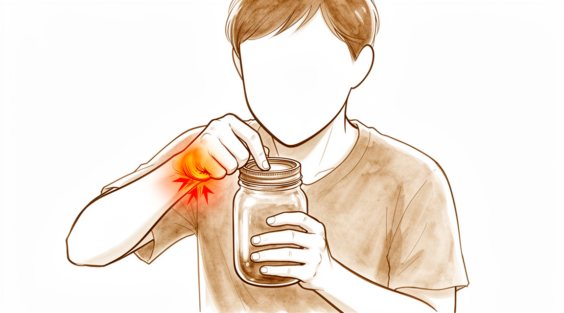
Viêm khớp gối ngón tay cái là một tình trạng phổ biến ảnh hưởng đến khớp ở gốc ngón tay cái. Loại viêm khớp do hao mòn này phát triển như một phần của quá trình lão hóa bình thường. Bạn có thể nhận thấy rằng cơn đau không chỉ ở bề mặt mà còn cảm thấy sâu bên trong khớp. Ở giai đoạn đầu của bệnh, bạn có thể thấy khó khăn hơn khi nắm chặt các vật. Những công việc đơn giản như xoay chìa khóa hoặc mở lọ có thể trở nên khó khăn do sức mạnh của bàn tay bạn suy giảm.
Cơn đau thường bùng phát sau khi bạn sử dụng tay trong một thời gian. Bạn có thể cảm thấy đau âm ỉ và cứng khớp khi vừa thức dậy vào buổi sáng. Sự cứng khớp này thường giảm bớt khi bạn cử động tay, nhưng có thể quay trở lại nếu bạn thực hiện các chuyển động lặp đi lặp lại. Một số người bị đau vào ban đêm, điều này có thể khiến họ khó đi vào giấc ngủ hoặc khó duy trì giấc ngủ. Nằm nghiêng về phía bên bị đau có thể gây áp lực trực tiếp lên khớp bị viêm, làm trầm trọng thêm sự khó chịu.
Các hoạt động hàng ngày trở nên khó khăn hơn khi tình trạng tiến triển. Bạn có thể gặp khó khăn với các kỹ năng vận động tinh, chẳng hạn như cài cúc áo hoặc nhét áo vào quần. Việc với tay ra sau lưng để cài móc áo ngực có thể đặc biệt gây đau. Bạn cũng có thể nhận thấy rằng việc cầm điện thoại hoặc tách cà phê gây ra cơn đau nhói ở gốc ngón tay cái. Điều này là do những hành động này đòi hỏi một lực nắm chắc chắn và ổn định mà khớp bị viêm không còn có thể cung cấp một cách thoải mái.
Đau mạn tính và viêm là những đặc điểm phổ biến của tình trạng này. Bạn có thể cảm thấy một cơn đau âm ỉ liên tục ngay cả khi đang nghỉ ngơi. Khớp có thể cảm thấy ấm hoặc sưng khi chạm vào. Mặc dù cơn đau có thể gây khó chịu, nhưng đó là một tín hiệu rõ ràng từ cơ thể cho thấy khớp đang chịu áp lực. Bác sĩ phẫu thuật của bạn sẽ lắng nghe tiền sử bệnh và khám tay để xác nhận chẩn đoán. Hiểu rõ các triệu chứng này giúp giải thích tại sao một số cử động gây đau và tại sao việc nghỉ ngơi hoặc sử dụng các loại nẹp cụ thể có thể mang lại sự giảm đau.
Những gì thực sự đang xảy ra
Viêm khớp ngón cái gốc là một tình trạng thoái hóa phổ biến và tiến triển. Bệnh ảnh hưởng đến khớp ở gốc ngón cái, nơi ngón cái tiếp xúc với cổ tay. Theo thời gian, lớp sụn trơn láng đệm giữa các đầu xương bị mòn đi. Điều này dẫn đến đau mãn tính và viêm. Bạn có thể nhận thấy khả năng nắm chặt giảm sút. Tình trạng này xảy ra sớm trong quá trình tiến triển của bệnh, ngay cả khi phim X-quang chưa cho thấy tổn thương đáng kể.
Khớp dựa vào sự vận động chính xác và độ ổn định. Các dây chằng và gân hoạt động như những sợi dây để giữ các xương ở vị trí cố định. Khi cấu trúc khớp thay đổi, các yếu tố ổn định này gặp khó khăn. Các xương có thể bị lệch hoặc di chuyển lên trên. Sự sai lệch này làm thay đổi cách khớp vận động khi thực hiện động tác bóp hoặc nắm. Ma sát resulting gây thêm tổn thương cho bề mặt khớp. Bác sĩ phẫu thuật có thể chẩn đoán tình trạng này thông qua khai thác tiền sử và khám thực thể đơn giản.
Phẫu thuật nhằm khôi phục lại giải phẫu và sinh cơ học của khớp này. Các thủ thuật như cắt xương thuyền (trapeziectomy) loại bỏ xương bị tổn thương để giảm đau. Các lựa chọn khác sử dụng gân hoặc miếng đệm tổng hợp để ổn định khớp. Mặc dù các phương pháp điều trị này cải thiện chức năng và sức khỏe, chúng không hoàn toàn tái tạo lại chuyển động của một khớp khỏe mạnh. Một số thủ thuật có thể cho phép xương dịch chuyển nhẹ theo thời gian, nhưng điều này thường không ảnh hưởng đến chức năng hàng ngày của bạn. Mục tiêu là giảm đau và khôi phục khả năng sử dụng bàn tay một cách hiệu quả.
Những gì chúng tôi có thể làm về vấn đề này
Chúng tôi bắt đầu bằng điều trị không phẫu thuật, phương pháp này hiệu quả đối với các giai đoạn đầu của viêm xương khớp do hao mòn ở gốc ngón tay cái của bạn. Bác sĩ phẫu thuật của bạn có thể khuyến nghị các chiến lược tự quản lý và vật lý trị liệu để giúp bạn duy trì khả năng vận động và sức mạnh. Những biện pháp bảo thủ này nhằm giảm đau và cải thiện chức năng mà không cần phẫu thuật. Bạn nên thử những phương pháp này một cách nghiêm túc trước khi xem xét các lựa chọn xâm lấn hơn. Đối với nhiều bệnh nhân, cách tiếp cận này là đủ để kiểm soát các triệu chứng một cách hiệu quả.
Nếu các biện pháp đơn giản không mang lại đủ sự giảm đau, bác sĩ phẫu thuật của bạn có thể thảo luận về quản lý bằng thuốc. Điều này có thể bao gồm thuốc giảm đau và thuốc chống viêm để giúp kiểm soát sự khó chịu. Tiêm là một lựa chọn khác. Tiêm cortisone có thể giảm viêm và đau trong một thời gian hạn chế. Tiêm axit hyaluronic có thể giúp bôi trơn khớp, mặc dù hiệu quả thay đổi tùy trường hợp. Tiêm huyết tương giàu tiểu cầu (PRP) sử dụng các thành phần máu của chính bạn để thúc đẩy quá trình chữa lành, nhưng bằng chứng về lợi ích lâu dài của chúng vẫn đang được phát triển. Bác sĩ phẫu thuật của bạn sẽ tư vấn xem loại tiêm nào, nếu có, là phù hợp với tình trạng cụ thể của bạn và thời gian giảm đau có thể kéo dài bao lâu.
Phẫu thuật chỉ được dành cho các trường hợp mà các biện pháp bảo thủ đã thất bại trong việc mang lại sự giảm đau đầy đủ. Mục tiêu của phẫu thuật là loại bỏ nguồn gốc gây đau và khôi phục chức năng. Một thủ thuật phổ biến là cắt xương thang (trapeziectomy), trong đó xương bị mòn được loại bỏ. Điều này thường mang lại kết quả lâm sàng tích cực lâu dài. Các lựa chọn khác bao gồm thay khớp hoặc hợp nhất khớp, tùy thuộc vào nhu cầu cá nhân của bạn. Mặc dù hầu hết bệnh nhân trải qua sự cải thiện đáng kể, điều quan trọng là phải biết rằng bất chấp nhiều lựa chọn điều trị, một tỷ lệ bệnh nhân không đổi vẫn cảm thấy không hài lòng hoặc có triệu chứng sau phẫu thuật. Bác sĩ phẫu thuật của bạn sẽ thảo luận về các rủi ro, lợi ích và thời gian phục hồi dự kiến để giúp bạn quyết định xem phẫu thuật có phải là bước đi đúng đắn cho bạn hay không.
Những điều cần biết
Viêm xương khớp gốc ngón cái là một tình trạng phổ biến, thường tiến triển nặng hơn theo thời gian. Đây là một bệnh lý tiến triển, nghĩa là các thay đổi do hao mòn trong khớp của bạn có xu hướng gia tăng thay vì tự ổn định. Bạn có thể nhận thấy rằng các triệu chứng tồn tại dai dẳng và có thể dần trở nên khó chịu hơn khi bệnh tiến triển.
Nếu bạn chọn quản lý tình trạng này bằng các phương pháp không phẫu thuật như trị liệu tay, quá trình dẫn đến phẫu thuật của bạn có thể kéo dài hơn. Tuy nhiên, bằng chứng cho thấy rằng những bệnh nhân không thực hiện trị liệu thường đạt đến giai đoạn cần phẫu thuật sớm hơn. Đối với những bệnh nhân ở giai đoạn đầu của bệnh, các phương pháp điều trị ít xâm lấn hơn như rửa khớp hoặc các thủ thuật làm dịu dây thần kinh có thể mang lại sự giảm đau. Những phương pháp này có tỷ lệ biến chứng thấp và thời gian hồi phục ngắn, đóng vai trò là một lựa chọn thay thế nhẹ nhàng hơn so với các ca phẫu thuật lớn hơn.
Khi phẫu thuật là lựa chọn phù hợp, triển vọng dài hạn rất tích cực. Hầu hết bệnh nhân đạt được kết quả xuất sắc, cải thiện chức năng hàng ngày và giảm đau. Bác sĩ phẫu thuật của bạn sẽ thảo luận về kỹ thuật tốt nhất dành cho bạn, dù đó là việc loại bỏ xương bị mòn, sử dụng miếng đệm hoặc thay thế khớp. Bất kể phương pháp cụ thể nào được chọn, bạn có thể mong đợi những cải thiện đáng kể về sức khỏe và chất lượng cuộc sống.
Điều quan trọng cần biết là một số thay đổi có thể xuất hiện trên phim X-quang theo thời gian, chẳng hạn như sự dịch chuyển nhẹ của các thành phần khớp. Tuy nhiên, những thay đổi trên hình ảnh X-quang này không nhất thiết có nghĩa là kết quả điều trị của bạn sẽ kém. Nhiều bệnh nhân duy trì sức mạnh và sự hài lòng tốt trong nhiều năm. Ngay cả khi một thủ thuật trước đó cần được sửa đổi, vẫn có các lựa chọn hiệu quả có sẵn với tỷ lệ hài lòng cao.
Mặc dù biến chứng có thể xảy ra, nhưng các vấn đề nghiêm trọng là tương đối hiếm gặp với các kỹ thuật hiện đại. Một số loại cấy ghép cũ cụ thể đã không còn được sử dụng do tỷ lệ biến chứng cao, vì vậy bác sĩ phẫu thuật của bạn sẽ chọn các vật liệu có hồ sơ an toàn đã được chứng minh. Bạn cũng nên nhận thức rằng các yếu tố như hút thuốc hoặc phẫu thuật tay trước đó có thể ảnh hưởng đến cách tình trạng tiến triển, nhưng những yếu tố này không ngăn cản việc điều trị thành công. Nhìn chung, với sự chăm sóc thích hợp, bạn có thể mong đợi khôi phục khả năng sử dụng ngón cái và trở lại các hoạt động bình thường với tình trạng đau giảm đi.
Khi nào nên đi khám
Viêm khớp gốc ngón cái là một tình trạng phổ biến và tiến triển. Bạn nên đi khám bác sĩ đa khoa nếu nhận thấy cơn đau dai dẳng không thuyên giảm khi nghỉ ngơi. Hãy yêu cầu được bác sĩ chuyên khoa đánh giá nếu bạn gặp phải tình trạng yếu hoặc mất vững ở ngón cái. Các triệu chứng như kẹt khớp hoặc đột ngột mất sức cũng cần được kiểm tra. Hãy tìm kiếm sự chăm sóc y tế nếu các triệu chứng của bạn ảnh hưởng đến giấc ngủ hoặc công việc. Sự gia tăng đột ngột về mức độ đau cũng là một dấu hiệu rõ ràng cần hành động. Trong khi những thay đổi do hao mòn là một phần của quá trình lão hóa, thì cơn đau hạn chế chức năng lại ít phổ biến hơn. Bác sĩ phẫu thuật của bạn có thể chẩn đoán tình trạng này thông qua việc khai thác tiền sử bệnh và khám lâm sàng đơn giản. Phát hiện sớm giúp kiểm soát bệnh trước khi nó làm giảm đáng kể lực nắm của bạn.
Evidence & references
Overview
- Basal thumb arthritis is a common condition [1].
- A comprehensive history and clinical examination are sufficient for the diagnosis of basal thumb arthritis [1].
- Osteoarthritis is likely the most common indication for basal joint arthroscopy [13].
- Chronic pain and inflammation are useful indications for metacarpophalangeal arthroscopy [13].
- Basal thumb osteoarthritis surgery improves health state utility irrespective of the surgical technique used [6].
- Trapeziectomy for basal thumb osteoarthritis does not increase the risk of developing wrist osteoarthritis in the long term [2].
- Long-term clinical outcomes of trapeziectomy for basal thumb arthritis are very positive [5].
- Interpositioning as an isolated procedure appears clinically to be the preferred treatment for basal thumb arthritis despite greater radiological degradation compared to suspensionplasty [5].
- Patients who underwent suture-button suspensionplasty (SBS) surgery for thumb CMC osteoarthritis achieve excellent long-term outcomes by maintaining favorable subjective and objective results [9].
- Some radiographic subsidence occurs over time in patients who underwent suture-button suspensionplasty for thumb CMC osteoarthritis [9].
- The use of Swanson silastic interposition arthroplasty in revision thumb-base surgery for failed trapeziectomy yields good medium-term results and high satisfaction rates [8].
- Swanson silastic interposition arthroplasty is advocated as an effective treatment option for revision thumb-base surgery provided other treatable causes of poor outcome are excluded [8].
- Pyrocarbon implants are used for the surgical treatment of basal thumb arthritis [10].
- The Artelon CMC Spacer is no longer used for the management of basal joint arthritis of the thumb due to an unacceptably high complication rate [11].
- Porous Polyurethaneurea (Artelon) Joint Spacer use has been abandoned for the treatment of basilar thumb osteoarthritis [17].
- Denervation, joint lavage, and capsular imbrication could be a good alternative to more invasive surgical options in patients with earlier stages of thumb carpometacarpal joint osteoarthritis [20].
- Denervation, joint lavage, and capsular imbrication offer advantages including a low rate of complications, low invasiveness, and short recovery times [20].
Anatomy & Pathophysiology
- Thumb basal joint arthritis is a progressive disease [4].
- A reduction in cylindrical grasp is associated with early symptomatic and radiographic CMC OA [12].
- Gross grasp is not associated with early thumb CMC OA [12].
- Wrist biomechanics are significantly altered following trapeziectomy [30].
- Ligament reconstruction with tendon interposition (LRTI) most closely resembles intact wrist biomechanics in a cadaveric model [30].
- Total joint arthroplasty restores thumb function but cannot fully replicate the kinematics of the healthy TMC joint [31].
- Kinematic analysis of the thumb CMC joint differentiates surgical treatments used for end-stage OA [32].
- Thumb motion capability is unaffected by sex and handedness [33].
- A rationale for dynamic stabilization of the thumb is based on its unique anatomy [34].
- The inter-metacarpal distance method is the most reliable tool for measuring thumb abduction [35].
- Surgical treatment is usually indicated to restore the anatomy and biomechanics of the trapeziometacarpal joint in fractures and dislocations of the base of the thumb metacarpal, as conservative treatment often yields poor results [36].
- Thumbs in patients with TMC-OA have different kinematics during first dorsal interosseous (FDI) maneuvers compared to healthy thumbs [37].
- An atrophic FDI may not be an efficient dynamic stabilizer [37].
- During thumb oppositional motion, internal rotation of the first metacarpal occurs, with the palmar base rotating primarily with respect to the dorsal base [38].
- The position of the thumb metacarpophalangeal joint exerts a strong influence on contact-pressure patterns in the trapeziometacarpal joint [39].
- Metacarpophalangeal joint flexion shifts the center of pressure in the trapeziometacarpal joint dorsally [39].
- Metacarpophalangeal joint hyperextension produces the most palmar contact pattern in the trapeziometacarpal joint [39].
- Trapeziectomy results in proximal migration of the first metacarpal [40].
- Suture suspensionplasty mitigates proximal migration of the first metacarpal while maintaining normal motion [40].
- Proximal migration of the thumb metacarpal does not appear to influence functional outcome [41].
- Altered thumb rotation patterns during pinch may contribute to joint misalignment and the development of osteoarthritis [42].
- Automated analysis of TMC joint kinematics using four-dimensional computed tomography significantly decreases analysis time [43].
- Ergonomic solutions are necessary to decrease thumb motions or strenuous effort at work, especially for women, to reduce the risk of thumb CMC osteoarthritis [44].
- Carpometacarpal and metacarpophalangeal joint collapse is associated with increased pain but not functional impairment in persons with thumb CMC osteoarthritis [45].
- Directionally coupled motion patterns in the CMC joint are similar in men and women [46].
Classification
- Basal thumb arthritis is a common condition where a comprehensive history and clinical examination are sufficient for diagnosis [1].
- Thumb basal joint arthritis is a progressive disease with substantial new biomechanical and longitudinal clinical studies changing prevailing opinions on serial degenerative changes [4].
- Subjects presenting with early CMC OA had significantly lower bone density as assessed with HU at the thumb CMC joint (trapezium and first metacarpal base) [7].
- The metacarpal surface of the trapezium demonstrates three distinct patterns of wear in arthritic surgical specimens [26].
- Radiological imaging of the trapeziometacarpal joint involves various measurements and classifications used to evaluate the joint [19].
- The radiological classification does not describe all stages of carpometacarpal joint osteoarthritis accurately enough to permit reliable and consistent communication between clinicians [25].
- There is not a reliable system for classification of disease severity in CMC joint disease based on radiographs [28].
- Ulnar instability should be included in the classification of thumb CMCj osteoarthritis stages and considered in treatment options [55].
Clinical Presentation
- Basal thumb arthritis is a common condition [1].
- A comprehensive history and clinical examination are sufficient for the diagnosis of basal thumb arthritis [1].
- Thumb basal joint arthritis is a progressive disease [4].
- Serial degenerative changes in thumb basal joint arthritis are described by new biomechanical and longitudinal clinical studies [4].
- Radiographic development of trapeziometacarpal arthrosis is an expected part of human aging [27].
- Clinically significant, functionally limiting trapeziometacarpal arthrosis is less common than radiographic development [27].
- The development of clinically significant trapeziometacarpal arthrosis may be unrelated to hand use [27].
- Subjects with early thumb carpometacarpal joint osteoarthritis have significantly lower bone density at the trapezium and first metacarpal base as assessed with Hounsfield Units [7].
- A reduction in cylindrical grasp strength is associated with early symptomatic and radiographic thumb carpometacarpal osteoarthritis [12].
- Gross grasp is not associated with early thumb carpometacarpal osteoarthritis [12].
- Cylindrical grasp may be a better tool than gross grasp to detect changes in thumb and hand function during early disease stages [12].
- Power Doppler ultrasound has a significant relationship with pain severity in thumb base osteoarthritis, suggesting it may be useful for understanding pain aetiology [22].
- The high prevalence of other symptomatic hand disorders requires a complete and standardized clinical examination of the hand to consider these disorders during surgical planning [21].
- Osteoarthritis is likely the most common indication for basal joint arthroscopy [13].
- Chronic pain and inflammation are useful indications for metacarpophalangeal arthroscopy [13].
- The metacarpal surface of the trapezium demonstrates three distinct patterns of wear in arthritic surgical specimens [26].
Investigations
- A comprehensive history and clinical examination are sufficient for the diagnosis of basal thumb arthritis [1].
- Radiographic development of trapeziometacarpal arthrosis is an expected part of human aging [27, 62].
- Clinically significant, functionally limiting trapeziometacarpal arthrosis is less common than radiographic changes [27, 62].
- The development of clinically significant trapeziometacarpal arthrosis may be unrelated to hand use [27].
- Subjects with early CMC OA have significantly lower bone density at the thumb CMC joint (trapezium and first metacarpal base) as assessed with Hounsfield Units (HU) [7].
- The volar-ulnar quadrant of the trapezium has significantly greater trabecular bone volume, thickness, and connectivity compared to the dorsal-radial and dorsal-ulnar quadrants [58].
- The greatest compressive loads at the first carpometacarpal joint occur at the volar-ulnar quadrant of the trapezium [58].
- The volar-ulnar quadrant of the trapezium represents a consistently affected region of wear in both normal and arthritic states [58].
- A reduction in cylindrical grasp strength is associated with early symptomatic and radiographic CMC OA [12].
- Gross grasp is not associated with early thumb CMC OA [12].
- Cylindrical grasp may be a better tool than gross grasp to detect changes in thumb and hand function during early disease stages [12].
- Power Doppler ultrasound has a significant relationship with pain severity in thumb base OA, suggesting it may be useful in understanding pain aetiology [22].
- Radiological imaging reviews provide an overview of different radiological views, historical origins, positioning, measurements, and classifications used to evaluate the trapeziometacarpal joint [19].
- Radiographic classification of osteoarthritis at the trapeziometacarpal joint does not describe all stages of CMC joint OA accurately enough to permit reliable and consistent communication between clinicians [25].
- Radiographs assist in the assessment of CMC joint disease, but there is not a reliable system for classification of disease severity [28].
- A negative grind test does not necessarily reflect negative radiographic evidence of thumb CMC osteoarthritis [63].
- Wrist radiographs demonstrate 47% sensitivity and 94% specificity in predicting end-stage ST joint arthritis [56].
- Direct visualization of the ST joint is important after trapeziectomy due to the limitations of wrist radiographs in predicting end-stage ST joint arthritis [56].
Treatment
Non-Operative Management
- Nonoperative modalities are effective for early stages of degenerative arthritis of the thumb carpometacarpal (CMC) joint [48].
- Surgical options for thumb CMC arthritis are reserved for cases refractory to conservative measures [48].
- Denervation, joint lavage, and capsular imbrication are good alternative treatments for earlier stages of thumb CMC joint osteoarthritis, offering a low rate of complications, low invasiveness, and short recovery times [20].
Arthroscopic Techniques
- Osteoarthritis is likely the most common indication for basal joint arthroscopy [13].
- Chronic pain and inflammation are useful indications for metacarpophalangeal arthroscopy [13].
- The use of arthroscopic-assisted techniques for thumb CMC osteoarthritis is still limited but may be a reasonable option for patients who do not respond to non-operative treatment [53].
Trapeziectomy and Interposition/Suspensionplasty
- Trapeziectomy with interposition or suspensionplasty yields very positive long-term clinical outcomes [5].
- Interpositioning as an isolated procedure appears to be the preferred treatment clinically, despite greater radiological degradation compared to suspensionplasty [5].
- Suture-button suspensionplasty (SBS) achieves excellent long-term outcomes by maintaining favorable subjective and objective results, despite some radiographic subsidence over time [9].
- Suture-button suspensionplasty (SBS) achieves excellent long-term outcomes by maintaining favorable subjective and objective results, despite some radiographic subsidence over time [16].
- The multiplicity of treatment modalities for carpometacarpal joint arthritis suggests that underlying trapezium excision is probably the prime factor in patients' clinical improvement [15].
- Thumb index metacarpal stabilization needs to be based on each individual clinical scenario [15].
- Trapeziectomy for basal thumb osteoarthritis does not increase the risk of developing wrist osteoarthritis in the long term [2].
Joint Replacement and Implants
- Health state utility gains occur after basal thumb osteoarthritis surgery regardless of the surgical techniques used [6].
- The ISISVR prosthesis is a reliable implant for treating disabling thumb basal joint arthritis, with a low complication rate and long-lasting clinical and functional improvements [18].
- Pyrocarbon implants are used for the surgical treatment of basal thumb arthritis [10].
- The Artelon CMC Spacer is no longer used for the management of basal joint arthritis of the thumb due to an unacceptably high complication rate [11].
- The use of Porous Polyurethaneurea (Artelon) Joint Spacer has been abandoned for the treatment of basilar thumb osteoarthritis due to findings indicating poor outcomes or high complications [17].
Revision Surgery
- Swanson silastic interposition arthroplasty is an effective treatment option for revision thumb-base surgery for failed trapeziectomy, showing good medium-term results and high satisfaction rates, provided other treatable causes of poor outcome are excluded [8].
Complications and Outcomes
- Common complications after surgery for basal thumb arthritis include those associated with resection arthroplasty, joint replacement, and joint fusion, with specific management strategies available for different types of complications [3].
- Despite 70 years of research and numerous treatment options, the best management for trapeziometacarpal arthritis remains debated, with a constant proportion of patients remaining unhappy or symptomatic post-surgery [52].
Ongoing Research
- The SCOOTT trial is a multicentre, three-arm randomized controlled trial designed to determine the clinical and cost-effectiveness of treating basal osteoarthritis of the thumb with or without surgery, and to compare trapeziectomy versus thumb CMC joint arthrodesis (CMCJA) [51].
Complications
- Basal thumb arthritis surgery complications are reviewed for resection arthroplasty, joint replacement, and joint fusion, including management strategies [3].
- Trapeziectomy for basal thumb osteoarthritis does not increase the risk of developing wrist osteoarthritis in the long term [2].
- Long-term clinical outcomes of trapeziectomy are very positive, with interpositioning appearing clinically preferred despite greater radiological degradation compared to suspensionplasty [5].
- Health state utility gains after basal thumb osteoarthritis surgery occur irrespective of the surgical technique used [6].
- Revision thumb-base surgery using Swanson silastic interposition arthroplasty yields good medium-term results and high satisfaction rates, provided other treatable causes of poor outcome are excluded [8].
- The Artelon CMC Spacer is no longer used for the management of basal joint arthritis of the thumb due to an unacceptably high complication rate [11].
- Simultaneous dual prosthetic replacement of the trapeziometacarpal and scaphotrapezial-trapezoid joints in pantrapezial osteoarthritis achieves a low complication rate [14].
- Suture-button suspensionplasty (SBS) for thumb CMC osteoarthritis maintains favorable subjective and objective results despite some radiographic subsidence over time [9, 16].
- The ISISVR prosthesis is a reliable implant for treating disabling thumb basal joint arthritis, with a low complication rate and long-lasting clinical and functional improvements [18].
Recovery
- Basal thumb arthritis is a common condition where a comprehensive history and clinical examination are sufficient for diagnosis [1].
- Thumb basal joint arthritis is a progressive disease with substantial new biomechanical and longitudinal clinical studies changing prevailing opinions on serial degenerative changes [4].
- Subjects presenting with early CMC OA had significantly lower bone density as assessed with HU at the thumb CMC joint (trapezium and first metacarpal base) [7].
- Increased degenerate-like changes were observed after simple excision of the trapezium at 6-year followup but these did not influence the clinical outcome [64].
- Trapeziectomy for basal thumb osteoarthritis does not increase the risk of developing wrist osteoarthritis in the long term [2].
- Long-term clinical outcomes of trapeziectomy for basal thumb arthritis are very positive, with interpositioning as an isolated procedure appearing, clinically, to be the preferred treatment despite greater radiological degradation when compared to suspensionplasty [5].
- The use of Swanson silastic interposition arthroplasty in revision thumb-base surgery for failed trapeziectomy yields good medium-term results and high satisfaction rates, advocating the technique as an effective treatment option for revision thumb-base surgery provided other treatable causes of poor outcome are excluded [8].
- Patients who underwent SBS surgery for thumb CMC osteoarthritis achieve excellent long-term outcomes by maintaining favorable subjective and objective results, despite some radiographic subsidence over time [9].
- The SSA technique for thumb CMC arthritis reconstruction yields good to excellent long-term clinical outcomes at 12- to 14-year follow-up [54].
- Simultaneous dual prosthetic replacement of trapeziometacarpal and scaphotrapezial-trapezoid joints in pantrapezial osteoarthritis achieves favorable functional outcomes and a low complication rate, making it a potentially superior alternative for patients with high functional demands or those requiring durable long-term results [14].
- The ISISVR prosthesis is a reliable implant for treating disabling thumb basal joint arthritis, with a low complication rate and long-lasting clinical and functional improvements at a minimum follow-up of 5 years [18].
- Outcomes of denervation, joint lavage and capsular imbrication for painful thumb carpometacarpal joint osteoarthritis indicate that this treatment approach could be a good alternative to more invasive surgical options in patients with earlier stages of thumb carpometacarpal joint osteoarthritis, with advantages including a low rate of complications, low invasiveness, and short recovery times [20].
- Basal thumb osteoarthritis surgery improves health state utility irrespective of technique [6].
- Advanced radiographic arthritis, current smoking status, and a history of ipsilateral hand surgery were patient-specific factors that predicted progression to surgery following injection [29].
- Patients treated with hand therapy had significantly longer times to surgery, and the 2-year surgery rates were significantly higher in those who did not undergo therapy treatment [65].
Key Evidence
- [L4] Basal thumb arthritis is a common condition where a comprehensive history and clinical examination are sufficient for diagnosis. [1] (10.1136/pgmj.2006.046300)
- [L3] Removal of the trapezium as treatment for basal thumb osteoarthritis does not increase the risk of developing wrist osteoarthritis in the long term. [2] (10.1186/s13018-021-02856-x)
- [L5] The article reviews the most common complications after surgery for basal thumb arthritis, emphasizing resection arthroplasty, joint replacement, and joint fusion, and highlights possible management strategies for the different types of complications. [3] (10.1177/17531934231197787)
- [L5] Thumb basal joint arthritis is a progressive disease with substantial new biomechanical and longitudinal clinical studies changing prevailing opinions on serial degenerative changes. [4] (10.5435/jaaos-d-17-00374)
- [L3] Long-term clinical outcomes of trapeziectomy for basal thumb arthritis are very positive, with interpositioning as an isolated procedure appearing, clinically, to be the preferred treatment despite greater radiological degradation when compared to suspensionplasty. [5] (10.1016/j.otsr.2016.08.014)
- [L3] This study demonstrates health state utility gains after basal thumb osteoarthritis surgery regardless of surgical techniques used. [6] (10.1177/1753193420909753)
- [L2] Subjects presenting with early CMC OA had significantly lower bone density as assessed with HU at the thumb CMC joint (trapezium and first metacarpal base). [7] (10.1016/j.jhsa.2017.09.004)
- [L4] The study found good medium-term results and high satisfaction rates, advocating the technique as an effective treatment option for revision thumb-base surgery provided other treatable causes of poor outcome are excluded. [8] (10.1177/1753193412447496)
- [L4] Patients who underwent SBS surgery for thumb CMC osteoarthritis achieve excellent long-term outcomes by maintaining favorable subjective and objective results, despite some radiographic subsidence over time. [9] (10.1016/j.jhsg.2023.12.002)
- [L4] This paper focuses on the surgical techniques and outcomes of pyrocarbon implants for the treatment of basal thumb arthritis. [10] (10.1016/j.hansur.2020.08.012)
- [L4] Due to an unacceptably high complication rate, we no longer use the Artelon CMC Spacer for the management of basal joint arthritis of the thumb. [11] (10.1016/j.jht.2013.12.001)
- [L3] A reduction in cylindrical grasp is associated with early symptomatic and radiographic CMC OA, whereas gross grasp is not associated with early thumb CMC OA, suggesting that cylindrical grasp may be a better tool to detect changes in thumb and hand function seen during early disease stages. [12] (10.1007/s11999-016-5151-2)
- [L5] Osteoarthritis will likely remain the most common indication for basal joint arthroscopy while chronic pain and inflammation are useful indications for metacarpophalangeal arthroscopy. [13] (10.1016/j.jhsa.2007.02.020)
- [L4] By preserving carpal stability and thumb function, this approach achieves favorable functional outcomes and a low complication rate, making it a potentially superior alternative for patients with high functional demands or those requiring durable long-term results. [14] (10.1016/j.jhsa.2025.12.013)
- [L5] The multiplicity of treatment modalities for carpometacarpal joint arthritis shows that the underlying trapezium excision is probably the prime factor in patients' clinical improvement, and thumb index metacarpal stabilization needs to be based on each individual clinical scenario. [15] (10.1016/j.jhsa.2007.02.013)
- [L4] Our findings demonstrate that patients who underwent SBS surgery for thumb CMC osteoarthritis achieve excellent long-term outcomes by maintaining favorable subjective and objective results, despite some radiographic subsidence over time. [16] (10.1016/j.jhsg.2025.100855)
- [L3] Due to these findings, we have abandoned its use for treatment of basilar thumb osteoarthritis. [17] (10.1016/j.jhsa.2013.05.013)
- [L4] The ISISVR prosthesis is a reliable implant for treating disabling thumb basal joint arthritis, with a low complication rate and long-lasting clinical and functional improvements. [18] (10.1177/17531934221123166)
- [L5] This review provides an overview of different radiological views described for the thumb, emphasizing their historical origin and positioning, and describes various measurements and classifications used to evaluate the trapeziometacarpal joint. [19] (10.1177/17531934221137979)
- [L4] The findings indicate that the presented treatment approach could be a good alternative to more invasive surgical options in patients with earlier stages of thumb carpometacarpal joint osteoarthritis, with advantages including a low rate of complications, low invasiveness, and short recovery times. [20] (10.1177/1753193416632149)
- [L3] The high prevalence of other symptomatic disorders of the hand requires a complete and standardized clinical examination of the hand, as they must be considered during surgical planning. [21] (10.1177/17531934231220644)
- [L4] The significant relationship of power Doppler with pain severity in thumb base OA suggests this might be a useful tool in understanding pain aetiology. [22] (10.1186/s12891-019-2610-4)
- [L3] The radiological classification does not describe all stages of carpometacarpal joint osteoarthritis accurately enough to permit reliable and consistent communication between clinicians. [25] (10.1016/j.jhsa.2014.09.007)
- [L5] Radiographic development of trapeziometacarpal arthrosis is an expected part of human aging, although clinically significant, functionally limiting trapeziometacarpal arthrosis is less common, and its development may be unrelated to hand use. [27] (10.1016/j.jhsa.2015.04.038)
- [L1] Review of the literature demonstrates that radiographs assist in the assessment of CMC joint disease, but there is not a reliable system for classification of disease severity. [28] (10.1007/s11999-013-3208-z)
- [L4] Advanced radiographic arthritis, current smoking status, and a history of ipsilateral hand surgery were patient-specific factors that predicted progression to surgery following injection. [29] (10.1016/j.jhsa.2020.03.025)
- [L5] Wrist biomechanics were significantly altered following trapeziectomy, and of the reconstructions tested, LRTI most closely resembled the intact biomechanics in this cadaveric model. [30] (10.1016/j.jhsa.2019.10.003)
- [L4] We also showed that, whereas total joint arthroplasty is able to restore thumb function, it cannot fully replicate the kinematics of the healthy TMC joint. [31] (10.1016/j.jhsa.2017.10.011)
- [L5] Kinematic analysis of the thumb CMC joint is effective in differentiating surgical treatments used for end-stage OA. [32] (10.1016/j.jhsa.2007.02.009)
- [L3] Thumb motion capability was unaffected by sex and handedness. [33] (10.1016/j.jhsa.2014.08.012)
- [L5] A rationale for a dynamic stabilization approach is presented based on the unique anatomy of the thumb. [34] (10.1016/j.jht.2022.06.007)
- [L4] Currently, it is the most reliable tool for measuring thumb abduction. [35] (10.1016/j.jht.2021.03.001)
- [L4] Surgical treatment is usually indicated to restore the anatomy and biomechanics of the trapeziometacarpal joint, as conservative treatment often yields poor results. [36] (10.1177/1753193414554357)
- [L4] Thumbs in patients with TMC-OA and healthy thumbs have different kinematics during FDI maneuvers, and an atrophic FDI may not be an efficient dynamic stabilizer. [37] (10.1016/j.jhsa.2024.12.018)
- [L5] During thumb oppositional motion, internal rotation of the first metacarpal occurred, with the palmar base rotating primarily with respect to the dorsal base. [38] (10.1016/j.jhsa.2017.07.028)
- [L5] The position of the thumb metacarpophalangeal joint exerts a strong influence on contact-pressure patterns in the trapeziometacarpal joint, with flexion shifting the center of pressure dorsally and hyperextension producing the most palmar contact pattern. [39] (10.2106/00004623-200105000-00009)
- [L5] This biomechanical cadaver study supports the hypothesis that trapeziectomy results in proximal migration of the first metacarpal, which is mitigated by suture suspensionplasty while maintaining normal motion. [40] (10.1016/j.jhsa.2022.05.001)
- [L1] Furthermore, proximal migration of the thumb metacarpal does not appear to influence the functional outcome. [41] (10.2106/jbjs.d.02630)
- [L3] Altered thumb rotation patterns during pinch may contribute to joint misalignment and the development of osteoarthritis. [42] (10.1177/17531934251383073)
- [L4] The automated approach significantly decreased the time needed to analyse each case and makes this model applicable for further research on TMC kinematics. [43] (10.1177/17531934241229948)
- [L3] Ergonomic solutions are necessary to decrease thumb motions or strenuous effort encountered at work, especially for women. [44] (10.1016/j.jhsa.2007.01.014)
- [L3] Future studies should determine the relationship between thumb hypermobility and joint collapse and how to manage these conditions effectively. [45] (10.1016/j.jht.2020.07.003)
- [L4] Directionally coupled motion patterns in the CMC joint are similar in men and women. [46] (10.1007/s11999-013-3063-y)
- [Paper] Degenerative arthritis of the thumb CMC joint is a common treatable condition where nonoperative modalities are effective for early stages, while surgical options are reserved for cases refractory to conservative measures. [48] (10.1016/j.hcl.2008.03.001)
- [L2] The SCOOTT trial is a multicentre, three-arm randomized controlled trial designed to determine the clinical and cost-effectiveness of treating basal osteoarthritis of the thumb with or without surgery, and to compare trapeziectomy versus thumb CMCJA. [51] (10.1302/0301-620x.108b1.bjj-2025-0483.r1)
- [L5] The author notes that despite 70 years of research and numerous treatment options, the best management for trapeziometacarpal arthritis remains debated, with a constant proportion of patients remaining unhappy or symptomatic post-surgery. [52] (10.1177/17531934221122987)
- [L1] The use of arthroscopic-assisted techniques for thumb CMC OA is still limited; however, it may be a reasonable option for patients with thumb CMC OA who do not respond to non-operative treatment. [53] (10.1177/1753193418757122)
- [L4] The SSA technique for thumb CMC arthritis reconstruction yields good to excellent long-term clinical outcomes. [54] (10.1177/15589447211003176)
- [L3] Wrist radiographs demonstrate a 47% sensitivity and 94% specificity in predicting end-stage ST joint arthritis, emphasizing the importance of directly visualizing the ST joint after trapeziectomy. [56] (10.1177/1558944718765246)
- [L4] The significantly greater trabecular bone volume, thickness, and connectivity in the volar-ulnar quadrant compared with the dorsal-radial and dorsal-ulnar quadrants provides evidence that the greatest compressive loads at the first carpometacarpal joint occur at the volar-ulnar quadrant of the trapezium, representing a consistently affected region of wear in both normal and arthritic states. [58] (10.1016/j.jhsa.2012.10.038)
- [L5] Radiographic development of trapeziometacarpal arthrosis is an expected part of human aging, although clinically significant, functionally limiting trapeziometacarpal arthrosis is less common. [62] (10.1016/j.jhsa.2015.04.042)
- [L3] However, a negative grind test does not necessarily reflect negative radiographic evidence of thumb CMC osteoarthritis. [63] (10.1016/j.jht.2010.02.001)
- [L2] Increased degenerate-like changes were observed after simple excision of the trapezium but these did not influence the clinical outcome. [64] (10.1007/s11999-013-2956-0)
- [L2] Patients treated with hand therapy had significantly longer times to surgery, and the 2-year surgery rates were significantly higher in those who did not undergo therapy treatment. [65] (10.1016/j.jhsa.2023.05.019)
References
[1] Basal thumb arthritis. Postgraduate Medical Journal. 2007. DOI: 10.1136/pgmj.2006.046300 [2] Trapeziectomy for basal thumb osteoarthritis does not increase the risk of developing wrist osteoarthritis in the long term. Journal of Orthopaedic Surgery and Research. 2021. DOI: 10.1186/s13018-021-02856-x [3] Basal thumb arthritis surgery: complications and its management. Journal of Hand Surgery (European Volume). 2024. DOI: 10.1177/17531934231197787 [4] Thumb Basal Joint Arthritis. Journal of the American Academy of Orthopaedic Surgeons. 2018. DOI: 10.5435/jaaos-d-17-00374 [5] Minimum 10-year clinical and radiological follow-up of trapeziectomy with interposition or suspensionplasty for basal thumb arthritis. Orthopaedics & Traumatology: Surgery & Research. 2016. DOI: 10.1016/j.otsr.2016.08.014 [6] Basal thumb osteoarthritis surgery improves health state utility irrespective of technique: a study of UK Hand Registry data. Journal of Hand Surgery (European Volume). 2020. DOI: 10.1177/1753193420909753 [7] Changes in Local Bone Density in Early Thumb Carpometacarpal Joint Osteoarthritis. The Journal of Hand Surgery. 2018. DOI: 10.1016/j.jhsa.2017.09.004 [8] The use of Swanson silastic interposition arthroplasty in revision thumb-base surgery for failed trapeziectomy; a case series of 10 patients. Journal of Hand Surgery (European Volume). 2012. DOI: 10.1177/1753193412447496 [9] Long-Term Results of Suture-Button Suspensionplasty in the Treatment of Thumb Carpometacarpal Arthritis: A Minimum 10-Year Follow-Up. Journal of Hand Surgery Global Online. 2024. DOI: 10.1016/j.jhsg.2023.12.002 [10] Pyrocarbon implants for the basal thumb arthritis. Hand Surgery and Rehabilitation. 2021. DOI: 10.1016/j.hansur.2020.08.012 [11] The use of the Artelon CMC Spacer for osteoarthritis of the basal joint of the thumb. Journal of Hand Therapy. 2014. DOI: 10.1016/j.jht.2013.12.001 [12] Reduction in Cylindrical Grasp Strength Is Associated With Early Thumb Carpometacarpal Osteoarthritis. Clinical Orthopaedics & Related Research. 2017. DOI: 10.1007/s11999-016-5151-2 [13] Arthroscopy of the Trapeziometacarpal and Metacarpophalangeal Joints. The Journal of Hand Surgery. 2007. DOI: 10.1016/j.jhsa.2007.02.020 [14] Simultaneous Dual Prosthetic Replacement of Trapeziometacarpal and Scaphotrapezial-Trapezoid Joints in Pantrapezial Osteoarthritis: Midterm Results of a Combined Implant Strategy. The Journal of Hand Surgery. 2026. DOI: 10.1016/j.jhsa.2025.12.013 [15] Extensor Carpi Radialis Longus Technique for Thumb Arthritis. The Journal of Hand Surgery. 2007. DOI: 10.1016/j.jhsa.2007.02.013 [16] WITHDRAWN: Long-Term Results of Suture-Button Suspensionplasty in the Treatment of Thumb Carpometacarpal Arthritis: A Minimum 10-Year Follow-Up. Journal of Hand Surgery Global Online. 2025. DOI: 10.1016/j.jhsg.2025.100855 [17] Porous Polyurethaneurea (Artelon) Joint Spacer Compared to Trapezium Resection and Ligament Reconstruction. The Journal of Hand Surgery. 2013. DOI: 10.1016/j.jhsa.2013.05.013 [18] The ISIS® prosthesis in 77 cases of trapeziometacarpal arthritis: outcomes and survival at a minimum follow-up of 5 years. Journal of Hand Surgery (European Volume). 2022. DOI: 10.1177/17531934221123166 [19] Radiological imaging of the trapeziometacarpal joint: a historical and clinical perspective. Journal of Hand Surgery (European Volume). 2022. DOI: 10.1177/17531934221137979 [20] Outcomes of denervation, joint lavage and capsular imbrication for painful thumb carpometacarpal joint osteoarthritis. Journal of Hand Surgery (European Volume). 2016. DOI: 10.1177/1753193416632149 [21] Trapeziometacarpal osteoarthritis: do not forget other disorders. Journal of Hand Surgery (European Volume). 2023. DOI: 10.1177/17531934231220644 [22] Musculoskeletal ultrasound in symptomatic thumb-base osteoarthritis: clinical, functional, radiological and muscle strength associations. BMC Musculoskeletal Disorders. 2019. DOI: 10.1186/s12891-019-2610-4 [25] Inter- and Intrarater Reliability of Osteoarthritis Classification at the Trapeziometacarpal Joint. The Journal of Hand Surgery. 2015. DOI: 10.1016/j.jhsa.2014.09.007 [26] 10.1055-s-0033-1350088. n.d.. [27] Epidemiology of Trapeziometacarpal Arthrosis. The Journal of Hand Surgery. 2015. DOI: 10.1016/j.jhsa.2015.04.038 [28] Intra- and Interobserver Reliability of the Eaton Classification for Trapeziometacarpal Arthritis: A Systematic Review. Clinical Orthopaedics & Related Research. 2014. DOI: 10.1007/s11999-013-3208-z [29] Thumb Carpometacarpal Arthritis: Prognostic Indicators and Timing of Further Intervention Following Corticosteroid Injection. The Journal of Hand Surgery. 2020. DOI: 10.1016/j.jhsa.2020.03.025 [30] The Effect of Surgical Treatments for Trapeziometacarpal Osteoarthritis on Wrist Biomechanics: A Cadaver Study. The Journal of Hand Surgery. 2020. DOI: 10.1016/j.jhsa.2019.10.003 [31] Impact of Osteoarthritis and Total Joint Arthroplasty on the Kinematics of the Trapeziometacarpal Joint: A Pilot Study. The Journal of Hand Surgery. 2018. DOI: 10.1016/j.jhsa.2017.10.011 [32] Joint Kinematics After Thumb Carpometacarpal Joint Reconstruction: An In Vitro Comparison of Various Constructs. The Journal of Hand Surgery. 2007. DOI: 10.1016/j.jhsa.2007.02.009 [33] Effect of Carpometacarpal Joint Osteoarthritis, Sex, and Handedness on Thumb In Vivo Kinematics. The Journal of Hand Surgery. 2014. DOI: 10.1016/j.jhsa.2014.08.012 [34] Dynamic stabilization of the painful thumb: A historical and evidence-informed synthesis. Journal of Hand Therapy. 2022. DOI: 10.1016/j.jht.2022.06.007 [35] Thumb carpometacarpal palmar and radial abduction in adults with thumb carpometacarpal joint pain: Inter-rater reliability and precision of the inter-metacarpal distance method. Journal of Hand Therapy. 2022. DOI: 10.1016/j.jht.2021.03.001 [36] Fractures and dislocation of the base of the thumb metacarpal. Journal of Hand Surgery (European Volume). 2014. DOI: 10.1177/1753193414554357 [37] Kinematics of Trapeziometacarpal Joint During First Dorsal Interosseous Maneuver in Osteoarthritic Patients: An Imaging Study Using Real-Time Magnetic Resonance Imaging and Ultrasonography. The Journal of Hand Surgery. 2025. DOI: 10.1016/j.jhsa.2024.12.018 [38] In Vivo 3-Dimensional Kinematics of Thumb Carpometacarpal Joint During Thumb Opposition. The Journal of Hand Surgery. 2018. DOI: 10.1016/j.jhsa.2017.07.028 [39] Influence of Metacarpophalangeal Joint Position on Basal Joint-Loading in the Thumb. The Journal of Bone and Joint Surgery-American Volume. 2001. DOI: 10.2106/00004623-200105000-00009 [40] First Carpometacarpal Joint Motion and Proximal Migration of the First Metacarpal After Tensioning of a Suture Device Suspensionplasty Compared With Trapeziectomy: A Biomechanical Cadaver Study. The Journal of Hand Surgery. 2023. DOI: 10.1016/j.jhsa.2022.05.001 [41] Ligament Reconstruction with or without Tendon Interposition to Treat Primary Thumb Carpometacarpal Osteoarthritis. Journal of Bone and Joint Surgery. 2005. DOI: 10.2106/jbjs.d.02630 [42] Thumb rotation patterns during pinch in patients with trapeziometacarpal osteoarthritis. Journal of Hand Surgery (European Volume). 2025. DOI: 10.1177/17531934251383073 [43] Automated analysis of trapeziometacarpal joint kinematics using four-dimensional computed tomography. Journal of Hand Surgery (European Volume). 2024. DOI: 10.1177/17531934241229948 [44] Osteoarthritis of the Thumb Carpometacarpal Joint in Women and Occupational Risk Factors: A Case–Control Study. The Journal of Hand Surgery. 2007. DOI: 10.1016/j.jhsa.2007.01.014 [45] Carpometacarpal and metacarpophalangeal joint collapse is associated with increased pain but not functional impairment in persons with thumb carpometacarpal osteoarthritis. Journal of Hand Therapy. 2021. DOI: 10.1016/j.jht.2020.07.003 [46] In Vivo Kinematics of the Thumb Carpometacarpal Joint During Three Isometric Functional Tasks. Clinical Orthopaedics & Related Research. 2014. DOI: 10.1007/s11999-013-3063-y [48] Early Treatment of Degenerative Arthritis of the Thumb Carpometacarpal Joint. Hand Clinics. 2008. DOI: 10.1016/j.hcl.2008.03.001 [51] What is the most effective treatment for basal osteoarthritis of the thumb?. The Bone & Joint Journal. 2026. DOI: 10.1302/0301-620x.108b1.bjj-2025-0483.r1 [52] Trapeziometacarpal arthritis: 70 years after Gervis. Journal of Hand Surgery (European Volume). 2022. DOI: 10.1177/17531934221122987 [53] A systematic review and meta-analysis of arthroscopic assisted techniques for thumb carpometacarpal joint osteoarthritis. Journal of Hand Surgery (European Volume). 2018. DOI: 10.1177/1753193418757122 [54] Suture Suspension Arthroplasty for Thumb Carpometacarpal Arthritis Reconstruction: 12- to 14-Year Follow-up. HAND. 2021. DOI: 10.1177/15589447211003176 [55] 10.1055-s-0039-1697650. n.d.. [56] Comparison of Radiographic and Intraoperative Visual Assessment of Scaphotrapezoid Joint Arthritis in Patients With End-Stage Carpometacarpal Arthritis of the Thumb Base. HAND. 2018. DOI: 10.1177/1558944718765246 [58] Trapezium Trabecular Morphology in Carpometacarpal Arthritis. The Journal of Hand Surgery. 2013. DOI: 10.1016/j.jhsa.2012.10.038 [62] In Reply:. The Journal of Hand Surgery. 2015. DOI: 10.1016/j.jhsa.2015.04.042 [63] Diagnostic Value of Clinical Grind Test for Carpometacarpal Osteoarthritis of the Thumb. Journal of Hand Therapy. 2010. DOI: 10.1016/j.jht.2010.02.001 [64] Degenerative Change at the Pseudarthrosis After Trapeziectomy at 6-year Followup. Clinical Orthopaedics & Related Research. 2014. DOI: 10.1007/s11999-013-2956-0 [65] Is Hand Therapy Associated With a Delay in Surgical Treatment in Thumb Carpometacarpal Arthritis?. The Journal of Hand Surgery. 2025. DOI: 10.1016/j.jhsa.2023.05.019


