Các bệnh lý chèn ép dây thần kinh
Patients › Hand
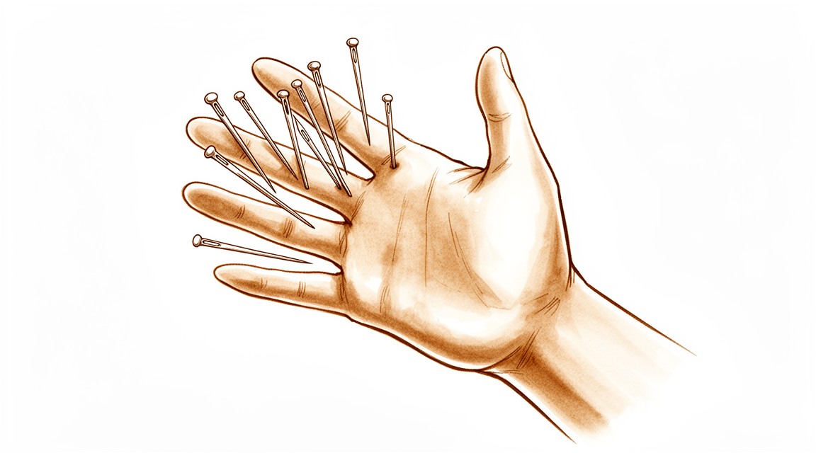
Hand numbness, tingling, or weakness – understanding carpal tunnel, cubital tunnel, and other compression neuropathies.
Những gì bạn đang cảm nhận
Bạn có thể nhận thấy đau, tê bì hoặc tê ở tay hoặc cánh tay. Các triệu chứng này thường tuân theo mô hình "chèn ép kép" (double-crush), nghĩa là một vấn đề ở một dây thần kinh có thể làm cho dây thần kinh khác nhạy cảm hơn với sự chèn ép. Ví dụ, các vấn đề với dây thần kinh trụ (dây thần kinh ở phía ngón út của cánh tay) có thể làm tăng nguy cơ phát triển hội chứng ống cổ tay (chèn ép dây thần kinh giữa ở cổ tay). Bạn có thể cảm thấy các triệu chứng ở nhiều vùng khác nhau do các yếu tố toàn thân, như sức khỏe tổng thể hoặc tình trạng viêm, góp phần vào cách phản ứng của các dây thần kinh này.
Vị trí khó chịu của bạn phụ thuộc vào dây thần kinh nào bị chèn ép. Nếu dây thần kinh trụ bị chèn ép ở khuỷu tay hoặc cổ tay, bạn có thể cảm thấy đau hoặc yếu ở ngón áp út và ngón út. Tại cổ tay, tình trạng này được gọi là hội chứng ống trụ. Các u nang, được gọi là u nang hoạt dịch (ganglia), là nguyên nhân phổ biến nhất của sự chèn ép cụ thể này. Nếu dây thần kinh giữa bị ảnh hưởng ở cổ tay, bạn có thể gặp các triệu chứng điển hình của hội chứng ống cổ tay. Hiếm khi, các tình trạng như giả gút (sự tích tụ tinh thể trong khớp) hoặc khối u có thể gây chèn ép dây thần kinh cấp tính đột ngột.
Các triệu chứng của bạn thường trở nên nghiêm trọng hơn khi vận động. Áp lực trong ống cổ tay tăng đáng kể khi bạn chủ động sử dụng tay, điều này có thể làm trầm trọng thêm hội chứng ống cổ tay. Bạn có thể thấy rằng việc với tay ra sau lưng để cài móc áo bra, nhét áo vào quần hoặc nâng vật trở nên khó khăn. Các đợt bùng phát vào ban đêm là phổ biến, vì nằm nghiêng có thể làm tăng áp lực lên các dây thần kinh. Vì các vị trí chèn ép ở gần gốc (proximal) thường bị bỏ qua, bạn có thể cảm thấy đau ở cẳng tay hoặc bàn tay ngay cả khi điểm chèn ép nằm cao hơn ở cánh tay. Bác sĩ phẫu thuật của bạn sẽ đánh giá các mô hình này để xác định xem vấn đề là cơ học, toàn thân, hay là sự kết hợp của cả hai.
Những gì thực sự đang xảy ra
Các dây thần kinh của bạn giống như các dây cáp điện, truyền tín hiệu từ não đến tay và các ngón tay của bạn. Khi các dây thần kinh này bị chèn ép hoặc nén, các tín hiệu sẽ bị chặn hoặc méo mó. Đây chính là nguyên nhân gây ra cảm giác đau, tê bì hoặc yếu ớt mà bạn cảm nhận. Ở cánh tay trên và bàn tay, tình trạng chèn ép này thường xảy ra do cơ chế "chèn ép kép" (double-crush). Điều này có nghĩa là một dây thần kinh có thể bị kích thích tại một vị trí, khiến nó nhạy cảm hơn với áp lực tại một vị trí khác ở gần đó.
Nhiều yếu tố có thể gây ra tình trạng chèn ép này. Đôi khi, đó chỉ đơn thuần là cách bạn sử dụng tay của mình. Ví dụ, áp lực bên trong ống cổ tay (carpal tunnel) ở cổ tay tăng lên đáng kể khi bạn chủ động sử dụng tay. Áp lực gia tăng này có thể chèn ép dây thần kinh giữa (median nerve). Vào những thời điểm khác, một vật cản vật lý là nguyên nhân. Một khối mỡ nhỏ, được gọi là u mỡ quanh thần kinh (perineural lipoma), có thể phát triển xung quanh dây thần kinh trụ (ulnar nerve) ở khuỷu tay. Hoặc, nếu bạn từng bị gãy xương cổ tay, các mảnh xương sắc nhọn hoặc thậm chí là thiết bị phẫu thuật có thể chèn ép trực tiếp lên dây thần kinh.
Tình trạng sức khỏe tổng thể của cơ thể bạn cũng đóng một vai trò quan trọng. Các yếu tố toàn thân, như bệnh tiểu đường hoặc viêm, có thể làm cho dây thần kinh của bạn dễ bị tổn thương hơn trước tình trạng chèn ép. Một cách thú vị là, các vấn đề với một dây thần kinh có thể dẫn đến các vấn đề với dây thần kinh khác. Ví dụ, nếu dây thần kinh trụ của bạn đã bị kích thích, bạn có thể có nhiều khả năng phát triển hội chứng ống cổ tay (carpal tunnel syndrome) sau này. Điều này là do cách tay bạn cử động và kiểm soát có thể thay đổi khi một dây thần kinh không hoạt động đúng cách.
Đôi khi, vấn đề bắt đầu từ cách các cơ kiểm soát tay của bạn. Sau một chấn thương cổ tay, bạn có thể mất đi một phần kiểm soát cảm giác-vận động, điều này làm thay đổi cách phân bổ áp lực lên các dây thần kinh của bạn. Trong những trường hợp hiếm gặp, các tình trạng như bệnh xơ cứng củ (tuberous sclerosis) có thể gây ra các khối u chèn ép lên dây thần kinh, ngay cả ở trẻ em. Bác sĩ phẫu thuật của bạn xem xét tất cả các yếu tố này—giải phẫu của bạn, tiền sử bệnh và các triệu chứng của bạn—để hiểu chính xác vị trí và lý do tại sao tình trạng chèn ép đang xảy ra. Điều này giúp lựa chọn phương pháp điều trị phù hợp để giảm áp lực và khôi phục chức năng bình thường.
Những gì chúng tôi có thể làm về vấn đề này
Điều trị bảo tồn mang lại lợi ích cho đa số bệnh nhân mắc hội chứng ống khuỷu tay có triệu chứng nhẹ hoặc trung bình. Bác sĩ phẫu thuật của bạn có thể sẽ bắt đầu bằng phương pháp này. Cách tiếp cận này tập trung vào việc giảm áp lực lên dây thần kinh. Bạn có thể được khuyên điều chỉnh các thói quen hàng ngày. Ví dụ, tránh gập khuỷu tay trong thời gian dài có thể giúp ích. Vật lý trị liệu nhằm duy trì tính linh hoạt của khớp và tăng cường các cơ xung quanh. Sự hỗ trợ này giúp bảo vệ dây thần kinh khỏi bị kích thích thêm. Hãy dành đủ thời gian cho kế hoạch không phẫu thuật này phát huy tác dụng. Hầu hết mọi người đều thấy cải thiện mà không cần phẫu thuật.
Nếu các biện pháp đơn giản không đủ hiệu quả, bác sĩ phẫu thuật của bạn có thể thảo luận về quản lý bằng thuốc. Điều này thường bao gồm thuốc giảm đau hoặc thuốc chống viêm để làm giảm sưng. Trong một số trường hợp, tiêm được sử dụng để đưa thuốc trực tiếp vào vùng bị ảnh hưởng. Tiêm cortisone làm giảm viêm và đau. Tiêm axit hyaluronic có thể bôi trơn không gian khớp. Tiêm huyết tương giàu tiểu cầu (PRP) sử dụng các thành phần máu của chính bạn để thúc đẩy quá trình lành thương. Những phương pháp điều trị này nhắm vào nguồn gốc gây kích thích. Hiệu quả của các mũi tiêm này khác nhau. Một số mang lại sự giảm đau trong vài tuần, trong khi những số khác có thể kéo dài trong nhiều tháng. Bác sĩ phẫu thuật của bạn sẽ giúp bạn quyết định xem bước này có phù hợp với bạn hay không dựa trên các triệu chứng cụ thể của bạn.
Phẫu thuật được xem xét khi điều trị bảo tồn đã đạt đến giới hạn. Điều này thường xảy ra khi cơn đau dai dẳng hoặc chức năng dây thần kinh xấu đi mặc dù đã điều trị các phương pháp khác. Phương án phẫu thuật liên quan đến giải phóng chèn ép. Điều này có nghĩa là bác sĩ phẫu thuật của bạn sẽ nới lỏng các cấu trúc chèn ép lên dây thần kinh. Điều này tạo ra nhiều không gian hơn cho dây thần kinh lành lại. Trong một số trường hợp, chẳng hạn như khi có khối u, cắt bỏ khối u cùng với giải phóng chèn ép sẽ mang lại sự giảm đau. Các kỹ thuật xâm lấn tối thiểu có thể được sử dụng để tạo các vết rạch nhỏ hơn. Những phương pháp này nhằm mục đích giảm thiểu mất máu và thời gian hồi phục. Bác sĩ phẫu thuật của bạn sẽ giải thích thủ thuật cụ thể nếu trở nên cần thiết. Mục tiêu là ngăn chặn sự chèn ép và khôi phục chức năng bình thường của dây thần kinh.
Những điều cần biết
Tiên lượng của bạn phụ thuộc phần lớn vào tốc độ được giải phóng chèn ép dây thần kinh. Khi được chẩn đoán sớm và điều trị cẩn thận, hầu hết bệnh nhân đều có khả năng phục hồi chức năng tốt. Bạn có thể mong đợi các triệu chứng của mình thuyên giảm khi dây thần kinh lành lại. Đối với nhiều người, điều này có nghĩa là việc trở lại chức năng bình thường của tay và cánh tay. Tuy nhiên, nếu các triệu chứng đã tồn tại trong thời gian dài, việc phục hồi hoàn toàn có thể không xảy ra. Các tín hiệu thần kinh cần thời gian để khôi phục, và tình trạng chèn ép kéo dài có thể gây ra những thay đổi vĩnh viễn.
Các quyết định điều trị khác nhau tùy thuộc vào dây thần kinh cụ thể liên quan. Đối với các vấn đề phổ biến như hội chứng ống cổ tay, phẫu thuật thường mang lại sự giảm đau lâu dài. Lợi ích này vẫn đúng ngay cả khi bạn mắc bệnh tiểu đường. Sự cải thiện lâu dài của bạn có khả năng tương tự như những bệnh nhân không mắc bệnh tiểu đường. Trong các trường hợp phức tạp hơn, chẳng hạn như chèn ép dây thần kinh trụ nặng ở khuỷu tay, các kỹ thuật xâm lấn tối thiểu là an toàn và hiệu quả. Những phương pháp này nhằm mục đích giải phóng dây thần kinh với sự gián đoạn tối thiểu đối với các mô xung quanh. Bạn có thể nhận thấy những cải thiện bền vững về cả sức mạnh và cảm giác theo thời gian.
Điều quan trọng là phải hiểu rằng việc quản lý không phải lúc nào cũng đơn giản. Các biến chứng có thể xảy ra, bao gồm tổn thương các cấu trúc lân cận, thất bại trong điều trị hoặc sự phát triển của các hội chứng đau mạn tính. Những rủi ro này được giảm thiểu khi bác sĩ phẫu thuật của bạn có hiểu biết sâu sắc về giải phẫu độc đáo của bạn. Trong một số trường hợp, phương pháp điều trị ban đầu có thể không giải quyết triệt để vấn đề. Tình trạng chèn ép tái phát hoặc dai dẳng có thể khó quản lý. Nếu các triệu chứng quay trở lại, bác sĩ phẫu thuật của bạn có thể thảo luận về các lựa chọn bổ sung, chẳng hạn như sử dụng lớp bọc collagen để bảo vệ dây thần kinh hoặc chuyển một dây thần kinh khác để khôi phục chức năng.
Nếu không được điều trị, các bệnh lý thần kinh do chèn ép thường dai dẳng hoặc trầm trọng hơn. Áp lực lên dây thần kinh thường không tự giải quyết. Trong một số trường hợp, một dây thần kinh bị chèn ép có thể khiến bạn dễ bị chèn ép hơn ở một khu vực khác. Ví dụ, các vấn đề về dây thần kinh trụ đôi khi có thể dẫn trước các vấn đề về dây thần kinh giữa. Do đó, việc đánh giá kịp thời là chìa khóa. Trong khi hầu hết các ấn phẩm về các tình trạng chi trên hiếm gặp đều dựa trên các nghiên cứu nhỏ hơn, nguyên tắc chung vẫn là: việc giải phóng chèn ép sớm và chính xác mang lại cơ hội tốt nhất để quay lại hoàn toàn các hoạt động hàng ngày của bạn.
Khi nào cần gặp bác sĩ
Hãy gặp bác sĩ đa khoa nếu bạn có tình trạng đau dai dẳng không cải thiện khi nghỉ ngơi. Hãy yêu cầu đánh giá bởi bác sĩ chuyên khoa nếu bạn nhận thấy tình trạng yếu hoặc mất ổn định ở bàn tay. Hãy tìm kiếm sự chăm sóc y tế nếu các triệu chứng của bạn cản trở giấc ngủ hoặc công việc. Tình trạng xấu đi đột ngột của các triệu chứng cũng cần được chú ý. Các bệnh lý chèn ép dây thần kinh có thể liên quan đến cơ chế "chèn ép kép", trong đó một vấn đề về dây thần kinh làm tăng tính nhạy cảm với một vấn đề khác. Các yếu tố toàn thân cũng có thể góp phần vào các tình trạng này. Ví dụ, các vấn đề về dây thần kinh trụ có thể xuất hiện trước khi có chèn ép dây thần kinh giữa. Hãy lưu ý rằng các vấn đề đồng thời ở cổ tay và cẳng tay thường bị bỏ qua. Việc đánh giá sớm giúp ngăn ngừa các biến chứng như hội chứng đau bệnh lý hoặc thất bại trong điều trị. Bác sĩ phẫu thuật của bạn dựa vào việc hiểu biết về giải phẫu bình thường để quản lý an toàn các trường hợp phức tạp này.
Evidence & references
Overview
- Compression neuropathies of the upper extremity involve pathophysiology, clinical evaluation, and management considerations including the double-crush mechanism and systemic factors [1].
- Validated patient-reported outcome measures are useful in the evaluation and management of upper extremity compression neuropathies [1].
- Complications of compressive neuropathy management include iatrogenic injury, treatment failure, and pathologic pain syndromes [2].
- Prevention of complications in compressive neuropathy management relies on a solid understanding of normal anatomy and anatomic variations [2].
- Diagnosis and treatment of compressive neuropathies are evolving with technology, specifically shifting towards preoperative imaging with ultrasound and MRN [3].
- Management of failed decompressions for compressive neuropathies remains challenging [3].
- Most publications on uncommon upper extremity compression syndromes (radial, ulnar, and median nerves) are small retrospective series or case reports [4].
- Treatment decisions for uncommon upper extremity compression syndromes are not typically based on high levels of evidence [4].
- Debulking of a tumor along with median nerve decompression relieved neurological symptoms in a child with tuberous sclerosis complex causing carpal tunnel syndrome [5].
- Minimally invasive in situ decompression is technically simple, safe, and provides good results for severe ulnar nerve entrapment at the elbow [6].
- Ulnar nerve pathology may precede and increase susceptibility to median nerve compression, as indicated by the incidence of carpal tunnel syndrome after ulnar neuropathy diagnosis [8].
- Use of a collagen matrix wrap in recurrent compression neuropathies of the upper extremity has shown good success [9].
- Surgical decompression for carpal tunnel syndrome is associated with a greater decrease in median nerve cross-sectional area than nonsurgical treatment [10].
- Anterior interosseous nerve transfer combined with cubital and ulnar tunnel release results in sustained clinical and electrophysiological improvements in severe chronic ulnar nerve compression [14].
- Anterior interosseous nerve transfer combined with cubital and ulnar tunnel release is encouraged as a standard treatment for severe chronic ulnar nerve compression [14].
- Endoscopic decompression for anterior interosseous nerve syndrome can achieve the same proximal and distal extents of the nerve as open techniques [15].
- Endoscopic decompression for anterior interosseous nerve syndrome uses an incision nearly one fourth the size of open techniques, minimizing morbidity, blood loss, and recovery time [15].
- Extensive decompression of the ulnar nerve beyond the cubital tunnel is not routinely needed, supported by satisfactory outcomes with endoscopic detection of compressing fascial bands within the FCU [17].
Anatomy & Pathophysiology
- Compression neuropathies of the upper extremity involve a double-crush mechanism [1].
- Systemic factors play a role in the pathophysiology of compression neuropathies of the upper extremity [1].
- Intracarpal tunnel pressures during active hand use are substantially greater than previously reported in patients with carpal tunnel syndrome [21].
- Perineural lipoma of the ulnar nerve can occur within the cubital tunnel [25].
- Sensorimotor control impairment can occur after wrist trauma [27].
- Distal radius fracture management requires evaluation of all potential causes for early carpal tunnel syndrome findings, including prominent volar cortical fragments causing direct pressure or prominently placed hardware [34].
- Ulnar nerve entrapment neuropathy at the elbow is associated with non-task-specific focal hand dystonia [41].
Classification
- Compression neuropathies of the upper extremity involve a double-crush mechanism [1].
- Systemic factors contribute to the pathophysiology of compression neuropathies of the upper extremity [1].
- Compressive neuropathy management complications include iatrogenic injury, treatment failure, and pathologic pain syndromes [2].
- Prevention of compressive neuropathy complications relies on understanding normal anatomy and anatomic variations [2].
- Diagnosis and treatment of compressive neuropathies are shifting towards preoperative imaging with ultrasound and MRN [3].
- Management of failed decompressions for compressive neuropathies remains challenging [3].
- Most publications on uncommon upper extremity compression syndromes (radial, ulnar, median nerves) are small retrospective series or case reports [4].
- Treatment decisions for uncommon upper extremity compression syndromes are not typically based on high levels of evidence [4].
- Debulking of a tumor along with median nerve decompression relieved neurological symptoms in a child with tuberous sclerosis complex causing carpal tunnel syndrome and thumb overgrowth [5].
- Minimally invasive in situ decompression is technically simple, safe, and provides good results for severe ulnar nerve entrapment at the elbow [6].
- Pseudogout is a rare cause of acute carpal tunnel syndrome and acute Guyon canal syndrome [7].
- Ulnar nerve pathology may precede and increase susceptibility to median nerve compression [8].
- Surgical decompression for carpal tunnel syndrome is associated with a greater decrease in median nerve cross-sectional area than nonsurgical treatment [10].
- Concurrent carpal tunnel syndrome and pronator syndrome are rarely considered, and proximal compression sites are easily overlooked [11].
- Ganglia are the most common cause of ulnar tunnel syndrome [12].
- Symptoms of ulnar tunnel syndrome vary based on the anatomic location of the compression within Guyon's canal [12].
- The term double crush syndrome is proposed to be expanded to multifocal neuropathy to describe the complex interplay of mechanical, systemic, pharmacological, and environmental factors contributing to nerve dysfunction [13].
- Unusual compression neuropathies of the forearm include posterior interosseous nerve syndrome, radial tunnel syndrome, and superficial radial nerve compression (Wartenberg's syndrome) [16].
- In-situ release is an alternative for managing McGowen grade 3 ulnar nerve compression neuropathy at the elbow, with a similar success rate to submuscular and intramuscular transpositions but a lower complication rate [23].
Clinical Presentation
- Compression neuropathies of the upper extremity involve a double-crush mechanism [1].
- Systemic factors contribute to the pathophysiology of compression neuropathies of the upper extremity [1].
- Ulnar nerve pathology may precede and increase susceptibility to median nerve compression [8].
- Concurrent carpal tunnel syndrome and pronator syndrome are rarely considered, and proximal compression sites are easily overlooked [11].
- Intracarpal tunnel pressures during active hand use in patients with carpal tunnel syndrome are substantially greater than previously reported [21].
- Ganglia are the most common cause of ulnar tunnel syndrome [12].
- Symptoms of ulnar tunnel syndrome vary based on the anatomic location of the compression within Guyon's canal [12].
- Pseudogout is a rare cause of acute neuropathic compression of the hand, including acute carpal tunnel syndrome and acute Guyon canal syndrome [7].
- Collagenoma in a child with tuberous sclerosis complex can cause carpal tunnel syndrome and thumb overgrowth [5].
- Uncommon compression syndromes of the radial, ulnar, and median nerves exist, with most publications being small retrospective series or case reports [4].
- Unusual compression neuropathies of the forearm include posterior interosseous nerve syndrome, radial tunnel syndrome, and superficial radial nerve compression (Wartenberg's syndrome) [16].
- A punched nerve syndrome of the deep motor branch of the ulnar nerve is a rare presentation [18].
- Multifocal neuropathy describes the complex interplay of mechanical, systemic, pharmacological, and environmental factors contributing to nerve dysfunction [13].
Investigations
- Diagnosis and treatment of compressive neuropathies are shifting towards preoperative imaging with ultrasound and MRN [3].
- Most publications on uncommon upper extremity compression syndromes are small retrospective series or case reports, and treatment decisions are not typically based on high levels of evidence [4].
- Debulking of a tumor along with median nerve decompression relieved neurological symptoms in a child with tuberous sclerosis complex causing carpal tunnel syndrome [5].
- Pseudogout is a rare cause of acute neuropathic compression of the hand, including acute carpal tunnel syndrome and acute Guyon canal syndrome [7].
- Ulnar nerve pathology may precede and increase susceptibility to median nerve compression [8].
- Concurrent carpal tunnel syndrome and pronator syndrome are rarely considered, and proximal compression sites are easily overlooked [11].
- Ganglia are the most common cause of ulnar tunnel syndrome, and symptoms vary based on the anatomic location of the compression within Guyon's canal [12].
- Endoscopic decompression for anterior interosseous nerve syndrome can be achieved over the same proximal and distal extents of the nerve as open techniques but with an incision nearly one fourth the size, minimizing morbidity, blood loss, and recovery time [15].
- Unusual compression neuropathies of the forearm specifically include posterior interosseous nerve syndrome, radial tunnel syndrome, and superficial radial nerve compression (Wartenberg's syndrome) [16].
- High-resolution ultrasound (HRUS) is a viable method to demonstrate a punched nerve syndrome of the deep motor branch of the ulnar nerve [18].
- Ultrasound measurements have limited value in predicting clinical results of patients treated for entrapment neuropathy of the ulnar nerve [19].
- After surgery for perineural lipoma of the ulnar nerve within the cubital tunnel, shooting pain resolved, sensation normalized in digits four and five, and hand strength gradually improved [25].
- The diagnostic accuracy of nerve conduction studies for ulnar neuropathy at the elbow may be lower than 80%–90% and depends on the severity of the neuropathy [33].
- Short segment testing is suggested to improve the diagnostic accuracy of nerve conduction studies for ulnar neuropathy at the elbow [33].
Treatment
- Conservative treatment benefits the majority of patients with cubital tunnel syndrome who present with mild or moderate symptoms [22].
- Surgical decompression is associated with a greater decrease in median nerve cross-sectional area compared to nonsurgical treatment [10].
- Debulking of a tumor along with median nerve decompression provides relief of neurological symptoms in cases such as collagenoma causing carpal tunnel syndrome [5].
- Minimally invasive in situ decompression is technically simple, safe, and yields good results for severe ulnar nerve entrapment at the elbow [6].
- In-situ release is an alternative for managing McGowen grade 3 ulnar nerve compression neuropathy at the elbow, offering a similar success rate to submuscular and intramuscular transpositions with a lower complication rate [23].
- Anterior interosseous nerve transfer combined with cubital and ulnar tunnel release results in sustained clinical and electrophysiological improvements in patients with severe chronic ulnar nerve compression [14].
- Minimally invasive endoscopic decompression for anterior interosseous nerve syndrome achieves the same proximal and distal extents of the nerve as open techniques but with an incision nearly one-fourth the size, minimizing morbidity, blood loss, and recovery time [15].
- Extensive decompression of the ulnar nerve beyond the cubital tunnel is not routinely needed, as satisfactory outcomes are supported by endoscopic detection of compressing fascial bands within the flexor carpi ulnaris [17].
- A novel technique using a collagen matrix wrap in recurrent compression neuropathies has shown good success [9].
- Pseudogout should be considered a rare cause of acute neuropathic compression of the hand, including acute carpal tunnel syndrome and acute Guyon canal syndrome [7].
- Complications of compressive neuropathy management include iatrogenic injury, treatment failure, and pathologic pain syndromes, with prevention relying on a solid understanding of normal anatomy and anatomic variations [2].
- The management of failed decompressions remains challenging as diagnosis and treatment evolve with technology, shifting towards preoperative imaging with ultrasound and MRN [3].
Complications
- Complications of compressive neuropathy management include iatrogenic injury [2].
- Complications of compressive neuropathy management include treatment failure [2].
- Complications of compressive neuropathy management include pathologic pain syndromes [2].
- Prevention of complications relies on a solid understanding of normal anatomy and anatomic variations [2].
- Management of failed decompressions remains challenging [3].
- Nerve injuries following elbow arthroscopy are likely under-reported in the literature [29].
- The number of severe nerve injuries following elbow arthroscopy may be much higher than previously thought [29].
Recovery
- Minimally invasive in situ decompression for severe ulnar nerve entrapment at the elbow is technically simple, safe, and provides good functional outcomes [6].
- Endoscopic decompression of the anterior interosseous nerve achieves the same proximal and distal extents as open techniques but with an incision nearly one-fourth the size, minimizing morbidity, blood loss, and recovery time [15].
- Extensive decompression of the ulnar nerve beyond the cubital tunnel is not routinely needed, as satisfactory outcomes are supported by endoscopic detection of compressing fascial bands within the flexor carpi ulnaris [17].
- Revision decompression combined with a collagen nerve wrap demonstrates good success in managing recurrent and persistent compression neuropathies of the upper extremity [9].
- Anterior interosseous nerve transfer combined with cubital and ulnar tunnel release results in sustained clinical and electrophysiological improvements in patients with severe chronic ulnar nerve compression [14].
- Early diagnosis and careful excision of epineural ganglia causing ulnar nerve compression in the cubital tunnel are associated with satisfactory outcomes, although complete electrophysiological recovery may not occur if symptoms have been present for a prolonged period [20].
- Debulking of a tumor along with median nerve decompression provides relief of neurological symptoms in cases such as collagenoma-induced carpal tunnel syndrome [5].
- Long-term improvement following carpal tunnel release in patients with diabetes is maintained to the same extent as in patients without diabetes [24].
- Treatment decisions for uncommon upper extremity compression syndromes are not typically based on high levels of evidence, as most publications are small retrospective series or case reports [4].
- Management of failed decompressions remains challenging despite evolving diagnostic and treatment technologies such as preoperative ultrasound and MRN [3].
Key Evidence
- [L5] Complications of compressive neuropathy management include iatrogenic injury, treatment failure, and pathologic pain syndromes, with prevention relying on a solid understanding of normal anatomy and anatomic variations. [2] (10.1016/j.hcl.2015.01.012)
- [L5] The diagnosis and treatment of compressive neuropathies continue to evolve with technology, shifting towards preoperative imaging with ultrasound and MRN, while the management of failed decompressions remains challenging. [3] (10.1016/j.jhsg.2022.10.009)
- [L4] This article reviews uncommon compression syndromes of the radial, ulnar, and median nerves, noting that most publications are small retrospective series or case reports and treatment decisions are not typically based on high levels of evidence. [4] (10.1016/j.hcl.2013.04.014)
- [Case_report] Debulking of the tumor along with median nerve decompression was performed with relief of neurological symptoms. [5] (10.1016/j.jhsa.2013.07.004)
- [L3] Minimally invasive in situ decompression is technically simple, safe and gives good results in patients with severe nerve compression. [6] (10.1177/1753193411416426)
- [L4] Pseudogout should be considered a rare cause of acute neuropathic compression of the hand. [7] (10.1016/j.jhsg.2022.07.010)
- [L2] This supports the hypothesis that ulnar nerve pathology may precede and increase susceptibility to median nerve compression. [8] (10.1016/j.jhsg.2026.100970)
- [L4] The authors report on the novel technique of using a collagen matrix wrap in recurrent compression neuropathies with good success. [9] (10.1097/sap.0b013e3182956475)
- [L3] Surgical decompression was associated with a greater decrease in median nerve cross-sectional area than nonsurgical treatment. [10] (10.1016/j.jhsa.2010.06.010)
- [L4] Concurrent carpal tunnel syndrome and pronator syndrome are rarely considered and proximal compression sites are easily overlooked. [11] (10.1016/j.otsr.2016.10.009)
- [L5] The article provides a comprehensive review of the anatomy, pathophysiology, and causes of ulnar tunnel syndrome, noting that ganglia are the most common cause and that symptoms vary based on the anatomic location of the compression within Guyon's canal. [12] (10.1016/j.hcl.2007.06.006)
- [L5] The authors propose expanding the term from double crush syndrome to multifocal neuropathy to better describe the complex interplay of mechanical, systemic, pharmacological, and environmental factors contributing to nerve dysfunction. [13] (10.1016/j.jhsa.2016.09.009)
- [L4] Anterior interosseous nerve transfer, along with cubital and ulnar tunnel release, results in sustained clinical and electrophysiological improvements in patients with severe chronic ulnar nerve compression, which encourages its adoption as a standard treatment for severe chronic ulnar nerve compression. [14] (10.1177/17531934251381023)
- [L4] Endoscopic decompression can be achieved over the same proximal and distal extents of the nerve as open techniques but with an incision nearly one fourth the size, minimizing morbidity, blood loss, and recovery time. [15] (10.1016/j.jhsa.2013.07.026)
- [L5] This article is a review examining unusual compression neuropathies of the forearm, specifically focusing on the radial nerve, including posterior interosseous nerve syndrome, radial tunnel syndrome, and superficial radial nerve compression (Wartenberg's syndrome). [16] (10.1016/j.jhsa.2009.10.016)
- [L4] The satisfactory outcomes support the perception that extensive decompression of the ulnar nerve beyond the cubital tunnel is not routinely needed. [17] (10.1007/s11552-011-9377-x)
- [L4] HRUS is a viable method to demonstrate a punched nerve syndrome. [18] (10.1007/s00402-015-2216-8)
- [L3] Ultrasound (US) measurements seem to have a limited value in clinical results of patients treated for entrapment neuropathy of the ulnar nerve. [19] (10.1177/1558944719857816)
- [Case_report] Early diagnosis and careful excision of epineural ganglia are associated with satisfactory outcomes, although complete electrophysiological recovery may not occur if symptoms have been present for a prolonged period. [20] (10.1007/s11552-006-9013-3)
- [L4] In patients with carpal tunnel syndrome, intracarpal tunnel pressures during active hand use are substantially greater than previously reported. [21] (10.1016/j.jhsa.2009.09.019)
- [L2] The majority of patients suffering from cubital tunnel syndrome with mild or moderate symptoms benefit from conservative treatment. [22] (10.1177/1753193408098480)
- [L4] Thus, in-situ release could be an alternative in management of patients with McGowen grade 3 ulnar nerve compression neuropathy at the elbow with a similar success rate as the submuscular and intramuscular transpositions with a lower complication rate. [23] (10.1016/j.jhsa.2015.06.068)
- [L2] Long-term improvement in patients with diabetes remained after carpal tunnel release to the same extent as for patients without diabetes. [24] (10.1016/j.jhsa.2014.01.012)
- [L4] After surgery, shooting pain resolved, sensation normalized in digits four and five, and hand strength gradually improved. [25] (10.1016/j.jhsg.2025.100889)
- [L5] This clinical review discusses the organization, neuroanatomy, assessment, clinical relevance, and rehabilitation of sensorimotor control impairment after wrist trauma, proposing promising rehabilitation strategies that require more rigorous evaluation in clinical trials. [27] (10.1016/j.jht.2015.12.003)
- [L4] Nerve injuries are likely under-reported in the literature, and this study indicates that the number of severe nerve injuries may be much higher than previously thought. [29] (10.1016/j.jhsa.2013.08.025)
- [L5] The diagnostic accuracy of nerve conduction studies for ulnar neuropathy at the elbow may be lower than 80%–90% and depends on the severity of the neuropathy; short segment testing is suggested to improve accuracy. [33] (10.1177/17531934241288802)
- [Paper] If early carpal tunnel syndrome findings are noted during distal radius fracture management, all potential causes should be evaluated, including prominent volar cortical fragments causing direct prominently placed hardware. [34] (10.1016/j.ocl.2012.07.021)
- [L4] This case establishes a clear-cut relationship between ulnar nerve entrapment neuropathy at the elbow and non-task-specific focal hand dystonia, demonstrated by the dramatic recovery of clinical and electrophysiological parameters after surgical decompression. [41] (10.1007/s11552-010-9280-x)
References
[1] Compression Neuropathies of the Upper Extremity. 2021. [2] Complications of Compressive Neuropathy. Hand Clinics. 2015. DOI: 10.1016/j.hcl.2015.01.012 [3] Future Considerations in the Diagnosis and Treatment of Compressive Neuropathies of the Upper Extremity. Journal of Hand Surgery Global Online. 2023. DOI: 10.1016/j.jhsg.2022.10.009 [4] Uncommon Upper Extremity Compression Neuropathies. Hand Clinics. 2013. DOI: 10.1016/j.hcl.2013.04.014 [5] Collagenoma in a Child With Tuberous Sclerosis Complex Causing Carpal Tunnel Syndrome and Thumb Overgrowth: Case Report. The Journal of Hand Surgery. 2013. DOI: 10.1016/j.jhsa.2013.07.004 [6] Severe ulnar nerve entrapment at the elbow: functional outcome after minimally invasive in situ decompression. Journal of Hand Surgery (European Volume). 2012. DOI: 10.1177/1753193411416426 [7] Pseudogout: A Rare Cause of Acute Carpal Tunnel Syndrome and Acute Guyon Canal Syndrome. Journal of Hand Surgery Global Online. 2022. DOI: 10.1016/j.jhsg.2022.07.010 [8] Incidence of Carpal Tunnel Syndrome After the Diagnosis of Ulnar Neuropathy. Journal of Hand Surgery Global Online. 2026. DOI: 10.1016/j.jhsg.2026.100970 [9] Revision Decompression and Collagen Nerve Wrap for Recurrent and Persistent Compression Neuropathies of the Upper Extremity. Annals of Plastic Surgery. 2014. DOI: 10.1097/sap.0b013e3182956475 [10] Sonographic Follow-Up of Patients With Carpal Tunnel Syndrome Undergoing Surgical or Nonsurgical Treatment: Prospective Cohort Study. The Journal of Hand Surgery. 2010. DOI: 10.1016/j.jhsa.2010.06.010 [11] Concurrent carpal tunnel syndrome and pronator syndrome: A retrospective study of 21 cases. Orthopaedics & Traumatology: Surgery & Research. 2017. DOI: 10.1016/j.otsr.2016.10.009 [12] Ulnar Tunnel Syndrome. Hand Clinics. 2007. DOI: 10.1016/j.hcl.2007.06.006 [13] Multifocal Neuropathy: Expanding the Scope of Double Crush Syndrome. The Journal of Hand Surgery. 2016. DOI: 10.1016/j.jhsa.2016.09.009 [14] Anterior interosseous nerve transfer combined with cubital and ulnar tunnel release for severe ulnar nerve compression. Journal of Hand Surgery (European Volume). 2025. DOI: 10.1177/17531934251381023 [15] Minimally Invasive Endoscopic Decompression for Anterior Interosseous Nerve Syndrome: Technical Notes. The Journal of Hand Surgery. 2013. DOI: 10.1016/j.jhsa.2013.07.026 [16] Unusual Compression Neuropathies of the Forearm, Part I: Radial Nerve. The Journal of Hand Surgery. 2009. DOI: 10.1016/j.jhsa.2009.10.016 [17] Endoscopic Detection of Compressing Fascial Bands around the Ulnar Nerve within the FCU. HAND. 2011. DOI: 10.1007/s11552-011-9377-x [18] A rare case of a punched nerve syndrome of the deep motor branch of the ulnar nerve. Archives of Orthopaedic and Trauma Surgery. 2015. DOI: 10.1007/s00402-015-2216-8 [19] Sonographic Follow-Up of Patients With Cubital Tunnel Syndrome Undergoing in Situ Open Neurolysis or Endoscopic Release: The SPECTRE Study. HAND. 2019. DOI: 10.1177/1558944719857816 [20] Ulnar Nerve Compression in the Cubital Tunnel by an Epineural Ganglion: A Case Report. HAND. 2007. DOI: 10.1007/s11552-006-9013-3 [21] Dynamics of Intracarpal Tunnel Pressure in Patients With Carpal Tunnel Syndrome. The Journal of Hand Surgery. 2010. DOI: 10.1016/j.jhsa.2009.09.019 [22] Conservative Treatment of the Cubital Tunnel Syndrome. Journal of Hand Surgery (European Volume). 2009. DOI: 10.1177/1753193408098480 [23] The Efficacy of In-Situ Cubital Tunnel Release in Management of Elbow Ulnar Compression Neuropathy in McGowen Grade 3. The Journal of Hand Surgery. 2015. DOI: 10.1016/j.jhsa.2015.06.068 [24] Carpal Tunnel Release in Patients With Diabetes: A 5-Year Follow-Up With Matched Controls. The Journal of Hand Surgery. 2014. DOI: 10.1016/j.jhsa.2014.01.012 [25] Perineural Lipoma of the Ulnar Nerve Within the Cubital Tunnel: A Brief Review of the Literature. Journal of Hand Surgery Global Online. 2026. DOI: 10.1016/j.jhsg.2025.100889 [27] Rehabilitation strategies for wrist sensorimotor control impairment: From theory to practice. Journal of Hand Therapy. 2016. DOI: 10.1016/j.jht.2015.12.003 [29] Nerve Injuries Following Elbow Arthroscopy. The Journal of Hand Surgery. 2013. DOI: 10.1016/j.jhsa.2013.08.025 [33] Re: Bourke G, Wade R, van Alfen N. Updates in diagnostic tools for diagnosing nerve injuries and compressions. J Hand Surg Eur. 2024, 49: 668–80. Journal of Hand Surgery (European Volume). 2024. DOI: 10.1177/17531934241288802 [34] Carpal Tunnel Syndrome After Distal Radius Fracture. Orthopedic Clinics of North America. 2012. DOI: 10.1016/j.ocl.2012.07.021 [41] Focal Hand Dystonia in a Patient with Ulnar Nerve Neuropathy at the Elbow. HAND. 2010. DOI: 10.1007/s11552-010-9280-x


