Bất ổn định khớp vai
Patients › Shoulder
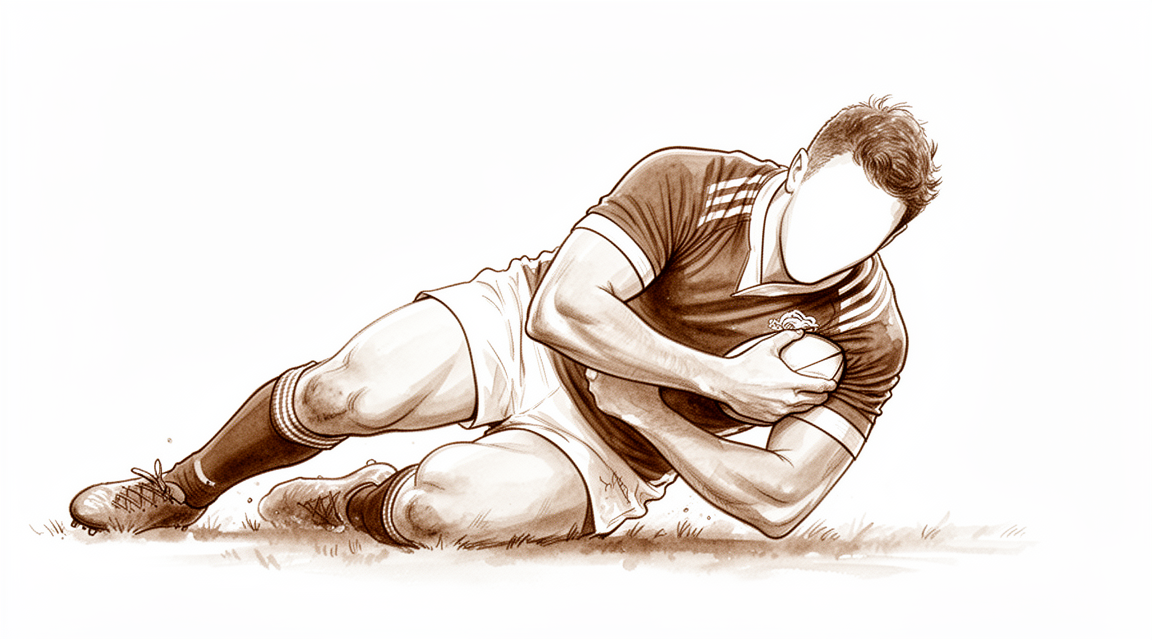
Shoulder instability — understanding the feeling of looseness, causes, and treatment options.
Những gì bạn đang cảm nhận
Bạn có thể cảm thấy đau ở phía trước hoặc phía sau vai. Cơn đau này thường báo hiệu tổn thương mô lớn hơn bên trong khớp. Bạn có thể nhận thấy rằng một số cử động cụ thể gây ra sự khó chịu nhói lên. Việc với tay ra sau lưng để cài áo ngực có thể trở nên khó khăn. Việc nhét áo vào quần cũng có thể gây đau. Những nhiệm vụ hàng ngày này trở nên thách thức khi khớp vai không ổn định.
Các triệu chứng của bạn có thể bùng phát vào ban đêm. Bạn có thể thức dậy với một bên vai bị đau nhức. Cơn đau thường trở nên tồi tệ hơn sau khi vận động. Nó cũng có thể xuất hiện khi bạn vừa thức dậy vào buổi sáng. Ngủ nghiêng về phía bên bị ảnh hưởng có thể đặc biệt khó chịu. Bạn có thể cảm thấy một cảm giác lỏng lẻo hoặc không ổn định. Tuy nhiên, độ chùng lỏng của khớp vai không phải lúc nào cũng có nghĩa là có điều gì đó sai sót. Nó phải được phân biệt với tình trạng mất ổn định thực sự.
Trong một số trường hợp, bạn có thể có cơn đau mơ hồ trong khi vận động mà không bị trật khớp hoàn toàn. Điều này được gọi là mất ổn định vi thể. Nó phổ biến ở bệnh nhân trẻ. Bạn có thể không cảm thấy vai bị trượt ra khỏi vị trí. Thay vào đó, bạn cảm thấy đau hoặc một cảm giác khó chịu mơ hồ. Tình trạng này có thể khó chẩn đoán. Bạn cũng có thể trải qua các triệu chứng ngoại khớp cảm giác như bị chèn ép. Điều này được gọi là mất ổn định vai nhẹ.
Nếu bạn có mất ổn định phía sau tái phát, việc chẩn đoán có thể khó khăn. Cơn đau có thể sâu và khó xác định chính xác. Bạn có thể gặp khó khăn với các cử động cụ thể trên đầu. Mặc dù có những thách thức, các phương pháp điều trị hiện đại có thể giúp ích. Bác sĩ phẫu thuật của bạn sẽ tìm kiếm các dấu hiệu cụ thể để hướng dẫn việc chăm sóc bạn. Đánh giá đúng mức độ mất xương là rất quan trọng để xác định các lựa chọn phẫu thuật của bạn. Điều này giúp đảm bảo kết quả tốt nhất có thể cho quá trình hồi phục của bạn.
Những gì thực sự đang xảy ra
Vai của bạn là một khớp cầu-đế, được thiết kế để có phạm vi vận động rộng. Đầu xương hình cầu nằm trong một hốc nông được lót bởi một lớp mô mềm gọi là bao khớp. Bao khớp này hoạt động như một vòng đệm, giúp duy trì sự ổn định của khớp trong khi bạn vận động. Trong trường hợp mất ổn định khớp vai, cấu trúc ổn định này bị giãn, rách hoặc lỏng lẻo. Đầu xương có thể trượt một phần ra khỏi vị trí (trật một phần) hoặc bật ra hoàn toàn (trật khớp). Sự cố cơ học này cho phép đầu xương di chuyển bất thường, gây ra cảm giác đau và bị kẹt mà bạn cảm nhận được.
Vấn đề thường liên quan đến các mô cụ thể giữ khớp với nhau. Sụn môi (labrum) là một vòng sụn giúp làm sâu hốc khớp. Khi sụn môi bị rách, lớp niêm phong bị phá vỡ. Các cơ và gân của nhóm cơ xoay vai (rotator cuff) cũng đóng vai trò quan trọng trong việc giữ đầu xương luôn ở vị trí trung tâm. Các vết rách ở những gân này, đặc biệt là gân dưới gai (subscapularis), có thể làm thay đổi đáng kể cách khớp vai vận động khi chịu tải. Ngay cả những thay đổi nhỏ trong cách các xương khớp vai di chuyển cũng có thể dẫn đến tăng áp lực lên các mô này. Theo thời gian, chuyển động bất thường này có thể gây ra tình trạng mòn và hư hỏng thêm.
Bác sĩ phẫu thuật của bạn sẽ đánh giá những thay đổi này để xác định hướng điều trị tốt nhất. Các kỹ thuật hiện đại, chẳng hạn như nội soi khớp (phẫu thuật qua lỗ khóa), cho phép sửa chữa chính xác các mô mềm này. Đối với một số bệnh nhân, đặc biệt là những người bị mất xương đáng kể hoặc trật khớp tái phát, có thể được khuyến nghị thực hiện thủ thuật Latarjet. Phẫu thuật này sử dụng một mảnh xương nhỏ để tái tạo hốc khớp, cung cấp khả năng bảo vệ bền vững chống lại tình trạng mất ổn định trong tương lai. Mặc dù phẫu thuật có thể ổn định khớp, nhưng nó có thể không khôi phục hoàn toàn chất lượng vận động chính xác như một khớp vai không bị tổn thương. Mục tiêu là ngăn chặn tình trạng trượt khớp và cho phép bạn quay trở lại các hoạt động hàng ngày một cách tự tin.
Những gì chúng tôi có thể làm về vấn đề này
Hành trình của bạn bắt đầu với việc tự quản lý và vật lý trị liệu. Phương pháp này thường là bước đầu tiên, đặc biệt nếu nguy cơ vai bị trật lại của bạn thấp. Bác sĩ phẫu thuật của bạn có thể khuyên dùng con đường này nếu bạn muốn tránh phẫu thuật hoặc nếu khám lâm sàng cho thấy kết quả ổn định mà không cần phẫu thuật. Vật lý trị liệu tập trung vào việc tăng cường các cơ xung quanh vai để cải thiện sự ổn định và chức năng. Bạn sẽ thực hiện các bài tập giúp bạn lấy lại kiểm soát khớp. Việc chăm sóc bảo tồn này nhằm giảm đau và ngăn ngừa trật khớp trong tương lai. Tuy nhiên, hãy lưu ý rằng điều trị không phẫu thuật có thể gây ra chi phí xã hội đáng kể do thời gian nghỉ làm việc hoặc thể thao. Nó cũng có thể kém tin cậy hơn đối với một số loại mất ổn định nhất định, chẳng hạn như các vấn đề ở vai sau. Bạn nên thử nghiệm phương pháp này một cách công bằng, nhưng hiểu rằng nó có thể không ngăn chặn các sự kiện tái phát ở tất cả mọi người.
Nếu cơn đau vẫn tiếp diễn, quản lý y tế có thể giúp bạn duy trì hoạt động trong khi bạn hồi phục. Bác sĩ phẫu thuật của bạn có thể đề xuất thuốc giảm đau hoặc thuốc chống viêm để quản lý khó chịu và sưng tấy. Những loại thuốc này không sửa chữa vấn đề cấu trúc cơ bản, nhưng chúng có thể khiến các hoạt động hàng ngày và liệu pháp trở nên thoải mái hơn. Trong một số trường hợp, tiêm có thể được xem xét để giảm viêm trong khớp. Trong khi các loại tiêm cụ thể như cortisone hoặc axit hyaluronic đôi khi được sử dụng trong chăm sóc chỉnh hình rộng rãi hơn, bằng chứng về mất ổn định vai tập trung mạnh mẽ vào việc liệu chăm sóc bảo tồn có hiệu quả hay không. Mục tiêu ở đây là giảm triệu chứng, không phải sửa chữa cấu trúc. Bạn nên thảo luận với bác sĩ phẫu thuật về những gì phù hợp với trường hợp cụ thể của bạn, vì trọng tâm chính vẫn là khôi phục sự ổn định thông qua chuyển động và sức mạnh hơn là chỉ che giấu cơn đau.
Phẫu thuật được xem xét khi chăm sóc bảo tồn đã đạt đến giới hạn hoặc nếu bạn có nguy cơ cao bị tái phát. Điều này đặc biệt đúng đối với thanh thiếu niên và người trẻ dưới 40 tuổi bị trật khớp vai trước lần đầu, nơi phẫu thuật hiệu quả hơn các lựa chọn bảo tồn trong việc ngăn ngừa mất ổn định tái phát. Bác sĩ phẫu thuật của bạn sẽ đánh giá bạn kỹ lưỡng, vì khám lâm sàng là yếu tố quan trọng nhất trong việc quyết định phẫu thuật có phù hợp với bạn hay không. Chẩn đoán hình ảnh, chẳng hạn như MRI hoặc quét CT, giúp đánh giá mất xương và tổn thương mô mềm. Nếu vai của bạn tiếp tục bị trượt mặc dù đã điều trị, hoặc nếu bạn bị mất xương đáng kể, ổn định phẫu thuật có thể được khuyến nghị. Phẫu thuật nhằm khôi phục sự ổn định của khớp trong khi giảm thiểu mất chuyển động. Quyết định này nên dựa trên chỉ định lâm sàng, không chỉ là mong muốn trở lại thể thao nhanh hơn.
Những điều cần mong đợi
Tiên lượng của bạn phụ thuộc phần lớn vào việc mất vững khớp vai của bạn có do một chấn thương cụ thể hay phát triển mà không có nguyên nhân rõ ràng. Nếu bạn bị trật khớp vai chấn thương lần đầu, nguy cơ tái phát là rất đáng kể. Ở những bệnh nhân dưới 40 tuổi, khoảng một phần ba trải qua tình trạng mất vững tái phát sau lần khám bác sĩ ban đầu. Nếu không được điều trị, phẫu thuật có thể làm giảm tỷ lệ tái phát so với chăm sóc không phẫu thuật trong vòng 10 năm.
Nếu bạn phẫu thuật để điều trị mất vững khớp vai hướng trước, bạn có thể mong đợi lợi ích lâu dài về độ vững và chức năng, ngay cả khi bạn được coi là có nguy cơ cao. Tuy nhiên, kết quả điều trị có sự khác biệt. Trong một số nghiên cứu, tỷ lệ mất vững tái phát sau sửa chữa nội soi nguyên phát là 30% ở giai đoạn theo dõi trung hạn. Các nghiên cứu khác cho thấy tỷ lệ tái phát thấp hơn, chẳng hạn như 18% sau tám năm với một số kỹ thuật nhất định. Đối với mất vững khớp vai hướng sau, quản lý bằng nội soi hiện đại mang lại sự phục hồi đáng tin cậy và bền vững, với các dữ liệu mới nổi cho thấy khả năng bảo vệ bền vững chống lại tái phát và duy trì tham gia hoạt động thể thao.
Nếu bạn bị mất xương nặng hoặc mất vững tái phát đòi hỏi phẫu thuật tái tạo phức tạp, bác sĩ phẫu thuật của bạn có thể giới thiệu bạn đến một chuyên gia có khối lượng phẫu thuật lớn. Các thủ thuật như sửa chữa Latarjet có lợi ích lâu dài dường như bền vững. Ngay cả 33 đến 35 năm sau khi sửa chữa này, thoái hóa khớp tuân theo diễn biến tự nhiên của trật khớp vai chứ không phải do chính phẫu thuật. Đối với những người cần thay khớp vai toàn phần do mất vững trước đó, chức năng thường tiếp tục cải thiện so với các giá trị trước phẫu thuật.
Cần lưu ý rằng không phải tất cả các trường hợp đều dễ dàng ổn định. Thất bại của cố định nguyên phát thường liên quan đến các vấn đề giải phẫu chưa được sửa chữa. Bác sĩ phẫu thuật của bạn sẽ đánh giá các nguy cơ cụ thể của bạn, chẳng hạn như mất xương hoặc tần suất trật khớp, để cá nhân hóa việc điều trị của bạn. Trong khi nhiều bệnh nhân đạt được khớp vai vững và trở lại hoạt động, một số người có thể gặp phải các triệu chứng dai dẳng hoặc cần can thiệp thêm. Dữ liệu dài hạn vẫn rất quan trọng để hiểu toàn cảnh quá trình phục hồi và các biến chứng tiềm ẩn.
Khi nào cần gặp bác sĩ
Hãy gặp bác sĩ đa khoa nếu bạn có đau vai dai dẳng không cải thiện khi nghỉ ngơi. Hãy yêu cầu đánh giá bởi bác sĩ chuyên khoa nếu bạn cảm thấy yếu, mất ổn định, hoặc nếu vai bị khóa hoặc đột ngột mất sức. Những triệu chứng này có thể ảnh hưởng đến giấc ngủ hoặc công việc của bạn. Hãy tìm kiếm chăm sóc y tế nếu bạn trải qua tình trạng xấu đi đột ngột của bệnh. Một cuộc khám lâm sàng toàn diện là yếu tố quan trọng nhất để xác định liệu bạn có cần phẫu thuật hay không. Việc đánh giá chính xác tình trạng mất xương cũng giúp quyết định các chỉ định phẫu thuật. Bác sĩ phẫu thuật của bạn sẽ sử dụng thông tin này để hướng dẫn kế hoạch điều trị và tối ưu hóa tiên lượng của bạn.
Evidence & references
Overview
- Nearly 40% of patients treated non-operatively for posterior shoulder instability eventually require surgery at long-term follow-up [2].
- The MOON Shoulder Instability Study has enrolled the largest cohort of patients undergoing shoulder stabilization to date [3].
- At long-term follow-up of 17 years, a high rate of poor outcomes was observed following nonoperative management of anterior shoulder instability [4].
- Proper evaluation of bone loss best determines shoulder instability surgical indications and outcomes [5].
- The 1-year outcomes in a prospective study suggest superiority of operative over non-operative treatment for posterior shoulder instability [6].
- Free bone block procedures are considered safe and clinically effective for the management of anterior shoulder instability with glenoid bone loss [14].
- The thresholds defined in a 2025 study can provide a guideline for interpreting patient outcomes following arthroscopic stabilization for posterior shoulder instability, allowing for earlier detection of recurrent posterior instability [19].
- With modern arthroscopic management, posterior shoulder instability represents a condition where reliable and lasting recovery may be achievable, supported by emerging data suggesting durable protection against recurrent instability and sustained athletic participation [20].
- Management of shoulder instability should be based on clinical indication, and surgical stabilization should not be done prophylactically in the hope of increasing the number of future games played or enhancing performance [23].
- Despite the wide array of available PROMs for assessing shoulder instability surgery outcomes, the availability of clinically significant outcome thresholds such as MCID and PASS remains relatively limited [82].
- Arthroscopic capsulolabral repair for posterior shoulder instability was a durable treatment option that improved long-term shoulder pain and function and facilitated return to sport in the majority of patients at a mean follow-up of 15.4 years, although a notable proportion of patients met various criteria for failure [83].
- RCTs reporting on shoulder instability surgery are well performed but poorly reported [84].
Anatomy & Pathophysiology
- The shoulder depends on dynamic and static stabilizers because it has little inherent stability, making it prone to instability [63].
- Shoulder instability results from an imbalance between static and dynamic stabilizers [76].
- A thorough understanding of normal anatomy and anatomic variations is critical to differentiate them from pathologic findings [76].
- Biomechanical studies on posterior shoulder instability remain limited in the literature [12].
- Current biomechanical models for posterior shoulder instability are performed in a static manner, which limits their translation for explaining a dynamic pathology [12].
- Time-zero biomechanical shoulder instability studies are valuable but limited because they do not replicate clinical dynamics [62].
- Observed results from time-zero biomechanical studies do not confirm that the surgical approach would provide sufficient long-term noncontractile shoulder stability to withstand repetitive soft-tissue loading in a dynamic, clinical situation [62].
- Influential articles in shoulder instability included a high proportion of biomechanical/cadaveric studies [48].
- The Latarjet procedure leads to anatomic and biomechanical changes in the shoulder [33].
- A more inferior graft position (fixed at 4-6 o'clock) in the Latarjet procedure may improve shoulder biomechanics, but additional work is needed to establish clinical relevance [67].
- In the setting of shoulder instability without evidence of a labral tear, the capsulolabral advancement technique may be considered biomechanically superior to suture capsulorrhaphy [53].
- The observed changes in scapular kinematics after rotator cuff repair are associated with an increased overall range of motion and suggest restored function of shoulder muscles [35].
- Scapular kinematics of patients with shoulder arthroplasty were influenced by implementation of external loads, but not by the type of load [36].
- Arm kinematic analyses suggest that open surgery stabilizes the shoulder but does not necessarily restore normal movement quality [60].
- Integrating digital dynamic radiography (DDR) into the clinical workflow allows dynamic noninvasive examination of shoulder kinematics and provides an inexpensive method to objectively quantify disease severity with low radiation dosage [52].
- A validated finite-element shoulder numerical model is suitable for shoulder articular contact evaluation [57].
- Current glenoid bone loss measurements are unable to provide an adequate estimation on the actual biomechanical effect of glenoid defects because the relation between the glenoid defect size and its biomechanical effect is nonlinear [69].
- Patients with shoulder instability have constitutional biomechanically relevant glenoid concavity shape differences [69].
- Current glenoid defect extent measurements are precise but not accurate because they do not account for the 3-dimensional shape of the glenoid concavity or the native glenoid shape, which are critical for expressing the loss of biomechanical stability [74].
- While more advanced measurement techniques that take glenoid concavity into account are more accurate in determining the biomechanical relevance of glenoid bone loss, the reliability of manually performed, more complex measurements was moderate [64].
- Most patients undergoing shoulder stabilization procedures regained fundamental strength and range of motion [75].
Classification
- A proposed classification system for shoulder instability is all-inclusive and recognizes that more than one pathology can occur in an individual shoulder [7].
- There is a high variety in the use of diagnostic tools and examinations for assessing shoulder instability [8].
- The FEDS classification, particularly the frequency and etiology of shoulder instability, may be helpful in identifying patients with a higher likelihood of undergoing surgical treatment [16].
- The FEDS classification, particularly the frequency and etiology of shoulder instability, may be helpful in identifying patients with a higher likelihood of undergoing surgical treatment [17].
- The FEDS classification, particularly the frequency and etiology of shoulder instability, may be helpful in identifying patients with a higher likelihood of undergoing surgical treatment [18].
- A new classification system for shoulder instability categorizes instability based on frequency, aetiology, direction, and severity [37].
- Shoulder instability cannot reliably be classified using the ICD-9 coding system [43].
- The ABC classification distinguishes three groups of posterior glenohumeral instability with two different subtypes based on the pathomechanical type of instability and the current standard of treatment [56].
- A resource on shoulder instability reviews the classification of shoulder instability, pathoanatomy, the concept of the glenoid track, and evaluation of bone loss [58].
- The ABC classification distinguishes three groups of posterior shoulder instability based on the nature of pathology and two subtypes based on pathomechanical causes [65].
- An expanded assessment framework is useful to estimate the contribution of each component of non-traumatic shoulder instability and offer a framework for targeted rehabilitation [66].
- The validity of testing specific subgroups within the expanded assessment framework for non-traumatic shoulder instability remains to be established [66].
Clinical Presentation
- Non-traumatic shoulder instability has multifactorial aetiologies and clinical manifestations [1].
- The MOON Shoulder Instability Study has enrolled the largest cohort of patients undergoing shoulder stabilization to date [3].
- Proper evaluation of bone loss best determines shoulder instability surgical indications and outcomes [5].
- A proposed classification system for shoulder instability is all-inclusive and recognizes that more than one pathology can occur in an individual shoulder [7].
- There is a high variety in the use of diagnostic tools for assessing shoulder instability [8].
- A thorough clinical exam is the most important factor when determining indication for shoulder instability surgery, with no difference in outcomes for posterior shoulder instability surgery between patients with normal vs. pathological radiologist-reported magnetic resonance arthrogram studies [9].
- Recurrent posterior shoulder instability is an uncommon condition that is often unrecognized, leading to incorrect diagnoses and delays [10].
- Identification of critical radiographic variables on magnetic resonance arthrography assists in the accurate diagnosis and management of clinically significant posterior shoulder instability [11].
- Biomechanical studies on posterior shoulder instability remain limited, with current models performed in a static manner which limits their translation for explaining a dynamic pathology [12].
- Detailed and specific information about prognosis is critical in the management of a first-time anterior shoulder dislocation [13].
- Failure of primary shoulder stabilization procedures is often related to uncorrected anatomic pathology, and the instability severity index score permits precise identification of patients at risk [15].
- Existing data on the presentation of shoulder instability in men and women is evaluated to determine if there are differences in occurrence, treatment, or functional outcome following management [31].
- The consensus statement on shoulder instability aims to improve diagnosis and treatment through universal agreement on outcome measurement tools and tailored treatment based on pathology, patient age, activity demands, and surgeon skills [32].
- Traumatic shoulder instability in patients older than 35 years may result in a wide array of pathologic findings as well as a diversity of clinical presentations [34].
- Proper identification and treatment of osseous defects resulting in complex shoulder instability is critical in minimizing recurrence [38].
- Current literature concerning shoulder anatomy and pathology related to shoulder stability/instability is reviewed to improve clinical diagnosis and surgical treatment [40].
- Minor or occult shoulder instability is an intra-articular pathology presenting with extra-articular subacromial impingement symptoms [41].
- The Delphi method was used to achieve an international consensus statement on shoulder instability covering diagnosis, nonoperative management, surgical options, rehabilitation, and clinical follow-up [42].
- Microinstability is diagnostically challenging and can be diagnosed in young patients with ambiguous shoulder pain during motion, without instability [44].
- HAGL lesions are a rare and underdiagnosed cause of anterior shoulder instability that can lead to recurrent dislocations if unaddressed [47].
Investigations
- Non-traumatic shoulder instability has multifactorial aetiologies and clinical manifestations [1].
- Proper evaluation of bone loss best determines shoulder instability surgical indications and outcomes [5].
- A high variety of diagnostic examinations and tools are used for assessing shoulder instability [8].
- A thorough clinical exam is the most important factor when determining indication for shoulder instability surgery [9].
- Recurrent posterior shoulder instability is an uncommon condition often unrecognized, leading to incorrect diagnoses and delays [10].
- Identification of critical radiographic variables on magnetic resonance arthrography assists in the accurate diagnosis and management of clinically significant posterior shoulder instability [11].
- Magnetic resonance arthrography is regarded as the gold-standard imaging modality for shoulder instability [85].
- CT imaging is more important than MRI for evaluating glenoid defects in recurrent anterior shoulder instability [86].
- Advanced imaging modalities are essential for identifying associated lesions in shoulder instability [87].
- Substantial variability exists in the scoring of important elements in radiological reports for the evaluation of anterior shoulder instability, regardless of modality [89].
- MR-arthrography is identified as the main tool in diagnosing shoulder instability injuries [90].
- Radiography can be used for screening patients for significant glenoid bone loss [91].
- Superior-capsular elongation and its diagnostic criteria of measurements by MR arthrography serve as references for diagnosing atraumatic posteroinferior shoulder instability [92].
- Radiographic progression of glenohumeral arthritis occurred in 14% of patients with posterior shoulder instability [93].
- ZTE MRI demonstrated high reproducibility for the evaluation of glenoid bone defect in shoulders with anterior instability [94].
- MRI is a valid imaging tool to diagnose and measure osseous lesions of the shoulder [95].
- Arthrotomography of the glenoid labrum is a helpful adjunct in substantiating the diagnosis of shoulder instability and in planning the choice of surgical reconstruction [96].
- While CT and MRI measurements of bone loss differ statistically, the differences are clinically imperceptible when using the circle technique [97].
Treatment
Non-Operative Management
- Non-traumatic shoulder instability has multifactorial aetiologies and clinical manifestations [1].
- Nonoperative management of anterior shoulder instability can result in high rates of recurrent instability and pain at long-term follow-up [4].
- At long-term follow-up of 17 years, a high rate of poor outcomes was observed following nonoperative management of anterior shoulder instability [4].
- Primary non-operative management is a prominent risk factor for recurrence of shoulder instability in young and adolescent athletes [39].
- Nonoperative treatment of shoulder instability has substantial societal costs [71].
- Recent studies continue to demonstrate a role for nonoperative treatment in the successful long-term management of anterior glenohumeral instability [77].
- NHL team physicians strongly favor nonoperative management in-season for initial posterior instability events of the shoulder [81].
- The study group achieved strong or unanimous consensus on 63% of statements related to the diagnosis, nonoperative treatment, and labrum repair for posterior shoulder instability [78].
Operative Management
- Proper evaluation of bone loss best determines shoulder instability surgical indications and outcomes [5].
- The success of treating anterior glenohumeral instability relies on multiple factors, including glenoid bone loss [51].
- Free bone block procedures are considered safe and clinically effective for the management of anterior shoulder instability with glenoid bone loss [14].
- Recurrent anterior shoulder instability with glenoid bone loss requires restoring the bone [51].
- Successful treatment of anterior instability of the shoulder requires a balance between restoring joint stability and minimizing loss of glenohumeral motion [59].
- Surgical treatment of primary, traumatic, anterior shoulder instability results in reduced rates of recurrence compared with nonsurgical treatment at 10-year follow-up [50].
- Successful results were obtained in patients younger than 40 years with both primary and recurrent anterior shoulder instability after arthroscopic treatment [55].
- To assess the effectiveness of an arthroscopic stabilization procedure for anterior shoulder instability using the Rowe score, a difference of at least 9.7 in the score is clinically relevant [46].
- Adolescent multidirectional shoulder instability refractory to non-surgical management appears to have long-term outcomes after surgical intervention that are comparable to adolescent patients with unidirectional instability [61].
- Diagnostic and therapeutic arthroscopy is useful for soft tissue instability complicating a previously successful total shoulder arthroplasty [79].
- The 1-year outcomes in a prospective study suggest superiority of operative over non-operative treatment for posterior shoulder instability [6].
- Long-term follow-up demonstrates that nearly 40% of patients treated non-operatively for posterior shoulder instability eventually require surgery [2].
- Most patients younger than 40 years with shoulder instability who were initially treated nonoperatively for 6 months were definitively treated without surgery [30].
- Multiple instability events at initial presentation are the major predictor of failure of nonoperative treatment for anterior shoulder instability [30].
- Management of shoulder instability should be based on clinical indication, and surgical stabilization should not be done prophylactically in the hope of increasing the number of future games played or enhancing performance [23].
- A thorough clinical exam is the most important factor when determining indication for shoulder instability surgery [9].
- Arthroscopic stabilization of the shoulder for posterior instability has promising early and midterm results [21].
- Primary arthroscopic treatment of posterior shoulder instability is associated with favorable outcomes and high return to sport and work rates [54].
- With modern arthroscopic management, posterior shoulder instability represents a condition where reliable and lasting recovery may be achievable, supported by emerging data suggesting durable protection against recurrent instability and sustained athletic participation [20].
- Treatment of posterior shoulder instability by capsulolabral reconstruction leads to good clinical outcomes; however the recurrence rate is high [49].
- The thresholds defined in this study can provide a guideline for interpreting patient outcomes following arthroscopic stabilization for posterior shoulder instability, allowing for earlier detection of recurrent posterior instability [19].
Complications
- Non-traumatic shoulder instability has multifactorial aetiologies and clinical manifestations [1].
- Nearly 40% of patients treated non-operatively for posterior shoulder instability eventually require surgery at long-term follow-up [2].
- Nonoperative management of anterior shoulder instability results in high rates of recurrent instability and pain at long-term follow-up [4].
- At long-term follow-up of 17 years, a high rate of poor outcomes was observed following nonoperative management of anterior shoulder instability [4].
- Operative treatment shows superiority over non-operative treatment for posterior shoulder instability at 1-year outcomes [6].
- Failure of primary shoulder stabilization procedures is often related to uncorrected anatomic pathology [15].
- The instability severity index score permits precise identification of patients at risk for failure of primary shoulder stabilization [15].
- Early and midterm results of arthroscopic stabilization for posterior instability are promising [21].
- About one third of stabilized shoulders experienced at least one redislocation after 8 to 10 years following arthroscopic shoulder stabilization using suture anchors [22].
- Patients with a history of anterior shoulder instability undergoing total shoulder arthroplasty can expect continued improvement in function compared with preoperative values at mid-term follow-up [25].
- The natural history of first-time shoulder dislocations is bound up with arthropathy [26].
- The open Latarjet procedure is a safe and reliable technique for recurrent anterior shoulder instability at 24-year follow-up [27, 28].
- A history of multiple instability episodes prior to presentation was the greatest predictor of recurrent instability and failure of nonoperative treatment and progression to surgery [29].
- Most patients younger than 40 years with shoulder instability who were initially treated nonoperatively for 6 months were definitively treated without surgery [30].
- The Latarjet procedure for anterior shoulder instability results in an overall complication rate of 16.1% and a reoperation rate of 2.6% [72].
- Serious complications at short-term follow-up after the Latarjet procedure appear rare [72].
- Approximately one-fourth of patients younger than 40 years with anterior shoulder instability developed symptomatic osteoarthritis at a mean follow-up of 15 years from their first instability event [88].
Recovery
- Nearly 40% of patients treated non-operatively for posterior shoulder instability eventually require surgery at long-term follow-up [2].
- Nonoperative management of anterior shoulder instability results in high rates of recurrent instability and pain at long-term follow-up [4].
- At long-term follow-up of 17 years, a high rate of poor outcomes was observed following nonoperative management of anterior shoulder instability [4].
- The 1-year outcomes in a prospective study suggest superiority of operative over non-operative treatment for posterior shoulder instability [6].
- About one third of stabilized shoulders experienced at least one redislocation after 8 to 10 years following arthroscopic shoulder stabilization using suture anchors [22].
- Patients with a history of anterior shoulder instability undergoing total shoulder arthroplasty can expect continued improvement in function compared with preoperative values at mid-term follow-up [25].
- The natural history of first-time shoulder dislocations is bound up with arthropathy [26].
- The open Latarjet procedure is a safe and reliable technique for recurrent anterior shoulder instability at 24-year follow-up [27].
- The open Latarjet procedure is a safe and reliable technique for recurrent anterior shoulder instability at 24-year follow-up [28].
- Patients aged >50 years with anterior shoulder instability have a decreased risk of recurrent dislocation after operative treatment compared with non-operative treatment [29].
- A history of multiple instability episodes prior to presentation was the greatest predictor of recurrent instability and failure of nonoperative treatment and progression to surgery [29].
- Outcomes at 3 years' follow-up for revision of failed Latarjet with the Eden-Hybinette surgical technique were satisfactory in 80% of patients, with 86% having stable shoulders [70].
- The combination of arthroscopic remplissage and classic Bankart repair for recurrent anterior shoulder instability with engaging Hill–Sachs lesions has long-term outcomes in terms of recurrence rate and does not significantly influence the range of motion of the shoulder [80].
- The number of episodes of dislocation before surgery and delayed surgical intervention did not increase the recurrent anterior shoulder instability rates postoperatively following an open Latarjet-Bristow procedure [99].
- There was no significant difference in reoperation rate and recurrence of symptoms between athletes who underwent objective return to sport testing and those released to sport on a time-based protocol after arthroscopic surgery for posterior shoulder instability [100].
- There was no significant difference in reoperation rate and recurrence of symptoms between athletes who underwent objective return to sport testing and those released to sport on a time-based protocol after arthroscopic surgery for posterior shoulder instability [101].
Key Evidence
- [L5] Non-traumatic shoulder instability's aetiologies and clinical manifestations are multifactorial. [1] (10.1177/17585732251320070)
- [L3] Long-term follow-up demonstrates that nearly 40% of patients treated non-operatively for posterior shoulder instability eventually require surgery. [2] (10.1177/2325967118s00098)
- [L4] The MOON Shoulder Instability Study has enrolled the largest cohort of patients undergoing shoulder stabilization to date. [3] (10.1177/0363546518755752)
- [L4] At long-term follow-up of 17 years, a high rate of poor outcomes was observed following nonoperative management of anterior shoulder instability. [4] (10.1016/j.jse.2021.07.016)
- [L5] Proper evaluation of bone loss best determines shoulder instability surgical indications and outcomes. [5] (10.1016/j.arthro.2021.01.004)
- [L3] The 1-year outcomes in this prospective study suggest superiority of operative over non-operative treatment for posterior shoulder instability. [6] (10.1016/j.otsr.2017.08.004)
- [L5] The authors propose a classification system, which challenges previous systems by being all inclusive and recognises that more than one pathology can occur in an individual shoulder. [7] (10.1016/j.cuor.2004.04.002)
- [L4] Many different diagnostic examinations for assessing shoulder instability are used and a high variety is seen in the use of diagnostic tools. [8] (10.1007/s00402-016-2443-7)
- [L3] A thorough clinical exam is the most important factor when determining indication for shoulder instability surgery. [9] (10.1016/j.xrrt.2026.100675)
- [L5] Recurrent posterior shoulder instability is an uncommon condition often unrecognized, leading to incorrect diagnoses and delays. [10] (10.5435/00124635-200608000-00004)
- [L3] Identification of these critical radiographic variables on magnetic resonance arthrography assists in the accurate diagnosis and management of clinically significant posterior shoulder instability. [11] (10.1177/0363546516660076)
- [L4] Biomechanical studies on posterior shoulder instability remain limited in the literature, with current models performed in a static manner which limits their translation for explaining a dynamic pathology. [12] (10.5312/wjo.v9.i11.245)
- [L2] Detailed and specific information about prognosis is critical in the management of a first-time anterior shoulder dislocation. [13] (10.1016/j.jse.2010.10.037)
- [L4] They are considered safe and clinically effective for the management of anterior shoulder instability with glenoid bone loss. [14] (10.5435/jaaos-d-22-00837)
- [L5] Failure of primary shoulder stabilization procedures is often related to uncorrected anatomic pathology, and the instability severity index score permits precise identification of patients at risk. [15] (10.1016/j.arthro.2010.11.057)
- [L2] The FEDS classification, particularly the frequency and etiology of the patient's shoulder instability, may be helpful in identifying patients with a higher likelihood of undergoing surgical treatment. [16] (10.1016/j.jse.2016.07.054)
- [L2] The FEDS classification, particularly the frequency and etiology of the patient's shoulder instability, may be helpful in identifying patients with a higher likelihood of undergoing surgical treatment. [17] (10.1177/2325967115607434)
- [L2] The FEDS classification, particularly the frequency and etiology of the patient's shoulder instability, may be helpful in identifying patients with a higher likelihood of undergoing surgical treatment. [18] (10.1016/j.jse.2016.07.053)
- [L4] The thresholds defined in this study can provide a guideline for interpreting patient outcomes following arthroscopic stabilization for posterior shoulder instability, allowing for earlier detection of recurrent posterior instability. [19] (10.1016/j.jseint.2025.08.006)
- [Commentary] With modern arthroscopic management, posterior shoulder instability represents a condition where reliable and lasting recovery may be achievable, supported by emerging data suggesting durable protection against recurrent instability and sustained athletic participation. [20] (10.1016/j.arthro.2025.09.003)
- [L1] The early and midterm results of arthroscopic stabilization of the shoulder for posterior instability are promising. [21] (10.1016/j.arthro.2014.11.009)
- [L4] With a follow-up of 97%, about one third of the stabilized shoulders experienced at least one redislocation after 8 to 10 years. [22] (10.1177/0363546511415657)
- [Commentary] Management of shoulder instability should be based on clinical indication, and surgical stabilization should not be done prophylactically in the hope of increasing the number of future games played or enhancing performance. [23] (10.1016/j.arthro.2021.01.053)
- [L3] At mid-term follow-up, patients with a history of anterior shoulder instability undergoing total shoulder arthroplasty can expect continued improvement in function compared with preoperative values. [25] (10.1016/j.jse.2023.07.005)
- [Abstract] The natural history of the first time shoulder dislocations is bound up with arthropathy. [26] (10.1016/j.jse.2007.02.100)
- [L3] This long-term follow-up study demonstrated that the open Latarjet procedure is a safe and reliable technique for recurrent anterior shoulder instability. [27] (10.1007/s00402-020-03426-2)
- [L3] This long-term follow-up study demonstrated that the open Latarjet procedure is a safe and reliable technique for recurrent anterior shoulder instability. [28] (10.1016/j.jse.2021.03.097)
- [L3] A history of multiple instability episodes prior to presentation was the greatest predictor of recurrent instability and failure of nonoperative treatment and progression to surgery. [29] (10.1016/j.asmr.2023.03.014)
- [L3] Most patients younger than 40 years with shoulder instability who were initially treated nonoperatively for 6 months were definitively treated without surgery. [30] (10.1016/j.arthro.2021.03.047)
- [L4] This review evaluates existing data on the presentation of shoulder instability in men and women to determine if there are differences in occurrence, treatment, or functional outcome following management. [31] (10.2106/jbjs.rvw.19.00007)
- [L5] The consensus statement aims to improve diagnosis and treatment of shoulder instability through universal agreement on outcome measurement tools and tailored treatment based on pathology, patient age, activity demands, and surgeon skills. [32] (10.1016/j.arthro.2009.06.022)
- [L4] The Latarjet procedure leads to anatomic and biomechanical changes in the shoulder. [33] (10.1016/j.asmr.2023.100804)
- [L4] Traumatic shoulder instability in the older patient may result in a wide array of pathologic findings as well as a diversity of clinical presentations. [34] (10.1177/2325967115584318)
- [L4] The observed changes in scapular kinematics are associated with an increased overall range of motion and suggest restored function of shoulder muscles. [35] (10.1016/j.jse.2015.10.021)
- [L4] Scapular kinematics of patients with shoulder arthroplasty was influenced by implementation of external loads, but not by the type of load. [36] (10.1016/j.clinbiomech.2012.04.009)
- [L5] The system categorizes instability based on frequency, aetiology, direction, and severity. [37] (10.1136/bjsm.2009.071183)
- [Paper] Proper identification and treatment of osseous defects resulting in complex shoulder instability is critical in minimizing recurrence. [38] (10.1016/j.csm.2013.07.002)
- [L2] Primary non-operative management is a prominent risk factor for recurrence of shoulder instability. [39] (10.1136/bjsports-2016-096895)
- [L5] The purpose of this article is to review the current literature concerning shoulder anatomy/pathology related to shoulder stability/instability to improve clinical diagnosis and surgical treatment of our patients. [40] (10.1016/j.arthro.2011.05.017)
- [L3] Minor shoulder instability is an intra-articular pathology presenting with extra-articular subacromial impingement symptoms. [41] (10.1007/s00167-011-1552-7)
- [L5] The Delphi method is a structured communication technique used to allow a panel of experts to achieve a consensus in a systematic manner, resulting in an international consensus statement on shoulder instability covering diagnosis, nonoperative management, surgical options, rehabilitation, and clinical follow-up. [42] (10.1016/j.arthro.2021.11.052)
- [L1] Shoulder instability cannot reliably be classified using the ICD-9 coding system. [43] (10.1016/j.jse.2008.10.005)
- [L3] Microinstability is diagnostically challenging and can be diagnosed in young patients with ambiguous shoulder pain during motion, without instability. [44] (10.1007/s00167-022-06941-4)
- [L4] To assess the effectiveness of an arthroscopic stabilization procedure for anterior shoulder instability using the Rowe score, a difference of at least 9.7 in the score is clinically relevant. [46] (10.1016/j.jse.2017.10.032)
- [Paper] HAGL lesions are a rare and underdiagnosed cause of anterior shoulder instability that can lead to recurrent dislocations if unaddressed. [47] (10.1016/j.eats.2020.10.053)
- [L4] Influential articles in shoulder instability included a high proportion of biomechanical/cadaveric studies. [48] (10.1177/2325967121992577)
- [Paper] Treatment of posterior shoulder instability by capsulolabral reconstruction leads to good clinical outcomes; however the recurrence rate is high. [49] (10.1016/j.otsr.2017.08.002)
- [L1] Surgical treatment of primary, traumatic, anterior shoulder instability results in reduced rates of recurrence compared with nonsurgical treatment at 10-year follow-up. [50] (10.1016/j.arthro.2006.11.026)
- [L5] The success of treating anterior glenohumeral instability relies on multiple factors, including glenoid bone loss. [51] (10.1016/j.arthro.2021.09.002)
- [Case_report] Integrating DDR into the clinical workflow allows dynamic noninvasive examination of shoulder kinematics and provides an inexpensive method to objectively quantify disease severity with low radiation dosage. [52] (10.1016/j.jseint.2023.02.015)
- [L5] In the setting of shoulder instability without evidence of a labral tear, the capsulolabral advancement technique may be considered biomechanically superior. [53] (10.1016/j.arthro.2012.04.140)
- [L1] Primary arthroscopic treatment of posterior shoulder instability is associated with favorable outcomes and high return to sport and work rates. [54] (10.1016/j.asmr.2024.101032)
- [L3] Successful results were obtained in patients younger than 40 years with both primary and recurrent anterior shoulder instability after arthroscopic treatment. [55] (10.1016/j.jse.2023.05.029)
- [L5] The ABC classification distinguishes three groups of posterior glenohumeral instability with two different subtypes based on the pathomechanical type of instability and the current standard of treatment. [56] (10.1007/s11678-017-0404-6)
- [L5] The numerical model is suitable for the shoulder articular contact evaluation. [57] (10.1016/j.otsr.2020.03.004)
- [L5] Shoulder Instability: Alternative Surgical Techniques represents a detailed resource that reviews classification of shoulder instability, pathoanatomy, the concept of glenoid track, and evaluation of bone loss and offers a description of various procedures designed to address bone loss and restore stability. [58] (10.1016/j.arthro.2012.09.003)
- [L5] Successful treatment of anterior instability of the shoulder requires a balance between restoring joint stability and minimizing loss of glenohumeral motion. [59] (10.1177/03635465030310011001)
- [L3] Arm kinematic analyses suggest that open surgery stabilizes the shoulder but does not necessarily restore normal movement quality. [60] (10.1016/j.jse.2013.09.021)
- [L4] Adolescent multidirectional shoulder instability refractory to non-surgical management appears to have long-term outcomes after surgical intervention that are comparable to adolescent patients with unidirectional instability. [61] (10.1177/2325967121s00021)
- [L5] Time-zero biomechanical shoulder instability studies are valuable but limited because they do not replicate clinical dynamics, and the observed results do not confirm that the surgical approach would provide sufficient long-term noncontractile shoulder stability to withstand repetitive soft-tissue loading in a dynamic, clinical situation. [62] (10.1016/j.arthro.2022.04.006)
- [L5] The shoulder depends on dynamic and static stabilizers because it has little inherent stability, making it prone to instability. [63] (10.1016/j.ocl.2019.11.008)
- [L3] While more advanced measurement techniques that take glenoid concavity into account are more accurate in determining the biomechanical relevance of glenoid bone loss, the reliability of manually performed, more complex measurements was moderate. [64] (10.1177/23259671231222938)
- [L5] This review guides the reader to correctly identify posterior shoulder instability (PSI) by providing diagnostic criteria and treatment strategies based on the ABC classification, which distinguishes three groups of PSI based on the nature of pathology and two subtypes based on pathomechanical causes. [65] (10.1530/eor-24-0025)
- [L5] An expanded assessment framework is useful to estimate the contribution of each component of non-traumatic shoulder instability and offer a framework for targeted rehabilitation, though the validity of testing specific subgroups remains to be established. [66] (10.1177/1758573214548934)
- [L5] A more inferior graft position (fixed at 4-6 o'clock) may improve shoulder biomechanics, but additional work is needed to establish clinical relevance. [67] (10.1177/23259671231202533)
- [L5] Current glenoid bone loss measurements are unable to provide an adequate estimation on the actual biomechanical effect of glenoid defects because the relation between the glenoid defect size and its biomechanical effect is nonlinear and patients with shoulder instability have constitutional biomechanically relevant glenoid concavity shape differences. [69] (10.1177/0363546518819102)
- [L4] The outcomes at 3 years' follow-up were satisfactory in 80% of patients and 86% had stable shoulders. [70] (10.1016/j.otsr.2019.12.009)
- [L3] Nonoperative treatment of shoulder instability has substantial societal costs. [71] (10.1177/1758573218773543)
- [L4] The Latarjet procedure for anterior shoulder instability results in an overall complication rate of 16.1% and a reoperation rate of 2.6%, though serious complications at short-term follow-up appear rare. [72] (10.1177/03635465211042314)
- [L5] Current glenoid defect extent measurements are precise but not accurate because they do not account for the 3-dimensional shape of the glenoid concavity or the native glenoid shape, which are critical for expressing the loss of biomechanical stability. [74] (10.1016/j.arthro.2020.05.006)
- [L1] Most patients undergoing shoulder stabilization procedures regained fundamental strength and range of motion. [75] (10.1016/j.asmr.2024.100978)
- [L5] Shoulder instability results from an imbalance between static and dynamic stabilizers, and a thorough understanding of normal anatomy and anatomic variations is critical to differentiate them from pathologic findings. [76] (10.1177/03635465000280062501)
- [L4] Recent studies continue to demonstrate a role for nonoperative treatment in the successful long-term management of anterior glenohumeral instability. [77] (10.1007/s12178-017-9432-5)
- [L5] The study group achieved strong or unanimous consensus on 63% of statements related to the diagnosis, nonoperative treatment, and labrum repair for posterior shoulder instability. [78] (10.1016/j.arthro.2024.04.035)
- [L5] This case demonstrates a clear indication for the usefulness of diagnostic and therapeutic arthroscopy in the situation of soft tissue instability complicating a previously successful total shoulder arthroplasty. [79] (10.1007/s11420-013-9373-5)
- [L4] This combination has long-term outcomes in terms of the recurrence rate and does not significantly influence the range of motion of the shoulder. [80] (10.1007/s00167-018-5261-3)
- [L4] NHL team physicians strongly favor nonoperative management in-season for initial posterior instability events of the shoulder. [81] (10.1177/23259671261440208)
- [L1] Despite the wide array of available PROMs for assessing shoulder instability surgery outcomes, the availability of clinically significant outcome thresholds such as MCID and PASS remains relatively limited. [82] (10.1016/j.arthro.2024.07.039)
- [L4] Arthroscopic capsulolabral repair for posterior shoulder instability was a durable treatment option that improved long-term shoulder pain and function and facilitated return to sport in the majority of patients at a mean follow-up of 15.4 years, although a notable proportion of patients met various criteria for failure. [83] (10.1177/03635465231162271)
- [L2] RCTs reporting on shoulder instability surgery are well performed but poorly reported. [84] (10.1177/1758573218754370)
- [L5] Magnetic resonance arthrography is regarded as the gold-standard imaging modality for shoulder instability. [85] (10.1016/j.mric.2019.12.005)
- [L3] Despite the advantages of MRI in the detection of soft tissue damages in recurrent anterior shoulder instability CT imaging proved to be more important for glenoid defects. [86] (10.1007/s00402-012-1656-7)
- [Paper] Advanced imaging modalities are essential for identifying associated lesions, and bony reconstruction procedures should be considered for patients with significant glenoid bone loss or recurrent instability after soft tissue reconstruction. [87] (10.1016/j.csm.2014.06.006)
- [L3] In a US geographic population of patients younger than 40 years with anterior shoulder instability, approximately one-fourth of patients developed symptomatic osteoarthritis at a mean follow-up of 15 years from their first instability event. [88] (10.1177/2325967120962515)
- [L5] Substantial variability was observed in the scoring of important elements in the radiological report for the evaluation of anterior shoulder instability, regardless of modality. [89] (10.1016/j.jseint.2024.03.012)
- [L5] MR-arthrography is identified as the main tool in diagnosing shoulder instability injuries. [90] (10.21037/qims.2017.08.05)
- [L4] Radiography can be used for screening patients for significant glenoid bone loss. [91] (10.1186/s12891-015-0607-1)
- [L3] The superior-capsular elongation as well as its diagnostic criteria of measurements by MR arthrography revealed in the present study could serve as references for diagnosing atraumatic posteroinferior shoulder instability and offer insight into the spectrum of imaging findings corresponding to the pathologies encountered at clinical presentation. [92] (10.3109/02841850903524421)
- [L3] Radiographic progression of glenohumeral arthritis occurred in 14% of patients with posterior shoulder instability. [93] (10.1177/2325967118s00154)
- [L3] ZTE MRI demonstrated high reproducibility for the evaluation of glenoid bone defect in shoulders with anterior instability. [94] (10.1016/j.jseint.2024.03.003)
- [L4] Additionally, MRI is a valid imaging tool to diagnose and measure osseous lesions of the shoulder. [95] (10.1007/s00247-018-4318-2)
- [L4] Arthrotomography of the glenoid labrum is a helpful adjunct in substantiating the diagnosis of shoulder instability and in planning the choice of surgical reconstruction. [96] (10.2106/00004623-198264040-00005)
- [Commentary] The authors conclude that while CT and MRI measurements of bone loss differ statistically, the differences are clinically imperceptible when using the circle technique, and they recommend continuing to use the circle technique for determining individual patient treatment for recurrent shoulder instability. [97] (10.1016/j.arthro.2019.10.001)
- [L4] The number of episodes of dislocation before surgery and the delayed surgical intervention did not increase the recurrent anterior shoulder instability rates postoperatively. [99] (10.1016/j.jseint.2022.12.003)
- [L3] In our cohort of young patients undergoing arthroscopic surgery for posterior shoulder instability, we detected no significant difference in reoperation rate and recurrence of symptoms between athletes who underwent objective return to sport testing and those who were released to sport on a time-based protocol. [100] (10.1177/2325967121s00549)
- [L3] In our cohort of young patients undergoing arthroscopic surgery for posterior shoulder instability, we detected no significant difference in reoperation rate and recurrence of symptoms between athletes who underwent objective return to sport testing and those who were released to sport on a time‐based protocol. [101] (10.1177/2325967121s00593)
References
[1] Assessment and diagnosis of non-traumatic shoulder instability: A scoping review. Shoulder & Elbow. 2025. DOI: 10.1177/17585732251320070 [2] Non-operative Management of Posterior Shoulder Instability: An Assessment of Survival and Predictors for Conversion to Surgery at 1 to 13 Years After Diagnosis. Orthopaedic Journal of Sports Medicine. 2018. DOI: 10.1177/2325967118s00098 [3] Descriptive Epidemiology of the MOON Shoulder Instability Cohort. The American Journal of Sports Medicine. 2018. DOI: 10.1177/0363546518755752 [4] Nonoperative management of anterior shoulder instability can result in high rates of recurrent instability and pain at long-term follow-up. Journal of Shoulder and Elbow Surgery. 2022. DOI: 10.1016/j.jse.2021.07.016 [5] Proper Evaluation of Bone Loss Determines Shoulder Instability Indications and Outcomes. Arthroscopy: The Journal of Arthroscopic & Related Surgery. 2021. DOI: 10.1016/j.arthro.2021.01.004 [6] Posterior shoulder instability: Prospective non-randomised comparison of operative and non-operative treatment in 51 patients. Orthopaedics & Traumatology: Surgery & Research. 2017. DOI: 10.1016/j.otsr.2017.08.004 [7] (ii) The classification of shoulder instability: new light through old windows!. Current Orthopaedics. 2004. DOI: 10.1016/j.cuor.2004.04.002 [8] International survey and surgeon’s preferences in diagnostic work-up towards treatment of anterior shoulder instability. Archives of Orthopaedic and Trauma Surgery. 2016. DOI: 10.1007/s00402-016-2443-7 [9] No difference in outcomes for posterior shoulder instability surgery in patients with a normal vs. pathological radiologist reported magnetic resonance arthrogram study. JSES Reviews, Reports, and Techniques. 2026. DOI: 10.1016/j.xrrt.2026.100675 [10] Recurrent Posterior Shoulder Instability. Journal of the American Academy of Orthopaedic Surgeons. 2006. DOI: 10.5435/00124635-200608000-00004 [11] Critical Findings on Magnetic Resonance Arthrograms in Posterior Shoulder Instability Compared With an Age-Matched Controlled Cohort. The American Journal of Sports Medicine. 2016. DOI: 10.1177/0363546516660076 [12] Biomechanics of posterior shoulder instability - current knowledge and literature review. World Journal of Orthopedics. 2018. DOI: 10.5312/wjo.v9.i11.245 [13] A predictive model of shoulder instability after a first-time anterior shoulder dislocation. Journal of Shoulder and Elbow Surgery. 2011. DOI: 10.1016/j.jse.2010.10.037 [14] Free Bone Block Procedures for Glenoid Reconstruction in Anterior Shoulder Instability. Journal of the American Academy of Orthopaedic Surgeons. 2023. DOI: 10.5435/jaaos-d-22-00837 [15] Failure of Operative Treatment for Glenohumeral Instability: Etiology and Management. Arthroscopy. 2011. DOI: 10.1016/j.arthro.2010.11.057 [16] Predictors for surgery in shoulder instability: a retrospective cohort study using the FEDS system. Journal of Shoulder and Elbow Surgery. 2016. DOI: 10.1016/j.jse.2016.07.054 [17] Predictors for Surgery in Shoulder Instability. Orthopaedic Journal of Sports Medicine. 2015. DOI: 10.1177/2325967115607434 [18] Clinical and radiographic outcomes of distal tibia allograft reconstruction for glenoid bone defects in recurrent anterior shoulder instability. Journal of Shoulder and Elbow Surgery. 2016. DOI: 10.1016/j.jse.2016.07.053 [19] Defining clinical significance following primary stabilization of posterior shoulder instability. JSES International. 2025. DOI: 10.1016/j.jseint.2025.08.006 [20] Editorial Commentary: Posterior Shoulder Instability in Athletes: Durable Recovery May Be Achievable With Arthroscopic Management. Arthroscopy: The Journal of Arthroscopic & Related Surgery. 2025. DOI: 10.1016/j.arthro.2025.09.003 [21] Arthroscopic Treatment of Posterior Shoulder Instability: A Systematic Review. Arthroscopy. 2014. DOI: 10.1016/j.arthro.2014.11.009 [22] Long-term Results After Arthroscopic Shoulder Stabilization Using Suture Anchors. The American Journal of Sports Medicine. 2011. DOI: 10.1177/0363546511415657 [23] Editorial Commentary: Operative Versus Nonoperative Management of Shoulder Instability in the National Football League Athlete: Do What Needs to Be Done—Treatment Choice Does Not Affect Future Performance or Games Played. Arthroscopy: The Journal of Arthroscopic & Related Surgery. 2021. DOI: 10.1016/j.arthro.2021.01.053 [25] Outcomes of total shoulder arthroplasty in patients with prior anterior shoulder instability: minimum 5-year follow-up. Journal of Shoulder and Elbow Surgery. 2024. DOI: 10.1016/j.jse.2023.07.005 [26] Long-Term Prognosis Of First Time Anterior Shoulder Dislocation In The Young: 229 Shoulders Prospectively Followed For 25 Years. Journal of Shoulder and Elbow Surgery. 2007. DOI: 10.1016/j.jse.2007.02.100 [27] Latarjet procedure for anterior shoulder instability: a 24-year follow-up study. Archives of Orthopaedic and Trauma Surgery. 2020. DOI: 10.1007/s00402-020-03426-2 [28] Latarjet Procedure for Anterior Shoulder Instability: A 24-Year Follow Up Study. Journal of Shoulder and Elbow Surgery. 2021. DOI: 10.1016/j.jse.2021.03.097 [29] Patients Aged >50 Years With Anterior Shoulder Instability Have a Decreased Risk of Recurrent Dislocation After Operative Treatment Compared With Non‐Operative Treatment. Arthroscopy, Sports Medicine, and Rehabilitation. 2023. DOI: 10.1016/j.asmr.2023.03.014 [30] Multiple Instability Events at Initial Presentation Are the Major Predictor of Failure of Nonoperative Treatment for Anterior Shoulder Instability. Arthroscopy: The Journal of Arthroscopic & Related Surgery. 2021. DOI: 10.1016/j.arthro.2021.03.047 [31] Shoulder Instability in Women Compared with Men. JBJS Reviews. 2019. DOI: 10.2106/jbjs.rvw.19.00007 [32] Consensus Statement on Shoulder Instability. Arthroscopy. 2009. DOI: 10.1016/j.arthro.2009.06.022 [33] Changes in Scapular Function, Shoulder Strength, and Range of Motion Occur After Latarjet Procedure. Arthroscopy, Sports Medicine, and Rehabilitation. 2023. DOI: 10.1016/j.asmr.2023.100804 [34] Arthroscopic Findings After Traumatic Shoulder Instability in Patients Older Than 35 Years. Orthopaedic Journal of Sports Medicine. 2015. DOI: 10.1177/2325967115584318 [35] Three-dimensional shoulder kinematics normalize after rotator cuff repair. Journal of Shoulder and Elbow Surgery. 2016. DOI: 10.1016/j.jse.2015.10.021 [36] Kinematic evaluation of patients with total and reverse shoulder arthroplasty during rehabilitation exercises with different loads. Clinical Biomechanics. 2012. DOI: 10.1016/j.clinbiomech.2012.04.009 [37] A new classification system for shoulder instability. British Journal of Sports Medicine. 2010. DOI: 10.1136/bjsm.2009.071183 [38] Biomechanics of Complex Shoulder Instability. Clinics in Sports Medicine. 2013. DOI: 10.1016/j.csm.2013.07.002 [39] Recurrence and return to play after shoulder instability events in young and adolescent athletes: a systematic review and meta-analysis. British Journal of Sports Medicine. 2016. DOI: 10.1136/bjsports-2016-096895 [40] Arthroscopic Anatomy, Variants, and Pathologic Findings in Shoulder Instability. Arthroscopy. 2011. DOI: 10.1016/j.arthro.2011.05.017 [41] Minor or occult shoulder instability: an intra‐articular pathology presenting with extra‐articular subacromial impingement symptoms. Knee Surgery, Sports Traumatology, Arthroscopy. 2011. DOI: 10.1007/s00167-011-1552-7 [42] Comprehensive Review of Shoulder Instability Includes Diagnosis, Nonoperative Management, Bankart, Latarjet, Remplissage, Glenoid Bone‐Grafting, Revision Surgery, Rehabilitation and Return to Play, and Clinical Follow‐Up. Arthroscopy. 2022. DOI: 10.1016/j.arthro.2021.11.052 [43] Intraobserver and interobserver agreement of International Classification of Diseases, Ninth Revision codes in classifying shoulder instability. Journal of Shoulder and Elbow Surgery. 2009. DOI: 10.1016/j.jse.2008.10.005 [44] Microinstability characterised by small and easily overlooked anterior labral or Hill–Sachs lesions can be managed with arthroscopic anterior labral repair. Knee Surgery, Sports Traumatology, Arthroscopy. 2022. DOI: 10.1007/s00167-022-06941-4 [46] Minimal clinically important differences in Rowe and Western Ontario Shoulder Instability Index scores after arthroscopic repair of anterior shoulder instability. Journal of Shoulder and Elbow Surgery. 2018. DOI: 10.1016/j.jse.2017.10.032 [47] Arthroscopic Repair of Humeral Avulsion of the Glenohumeral Ligament for Anterior Shoulder Instability. Arthroscopy Techniques. 2021. DOI: 10.1016/j.eats.2020.10.053 [48] Influential and Highest Cited Shoulder Instability Articles: A Bibliometric Analysis. Orthopaedic Journal of Sports Medicine. 2021. DOI: 10.1177/2325967121992577 [49] Outcomes of capsulolabral reconstruction for posterior shoulder instability. Orthopaedics & Traumatology: Surgery & Research. 2017. DOI: 10.1016/j.otsr.2017.08.002 [50] Shoulder Instability: Surgical Versus Nonsurgical Treatment. Arthroscopy. 2007. DOI: 10.1016/j.arthro.2006.11.026 [51] Editorial Commentary: Recurrent Anterior Shoulder Instability With Glenoid Bone Loss Requires Restoring the Bone. Arthroscopy. 2022. DOI: 10.1016/j.arthro.2021.09.002 [52] Digital dynamic radiography—a novel diagnostic technique for posterior shoulder instability: a case report. JSES International. 2023. DOI: 10.1016/j.jseint.2023.02.015 [53] Suture Capsulorrhaphy Versus Capsulolabral Advancement for Shoulder Instability. Arthroscopy. 2012. DOI: 10.1016/j.arthro.2012.04.140 [54] Arthroscopic Repair for Posterior Shoulder Instability Is Associated With Favorable Outcomes and High Return to Sport or Work: A Systematic Review and Meta‐Analysis. Arthroscopy, Sports Medicine, and Rehabilitation. 2024. DOI: 10.1016/j.asmr.2024.101032 [55] Lesion prevalence and patient outcome comparison between primary and recurrent anterior shoulder instability. Journal of Shoulder and Elbow Surgery. 2023. DOI: 10.1016/j.jse.2023.05.029 [56] ABC classification of posterior shoulder instability. Obere Extremität. 2017. DOI: 10.1007/s11678-017-0404-6 [57] Glenoid track evaluation by a validated finite-element shoulder numerical model. Orthopaedics & Traumatology: Surgery & Research. 2020. DOI: 10.1016/j.otsr.2020.03.004 [58] Shoulder Instability: Alternative Surgical Techniques. GiovanniDi Giacomo, AlbertoCostantini, AndreaDe Vita, NicoladeGasperis. 2011. Springer, Milan, Italy, 194 pp. Arthroscopy. 2012. DOI: 10.1016/j.arthro.2012.09.003 [59] Open Repairs for the Treatment of Anterior Shoulder Instability. The American Journal of Sports Medicine. 2003. DOI: 10.1177/03635465030310011001 [60] Movement control in patients with shoulder instability: a comparison between patients after open surgery and nonoperated patients. Journal of Shoulder and Elbow Surgery. 2014. DOI: 10.1016/j.jse.2013.09.021 [61] MEAN 6 YEAR CLINICAL OUTCOMES, SURVIVORSHIP, AND RETURN TO SPORTS AFTER ARTHROSCOPIC CAPSULAR REPAIR WITH SUTURE ANCHORS FOR ADOLESCENT MULTIDIRECTIONAL SHOULDER INSTABILITY. Orthopaedic Journal of Sports Medicine. 2021. DOI: 10.1177/2325967121s00021 [62] Editorial Commentary : Time‐Zero Biomechanical Shoulder Instability Studies Are Valuable But Limited Because They Do Not Replicate Clinical Dynamics. Arthroscopy. 2022. DOI: 10.1016/j.arthro.2022.04.006 [63] Global Perspectives on Management of Shoulder Instability. Orthopedic Clinics of North America. 2020. DOI: 10.1016/j.ocl.2019.11.008 [64] Reliability of Manual Measurements Versus Semiautomated Software for Glenoid Bone Loss Quantification in Patients With Anterior Shoulder Instability. Orthopaedic Journal of Sports Medicine. 2024. DOI: 10.1177/23259671231222938 [65] Diagnosis and treatment of posterior shoulder instability based on the ABC classification. EFORT Open Reviews. 2024. DOI: 10.1530/eor-24-0025 [66] The clinical physiotherapy assessment of non-traumatic shoulder instability. Shoulder & Elbow. 2014. DOI: 10.1177/1758573214548934 [67] Biomechanical Evaluation of the 2 Different Levels of Coracoid Graft Positions in the Latarjet Procedure for Anterior Shoulder Instability. Orthopaedic Journal of Sports Medicine. 2023. DOI: 10.1177/23259671231202533 [69] Challenging the Current Concept of Critical Glenoid Bone Loss in Shoulder Instability: Does the Size Measurement Really Tell It All?. The American Journal of Sports Medicine. 2019. DOI: 10.1177/0363546518819102 [70] Revision of failed Latarjet with the Eden-Hybinette surgical technique. Orthopaedics & Traumatology: Surgery & Research. 2020. DOI: 10.1016/j.otsr.2019.12.009 [71] Direct and indirect costs associated with nonoperative treatment for shoulder instability: an observational study in 132 patients. Shoulder & Elbow. 2018. DOI: 10.1177/1758573218773543 [72] Complications Related to Latarjet Shoulder Stabilization: A Systematic Review. The American Journal of Sports Medicine. 2021. DOI: 10.1177/03635465211042314 [74] Editorial Commentary: Glenoid Bone Loss Measurements in Shoulder Instability—Precise but Not Accurate. Arthroscopy. 2020. DOI: 10.1016/j.arthro.2020.05.006 [75] Objective Measures for Assessing Readiness to Return to Sport After Shoulder Instability Procedures Are Not Standardized: A Systematic Review. Arthroscopy, Sports Medicine, and Rehabilitation. 2024. DOI: 10.1016/j.asmr.2024.100978 [76] The Pathophysiology of Shoulder Instability. The American Journal of Sports Medicine. 2000. DOI: 10.1177/03635465000280062501 [77] The Epidemiology and Natural History of Anterior Shoulder Instability. Current Reviews in Musculoskeletal Medicine. 2017. DOI: 10.1007/s12178-017-9432-5 [78] Posterior Shoulder Instability, Part I—Diagnosis, Nonoperative Management, and Labral Repair for Posterior Shoulder Instability—An International Expert Delphi Consensus Statement. Arthroscopy. 2024. DOI: 10.1016/j.arthro.2024.04.035 [79] Shoulder Instability after Total Shoulder Arthroplasty: A Case of Arthroscopic Repair. HSS Journal®: The Musculoskeletal Journal of Hospital for Special Surgery. 2014. DOI: 10.1007/s11420-013-9373-5 [80] Long‐term outcome of arthroscopic remplissage in addition to the classic Bankart repair for the management of recurrent anterior shoulder instability with engaging Hill–Sachs lesions. Knee Surgery, Sports Traumatology, Arthroscopy. 2018. DOI: 10.1007/s00167-018-5261-3 [81] Treatment of Posterior Shoulder Instability in National Hockey League Players: A Survey of NHL Team Physicians. Orthopaedic Journal of Sports Medicine. 2026. DOI: 10.1177/23259671261440208 [82] High Variability in Standardized Outcome Thresholds of Clinically Important Changes in Shoulder Instability Surgery: A Systematic Review. Arthroscopy. 2024. DOI: 10.1016/j.arthro.2024.07.039 [83] Minimum 10-Year Clinical Outcomes After Arthroscopic Capsulolabral Repair for Isolated Posterior Shoulder Instability. The American Journal of Sports Medicine. 2023. DOI: 10.1177/03635465231162271 [84] An assessment of quality of randomized controlled trials in shoulder instability surgery using a modification of the clear CLEAR-NPT score. Shoulder & Elbow. 2018. DOI: 10.1177/1758573218754370 [85] Posterior Shoulder Instability. Magnetic Resonance Imaging Clinics of North America. 2020. DOI: 10.1016/j.mric.2019.12.005 [86] The importance of CT for the pre-operative surgical planning in recurrent anterior shoulder instability. Archives of Orthopaedic and Trauma Surgery. 2012. DOI: 10.1007/s00402-012-1656-7 [87] Shoulder Instability in the Military. Clinics in Sports Medicine. 2014. DOI: 10.1016/j.csm.2014.06.006 [88] Incidence of and Risk Factors for Glenohumeral Osteoarthritis After Anterior Shoulder Instability: A US Population–Based Study With Average 15-Year Follow-up. Orthopaedic Journal of Sports Medicine. 2020. DOI: 10.1177/2325967120962515 [89] Substantial variability in what is considered important in the radiological report for anterior shoulder instability: a Delphi study with Dutch musculoskeletal radiologists and orthopedic surgeons. JSES International. 2024. DOI: 10.1016/j.jseint.2024.03.012 [90] Imaging of shoulder instability. Quantitative Imaging in Medicine and Surgery. 2017. DOI: 10.21037/qims.2017.08.05 [91] Imaging methods for quantifying glenoid and Hill-Sachs bone loss in traumatic instability of the shoulder: a scoping review. BMC Musculoskeletal Disorders. 2015. DOI: 10.1186/s12891-015-0607-1 [92] Superior-capsular elongation and its significance in atraumatic posteroinferior multidirectional shoulder instability in magnetic resonance arthrography. Acta Radiologica. 2010. DOI: 10.3109/02841850903524421 [93] Does Labral Reconstruction Prevent Osteoarthritis After Posterior Shoulder Instability: A Population-Based Study. Orthopaedic Journal of Sports Medicine. 2018. DOI: 10.1177/2325967118s00154 [94] Evaluation of glenoid morphology and bony Bankart lesion in shoulders with traumatic anterior instability using zero echo time magnetic resonance imaging. JSES International. 2024. DOI: 10.1016/j.jseint.2024.03.003 [95] Magnetic resonance imaging predictors of shoulder instability in adolescents. Pediatric Radiology. 2018. DOI: 10.1007/s00247-018-4318-2 [96] Arthrotomography of the glenoid labrum in shoulder instability.. The Journal of Bone & Joint Surgery. 1982. DOI: 10.2106/00004623-198264040-00005 [97] Editorial Commentary: Methodology of Measuring Bone Loss in Recurrent Shoulder Instability Surgery: Traditional Computed Tomography Scan and Magnetic Resonance Imaging Do Not Tell the Full Story. Arthroscopy: The Journal of Arthroscopic & Related Surgery. 2020. DOI: 10.1016/j.arthro.2019.10.001 [99] Quality of life following an open Latarjet-Bristow procedure in a general population with recurrent anterior shoulder instability. JSES International. 2023. DOI: 10.1016/j.jseint.2022.12.003 [100] Paper 11: Return to Sport Testing vs Time-Based Clearance in Posterior Shoulder Instability. Orthopaedic Journal of Sports Medicine. 2022. DOI: 10.1177/2325967121s00549 [101] Paper 29: Return to Sport Testing vs Time-Based Clearance in Posterior Shoulder Instability. Orthopaedic Journal of Sports Medicine. 2022. DOI: 10.1177/2325967121s00593


