Chèn ép dưới mỏm cùng vai và viêm bao hoạt dịch
Patients › Shoulder
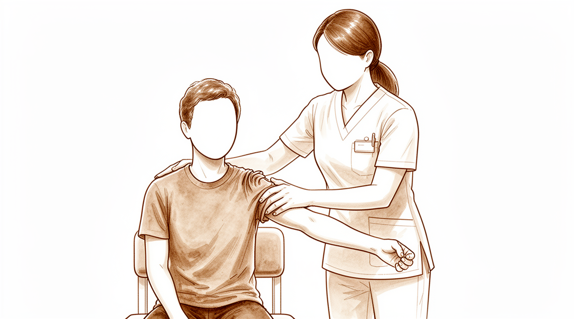
Subacromial impingement — causes of shoulder pain with overhead activity, diagnosis, and treatment options.
Những gì bạn đang cảm thấy
Đau vai là lý do phổ biến nhất khiến mọi người tìm kiếm sự chăm sóc y tế cho vấn đề này. Bạn có thể đang mắc hội chứng chèn ép dưới mỏm cùng vai, một tình trạng trong đó các cấu trúc trong vai của bạn bị chèn ép. Điều này thường liên quan đến các gân của nhóm cơ xoay vai hoặc bao hoạt dịch dưới mỏm cùng vai, một túi nhỏ chứa dịch giúp đệm cho khớp của bạn. Bạn có thể cảm thấy đau khi cử động cánh tay, đặc biệt là khi với lên cao hoặc đưa ra sau lưng. Các nhiệm vụ đơn giản như cài áo vào trong quần hoặc cài dây áo ngực có thể trở nên khó khăn và gây đau.
Cơn đau thường bùng phát vào ban đêm, khiến bạn khó ngủ ở bên bị ảnh hưởng. Bạn có thể nhận thấy cảm giác cứng khớp khi vừa thức dậy, giảm nhẹ một chút khi bạn di chuyển. Các hoạt động có xu hướng làm tăng sự khó chịu, đặc biệt là khi nâng vật hoặc với lên các kệ cao. Trong nhiều trường hợp, tình trạng viêm không chỉ xảy ra ở bao hoạt dịch mà còn lan vào khớp vai chính. Tình trạng viêm lan rộng này có thể gây ra cơn đau dữ dội ngay cả với những cử động nhỏ.
Mặc dù tình trạng này phổ biến, bác sĩ phẫu thuật của bạn sẽ đảm bảo rằng các vấn đề khác không phải là nguyên nhân gây ra các triệu chứng của bạn. Ví dụ, họ sẽ kiểm tra tình trạng mất vững hoặc các nguyên nhân hiếm gặp như khối u lành tính nhỏ hoặc lắng đọng canxi. Phụ nữ từ 30 đến 60 tuổi có lắng đọng canxi lớn hơn 1,5 cm có nguy cơ cao hơn đối với các triệu chứng đáng kể. Tuy nhiên, hình ảnh học có thể cho thấy các dấu hiệu của chèn ép ngay cả khi độ dày của gân trông bình thường so với vai bên kia của bạn.
Tin tốt là các bài tập cụ thể có hiệu quả và có thể giảm nhu cầu phẫu thuật. Những kết quả này thường kéo dài trong nhiều năm. Nếu các phương pháp điều trị bảo tồn như vật lý trị liệu không giúp ích sau ít nhất 6 tuần, bác sĩ phẫu thuật của bạn có thể thảo luận về các lựa chọn khác. Các mũi tiêm có thể cung cấp sự giảm đau tạm thời bằng cách giảm viêm. Kế hoạch chăm sóc của bạn sẽ được cá nhân hóa theo nhu cầu cụ thể, tập trung vào việc giúp bạn trở lại các hoạt động hàng ngày với ít đau đớn hơn.
Những gì thực sự đang xảy ra
Vai của bạn là một khớp cầu-đáy được bao bọc bởi một lớp vỏ chặt chẽ gọi là bao khớp. Trong không gian này, các gân và một túi nhỏ chứa dịch gọi là bao hoạt dịch trượt mượt mà khi bạn nâng cánh tay lên. Trong tình trạng chèn ép dưới mỏm cùng vai, các cấu trúc này bị ép chặt vào xương nằm phía trên. Sự chèn ép này gây viêm và đau khi bạn di chuyển cánh tay lên cao.
Bạn có thể cảm thấy sự chèn ép này do cách các cơ vai phối hợp với nhau. Bình thường, các cơ vòng xoay (rotator cuff) giữ cho đầu xương cánh tay nằm chính giữa ổ khớp. Nếu các cơ này yếu hoặc không phối hợp nhịp nhàng, đầu xương cánh tay sẽ dịch chuyển lên trên. Điều này làm giảm không gian cho các gân di chuyển. Kết quả là ma sát gây kích ứng các mô. Sự kích ứng này chính là nguyên nhân gây ra cơn đau nhói và cứng khớp.
Chẩn đoán hình ảnh giúp bác sĩ phẫu thuật xác định chính xác vị trí bị chèn ép. Nó cho thấy có sưng trong bao hoạt dịch hay dày lên của các gân hay không. Tuy nhiên, không phải ai bị đau cũng có những thay đổi rõ ràng trên phim chụp. Một số người có gân trông bình thường nhưng vẫn cảm thấy đau do cách vận động vai của họ. Đó là lý do tại sao bác sĩ phẫu thuật xem xét cả triệu chứng của bạn và các kiểu vận động.
Điều trị tập trung vào việc khắc phục sự vận động sai lệch này. Vật lý trị liệu giúp tăng cường các cơ ổn định khớp. Điều này tạo thêm không gian cho các gân trượt mà không bị chèn ép. Tiêm thuốc cũng có thể giúp ích bằng cách giảm viêm nhanh chóng. Điều này tạo ra một khoảng thời gian giảm đau để bạn bắt đầu tập luyện. Hầu hết mọi người đều cải thiện với các bước điều trị không phẫu thuật này. Phẫu thuật hiếm khi cần thiết và chỉ được xem xét nếu các phương pháp điều trị khác thất bại sau sáu tuần.
Những gì chúng tôi có thể làm về vấn đề này
Bắt đầu bằng tự chăm sóc và vật lý trị liệu. Bác sĩ phẫu thuật của bạn có thể sẽ khuyến nghị các bài tập cụ thể để tăng cường sức mạnh cho các cơ xung quanh vai của bạn. Phương pháp này hiệu quả và có thể giảm nhu cầu phẫu thuật. Các lợi ích của liệu pháp tập luyện này được duy trì trong thời gian dài, với kết quả kéo dài sau 10 năm. Bạn nên dành cho phương pháp điều trị bảo tồn này một cơ hội hợp lý để phát huy tác dụng. Nếu bạn không cải thiện sau ít nhất 6 tuần điều trị không phẫu thuật, bác sĩ phẫu thuật của bạn có thể thảo luận về các lựa chọn khác. Tuổi trẻ hơn, chỉ số khối cơ thể (BMI) thấp hơn và thời gian xuất hiện triệu chứng ngắn hơn trước khi bắt đầu điều trị là những dấu hiệu tốt cho quá trình hồi phục.
Nếu các bài tập đơn thuần không mang lại đủ sự giảm đau, bác sĩ phẫu thuật của bạn có thể đề xuất quản lý bằng thuốc. Điều này thường bao gồm thuốc giảm đau và thuốc chống viêm. Tiêm vào không gian dưới xương bả vai (không gian dưới mỏm quạ) cũng có thể giúp ích. Tiêm corticosteroid là liệu pháp hiệu quả trong ngắn hạn để giảm đau và cải thiện chức năng. Một số bệnh nhân cũng có thể được hưởng lợi từ việc tiêm axit hyaluronic, cung cấp giảm đau tương tự như steroid trong ngắn hạn. Một lựa chọn khác là huyết tương điều kiện hóa tự thân (ACP), sử dụng các thành phần máu của chính bạn và là một lựa chọn thay thế tốt nếu bạn không thể dùng steroid. Một liều tiêm ketorolac đơn lẻ có thể mang lại sự cải thiện lớn hơn ở tuần thứ tư so với tiêm steroid tiêu chuẩn. Mặc dù hướng dẫn siêu âm không vượt trội hơn so với tiêm không định hướng (blind) cho khu vực này, nhưng chẩn đoán chính xác và kỹ thuật đúng cách là rất quan trọng để có kết quả tốt.
Phẫu thuật chỉ được xem xét khi điều trị bảo tồn đã đạt đến giới hạn. Phẫu thuật được chỉ định nếu bạn vẫn còn đau dai dẳng và mất chức năng mặc dù đã thử các phương pháp điều trị không phẫu thuật. Bác sĩ phẫu thuật của bạn sẽ đánh giá xem cắt bỏ chóp dưới mỏm quạ nội soi có phải là một lựa chọn khả thi cho bạn hay không, đặc biệt nếu gân quay của bạn còn nguyên vẹn. Lưu ý rằng bằng chứng gần đây cho thấy phẫu thuật có thể không mang lại lợi ích rõ ràng cho tất cả những người bị chèn ép và có thể gây hại. Do đó, bác sĩ phẫu thuật của bạn sẽ cân nhắc cẩn thận các nguy cơ và lợi ích trước khi khuyến nghị phẫu thuật. Các công cụ hình ảnh như MRI giúp xác định mức độ tổn thương, nhưng cần thận trọng khi giải thích kết quả chụp sớm sau khi tiêm steroid, vì chúng đôi khi có thể bắt chước hình ảnh của một vết rách.
Những điều cần biết
Đau vai của bạn thường bắt nguồn từ tình trạng sưng ở bao hoạt dịch, một túi nhỏ chứa dịch giúp giảm xóc cho khớp của bạn. Tình trạng này được gọi là chèn ép dưới mỏm cùng vai. Tin tốt là cơ thể bạn thường tự chữa lành tình trạng này. Trên thực tế, 94% bệnh nhân mắc vai đông lạnh tự phát đã hồi phục mức độ chức năng và vận động bình thường mà không cần bất kỳ điều trị nào. Ngay cả khi bạn không mắc vai đông lạnh, diễn biến tự nhiên của cơn đau này có xu hướng cải thiện theo thời gian. Nhiều người nhận thấy rằng các phương pháp điều trị bằng bài tập cụ thể là hiệu quả và làm giảm nhu cầu phẫu thuật. Những lợi ích này được duy trì trong ít nhất 10 năm.
Nếu cơn đau của bạn vẫn tiếp diễn, bác sĩ phẫu thuật có thể đề xuất các lựa chọn không phẫu thuật. Tiêm thuốc có thể cung cấp sự giảm đau trong thời gian ngắn. Tiêm steroid vào vai hiệu quả trong việc giảm đau và cải thiện chức năng trong ngắn hạn. Bạn không cần hướng dẫn bằng siêu âm cho các mũi tiêm này; chúng hoạt động tốt như nhau khi không có sự hỗ trợ đó. Các loại tiêm khác, chẳng hạn như những loại sử dụng dịch thủy phân nhau thai người hoặc hyaluronate, cũng cho thấy những cải thiện đáng kể về mức độ đau và chất lượng cuộc sống. Vật lý trị liệu là một phần quan trọng của quá trình này. Nó giúp bạn lấy lại sức mạnh và tầm vận động.
Phẫu thuật thường không phải là lựa chọn đầu tiên. Bằng chứng ủng hộ việc quản lý không phẫu thuật hoặc không điều trị đối với chèn ép dưới mỏm cùng vai. Điều trị nội soi khớp không mang lại lợi ích rõ ràng và có thể gây hại. Ngay cả khi bạn có các lắng đọng canxi, việc loại bỏ chúng không yêu cầu loại bỏ thêm xương để đạt được kết quả tốt trong ngắn hạn. Nếu bạn cần phẫu thuật, nó thường chỉ được xem xét sau khi điều trị không phẫu thuật trong ít nhất 6 tuần. Bác sĩ phẫu thuật của bạn sẽ xem xét cẩn thận tiến triển của bạn.
Một số yếu tố ảnh hưởng đến tốc độ hồi phục của bạn. Tuổi trẻ hơn, chỉ số khối cơ thể (BMI) thấp hơn và thời gian ngắn hơn của các triệu chứng trước khi bắt đầu điều trị là những dấu hiệu tốt. Những thay đổi có thể đảo ngược trên MRI cũng dự đoán kết quả tốt hơn. Tuy nhiên, hãy lưu ý rằng các mũi tiêm vai trước phẫu thuật có liên quan đến tỷ lệ phẫu thuật lại tăng cao. Nguy cơ này phụ thuộc vào số lượng mũi tiêm bạn nhận được và thời điểm chúng được thực hiện. Nhìn chung, hầu hết bệnh nhân đều cải thiện với chăm sóc bảo tồn. Bác sĩ phẫu thuật của bạn sẽ giúp bạn tìm thấy sự cân bằng đúng đắn giữa nghỉ ngơi, tập thể dục và thuốc men để đưa bạn trở lại các hoạt động hàng ngày.
Khi nào nên gặp bác sĩ
Đau vai là tình trạng phổ biến, thường do chèn ép hoặc viêm bao hoạt dịch. Hãy gặp bác sĩ đa khoa nếu cơn đau dai dẳng dù đã nghỉ ngơi. Hãy tìm kiếm đánh giá từ bác sĩ chuyên khoa nếu bạn gặp phải tình trạng yếu cơ, mất ổn định, hoặc nếu vai của bạn bị khóa hoặc đột ngột mất sức. Hãy liên hệ với bác sĩ phẫu thuật nếu các triệu chứng cản trở giấc ngủ hoặc công việc. Tình trạng đau tăng lên đột ngột cũng cần được chú ý và xử trí kịp thời. Trong khi nhiều trường hợp cải thiện với điều trị bảo tồn, một số trường hợp liên quan đến khối u hiếm hoặc các khối lắng đọng xương lớn cần phải phẫu thuật loại bỏ. Bác sĩ của bạn sẽ kiểm tra các vấn đề cụ thể này nếu các phương pháp điều trị tiêu chuẩn không hiệu quả. Đánh giá sớm giúp phân biệt chèn ép với các tình trạng khác như mất ổn định khớp. Chẩn đoán đúng đắn đảm bảo bạn nhận được sự chăm sóc phù hợp để khôi phục khả năng vận động và giảm viêm.
Evidence & references
Overview
- Blind subacromial corticosteroid injections are as effective as ultrasound-guided injections for improving pain and function in subacromial impingement syndrome after short-term follow-up [1].
- Ultrasound guidance is not superior to blind injection for subacromial bursa injections regarding pain or function outcomes [4].
- Ultrasound guidance is superior to blind injection for bicipital groove injections [4].
- Subacromial injections of human placenta hydrolysate show significant improvement in pain, functional level, and quality of life in patients with shoulder impingement syndrome [3].
- Subacromial injection of corticosteroids is an effective short-term therapy for symptomatic subacromial impingement syndrome [8].
- Subacromial steroid injection is an alternative modality for primary frozen shoulder, and treatment should be individualized [12].
- Management of subacromial impingement syndrome includes physical therapy, injections, and surgery for some patients [2].
- There remains a need for high-quality studies of the pathology, etiology, and management of subacromial impingement syndrome [2].
- In patients with isolated unilateral subacromial pain syndrome and intact rotator cuff tendons, there are more cases of ultrasonographic impingement in affected shoulders compared to unaffected shoulders [5].
- There are no significant differences in supraspinatus tendon thickness, subacromial bursa thickness, or acromio-humeral distance between affected and unaffected shoulders in patients with isolated unilateral subacromial pain syndrome [5].
- Arthroscopic bursectomy and debridement of calcific deposits for calcific tendonitis yields short-term functional outcomes that are not influenced by the addition of subacromial decompression [10].
- Arthroscopic treatment should no longer be offered to people with subacromial impingement as surgery offers no discernible benefits but may result in harm [15].
- The weight of evidence supports nonoperative management or no treatment for subacromial impingement [15].
- Specific exercise treatment for patients with subacromial pain is effective and reduces the need for surgery, with maintained results after 10 years [18].
- There is no uniform definition for any of the diagnostic labels for shoulder pain across different randomized controlled trials [19].
- Following nonoperative treatment for at least 6 weeks, subacromial decompression is a viable and good surgical option for shoulder impingement with an intact rotator cuff [24].
Anatomy & Pathophysiology
- Ultrasound guidance is not superior to non-guided injection for the subacromial bursa in terms of pain or function outcomes [4].
- Ultrasound guidance is not superior to non-guided injection for the glenohumeral joint in terms of pain or function outcomes [4].
- Ultrasound guidance is superior to non-guided injection for the bicipital groove [4].
- In patients with isolated unilateral subacromial pain syndrome and intact rotator cuff tendons, affected shoulders show more cases of ultrasonographic impingement compared to unaffected shoulders [5].
- In patients with isolated unilateral subacromial pain syndrome and intact rotator cuff tendons, there are no significant differences in supraspinatus tendon thickness between affected and unaffected shoulders [5].
- In patients with isolated unilateral subacromial pain syndrome and intact rotator cuff tendons, there are no significant differences in subacromial bursa thickness between affected and unaffected shoulders [5].
- In patients with isolated unilateral subacromial pain syndrome and intact rotator cuff tendons, there are no significant differences in acromio-humeral distance between affected and unaffected shoulders [5].
- Intra-articular corticosteroid intervention provides clinically meaningful short-term improvements in adhesive capsulitis [6].
- Intra-articular corticosteroid intervention administered after distension of the shoulder capsule provides clinically meaningful short-term improvements in adhesive capsulitis [6].
- The acromial morphology classification system is an unreliable method to assess the acromion [23].
- The acromial index shows no association with the presence of rotator cuff disease [23].
- Imaging is an essential tool for the evaluation of patients with shoulder pain [26].
- Understanding the extent of an injury with imaging is key to successful management of shoulder pain [26].
- 94% of patients with spontaneous frozen shoulder recover to normal levels of function and motion without treatment [29].
- Biomechanical studies indicate that the long head of the biceps contributes to stability of the glenohumeral joint in all directions [32].
- In vivo studies have not yet established the stabilizing effect of the long head of the biceps on the glenohumeral joint [32].
- The physiologic load required for the long head of the biceps to stabilize the glenohumeral joint remains unknown [32].
- Pain reduction from subacromial injection causes shifts in scapulohumeral rhythm in patients with full-thickness rotator cuff tears [33].
- Pain reduction from subacromial injection results in an increase in glenohumeral motion in patients with full-thickness rotator cuff tears [33].
- Pain reduction from subacromial injection results in reduced reliance on scapular rotation in patients with full-thickness rotator cuff tears [33].
- Addressing aberrant movement patterns and facilitating balanced activation of all shoulder muscles may be an appropriate treatment direction for subacromial pain syndrome [34].
- Exercise protocols targeting the rotator cuff and scapular stabilizers are effective in improving pain, function, and shoulder active range of motion in patients with subacromial syndrome [35].
- There are no between-group differences in shoulder maximal voluntary contraction (MVC) in subjects with subacromial impingement syndrome [36].
- The use of a triaxial gyroscope is a simple, non-invasive, and reproducible method for recording shoulder anteflexion and abduction [37].
- The Korean Shoulder Scoring System (KSS) is a useful measurement tool that combines subjective and objective evaluations for shoulder function related to rotator cuff disorders [38].
- Isometric measurement of shoulder rotation strength provides reliable information on the functional integrity of the rotator cuff muscles [39].
- Functional integrity of the rotator cuff muscles, as measured by isometric shoulder rotation strength, is significantly related to patients' function and quality of life [39].
- The majority of questions in commonly adopted shoulder-specific functional outcome measurement tools are subjective in nature [40].
- The Shoulder Intervention Project (SIP) presents the rationale, design, methods, and operational aspects of a new rehabilitation approach to evaluate shoulder function and work disability after decompression surgery for subacromial impingement syndrome [47].
- Acute experimental shoulder pain has an inhibitory effect on the activity of the infraspinatus during arm elevation [51].
- All upper extremity-specific scales have acceptable psychometric properties for measuring rotator cuff tears [52].
Classification
- Subacromial impingement syndrome is a specific diagnosis that must be differentiated from other conditions such as glenohumeral instability, particularly in younger athletes [13].
- Impingement and rotator cuff syndromes were the most frequent diagnoses in population-based consultation patterns for shoulder pain [7].
- There is no uniform definition for any of the diagnostic labels for shoulder pain, as revealed by the comparison of selection criteria from different randomised controlled trials [19].
- Rotator cuff and subacromial bursa pathology were the most common findings on ultrasound and MRA in a prospective study of shoulder pain in primary care [14].
- In patients with isolated unilateral subacromial pain syndrome and intact rotator cuff tendons, there were more cases of ultrasonographic impingement in affected shoulders compared to unaffected shoulders [5].
- There were no significant differences in supraspinatus tendon thickness, subacromial bursa thickness, or acromio-humeral distance between affected and unaffected shoulders in patients with unilateral subacromial pain syndrome [5].
- The acromial morphology classification system is an unreliable method to assess the acromion [23].
- The acromial index shows no association with the presence of rotator cuff disease [23].
- An extended inflammatory process is present in patients with subacromial impingement syndrome, involving not only the subacromial bursa but also the glenohumeral joint [9].
- Increased levels of inflammatory markers are present in the subscapularis tendon and joint capsule in patients with subacromial impingement [9].
- Abundant hemodynamic activity within the Bursa and AP resulted in severe motion pain that reflected focal bursitis, probably due to subacromial impingement and secondary glenohumeral synovitis [11].
- A novel rat model of subacromial impingement creates cellular and molecular changes consistent with the development of rotator cuff tendinopathy [50].
Clinical Presentation
- Impingement and rotator cuff syndromes are the most frequent diagnoses in patients with shoulder pain [7].
- Rotator cuff and subacromial bursa pathology are the most common findings on ultrasound and magnetic resonance arthrography in patients with shoulder pain [14].
- Subacromial impingement syndrome is a specific diagnosis that must be differentiated from other conditions such as glenohumeral instability, particularly in younger athletes [13].
- Subacromial lipoma should be included in the differential diagnosis of rotator cuff impingement when conservative treatments fail [16].
- A large ossified mass attached to the rotator cuff tendon in the subacromial bursa can cause impingement pain and restricted shoulder motion [21].
- Symptoms of subacromial impingement can be caused by a rare benign soft tissue tumor, such as a collagenous fibroma located in the subacromial bursa [45].
- Atypical presentations of calcific tendinitis, such as involvement of the teres minor, can affect overhead movement and present with isolated posterior shoulder pain [43].
- Women aged between 30 and 60 years with subacromial pain syndrome and a calcific deposit of >1.5 cm in length have the highest chance of suffering from symptomatic calcific tendinopathy of the rotator cuff [17].
- In patients with isolated unilateral subacromial pain syndrome and intact rotator cuff tendons, there are more cases of ultrasonographic impingement in affected shoulders compared to unaffected shoulders [5].
- There are no significant differences in supraspinatus tendon thickness, subacromial bursa thickness, or acromio-humeral distance between affected and unaffected shoulders in patients with isolated unilateral subacromial pain syndrome [5].
- Abundant hemodynamic activity within the bursa and anterior portal region results in severe motion pain that reflects focal bursitis, probably due to subacromial impingement and secondary glenohumeral synovitis [11].
- An extended inflammatory process is present not only in the subacromial bursa but also in the glenohumeral joint in patients with subacromial impingement syndrome [9].
Investigations
- Blind subacromial corticosteroid injections are as effective as ultrasound-guided injections for improving pain and function in subacromial impingement syndrome after short-term follow-up [1].
- Ultrasound guidance is not superior to blind injection for subacromial bursa and glenohumeral joint injections regarding pain or function [4].
- Ultrasound guidance is superior to blind injection for bicipital groove injections [4].
- Patients with isolated unilateral subacromial pain syndrome have more cases of ultrasonographic impingement in the affected shoulder compared to the unaffected shoulder [5].
- There are no significant differences in supraspinatus tendon thickness, subacromial bursa thickness, or acromio-humeral distance between affected and unaffected shoulders in patients with isolated unilateral subacromial pain syndrome [5].
- Impingement and rotator cuff syndromes are the most frequent diagnoses in patients with shoulder pain [7].
- An extended inflammatory process is present in the subscapularis tendon and joint capsule, in addition to the subacromial bursa, in patients with subacromial impingement syndrome [9].
- Abundant hemodynamic activity within the bursa and anterior portal results in severe motion pain reflecting focal bursitis, likely due to subacromial impingement and secondary glenohumeral synovitis [11].
- Subacromial impingement syndrome is a specific diagnosis that must be differentiated from other conditions such as glenohumeral instability, particularly in younger athletes [13].
- Rotator cuff and subacromial bursa pathology are the most common findings on ultrasound and magnetic resonance arthrography (MRA) in patients with shoulder pain [14].
- Subacromial lipoma should be included in the differential diagnosis of rotator cuff impingement when conservative treatments fail [16].
- A large ossified mass attached to the rotator cuff tendon in the subacromial bursa can cause impingement pain and loss of motion, which resolves after surgical excision and repair [21].
- Younger age is a good prognostic factor for the natural course of subacromial impingement syndrome [22].
- Lower BMI is a good prognostic factor for the natural course of subacromial impingement syndrome [22].
- More functional capacity is a good prognostic factor for the natural course of subacromial impingement syndrome [22].
- A shorter symptomatic period is a good prognostic factor for the natural course of subacromial impingement syndrome [22].
- Reversible changes on MRI are a good prognostic factor for the natural course of subacromial impingement syndrome [22].
- Higher Constant and ASES scores at the first evaluation are good prognostic factors for the natural course of subacromial impingement syndrome [22].
- Accurate diagnosis of the etiology of shoulder pain and proper injection technique are important in achieving satisfactory clinical outcomes with subacromial corticosteroid injections [25].
- Imaging is an essential tool for the evaluation of patients with shoulder pain [26].
- Understanding the extent of an injury with imaging is key to successful management of shoulder pain [26].
- Magnetic resonance imaging (MRI) appearance of the shoulder after subacromial injection with corticosteroids can mimic a rotator cuff tear [41].
- Caution should be used in the interpretation of MRI scans of the shoulder soon after the injection of corticosteroids [41].
- MRI findings are significantly associated with the change in SPADI score from baseline to one-year follow-up in subacromial pain syndrome [53].
- Patients with higher MRI total scores have a poorer outcome after treatment for subacromial pain syndrome [53].
- Patients with tendinosis on MRI have a poorer outcome after treatment for subacromial pain syndrome [53].
- Patients with bursitis on MRI have a poorer outcome after treatment for subacromial pain syndrome [53].
Treatment
Non-Operative Management
- Blind subacromial corticosteroid injections are as effective as ultrasound-guided injections for improving pain and function in subacromial impingement syndrome after short-term follow-up [1].
- Ultrasound guidance is not superior to blind injection for subacromial bursa injections regarding pain or function outcomes [4].
- Subacromial injections of human placenta hydrolysate show significant improvement in pain, functional level, and quality of life in patients with shoulder impingement syndrome [3].
- Subacromial injection of corticosteroids is an effective short-term therapy for symptomatic subacromial impingement syndrome [8].
- There is little reproducible evidence to support the efficacy of subacromial corticosteroid injection in managing rotator cuff disease [27].
- A single injection of 60 mg of ketorolac resulted in greater improvements in outcomes than a single injection of 40 mg triamcinolone for subacromial impingement at four weeks [44].
- Subacromial hyaluronate injection produces similar pain and functional improvement to corticosteroid at short-term follow-up for impingement syndrome [20].
- Subacromial autologous conditioned plasma (ACP) injections are a good alternative to subacromial cortisone injections, especially in patients with contraindications to cortisone [42].
- Subacromial steroid injection is an alternative modality for primary frozen shoulder, and treatment should be individualized [12].
- Specific exercise treatment for subacromial pain is effective and reduces the need for surgery, with maintained results after 10 years [18].
- Management of subacromial impingement syndrome includes physical therapy, injections, and surgery for some patients [2].
- The diagnostic labeling of shoulder pain lacks uniformity across randomized controlled trials [19].
Operative Management
- Arthroscopic treatment should no longer be offered to people with subacromial impingement as surgery offers no discernible benefits but may result in harm, with evidence supporting nonoperative management or no treatment [15].
- Following nonoperative treatment for at least 6 weeks, arthroscopic subacromial decompression (SAD) is a viable and good surgical option for shoulder impingement with an intact rotator cuff [24].
- Surgery is indicated for persistent pain and loss of function despite conservative treatment in the patient care pathway for subacromial shoulder pain [49].
- The short-term functional outcome of patients with calcific tendonitis after arthroscopic bursectomy and debridement is not influenced by whether it is performed in combination with or without subacromial decompression [10].
Differential Diagnosis
- Subacromial lipoma should be included in the differential diagnosis of rotator cuff impingement when conservative treatments fail [16].
Complications
- Arthroscopic treatment for subacromial impingement offers no discernible benefits and may result in harm [15].
- Abundant hemodynamic activity within the subacromial bursa and anterior portal resulted in severe motion pain, reflecting focal bursitis likely due to subacromial impingement and secondary glenohumeral synovitis [11].
- An extended inflammatory process is present not only in the subacromial bursa but also in the glenohumeral joint in patients with subacromial impingement syndrome [9].
- A large ossified mass attached to the rotator cuff tendon in the subacromial bursa can cause impingement pain and loss of shoulder motion, requiring surgical excision and repair [21].
Recovery
- Blind subacromial corticosteroid injections are as effective as ultrasound-guided injections for improving pain and function in subacromial impingement syndrome after short-term follow-up [1].
- Subacromial injections of human placenta hydrolysate show significant improvement in pain, functional level, and quality of life in patients with shoulder impingement syndrome [3].
- Intra-articular corticosteroid intervention, administered alone or after distension of the shoulder capsule, provides clinically meaningful short-term improvements in adhesive capsulitis of the shoulder [6].
- Subacromial injection of corticosteroids is an effective short-term therapy for symptomatic subacromial impingement syndrome [8].
- An extended inflammatory process is present in both the subacromial bursa and the glenohumeral joint capsule in patients with subacromial impingement syndrome [9].
- The short-term functional outcome of patients with calcific tendonitis after arthroscopic bursectomy and debridement is not influenced by the addition of subacromial decompression [10].
- Women aged 30 to 60 years with subacromial pain syndrome and a calcific deposit greater than 1.5 cm in length have the highest chance of suffering from symptomatic calcific tendinopathy of the rotator cuff [17].
- Specific exercise treatment for subacromial pain is effective and reduces the need for surgery, with maintained results after 10 years [18].
- Subacromial hyaluronate injection produces similar short-term pain and functional improvement to corticosteroid for impingement syndrome [20].
- Younger age, lower BMI, more functional capacity, a shorter symptomatic period, reversible changes on MRI, and higher Constant and ASES scores at initial evaluation are good prognostic factors for the natural course of subacromial impingement syndrome [22].
- The natural history of rotator cuff tendinopathy likely plays a significant role in long-term results, supporting the view that arthroscopic decompression is not recommended for its treatment [28].
- 94% of patients with spontaneous frozen shoulder recover to normal levels of function and motion without treatment [29].
- Arthroscopic acromioplasty provides no relevant additional clinical effects or impact on rotator cuff integrity compared to bursectomy alone at 12 years' follow-up for chronic subacromial pain syndrome [30].
- Intraoperative ultrasound facilitates arthroscopic debridement of calcific rotator cuff tendinitis, with highly significant clinical improvement observed 2 weeks post-surgery and excellent radiological results until 9 months follow-up [31].
- Arthroscopic acromioplasty significantly improves long-term clinical outcomes up to 2 years when combined with platelet-rich plasma injection for chronic rotator cuff tendinopathy [54].
- Preoperative shoulder injections are associated with increased rotator cuff revision rates, with a correlation observed that is dependent on injection frequency and time [55].
Key Evidence
- [L1] Blind injections into the subacromial bursa were as effective as ultrasound-guided injections for improving pain and function in subacromial impingement syndrome after a short-term follow-up. [1] (10.1177/0363546515618653)
- [L5] Management of subacromial impingement syndrome includes physical therapy, injections, and surgery for some patients, but there remains a need for high-quality studies of the pathology, etiology, and management of the condition. [2] (10.5435/00124635-201111000-00006)
- [L1] Subacromial injections showed significant improvement in pain, functional level, and quality of life in patients with shoulder impingement syndrome. [3] (10.1186/s12891-024-08266-4)
- [L1] Ultrasound guidance is not superior in the subacromial bursa and glenohumeral joint injections in pain or function. [4] (10.1016/j.arthro.2021.12.013)
- [L3] In this cohort of patients with isolated unilateral SAPS, we found more cases of ultrasonographic impingement in affected shoulders compared to unaffected, but no significant differences in supraspinatus tendon thickness, subacromial bursa thickness, or acromio-humeral distance. [5] (10.1016/j.jse.2025.02.020)
- [L1] Intra-articular corticosteroid intervention, administered either alone or after distension of the shoulder capsule, provided clinically meaningful improvements in the short term. [6] (10.1177/0363546518823337)
- [L3] Impingement and rotator cuff syndromes were the most frequent diagnoses. [7] (10.1186/1471-2474-13-238)
- [L1] Subacromial injection of corticosteroids is an effective short-term therapy for the treatment of symptomatic subacromial impingement syndrome. [8] (10.2106/00004623-199611000-00007)
- [L3] This study provides evidence that an extended inflammatory process is present, not only in the subacromial bursa but also in the glenohumeral joint in patients with subacromial impingement syndrome. [9] (10.1007/s00167-020-05992-9)
- [L1] This study has demonstrated that the short-term functional outcome of patients with calcific tendonitis after arthroscopic bursectomy and debridement of the calcific deposit is not influenced if performed in combination with or without a subacromial decompression. [10] (10.1016/j.arthro.2015.05.015)
- [L4] Abundant hemodynamic activity within the Bursa and AP resulted in severe motion pain that reflected focal bursitis, probably due to subacromial impingement and secondary glenohumeral synovitis. [11] (10.1016/j.jse.2025.04.023)
- [L1] Subacromial steroid injection is an alternative modality, and treatment should be individualized. [12] (10.1016/j.jse.2011.04.029)
- [L2] Rotator cuff and subacromial bursa pathology were the most common findings on ultrasound and MRA. [14] (10.1186/1471-2474-12-119)
- [L5] Arthroscopic treatment should no longer be offered to people with subacromial impingement as surgery offers no discernible benefits but may result in harm, and the weight of evidence supports nonoperative management or no treatment. [15] (10.1016/j.arthro.2022.03.017)
- [Case_report] Subacromial lipoma should be included in the differential diagnosis of rotator cuff impingement when conservative treatments fail. [16] (10.1016/j.jse.2008.09.017)
- [L3] This study demonstrates that women aged between 30 and 60 years with subacromial pain syndrome and a calcific deposit of >1.5 cm in length have the highest chance of suffering from symptomatic calcific tendinopathy of the rotator cuff. [17] (10.1016/j.jse.2015.02.024)
- [L2] Specific exercise treatment for patients with subacromial pain was effective and reduced the need for surgery with maintained results after 10 years. [18] (10.1016/j.jse.2024.10.027)
- [L2] The comparison of selection criteria from different randomised controlled trials revealed no uniform definition for any of the diagnostic labels for shoulder pain. [19] (10.1016/j.math.2008.04.005)
- [L2] A subacromial hyaluronate injection to treat impingement syndrome produces similar pain and functional improvement to corticosteroid at a short-term follow-up. [20] (10.1016/j.jse.2011.11.009)
- [L4] A large ossified mass attached to the rotator cuff tendon in the subacromial bursa was successfully treated with surgical excision and repair, resulting in the resolution of impingement pain and restoration of shoulder motion by 12 months. [21] (10.1097/01.blo.0000170720.91461.58)
- [L2] Younger age, lower BMI, more functional capacity, a shorter symptomatic period, reversible changes on MRI, and higher Constant and ASES scores at the first evaluation were good prognostic factors for the natural course of subacromial impingement syndrome. [22] (10.1016/j.jse.2015.06.007)
- [L3] The acromial morphology classification system is an unreliable method to assess the acromion, and the acromial index shows no association with the presence of rotator cuff disease. [23] (10.1016/j.jse.2011.09.028)
- [L5] Following nonoperative treatment for at least 6 weeks, SAD is a viable and good surgical option for the treatment of shoulder impingement with an intact rotator cuff. [24] (10.1016/j.arthro.2019.06.012)
- [L5] Accurate diagnosis of the etiology of a patient's shoulder pain and proper injection technique are important in achieving satisfactory clinical outcomes. [25] (10.1016/j.jse.2007.07.009)
- [L4] Imaging is an essential tool for evaluation of patients with shoulder pain; understanding the extent of an injury with imaging is key to successful management. [26] (10.1016/j.csm.2013.03.009)
- [L1] This systematic review of the available literature indicates that there is little reproducible evidence to support the efficacy of subacromial corticosteroid injection in managing rotator cuff disease. [27] (10.5435/00124635-200701000-00002)
- [L1] The natural history of rotator cuff tendinopathy probably plays a significant role in the results in the long-term. [28] (10.1302/0301-620x.99b6.bjj-2016-0569.r1)
- [L4] We found 94% of patients with spontaneous frozen shoulder recovered to normal levels of function and motion without treatment. [29] (10.1007/s11999-011-2176-4)
- [L2] There were no relevant additional effects of arthroscopic acromioplasty on bursectomy alone with respect to clinical outcomes and rotator cuff integrity at 12 years' follow-up. [30] (10.1016/j.jse.2017.03.021)
- [L1] Highly significant clinical improvement of the shoulder was already observed in the entire population 2 weeks after surgery, with excellent radiological results observed until the 9 months follow-up. [31] (10.1007/s00402-014-1927-6)
- [L5] Biomechanical studies indicate that the long head of the biceps contributes to stability of the glenohumeral joint in all directions, though in vivo studies have yet to establish this stabilizing effect and the physiologic load required remains unknown. [32] (10.1016/j.arthro.2010.10.014)
- [L3] Pain reduction caused shifts in scapulohumeral rhythm resulting in an increase in glenohumeral motion and a reduced reliance on scapular rotation. [33] (10.1016/j.jse.2007.05.010)
- [L1] Addressing aberrant movement patterns and facilitating balanced activation of all shoulder muscles may be a more appropriate treatment direction for the future. [34] (10.1177/1758573216660038)
- [L1] Both interventions are effective in terms of pain, function, and shoulder active range of motion. [35] (10.1016/j.jht.2017.11.041)
- [L3] No between-group differences in shoulder MVC were observed. [36] (10.1002/mus.20636)
- [L3] The use of a tri axial gyroscope is a simple non invasive and reproducible method for the recording of shoulder anteflexion and abduction. [37] (10.1186/1471-2474-13-135)
- [L4] The KSS is a useful measurement tool that combines subjective and objective evaluations for shoulder function related to rotator cuff disorders. [38] (10.1016/j.jse.2008.11.019)
- [L3] Isometric measurement of shoulder rotation strength provides reliable information on the functional integrity of the rotator cuff muscles, which is significantly related to patients' function and quality of life. [39] (10.1016/j.jse.2004.03.009)
- [L1] The majority of questions posed in the most commonly adopted shoulder-specific functional outcome measurement tools were subjective in nature and may account for part of the phenomenon. [40] (10.1007/s00264-007-0493-8)
- [L4] One should use caution in the interpretation of magnetic resonance imaging scans of the shoulder soon after the injection of corticosteroids. [41] (10.1016/j.arthro.2007.01.024)
- [L3] Therefore, subacromial ACP injections are a good alternative to subacromial cortisone injections, especially in patients with contraindication to cortisone. [42] (10.1007/s00167-015-3651-3)
- [Case_report] This case highlights the importance of considering atypical presentations of calcific tendinitis, particularly in the context of isolated posterior shoulder pain. [43] (10.1016/j.jisako.2025.101055)
- [L1] In this study, a single injection of 60 mg of ketorolac resulted in improvements in outcomes greater than a single injection of 40 mg triamcinolone for the treatment of subacromial impingement when assessed at four weeks. [44] (10.1016/j.jse.2012.08.026)
- [L4] In this case, the symptoms were caused by a rare benign soft tissue tumor: a collagenous fibroma located in the subacromial bursa. [45] (10.1016/j.jse.2010.04.009)
- [L1] The paper presents the rationale, design, methods, and operational aspects of the Shoulder Intervention Project (SIP) to evaluate a new rehabilitation approach. [47] (10.1186/1471-2474-15-215)
- [L5] The document outlines a patient care pathway for subacromial shoulder pain emphasizing shared decision-making, continuity of care, and a stepwise approach from primary to secondary care, noting that surgery is indicated for persistent pain and loss of function despite conservative treatment. [49] (10.1177/1758573215576456)
- [L5] This new rat subacromial impingement model creates cellular and molecular changes consistent with the development of rotator cuff tendinopathy. [50] (10.1016/j.jse.2022.02.041)
- [L5] This study demonstrates that acute experimental shoulder pain has an inhibitory effect on the activity of the infraspinatus during arm elevation. [51] (10.1016/j.jse.2016.09.005)
- [L3] All upper extremity-specific scales had acceptable psychometric properties. [52] (10.1097/corr.0000000000000800)
- [L2] In this study, MRI findings were significantly associated with the change in the SPADI score from baseline and to one year follow-up, with a poorer outcome after treatment for the patients with higher MRI total score, tendinosis and bursitis on MRI. [53] (10.1186/s12891-017-1827-3)
- [L1] Arthroscopic acromioplasty significantly improves long-term clinical outcomes up to 2 years. [54] (10.1177/0363546515608485)
- [L3] This study strongly suggests a correlation between preoperative shoulder injections and revision rotator cuff repair, with frequency and time dependence observed. [55] (10.1016/j.arthro.2018.10.116)
References
[1] Ultrasound-Guided Versus Blind Subacromial Corticosteroid Injections for Subacromial Impingement Syndrome. The American Journal of Sports Medicine. 2015. DOI: 10.1177/0363546515618653 [2] Subacromial Impingement Syndrome. American Academy of Orthopaedic Surgeon. 2011. DOI: 10.5435/00124635-201111000-00006 [3] Effectiveness and safety of human placenta hydrolysate injection into subacromial space in patients with shoulder impingement syndrome: a single-blind, randomized trial. BMC Musculoskeletal Disorders. 2025. DOI: 10.1186/s12891-024-08266-4 [4] Ultrasound Guidance Is Not Superior in Subacromial Bursa and Intraarticular Injections but Superior in Bicipital Groove: A Meta-analysis of Randomized Controlled Trials. Arthroscopy: The Journal of Arthroscopic & Related Surgery. 2022. DOI: 10.1016/j.arthro.2021.12.013 [5] Bilateral ultrasonographic findings in patients with unilateral subacromial pain syndrome and intact rotator cuff tendons. Journal of Shoulder and Elbow Surgery. 2025. DOI: 10.1016/j.jse.2025.02.020 [6] Efficacy of Pharmacological Therapies for Adhesive Capsulitis of the Shoulder: A Systematic Review and Network Meta-analysis. The American Journal of Sports Medicine. 2019. DOI: 10.1177/0363546518823337 [7] Population-based consultation patterns in patients with shoulder pain diagnoses. BMC Musculoskeletal Disorders. 2012. DOI: 10.1186/1471-2474-13-238 [8] Efficacy of Injections of Corticosteroids for Subacromial Impingement Syndrome. The Journal of Bone & Joint Surgery. 1996. DOI: 10.2106/00004623-199611000-00007 [9] Increased levels of inflammatory markers in the subscapularis tendon and joint capsule in patients with subacromial impingement. Knee Surgery, Sports Traumatology, Arthroscopy. 2020. DOI: 10.1007/s00167-020-05992-9 [10] Short‐Term Outcome After Arthroscopic Bursectomy Debridement of Rotator Cuff Calcific Tendonopathy With and Without Subacromial Decompression: A Prospective Randomized Controlled Trial. Arthroscopy. 2015. DOI: 10.1016/j.arthro.2015.05.015 [11] Impact of hemodynamics in individual shoulder structures on pain intensity in patients with rotator cuff tear. Journal of Shoulder and Elbow Surgery. 2026. DOI: 10.1016/j.jse.2025.04.023 [12] Comparison of glenohumeral and subacromial steroid injection in primary frozen shoulder: a prospective, randomized short-term comparison study. Journal of Shoulder and Elbow Surgery. 2011. DOI: 10.1016/j.jse.2011.04.029 [13] BIGLIANI, LOUIS U. M.D.+, NEW YORK, N.Y.; LEVINE, WILLIAM N. M.D.++, BALTIMORE, MARYLAND. The Journal of Bone and Joint Surgery. American Volume. 1997. [14] A prospective study of shoulder pain in primary care: Prevalence of imaged pathology and response to guided diagnostic blocks. BMC Musculoskeletal Disorders. 2011. DOI: 10.1186/1471-2474-12-119 [15] Editorial Commentary : Arthroscopic Treatment Should No Longer Be Offered to People With Subacromial Impingement. Arthroscopy. 2022. DOI: 10.1016/j.arthro.2022.03.017 [16] Lipoma of the supraspinatus muscle causing impingement syndrome: A case report. Journal of Shoulder and Elbow Surgery. 2009. DOI: 10.1016/j.jse.2008.09.017 [17] Prevalence of calcific deposits within the rotator cuff tendons in adults with and without subacromial pain syndrome: clinical and radiologic analysis of 1219 patients. Journal of Shoulder and Elbow Surgery. 2015. DOI: 10.1016/j.jse.2015.02.024 [18] No need for subacromial decompression in responders to specific exercise treatment: a 10-year follow-up of a randomized controlled trial. Journal of Shoulder and Elbow Surgery. 2025. DOI: 10.1016/j.jse.2024.10.027 [19] Lack of uniformity in diagnostic labeling of shoulder pain: Time for a different approach. Manual Therapy. 2008. DOI: 10.1016/j.math.2008.04.005 [20] Does hyaluronate injection work in shoulder disease in early stage? A multicenter, randomized, single blind and open comparative clinical study. Journal of Shoulder and Elbow Surgery. 2012. DOI: 10.1016/j.jse.2011.11.009 [21] Case Reports: Ossified Mass of the Rotator Cuff Tendon in the Subacromial Bursa. Clinical Orthopaedics and Related Research. 2005. DOI: 10.1097/01.blo.0000170720.91461.58 [22] Medium-term natural history of subacromial impingement syndrome. Journal of Shoulder and Elbow Surgery. 2015. DOI: 10.1016/j.jse.2015.06.007 [23] Relationship of radiographic acromial characteristics and rotator cuff disease: a prospective investigation of clinical, radiographic, and sonographic findings. Journal of Shoulder and Elbow Surgery. 2012. DOI: 10.1016/j.jse.2011.09.028 [24] Indications for Arthroscopic Subacromial Decompression. A Level V Evidence Clinical Guideline. Arthroscopy. 2019. DOI: 10.1016/j.arthro.2019.06.012 [25] Subacromial corticosteroid injections. Journal of Shoulder and Elbow Surgery. 2008. DOI: 10.1016/j.jse.2007.07.009 [26] Imaging of the Shoulder with Arthroscopic Correlation. Clinics in Sports Medicine. 2013. DOI: 10.1016/j.csm.2013.03.009 [27] The Efficacy of Subacromial Corticosteroid Injection in the Treatment of Rotator Cuff Disease: A Systematic Review. Journal of the American Academy of Orthopaedic Surgeons. 2007. DOI: 10.5435/00124635-200701000-00002 [28] Arthroscopic decompression not recommended in the treatment of rotator cuff tendinopathy. The Bone & Joint Journal. 2017. DOI: 10.1302/0301-620x.99b6.bjj-2016-0569.r1 [29] The Natural History of Idiopathic Frozen Shoulder: A 2- to 27-year Followup Study. Clinical Orthopaedics & Related Research. 2012. DOI: 10.1007/s11999-011-2176-4 [30] Does acromioplasty result in favorable clinical and radiologic outcomes in the management of chronic subacromial pain syndrome? A double-blinded randomized clinical trial with 9 to 14 years' follow-up. Journal of Shoulder and Elbow Surgery. 2017. DOI: 10.1016/j.jse.2017.03.021 [31] The intraoperative use of ultrasound facilitates significantly the arthroscopic debridement of calcific rotator cuff tendinitis. Archives of Orthopaedic and Trauma Surgery. 2014. DOI: 10.1007/s00402-014-1927-6 [32] Anatomy, Function, Injuries, and Treatment of the Long Head of the Biceps Brachii Tendon. Arthroscopy. 2011. DOI: 10.1016/j.arthro.2010.10.014 [33] Shoulder kinematics in patients with full-thickness rotator cuff tears after a subacromial injection. Journal of Shoulder and Elbow Surgery. 2008. DOI: 10.1016/j.jse.2007.05.010 [34] Electromyographic activity of the shoulder muscles during rehabilitation exercises in subjects with and without subacromial pain syndrome: a systematic review. Shoulder & Elbow. 2016. DOI: 10.1177/1758573216660038 [35] Pain, motion and function comparison of two exercise protocols for the rotator cuff and scapular stabilizers in patients with subacromial syndrome. Journal of Hand Therapy. 2018. DOI: 10.1016/j.jht.2017.11.041 [36] Force steadiness, muscle activity, and maximal muscle strength in subjects with subacromial impingement syndrome. Muscle & Nerve. 2006. DOI: 10.1002/mus.20636 [37] Reproducibility of a 3-dimensional gyroscope in measuring shoulder anteflexion and abduction. BMC Musculoskeletal Disorders. 2012. DOI: 10.1186/1471-2474-13-135 [38] The development and validation of an appraisal method for rotator cuff disorders: The Korean Shoulder Scoring System. Journal of Shoulder and Elbow Surgery. 2009. DOI: 10.1016/j.jse.2008.11.019 [39] The impact of rotator cuff pathology on isometric and isokinetic strength, function, and quality of life. Journal of Shoulder and Elbow Surgery. 2004. DOI: 10.1016/j.jse.2004.03.009 [40] Difference in outcome of shoulder surgery between workers’ compensation and nonworkers’ compensation populations. International Orthopaedics. 2007. DOI: 10.1007/s00264-007-0493-8 [41] Magnetic Resonance Imaging Appearance of the Shoulder After Subacromial Injection With Corticosteroids Can Mimic a Rotator Cuff Tear. Arthroscopy. 2007. DOI: 10.1016/j.arthro.2007.01.024 [42] The effect of subacromial injections of autologous conditioned plasma versus cortisone for the treatment of symptomatic partial rotator cuff tears. Knee Surgery, Sports Traumatology, Arthroscopy. 2015. DOI: 10.1007/s00167-015-3651-3 [43] Atypical calcific tendinitis involving teres minor which affects overhead movement: A case report. Journal of ISAKOS. 2026. DOI: 10.1016/j.jisako.2025.101055 [44] A double-blind randomized controlled trial comparing the effects of subacromial injection with corticosteroid versus NSAID in patients with shoulder impingement syndrome. Journal of Shoulder and Elbow Surgery. 2013. DOI: 10.1016/j.jse.2012.08.026 [45] An unusual cause of subacromial impingement: A collagenous fibroma in the bursa. Journal of Shoulder and Elbow Surgery. 2010. DOI: 10.1016/j.jse.2010.04.009 [47] Shoulder function and work disability after decompression surgery for subacromial impingement syndrome: a randomised controlled trial of physiotherapy exercises and occupational medical assistance. BMC Musculoskeletal Disorders. 2014. DOI: 10.1186/1471-2474-15-215 [49] Subacromial shoulder pain. Shoulder & Elbow. 2015. DOI: 10.1177/1758573215576456 [50] Evaluating the role of subacromial impingement in rotator cuff tendinopathy: development and analysis of a novel rat model. Journal of Shoulder and Elbow Surgery. 2022. DOI: 10.1016/j.jse.2022.02.041 [51] The influence of induced shoulder muscle pain on rotator cuff and scapulothoracic muscle activity during elevation of the arm. Journal of Shoulder and Elbow Surgery. 2017. DOI: 10.1016/j.jse.2016.09.005 [52] Which Is the Best Outcome Measure for Rotator Cuff Tears?. Clinical Orthopaedics & Related Research. 2019. DOI: 10.1097/corr.0000000000000800 [53] Shoulder MRI features with clinical correlations in subacromial pain syndrome: a cross-sectional and prognostic study. BMC Musculoskeletal Disorders. 2017. DOI: 10.1186/s12891-017-1827-3 [54] Platelet-Rich Plasma Injection With Arthroscopic Acromioplasty for Chronic Rotator Cuff Tendinopathy. The American Journal of Sports Medicine. 2015. DOI: 10.1177/0363546515608485 [55] Injections Prior to Rotator Cuff Repair Are Associated With Increased Rotator Cuff Revision Rates. Arthroscopy*. 2019. DOI: 10.1016/j.arthro.2018.10.116


