Gãy xương thuyền
Patients › Wrist
Scaphoid fractures — recognition, the high non-union risk, casting and percutaneous/open fixation.
Những gì bạn đang cảm nhận
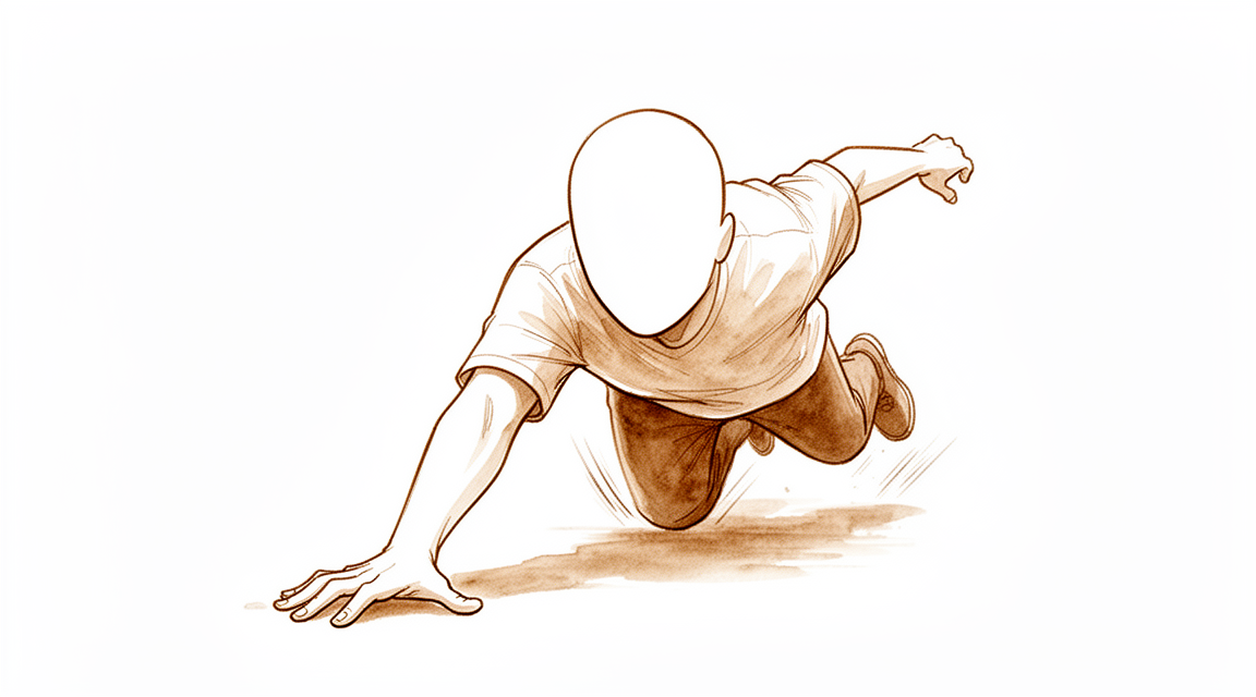
Bạn có thể cảm thấy đau và nhạy cảm ở gốc ngón cái, ngay dưới cổ tay. Khu vực này được gọi là hố snuffbox giải phẫu. Cơn đau thường bắt đầu sau khi ngã xuống bàn tay dang rộng. Bạn có thể nhận thấy sưng hoặc bầm tím xung quanh cổ tay. Các hoạt động đơn giản có thể trở nên khó khăn. Với tay ra sau lưng để cài áo ngực có thể gây đau. Nhét áo vào quần hoặc xoay tay nắm cửa có thể kích hoạt cơn đau nhói. Nâng vật, kể cả những vật nhẹ, có thể cảm thấy không ổn định hoặc đau.
Cơn đau thường trở nên tồi tệ hơn khi vận động. Sử dụng ngón cái và cổ tay cùng lúc gây căng thẳng cho xương bị tổn thương. Bạn có thể thấy khó cầm nắm đồ vật chắc chắn. Điều này có thể khiến việc giữ một tách cà phê hoặc điện thoại trở nên thách thức. Nghỉ ngơi tay thường giúp giảm bớt sự khó chịu. Tuy nhiên, cơn đau có thể bùng phát vào cuối ngày sau khi bạn đã hoạt động. Một số bệnh nhân báo cáo rằng sự khó chịu rõ rệt vào ban đêm, đặc biệt nếu bạn ngủ nghiêng về phía đó. Thức dậy với cổ tay cứng hoặc đau nhức là điều phổ biến.
Điều quan trọng là phải biết rằng X-quang tiêu chuẩn không phải lúc nào cũng cho thấy rõ gãy xương. Trên thực tế, chỉ khoảng 40% bệnh nhân được xác định có gãy xương thực sự dựa trên X-quang ban đầu và khám lâm sàng đơn thuần. Điều này có nghĩa là các triệu chứng của bạn có thể cảm thấy nghiêm trọng ngay cả khi lần quét đầu tiên trông bình thường. Nếu bác sĩ phẫu thuật nghi ngờ gãy xương nhưng X-quang không rõ ràng, họ có thể chỉ định chụp MRI. Chụp quét này có thể tìm thấy các tổn thương ẩn mà X-quang bỏ sót. Chẩn đoán sớm giúp ngăn ngừa biến chứng.
Nếu xương không lành đúng cách, nó có thể dẫn đến không liền xương. Điều này xảy ra khi xương không hàn gắn lại với nhau. Tỷ lệ không liền xương vẫn cao đối với gãy xương thuyền. Trong một số trường hợp, không liền xương hoặc liền xương muộn xảy ra ở hơn 6% bệnh nhân, ngay cả khi được điều trị ban đầu đúng cách. Hầu hết những bệnh nhân này sẽ cần phẫu thuật để sửa chữa xương. Cố định nội khoa sớm ngày càng được ưa chuộng để giảm thiểu rủi ro này. Bác sĩ phẫu thuật của bạn sẽ thảo luận về phương án tốt nhất cho bạn dựa trên các chi tiết cụ thể về chấn thương của bạn.
Những gì thực sự đang xảy ra
Xương thuyền (scaphoid) của bạn là một xương nhỏ, có hình dạng như một quả óc chó, nằm ở cổ tay. Nó đóng vai trò như một cầu nối quan trọng giữa các xương cẳng tay và xương bàn tay. Xương này có nguồn cung cấp máu phức tạp, nghĩa là nó không phải lúc nào cũng nhận đủ dưỡng chất để tự lành. Khi xương này bị gãy, quá trình lành xương có thể bị đình trệ. Tình trạng này được gọi là không liền xương (nonunion). Ngay cả với chẩn đoán và phẫu thuật hiện đại, tỷ lệ không liền xương vẫn còn cao.
Nếu xương không liền lại, chuyển động bình thường của cổ tay bạn sẽ thay đổi. Các xương ở cổ tay vốn di chuyển phối hợp nhịp nhàng như một điệu nhảy. Một xương thuyền bị gãy sẽ làm gián đoạn nhịp điệu này. Nó làm giảm bớt sự kết nối giữa hàng xương cổ tay trên và hàng xương cổ tay dưới. Điều này dẫn đến chuyển động bất thường và sự mài mòn trên các bề mặt khớp. Theo thời gian, sự mài mòn này có thể gây ra viêm xương khớp. Bạn có thể cảm thấy đau và cứng khớp khi các bề mặt khớp cọ xát vào nhau mà không có lớp bảo vệ trơn tru như bình thường.
Bác sĩ phẫu thuật của bạn có thể giúp khôi phục sự cân bằng này. Đối với một số trường hợp gãy xương, một chiếc vít đơn giản sẽ giữ các mảnh xương lại với nhau trong quá trình lành. Đối với các trường hợp phức tạp hơn, bác sĩ phẫu thuật có thể sử dụng ghép xương. Điều này liên quan đến việc lấy một mảnh xương nhỏ khỏe mạnh để lấp đầy các khoảng trống và thúc đẩy sự phát triển. Trong một số tình huống, bác sĩ phẫu thuật cũng có thể điều chỉnh hình dạng của xương cẳng tay. Điều này giúp phân tán trọng lượng ra xa xương thuyền bị tổn thương. Những bước này nhằm mục đích khôi phục chuyển động cổ tay bình thường và lực nắm tay.
Mục tiêu là ngăn chặn sự mài mòn bất thường trước khi nó gây ra tổn thương vĩnh viễn. Nếu được điều trị sớm, ngay cả các trường hợp gãy xương có di lệch cũng có thể lành tốt. Nếu xương liền lại, bạn nhiều khả năng sẽ có kết quả điều trị tốt, bất kể những thay đổi nhỏ về hình dạng. Trọng tâm là mang lại cho bạn một cổ tay không đau, hoạt động chức năng tốt, có thể thực hiện các công việc hàng ngày mà không bị cảm giác mài mòn hoặc mất ổn định do sự gián đoạn của cầu nối xương.
Những gì chúng tôi có thể làm về vấn đề này
Chăm sóc của bạn bắt đầu bằng việc theo dõi cẩn thận và nghỉ ngơi. Vì chụp X-quang tiêu chuẩn và hai lần khám lâm sàng chỉ xác định được gãy xương thực sự ở khoảng 40% bệnh nhân, bác sĩ phẫu thuật của bạn có thể sử dụng MRI sớm để có câu trả lời rõ ràng. Chụp cắt lớp này tìm chính xác các tổn thương ẩn và giúp loại trừ gãy xương khi kết quả ban đầu không rõ ràng. Trong khi chờ chẩn đoán hoặc trong quá trình điều trị bảo tồn, bạn nên tránh các hoạt động gây căng thẳng cho cổ tay. Vật lý trị liệu nhằm khôi phục tầm vận động và sức mạnh của bạn một khi xương đã lành. Đối với nhiều bệnh nhân, đặc biệt là những người có gãy xương không lệch, điều trị không phẫu thuật là hiệu quả. Tỷ lệ liền xương trong các trường hợp này đạt mức xấp xỉ hoặc thậm chí vượt qua tỷ lệ của phẫu thuật. Bạn có thể mong đợi phải đeo bột hoặc nẹp trong một khoảng thời gian do bác sĩ phẫu thuật quyết định. Không có một phác đồ tối ưu duy nhất về thời gian bất động cổ tay sau bất kỳ thủ thuật nào, vì vậy nhóm điều trị của bạn sẽ hướng dẫn bạn dựa trên tiến trình lành thương của bạn.
Quản lý đau là một phần quan trọng trong quá trình phục hồi của bạn. Bác sĩ phẫu thuật có thể khuyên dùng các loại thuốc giảm đau không kê đơn để giữ cho bạn cảm thấy thoải mái. Tuy nhiên, bạn phải thận trọng với các thuốc chống viêm không steroid (NSAIDs). Nếu bạn dùng các loại thuốc này trong tháng đầu tiên sau chấn thương, bạn đối mặt với nguy cơ tăng cao rằng xương sẽ không lành (liên xương giả). Sự thất bại này có thể dẫn đến các thủ thuật cứu chữa phức tạp hơn sau này. Đối với hầu hết các gãy xương cấp tính, chúng tôi không sử dụng tiêm cortisone, axit hyaluronic hoặc PRP như một phần của chăm sóc tiêu chuẩn. Thay vào đó, chúng tôi tập trung vào việc bảo vệ xương trong khi nó tự sửa chữa. Nếu bạn có một liên xương giả không lệch và không gập, ghép xương xâm lấn tối thiểu và cố định bằng vít nén có thể được xem xét. Phương pháp này an toàn và hiệu quả trong việc ổn định xương mà không cần phẫu thuật lớn.
Phẫu thuật thường được dành riêng cho các gãy xương lệch hoặc các trường hợp mà điều trị bảo tồn đã thất bại. Nếu gãy xương của bạn bị lệch, can thiệp phẫu thuật được khuyến nghị để căn chỉnh xương đúng vị trí. Đối với các liên xương giả gần đây không lành sau điều trị ban đầu, bác sĩ phẫu thuật của bạn có thể thực hiện cắt bỏ xương thuyền nhỏ hoặc sử dụng cố định bằng hai vít chống xoay với nội soi khớp. Các thủ thuật này nhằm mục đích ổn định xương và thúc đẩy quá trình lành thương. Trong khi cố định nội khoa sớm ngày càng được ưa chuộng cho một số gãy xương cấp tính, nó không phải lúc nào cũng cần thiết cho các chấn thương không lệch. Trên thực tế, không có lợi ích lâu dài thực sự nào từ phẫu thuật so với điều trị không phẫu thuật đối với các gãy xương cấp tính không lệch hoặc lệch tối thiểu. Bác sĩ phẫu thuật của bạn sẽ giúp bạn lựa chọn một phương pháp dựa trên các giá trị cá nhân và khả năng chịu đựng rủi ro của bạn. Hãy nhớ rằng, tỷ lệ liên xương giả vẫn cao ngay cả với các kỹ thuật cải tiến, vì vậy việc theo dõi chặt chẽ là rất quan trọng bất kể phương pháp điều trị nào được chọn.
Những điều cần biết
Hầu hết các gãy xương thuyền đều lành tốt, đặc biệt là ở trẻ em. Đối với người lớn, tiên lượng phụ thuộc vào việc bạn được điều trị nhanh chóng như thế nào và liệu các mảnh xương có bị dịch chuyển hay không. Nếu gãy xương không bị dịch chuyển hoặc chỉ dịch chuyển nhẹ, bác sĩ phẫu thuật có thể khuyến cáo bó bột hoặc phẫu thuật. Cả hai phương pháp đều dẫn đến chức năng lâu dài tương tự nhau. Tuy nhiên, phẫu thuật giúp bạn trở lại làm việc sớm hơn khoảng 7 tuần so với chỉ bó bột.
Nếu xương không lành, tình trạng này được gọi là không liền xương. Điều này xảy ra ở hơn 10% các trường hợp sau phẫu thuật cho các gãy xương kín. Tình trạng này cũng phổ biến hơn nếu bạn đến khám điều trị sau hơn 21 ngày kể từ khi bị chấn thương. Việc điều trị chậm trễ làm tăng nguy cơ bó bột thất bại. Không liền xương có thể dẫn đến viêm xương khớp do mòn dần tiến triển ở cổ tay. Mặc dù điều này nghe có vẻ nghiêm trọng, nhiều bệnh nhân vẫn báo cáo khả năng vận động và sức mạnh cổ tay tốt sau nhiều năm, ngay cả khi hình dạng xương bị thay đổi nhẹ.
Bác sĩ phẫu thuật sẽ theo dõi quá trình lành vết gãy của bạn một cách chặt chẽ. Nếu bạn bị không liền xương, có thể cần phẫu thuật thêm. Các thủ thuật lặp lại này ít thành công hơn so với lần đầu tiên. Trong một số trường hợp viêm xương khớp đã phát triển, việc loại bỏ một phần xương thuyền có thể giảm đau. Khoảng 94% bệnh nhân hài lòng với thủ thuật này, và nó ngăn ngừa cổ tay bị sụp đổ thêm.
Nhìn chung, hầu như tất cả các gãy xương thuyền liền thành công đều dẫn đến kết quả tốt. Chìa khóa là đảm bảo xương lành. Chẩn đoán sớm là rất quan trọng vì X-quang thông thường thường bỏ sót các chấn thương này. Nếu bạn thuộc nhóm có nguy cơ cao bị lành vết gãy chậm, chẳng hạn như những người mắc một số bệnh lý sức khỏe tâm thần hoặc sống trong các cộng đồng thiếu thốn, việc chú ý thêm đến các cuộc tái khám là rất quan trọng. Với sự chăm sóc thích hợp, bạn có thể mong đợi phục hồi chức năng và sức mạnh bàn tay bình thường trong vài tháng tới.
Khi nào cần gặp bác sĩ
Hãy gặp bác sĩ đa khoa nếu bạn có tình trạng đau dai dẳng, yếu hoặc mất ổn định ở cổ tay. Các triệu chứng ảnh hưởng đến giấc ngủ hoặc công việc cần được chú ý. Hãy yêu cầu đánh giá bởi bác sĩ chuyên khoa nếu cổ tay của bạn bị khóa hoặc đột ngột mất sức. Sự gia tăng đau đột ngột cũng là một dấu hiệu cảnh báo. Hãy lưu ý rằng việc đến khám muộn hơn 21 ngày hoặc lâu hơn sau khi bị chấn thương dự báo nguy cơ cao hơn về thất bại khi bó bột. Các trường hợp gãy xương bị chẩn đoán nhầm có thể dẫn đến các biến chứng nghiêm trọng. Chẩn đoán sớm giúp tránh những vấn đề này. Nếu kết quả chụp X-quang ban đầu không rõ ràng, chụp MRI sớm có thể xác định chính xác các tổn thương. Đừng bỏ qua các triệu chứng, vì các xương gãy không được điều trị có thể không lành lại đúng cách.
Evidence & references
Overview
- Pediatric scaphoid fractures have excellent outcomes [1].
- Some well-established and widely used principles of scaphoid fracture management are supported by an insufficient amount of evidence, with many decisions based on small case series [2].
- The clinical outcomes of malunited scaphoids after reconstruction for scaphoid fractures nonunion did not differ significantly from well-united scaphoids at a minimum 5-year follow-up [4].
- Internal fixation of scaphoid fractures is indicated in certain acute situations and in chronic nonunion cases [15].
- Appropriately performed acute percutaneous internal fixation is now a standard treatment option for a selected group of patients with acute scaphoid fracture [17].
- The definition of instability of scaphoid fractures and the indications for conservative treatment must be considered carefully [14].
- Nondisplaced scaphoid fractures can be effectively treated nonoperatively with union rates approaching or exceeding those of operative intervention, while operative intervention is recommended for displaced fractures [24].
- This study did not demonstrate a true long-term benefit of internal fixation, compared with nonoperative treatment, for acute nondisplaced or minimally displaced scaphoid fractures [5].
- For all indications, the scaphoid staple has a high union rate and a low complication rate [6].
- Patients with recent scaphoid fractures that failed treatment may also be treated with distal scaphoid resection [25].
- Despite improvements in diagnosis and surgical techniques, nonunion rates remain high and early internal fixation is increasingly favored even for nondisplaced fractures [28].
Anatomy & Pathophysiology
- The scaphoid is critical to the coordination of normal carpal kinematics [34].
- Scaphoid fracture has significant biomechanical consequences to the wrist [34].
- Scaphoid nonunions have a dramatic impact on carpal kinematics, partially uncoupling the proximal and distal carpal rows [35].
- Problem fractures and non-unions of the scaphoid are associated with major alterations in wrist kinematics [46].
- Problem fractures and non-unions of the scaphoid are associated with a higher incidence of premature carpal collapse and degenerative arthritis than previously appreciated [46].
- A foreshortened healed scaphoid will disrupt carpal kinematics [37].
- A foreshortened healed scaphoid negatively impacts results, including decreased wrist range of motion and diminished grip strength [37].
- Malunion or nonunion of an acute scaphoid fracture can lead to abnormal carpal kinematics and wrist arthrosis [61].
- Radiocarpal-based lunate morphology was not associated with scaphoid fracture [74].
- Anomalous carpal kinematics caused by lunotriquetral coalition may have predisposed both scaphoid bones to fracture, although causality cannot be proven [78].
Classification
- Pediatric scaphoid fractures have excellent outcomes [1].
- Even some well-established and widely used principles of scaphoid fracture management are supported by an insufficient amount of evidence, with many decisions based on small case series [2].
- The combination of conventional radiographs and two clinical examinations does not provide adequate diagnostic certainty for scaphoid fractures, as a true fracture was identified in only about 40% of patients [3].
- If there is a strong clinical suspicion of a scaphoid fracture which cannot be confirmed by conventional radiology, bone scintigraphy is a valuable diagnostic tool [8].
- The combination of conventional radiographs and clinical reassessment does not increase the accuracy of these diagnostic tests compared with the accuracy of conventional radiographs alone and is therefore also limited in diagnosing scaphoid fractures [9].
- There is no consensus regarding the imaging modality and measurements to use to define a scaphoid fracture as 'nondisplaced' [10].
- Due to low agreement between observers for the recognition of scaphoid fractures and poor diagnostic performance, 6-week radiographs are not adequate for evaluating suspected scaphoid fractures [12].
- Scaphoid fractures account for 2% of all fractures and are the most commonly injured carpal bone [13].
- The definition of instability of scaphoid fractures and the indications for conservative treatment must be considered carefully [14].
- Internal fixation of scaphoid fractures is indicated in certain acute situations and in chronic nonunion cases [15].
- Despite improvements in diagnosis and surgical techniques, nonunion rates remain high and early internal fixation is increasingly favored even for nondisplaced fractures [28].
- There is a need for a validated prognostic classification system for scaphoid nonunions that can allow comparisons between outcome studies [41].
- The authors hypothesise higher union rates in scaphoid fractures using more stable fixation systems [79].
- Scaphoid fracture and nonunion management continues to be an area of expanding evidence with opportunities to improve knowledge and familiarization with current evidence-based data [80].
Clinical Presentation
- Scaphoid fractures account for 2% of all fractures and are the most commonly injured carpal bone [13].
- Most scaphoid fractures are missed due to failure to consider the possibility of the injury and search for clinical signs [19].
- The combination of conventional radiographs and two clinical examinations does not provide adequate diagnostic certainty for scaphoid fractures, as a true fracture was identified in only about 40% of patients [3].
- The combination of conventional radiographs and clinical reassessment does not increase the accuracy of these diagnostic tests compared with the accuracy of conventional radiographs alone and is therefore also limited in diagnosing scaphoid fractures [9].
- 6-week radiographs are not adequate for evaluating suspected scaphoid fractures due to low agreement between observers for the recognition of scaphoid fractures and poor diagnostic performance [12].
- Ultrasonic assessment is not recommended for the early diagnosis of acute scaphoid fractures, with a sensitivity of only 50% and five missed scaphoid fractures in a small series [39].
- If there is a strong clinical suspicion of a scaphoid fracture which cannot be confirmed by conventional radiology, bone scintigraphy is a valuable diagnostic tool [8].
- Clinical examination along with early MRI scan should form the basis of diagnosing a suspected scaphoid fracture [20].
- The use of early MRI in patients with clinically suspected scaphoid fracture results in the accurate and reliable identification of a significant number of radiological occult injuries and early identification of patients without acute injuries [23].
- MRI-detected scaphoid fractures are not universally benign, with delayed or nonunion seen in over 6% despite appropriate initial immobilization, with most of these patients with nonunion requiring surgery to achieve union [22].
- The diagnosis of scaphoid and other fractures is reliable when using HRpQCT in patients with a clinically-suspected fracture [40].
- There is no consensus regarding the imaging modality and measurements to use to define a scaphoid fracture as 'nondisplaced' [10].
- Oblique scaphoid fractures are potentially unstable and may result in detrimental sequelae if overlooked in the acute stage [16].
Investigations
- Conventional radiographs combined with two clinical examinations provide inadequate diagnostic certainty for scaphoid fractures, identifying a true fracture in only about 40% of patients [3].
- The combination of conventional radiographs and clinical reassessment does not increase diagnostic accuracy compared to conventional radiographs alone [9].
- There is no consensus on the imaging modality or measurements used to define a scaphoid fracture as nondisplaced [10].
- Six-week radiographs are not adequate for evaluating suspected scaphoid fractures due to low inter-observer agreement and poor diagnostic performance [12].
- Most missed scaphoid fractures result from a failure to consider the injury possibility and search for clinical signs [19].
- Clinical examination combined with early MRI scan should form the basis for diagnosing suspected scaphoid fractures [20].
- MRI-detected scaphoid fractures are not universally benign, with delayed or nonunion occurring in over 6% of cases despite appropriate initial immobilization [22].
- Most patients with nonunion of MRI-detected scaphoid fractures require surgery to achieve union [22].
- Early MRI in patients with clinically suspected scaphoid fractures accurately and reliably identifies a significant number of radiological occult injuries [23].
- Early MRI in patients with clinically suspected scaphoid fractures allows for the early identification of patients without acute injuries [23].
- Early MRI provides an immediate diagnosis for suspected scaphoid fractures when initial radiographs are inconclusive [56].
- Early MRI for suspected scaphoid fractures when initial radiographs are inconclusive is cost-effective and minimizes complications [56].
- CT is a good way to screen for occult fractures but may not be superior to MRI or bone scanning in detecting scaphoid fractures without causing overtreatment [58].
- Multidetector computed tomography (MDCT) has a sensitivity of 86% and specificity of 100% for detecting occult scaphoid fractures in patients with negative radiographic examinations [59].
- MRI is the optimal second test for assessing a possible scaphoid fracture after a negative radiograph [60].
- CT is preferred over MRI when the fracture is visible for further assessment and surgical planning [60].
- There is variation in definitions of scaphoid fractures on MRI scans, highlighting a need for consensus to assess reliability and diagnostic performance [63].
- Bone scintigraphy is inappropriate for evaluating specificity and sensitivity against clinical examination [64].
- MRI is the recommended examination of choice for diagnosing occult scaphoid fractures over bone scintigraphy [64].
- MRI is considered the best diagnostic radiological test for triage of suspected scaphoid fractures according to existing literature [67].
- Bone scanning, CT, and ultrasound may be useful for suspected scaphoid fractures when MRI is not readily available [67].
- Routine MRI of suspected scaphoid fractures carries a notable risk of overdiagnosis and potential overtreatment [72].
- Nearly 70% of MRI findings in suspected scaphoid fractures are categorized as distracting and potentially misleading [72].
- Stopping the pursuit of occult fractures may prevent unnecessary treatment due to the risk of overdiagnosis with routine MRI [72].
- Better standardization of MRI definitions for scaphoid fractures is required to address diagnostic uncertainty [76].
- A definitive definition may not exist to solve the potentially unsolvable issue of diagnostic uncertainty in scaphoid fractures [76].
- Patients should participate in decisions regarding diagnostic and treatment strategies for scaphoid fractures due to diagnostic uncertainty [76].
- MRI is not 100% specific for diagnosing occult scaphoid fractures, with a specificity of 96% in healthy volunteers [77].
Treatment
Nonoperative Management
- Pediatric scaphoid fractures have excellent outcomes [1].
- Internal fixation of scaphoid fractures is indicated in certain acute situations and in chronic nonunion cases [15].
- Early treatment of acute scaphoid fractures is important, with union rates significantly greater when treatment is instituted prior to 4 weeks from injury [26].
- Nondisplaced scaphoid fractures can be effectively treated nonoperatively with union rates approaching or exceeding those of operative intervention, while operative intervention is recommended for displaced fractures [24].
- Surgical treatment for non-displaced and minimally displaced acute scaphoid fractures may be slightly favourable compared to conservative treatment for standardised functional outcome on the short term (within 2 years), with a significantly faster return to work (SMD of 7 weeks) [21].
- This study did not demonstrate a true long-term benefit of internal fixation, compared with nonoperative treatment, for acute nondisplaced or minimally displaced scaphoid fractures [5].
- We found no difference in functional outcome at 12 months for fractures of the waist of the scaphoid with ≤ 2 mm displacement treated operatively or nonoperatively [18].
- Non- and minimally displaced scaphoid waist fractures are best treated conservatively [36].
- Non-operative treatment of non-displaced scaphoid fractures may be preferred [53].
- A restricted period of cast immobilisation is usually adequate for the treatment of non-displaced scaphoid fractures [53].
- Nondisplaced fractures of the scaphoid heal with cast immobilization in most cases, but operative treatment is being offered with greater frequency to active patients to reduce the period of cast immobilization [57].
- The definition of instability of scaphoid fractures and the indications for conservative treatment must be considered carefully [14].
- Currently, there is insufficient evidence to support the most effective treatment for acute scaphoid fractures [51].
- Even some well-established and widely used principles of scaphoid fracture management are supported by an insufficient amount of evidence, with many decisions based on small case series [2].
- Among patients with nonoperatively managed scaphoid fractures, those prescribed NSAIDs within 1 month of diagnosis demonstrated an increased risk of nonunion and subsequent salvage procedures [54].
Operative Management
- Appropriately performed acute percutaneous internal fixation is now a standard treatment option for a selected group of patients with acute scaphoid fracture [17].
- The frequency of non-union after surgical management for closed scaphoid fractures exceeds 10% and remained consistent during the study period [11].
- For all indications, the scaphoid staple has a high union rate and a low complication rate [6].
- The use of 2 headless compression screws for the treatment of scaphoid nonunions is safe and effective [44].
- Patients with recent scaphoid fractures that failed treatment may also be treated with distal scaphoid resection [25].
- The optimal protocol for postoperative immobilization following operative treatment of scaphoid fractures remains controversial [62].
Special Populations and Considerations
- The authors prefer to treat nondisplaced acute scaphoid fractures in the athlete on an individualized basis [55].
- The clinical outcomes of malunited scaphoids after reconstruction for scaphoid fractures nonunion did not differ significantly from well-united scaphoids at a minimum 5-year follow-up [4].
- This case is interesting as the child is one of the youngest patients described in the literature with a scaphoid fracture, and the fracture went on to non-union despite immediate medical attention and rigorous treatment [49].
- The authors argue that comfort with uncertainty is key in suspected scaphoid fracture scenarios, as there is no best strategy; instead, clinicians should help patients choose a diagnostic and therapeutic course based on their individual values and risk tolerance [66].
Complications
- Pediatric scaphoid fractures have excellent outcomes [1].
- Many decisions regarding scaphoid fracture management are based on small case series due to insufficient evidence for well-established principles [2].
- The combination of conventional radiographs and two clinical examinations does not provide adequate diagnostic certainty for scaphoid fractures, as a true fracture was identified in only about 40% of patients [3].
- Clinical outcomes of malunited scaphoids after reconstruction for scaphoid fracture nonunion did not differ significantly from well-united scaphoids at a minimum 5-year follow-up [4].
- There is no true long-term benefit of internal fixation compared with nonoperative treatment for acute nondisplaced or minimally displaced scaphoid fractures [5].
- The scaphoid staple has a high union rate and a low complication rate for all indications [6].
- Subacute scaphoid fractures presenting within 6 months from injury can be expected to successfully heal with casting alone, even if the initial diagnosis is delayed [7].
- The frequency of nonunion after surgical management for closed scaphoid fractures exceeds 10% and remained consistent during the study period [11].
- Oblique scaphoid fractures are potentially unstable and may result in detrimental sequelae if overlooked in the acute stage [16].
- Appropriately performed acute percutaneous internal fixation is a standard treatment option for a selected group of patients with acute scaphoid fracture [17].
- There is no difference in functional outcome at 12 months for fractures of the waist of the scaphoid with ≤ 2 mm displacement treated operatively or nonoperatively [18].
- Persistent nonunion is common after surgery for scaphoid nonunion, and surgeries for persistent nonunion are even less successful [27].
- Delayed presentation of scaphoid fractures 21 days or more after injury predicts a greater risk of casting failure, although the union rate remains high with comparable time in cast [29].
- Nearly half of all patients with malunited acute scaphoid fractures demonstrated radiographic findings of early arthritis on CT imaging but had overall good clinical results on midterm follow-up [30].
- Increased likelihood for nonunion was found when the fracture was treated greater than 31 days from injury and when fracture volume was less than 38% of the entire scaphoid [32].
Recovery
- Pediatric scaphoid fractures have excellent outcomes [1].
- Clinical outcomes of malunited scaphoids after reconstruction for nonunion did not differ significantly from well-united scaphoids at a minimum 5-year follow-up [4].
- There is no true long-term benefit of internal fixation compared with nonoperative treatment for acute nondisplaced or minimally displaced scaphoid fractures [5].
- The scaphoid staple has a high union rate and a low complication rate for all indications [6].
- Subacute scaphoid fractures presenting within 6 months from injury can be expected to successfully heal with casting alone, even if the initial diagnosis is delayed [7].
- The frequency of nonunion after surgical management for closed scaphoid fractures exceeds 10% [11].
- Oblique scaphoid fractures are potentially unstable and may result in detrimental sequelae if overlooked in the acute stage [16].
- There is no difference in functional outcome at 12 months for fractures of the waist of the scaphoid with ≤ 2 mm displacement treated operatively or nonoperatively [18].
- Surgical treatment for non-displaced and minimally displaced acute scaphoid fractures may be slightly favourable compared to conservative treatment for standardised functional outcome on the short term (within 2 years) [21].
- Surgical treatment for non-displaced and minimally displaced acute scaphoid fractures results in a significantly faster return to work (SMD of 7 weeks) [21].
- Union rates are significantly greater when treatment is instituted prior to 4 weeks from injury [26].
- Persistent nonunion is common after surgery for scaphoid non-union, and surgeries for persistent nonunion are even less successful [27].
- Delayed presentation of scaphoid fractures 21 days or more after injury predicts a greater risk of casting failure [29].
- The union rate remains high with comparable time in cast despite delayed presentation of scaphoid fractures 21 days or more after injury [29].
- Nearly half of all patients with malunited acute scaphoid fractures demonstrated radiographic findings of early arthritis on CT imaging [30].
- Patients with malunited acute scaphoid fractures demonstrated overall good clinical results on midterm follow-up despite radiographic findings of early arthritis [30].
- From an 8- to 11-year perspective, patients with distal scaphoid fractures report normal self-assessed hand function [31].
- From an 8- to 11-year perspective, patients with distal scaphoid fractures report good wrist motion and strength [31].
- Increased likelihood for nonunion was found when the fracture was treated greater than 31 days from injury [32].
- Increased likelihood for nonunion was found when fracture volume was less than 38% of the entire scaphoid [32].
- Patients treated nonoperatively or with salvage procedures had similar long-term outcomes as those treated with a corrective scaphoid osteotomy [65].
- Patients with comorbid psychiatric conditions experienced increased rates of delayed scaphoid union [83].
- Dynamic imaging with time-intensity curve analysis does not provide additional predictive value over standard delayed enhanced imaging for acute scaphoid fracture viability assessment using contrast-enhanced MRI [84].
- Scaphoid nonunions demonstrate findings indicative of progression to union on CT at a mean of 6 weeks [86].
- Scaphoid nonunions demonstrate findings indicative of progression to union on CT as early as 3 weeks postoperatively [86].
Key Evidence
- [L1] Pediatric scaphoid fractures have excellent outcomes. [1] (10.1177/1558944717735948)
- [L5] Even some well-established and widely used principles of scaphoid fracture management are supported by an insufficient amount of evidence, with many decisions based on small case series. [2] (10.1177/1753193420977241)
- [L5] The combination of conventional radiographs and two clinical examinations does not provide adequate diagnostic certainty for scaphoid fractures, as a true fracture was identified in only about 40% of patients. [3] (10.1097/corr.0000000000002413)
- [L4] The clinical outcomes of malunited scaphoids after reconstruction for scaphoid fractures nonunion did not differ significantly from well-united scaphoids at a minimum 5-year follow-up. [4] (10.1016/j.otsr.2014.09.026)
- [L1] This study did not demonstrate a true long-term benefit of internal fixation, compared with nonoperative treatment, for acute nondisplaced or minimally displaced scaphoid fractures. [5] (10.2106/jbjs.g.00673)
- [L4] For all indications, the scaphoid staple has a high union rate and a low complication rate. [6] (10.1177/1558944716658747)
- [Paper] If there is a strong clinical suspicion of a scaphoid fracture which cannot be confirmed by conventional radiology, bone scintigraphy is a valuable diagnostic tool. [8] (10.1016/j.injury.2005.02.009)
- [L2] The combination of conventional radiographs and clinical reassessment does not increase the accuracy of these diagnostic tests compared with the accuracy of conventional radiographs alone and is therefore also limited in diagnosing scaphoid fractures. [9] (10.1097/corr.0000000000002310)
- [L5] There is no consensus regarding the imaging modality and measurements to use to define a scaphoid fracture as 'nondisplaced.' [10] (10.1016/j.jhsa.2012.10.025)
- [L3] The frequency of non-union after surgical management for closed scaphoid fractures exceeds 10% and remained consistent during the study period. [11] (10.1016/j.jhsa.2015.06.019)
- [L2] Due to low agreement between observers for the recognition of scaphoid fractures and poor diagnostic performance, 6-week radiographs are not adequate for evaluating suspected scaphoid fractures. [12] (10.1007/s00402-016-2438-4)
- [L5] Scaphoid fractures account for 2% of all fractures and are the most commonly injured carpal bone. [13] (10.1016/j.hcl.2017.04.003)
- [L5] The definition of instability of scaphoid fractures and the indications for conservative treatment must be considered carefully. [14] (10.1142/s0218810415400018)
- [L5] Internal fixation of scaphoid fractures is indicated in certain acute situations and in chronic nonunion cases. [15] (10.1016/s0749-0712(21)00118-9)
- [L4] Oblique scaphoid fractures are potentially unstable and may result in detrimental sequelae if overlooked in the acute stage. [16] (10.1016/j.injury.2009.07.078)
- [L4] Appropriately performed acute percutaneous internal fixation is now a standard treatment option for a selected group of patients with acute scaphoid fracture. [17] (10.5435/00124635-200708000-00004)
- [L1] We found no difference in functional outcome at 12 months for fractures of the waist of the scaphoid with ≤ 2 mm displacement treated operatively or nonoperatively. [18] (10.1302/0301-620x.104b8.bjj-2022-0085.r2)
- [L4] Most scaphoid fractures were missed due to failure to consider the possibility of the injury and search for clinical signs. [19] (10.1016/j.injury.2019.05.009)
- [L3] Clinical examination along with early MRI scan should form the basis of diagnosing a suspected scaphoid fracture. [20] (10.1177/1753193420979465)
- [L1] Surgical treatment for non-displaced and minimally displaced acute scaphoid fractures may be slightly favourable compared to conservative treatment for standardised functional outcome on the short term (within 2 years), with a significantly faster return to work (SMD of 7 weeks). [21] (10.1136/jisakos-2015-000024)
- [L3] MRI-detected scaphoid fractures are not universally benign, with delayed or nonunion seen in over 6% despite appropriate initial immobilization, with most of these patients with nonunion requiring surgery to achieve union. [22] (10.1302/0301-620x.106b4.bjj-2023-1171.r1)
- [L2] The use of early MRI in patients with clinically suspected scaphoid fracture results in the accurate and reliable identification of a significant number of radiological occult injuries and early identification of patients without acute injuries. [23] (10.1177/1753193412471008)
- [L1] Nondisplaced scaphoid fractures can be effectively treated nonoperatively with union rates approaching or exceeding those of operative intervention, while operative intervention is recommended for displaced fractures. [24] (10.2106/jbjs.rvw.15.00073)
- [L4] Patients with recent scaphoid fractures that failed treatment may also be treated with distal scaphoid resection. [25] (10.1016/j.jhsg.2024.03.013)
- [L5] Early treatment of acute scaphoid fractures is important, with union rates significantly greater when treatment is instituted prior to 4 weeks from injury. [26] (10.1016/s0749-0712(21)00580-1)
- [L4] Persistent nonunion is common after surgery for scaphoid non-union, and surgeries for persistent nonunion are even less successful. [27] (10.1016/j.jhsa.2015.06.022)
- [L5] This article reviews current concepts regarding the treatment of scaphoid fractures and nonunions, highlighting that despite improvements in diagnosis and surgical techniques, nonunion rates remain high and early internal fixation is increasingly favored even for nondisplaced fractures. [28] (10.1016/j.jhsa.2008.04.026)
- [L4] Delayed presentation of scaphoid fractures 21 days or more after injury predicts a greater risk of casting failure; however, the union rate remains high with comparable time in cast. [29] (10.1016/j.jhsa.2023.10.020)
- [L4] Nearly half of all patients with malunited acute scaphoid fractures demonstrated radiographic findings of early arthritis on CT imaging but overall good clinical results on midterm follow-up. [30] (10.1016/j.jhsa.2020.04.002)
- [L2] From an 8- to 11-year perspective, patients with distal scaphoid fractures report normal self-assessed hand function as well as good wrist motion and strength. [31] (10.1016/j.jhsa.2017.06.016)
- [L5] The scaphoid is critical to the coordination of normal carpal kinematics, and its fracture has significant biomechanical consequences to the wrist. [34] (10.1016/s0749-0712(21)01439-6)
- [L4] Scaphoid nonunions have a dramatic impact on carpal kinematics, partially uncoupling the proximal and distal carpal rows. [35] (10.1016/j.jhsa.2008.03.008)
- [L2] Non- and minimally displaced scaphoid waist fractures are best treated conservatively. [36] (10.1016/j.jhsa.2015.03.007)
- [L5] All scaphoid fractures that heal do not yield acceptable results, as a foreshortened healed scaphoid will disrupt carpal kinematics and negatively impact results, including decreased wrist range of motion and diminished grip strength. [37] (10.1016/s0749-0712(21)01437-2)
- [L4] With a sensitivity of only 50% and five missed scaphoid fractures in this small series, we can not recommend ultrasonic assessment for the early diagnosis of acute scaphoid fractures. [39] (10.1054/jhsb.2000.0432)
- [L4] The diagnosis of scaphoid and other fractures is reliable when using HRpQCT in patients with a clinically-suspected fracture. [40] (10.1302/0301-620x.102b4.bjj-2019-0632.r3)
- [L4] There is a need for a validated prognostic classification system for scaphoid nonunions that can allow comparisons between outcome studies. [41] (10.1177/1753193417739510)
- [L4] The use of 2 headless compression screws for the treatment of scaphoid nonunions is safe and effective. [44] (10.1016/j.jhsa.2014.02.030)
- [L5] Problem fractures and non-unions of the scaphoid are associated with major alterations in wrist kinematics and a higher incidence of premature carpal collapse and degenerative arthritis than previously appreciated. [46] (10.2106/00004623-199274030-00014)
- [L4] This case is interesting as the child is one of the youngest patients described in the literature with a scaphoid fracture, and the fracture went on to non-union despite immediate medical attention and rigorous treatment. [49] (10.2106/00004623-198365080-00026)
- [L1] Currently, there is insufficient evidence to support the most effective treatment for acute scaphoid fractures. [51] (10.1007/s11552-010-9276-6)
- [L4] A restricted period of cast immobilisation is usually adequate for the treatment of non-displaced scaphoid fractures. [53] (10.1016/j.injury.2008.10.028)
- [L2] Among patients with nonoperatively managed scaphoid fractures, those prescribed NSAIDs within 1 month of diagnosis demonstrated an increased risk of nonunion and subsequent salvage procedures. [54] (10.1016/j.jhsg.2026.100958)
- [L4] The authors prefer to treat nondisplaced acute scaphoid fractures in the athlete on an individualized basis. [55] (10.1016/s0749-0712(21)00181-5)
- [L5] Early magnetic resonance imaging (MRI) provides an immediate diagnosis for suspected scaphoid fractures when initial radiographs are inconclusive, which is cost-effective and minimizes complications. [56] (10.1016/j.jhsa.2013.03.055)
- [L5] Nondisplaced fractures of the scaphoid heal with cast immobilization in most cases, but operative treatment is being offered with greater frequency to active patients to reduce the period of cast immobilization. [57] (10.5435/00124635-200007000-00003)
- [Commentary] CT is a good way to screen occult fractures but may not be any better than MRI or bone scanning in detecting scaphoid fractures without some over treatment. [58] (10.1177/1753193412446273)
- [L2] Although MRI remains the best diagnostic tool after radiography for detecting occult scaphoid fractures, MDCT sensitivity was 86% and specificity was 100% in this study. [59] (10.1007/s11604-010-0520-3)
- [Paper] MRI is the optimal second test for assessing a possible scaphoid fracture after a negative radiograph, while CT is preferred when the fracture is visible for further assessment and surgical planning. [60] (10.1016/j.hcl.2019.03.001)
- [L5] Early diagnosis and vigilant care of an acute scaphoid fracture are warranted to prevent malunion or nonunion, which can lead to abnormal carpal kinematics and wrist arthrosis. [61] (10.2106/00004623-200612000-00026)
- [L4] The optimal protocol for postoperative immobilization following operative treatment of scaphoid fractures remains controversial. [62] (10.1177/15589447221093675)
- [L3] This review highlights the need for a consensus definition of scaphoid fractures on MRI scans to assess the reliability and diagnostic performance of MRI scans for diagnosing true scaphoid fractures, as well as their potential harms and benefits. [63] (10.1177/17531934251367541)
- [L5] The authors argue that bone scintigraphy is inappropriate for evaluating specificity and sensitivity against clinical examination, and that MRI is the recommended examination of choice for diagnosing occult scaphoid fractures. [64] (10.1016/j.injury.2007.12.013)
- [L4] Patients treated nonoperatively or with salvage procedures had similar long-term outcomes as those treated with a corrective scaphoid osteotomy. [65] (10.1177/1558944716643295)
- [L5] The authors argue that comfort with uncertainty is key in suspected scaphoid fracture scenarios, as there is no best strategy; instead, clinicians should help patients choose a diagnostic and therapeutic course based on their individual values and risk tolerance. [66] (10.1097/corr.0000000000003141)
- [L5] According to the existing literature, MRI is the best diagnostic radiological test for triage of suspected scaphoid fractures, but bone scanning, CT, and ultrasound may also be useful, particularly when MRI is not readily available. [67] (10.1016/j.jhsa.2008.04.016)
- [L5] Routine MRI of suspected scaphoid fractures carries a notable risk of overdiagnosis and potential overtreatment, with nearly 70% of MRI findings categorized as distracting and potentially misleading, suggesting that stopping the pursuit of occult fractures may prevent unnecessary treatment. [72] (10.1097/corr.0000000000002914)
- [L3] By contrast, radiocarpal-based lunate morphology was not associated with scaphoid fracture. [74] (10.1016/j.jhsa.2025.10.018)
- [L5] The authors argue that better standardization of MRI definitions for scaphoid fractures is required, but acknowledge that a definition may not exist to solve the potentially unsolvable issue of diagnostic uncertainty, suggesting patients should participate in decisions regarding diagnostic and treatment strategies. [76] (10.1177/17531934251394819)
- [Paper] MRI is not 100% specific for diagnosing an occult scaphoid fracture, with a specificity of 96% in healthy volunteers. [77] (10.1016/s0363-5023(10)60085-8)
- [L4] The patient may represent two isolated coexisting conditions, or the anomalous carpal kinematics caused by the lunotriquetral coalition may have predisposed both scaphoid bones to fracture, although causality cannot be proven. [78] (10.1016/j.jhsa.2015.07.003)
- [Paper] The authors hypothesise higher union rates in scaphoid fractures using more stable fixation systems. [79] (10.1007/s00402-016-2556-z)
- [L5] Scaphoid fracture and nonunion management continues to be an area of expanding evidence with opportunities to improve knowledge and familiarization with current evidence-based data. [80] (10.1016/j.jhsg.2024.06.013)
- [L3] Patients with comorbid psychiatric conditions experienced increased rates of delayed scaphoid union. [83] (10.1177/15589447221142894)
- [L4] Our data are consistent with previously reported data supporting contrast-enhanced MRI for assessment of viability, and showing that dynamic imaging with time-intensity curve analysis does not provide additional predictive value over standard delayed enhanced imaging for acute scaphoid fracture. [84] (10.1007/s00256-014-1981-8)
- [L4] Scaphoid nonunions demonstrate findings indicative of progression to union on CT at a mean of 6 weeks and as early as 3 weeks postoperatively. [86] (10.1016/j.jhsa.2016.07.051)
References
[1] Management Modalities and Outcomes Following Acute Scaphoid Fractures in Children: A Quantitative Review and Meta-Analysis. HAND. 2017. DOI: 10.1177/1558944717735948 [2] Questions regarding the evidence guiding treatment of displaced scaphoid fractures. Journal of Hand Surgery (European Volume). 2020. DOI: 10.1177/1753193420977241 [3] CORR Insights®: What Is the Diagnostic Performance of Conventional Radiographs and Clinical Reassessment Compared With HR-pQCT Scaphoid Fracture Diagnosis?. Clinical Orthopaedics & Related Research. 2022. DOI: 10.1097/corr.0000000000002413 [4] Clinical outcome of scaphoid malunion as a result of scaphoid fracture nonunion surgical treatment: A 5-year minimum follow-up study. Orthopaedics & Traumatology: Surgery & Research. 2015. DOI: 10.1016/j.otsr.2014.09.026 [5] Nonoperative Compared with Operative Treatment of Acute Scaphoid Fractures. The Journal of Bone & Joint Surgery. 2008. DOI: 10.2106/jbjs.g.00673 [6] The Scaphoid Staple: A Systematic Review. HAND. 2016. DOI: 10.1177/1558944716658747 [7] 10.1055-s-0035-1564983. n.d.. [8] Outcome of routine bone scintigraphy in suspected scaphoid fractures. Injury. 2005. DOI: 10.1016/j.injury.2005.02.009 [9] What Is the Diagnostic Performance of Conventional Radiographs and Clinical Reassessment Compared With HR-pQCT Scaphoid Fracture Diagnosis?. Clinical Orthopaedics & Related Research. 2022. DOI: 10.1097/corr.0000000000002310 [10] Diagnosis of Scaphoid Fracture Displacement. The Journal of Hand Surgery. 2013. DOI: 10.1016/j.jhsa.2012.10.025 [11] An Epidemiologic Perspective on Scaphoid Fracture Treatment and Frequency of Nonunion. The Journal of Hand Surgery. 2015. DOI: 10.1016/j.jhsa.2015.06.019 [12] 6-week radiographs unsuitable for diagnosis of suspected scaphoid fractures. Archives of Orthopaedic and Trauma Surgery. 2016. DOI: 10.1007/s00402-016-2438-4 [13] Treatment of Scaphoid Fractures. Hand Clinics. 2017. DOI: 10.1016/j.hcl.2017.04.003 [14] Scaphoid Fracture - Overview and Conservative Treatment. Hand Surgery. 2015. DOI: 10.1142/s0218810415400018 [15] INTERNAL FIXATION OF SCAPHOID FRACTURES. Hand Clinics. 1997. DOI: 10.1016/s0749-0712(21)00118-9 [16] Management of late-diagnosed scaphoid fractures. Injury. 2010. DOI: 10.1016/j.injury.2009.07.078 [17] Percutaneous Fixation of Scaphoid Fractures. Journal of the American Academy of Orthopaedic Surgeons. 2007. DOI: 10.5435/00124635-200708000-00004 [18] One-year outcome of surgery compared with immobilization in a cast for adults with an undisplaced or minimally displaced scaphoid fracture. The Bone & Joint Journal. 2022. DOI: 10.1302/0301-620x.104b8.bjj-2022-0085.r2 [19] Why scaphoid fractures are missed. A review of 52 medical negligence cases. Injury. 2019. DOI: 10.1016/j.injury.2019.05.009 [20] Reliability of clinical tests for prediction of occult scaphoid fractures and cost benefit analysis of a dedicated scaphoid pathway. Journal of Hand Surgery (European Volume). 2020. DOI: 10.1177/1753193420979465 [21] Surgical treatment of non- and minimally-displaced acute scaphoid fractures favours over-conservative treatment but only in the short term: an updated meta-analysis. Journal of ISAKOS. 2016. DOI: 10.1136/jisakos-2015-000024 [22] The rate of nonunion in the MRI-detected occult scaphoid fracture. The Bone & Joint Journal. 2024. DOI: 10.1302/0301-620x.106b4.bjj-2023-1171.r1 [23] Early magnetic resonance imaging in patients with a clinically suspected scaphoid fracture may identify occult wrist injuries. Journal of Hand Surgery (European Volume). 2012. DOI: 10.1177/1753193412471008 [24] Acute Scaphoid Fractures. JBJS Reviews. 2016. DOI: 10.2106/jbjs.rvw.15.00073 [25] Distal Scaphoid Excision for Chronic and Nonchronic Scaphoid Fracture Nonunion. Journal of Hand Surgery Global Online. 2024. DOI: 10.1016/j.jhsg.2024.03.013 [26] MANAGEMENT OF ACUTE SCAPHOID FRACTURES. Hand Clinics. 2000. DOI: 10.1016/s0749-0712(21)00580-1 [27] Importance of Computed Tomography in Determining Displacement of Scaphoid Fractures. The Journal of Hand Surgery. 2015. DOI: 10.1016/j.jhsa.2015.06.022 [28] Treatment of Scaphoid Fractures and Nonunions. The Journal of Hand Surgery. 2008. DOI: 10.1016/j.jhsa.2008.04.026 [29] Clinically Significant Treatment Delay in Pediatric Scaphoid Fractures. The Journal of Hand Surgery. 2024. DOI: 10.1016/j.jhsa.2023.10.020 [30] Scaphoid Malunion Clinical and Radiographic Outcomes at a Minimum of 4 Years Follow-Up. The Journal of Hand Surgery. 2020. DOI: 10.1016/j.jhsa.2020.04.002 [31] Long-Term Outcomes After Distal Scaphoid Fractures: A 10-Year Follow-Up. The Journal of Hand Surgery. 2017. DOI: 10.1016/j.jhsa.2017.06.016 [32] 10.1055-s-0039-3402769. n.d.. [34] EFFECTS OF SCAPHOID FRACTURES ON THE BIOMECHANICS OF THE WRIST. Hand Clinics. 2001. DOI: 10.1016/s0749-0712(21)01439-6 [35] Interfragmentary Motion in Patients With Scaphoid Nonunion. The Journal of Hand Surgery. 2008. DOI: 10.1016/j.jhsa.2008.03.008 [36] Conservative Treatment Versus Arthroscopic-Assisted Screw Fixation of Scaphoid Waist Fractures—A Randomized Trial With Minimum 4-Year Follow-Up. The Journal of Hand Surgery. 2015. DOI: 10.1016/j.jhsa.2015.03.007 [37] INCIDENCE, MECHANISM, AND NATURAL HISTORY OF SCAPHOID FRACTURES. Hand Clinics. 2001. DOI: 10.1016/s0749-0712(21)01437-2 [39] Ultrasound for Diagnosis of Scaphoid Fractures. Journal of Hand Surgery. 2000. DOI: 10.1054/jhsb.2000.0432 [40] The interobserver reliability of the diagnosis and classification of scaphoid fractures using high-resolution peripheral quantitative CT. The Bone & Joint Journal. 2020. DOI: 10.1302/0301-620x.102b4.bjj-2019-0632.r3 [41] Scaphoid nonunion: what is the role of the Zaidemberg 1,2 intercompartmental supraretinacular arterial flap?. Journal of Hand Surgery (European Volume). 2017. DOI: 10.1177/1753193417739510 [44] Scaphoid Nonunions Treated With 2 Headless Compression Screws and Bone Grafting. The Journal of Hand Surgery. 2014. DOI: 10.1016/j.jhsa.2014.02.030 [46] Compression-staple fixation for fractures, non-unions, and delayed unions of the carpal scaphoid.. The Journal of Bone & Joint Surgery. 1992. DOI: 10.2106/00004623-199274030-00014 [49] Carpal scaphoid fracture and non-union in an eight-year-old child. Report of a case.. The Journal of Bone & Joint Surgery. 1983. DOI: 10.2106/00004623-198365080-00026 [51] Treatment of Acute Scaphoid Fractures: A Systematic Review and Meta-Analysis. HAND. 2010. DOI: 10.1007/s11552-010-9276-6 [53] Non-operative treatment of non-displaced scaphoid fractures may be preferred. Injury. 2009. DOI: 10.1016/j.injury.2008.10.028 [54] Early Nonsteroidal Anti-Inflammatory Drug Prescriptions and Nonunion After Scaphoid Fractures: A TriNetX Matched Cohort Study. Journal of Hand Surgery Global Online. 2026. DOI: 10.1016/j.jhsg.2026.100958 [55] PERCUTANEOUS AND ARTHROSCOPIC SCREW FIXATION OF SCAPHOID FRACTURES IN THE ATHLETE. Hand Clinics. 1999. DOI: 10.1016/s0749-0712(21)00181-5 [56] The Role of Magnetic Resonance Imaging in Scaphoid Fractures. The Journal of Hand Surgery. 2013. DOI: 10.1016/j.jhsa.2013.03.055 [57] Acute Fractures of the Scaphoid. Journal of the American Academy of Orthopaedic Surgeons. 2000. DOI: 10.5435/00124635-200007000-00003 [58] Commentary on ‘Early CT for suspected occult scaphoid fractures’ by Stevenson et al. J Hand Surg Eur. 2012, 37: 447-51. Journal of Hand Surgery (European Volume). 2012. DOI: 10.1177/1753193412446273 [59] Diagnostic accuracy of multidetector computed tomography for patients with suspected scaphoid fractures and negative radiographic examinations. Japanese Journal of Radiology. 2011. DOI: 10.1007/s11604-010-0520-3 [60] Imaging for Acute and Chronic Scaphoid Fractures. Hand Clinics. 2019. DOI: 10.1016/j.hcl.2019.03.001 [61] Acute Fractures of the Scaphoid. The Journal of Bone & Joint Surgery. 2006. DOI: 10.2106/00004623-200612000-00026 [62] Postoperative Immobilization of Scaphoid Fractures: A Comprehensive Review of the Literature. HAND. 2022. DOI: 10.1177/15589447221093675 [63] Variation in definitions of scaphoid fracture on MRI scans for suspected fracture: a systematic review. Journal of Hand Surgery (European Volume). 2025. DOI: 10.1177/17531934251367541 [64] A prospective comparison for suspected scaphoid fractures: Bone scintigraphy versus clinical outcome. Injury. 2008. DOI: 10.1016/j.injury.2007.12.013 [65] Long-Term Outcomes of Scaphoid Malunion. HAND. 2016. DOI: 10.1177/1558944716643295 [66] Reply to the Letter to the Editor: Routine MRI Among Patients With a Suspected Scaphoid Fracture Risks Overdiagnosis. Clinical Orthopaedics & Related Research. 2024. DOI: 10.1097/corr.0000000000003141 [67] Imaging for Suspected Scaphoid Fracture. The Journal of Hand Surgery. 2008. DOI: 10.1016/j.jhsa.2008.04.016 [72] Editor’s Spotlight/Take 5: Routine MRI Among Patients With a Suspected Scaphoid Fracture Risks Overdiagnosis. Clinical Orthopaedics & Related Research. 2023. DOI: 10.1097/corr.0000000000002914 [74] The Association Between Lunate Morphology and Scaphoid Fractures: A Comparative Radiographic Assessment. The Journal of Hand Surgery. 2026. DOI: 10.1016/j.jhsa.2025.10.018 [76] Re: van Boxel et al. Variation in definitions of scaphoid fracture on MRI scans for suspected fracture: a systematic review. Journal of Hand Surgery (European Volume). 2025. DOI: 10.1177/17531934251394819 [77] False Positive MRI's for Scaphoid Fracture in Healthy Volunteers. The Journal of Hand Surgery. 2010. DOI: 10.1016/s0363-5023(10)60085-8 [78] A Rare Case of Bilateral Lunotriquetral Coalition and Bilateral Scaphoid Nonunion. The Journal of Hand Surgery. 2015. DOI: 10.1016/j.jhsa.2015.07.003 [79] Rotational stability in screw-fixed scaphoid fractures compared to plate-fixed scaphoid fractures. Archives of Orthopaedic and Trauma Surgery. 2016. DOI: 10.1007/s00402-016-2556-z [80] Scaphoid Fractures and Nonunion: A Survey-based Review of Hand Surgeon’s Practice and the Evidence. Journal of Hand Surgery Global Online. 2024. DOI: 10.1016/j.jhsg.2024.06.013 [83] Delayed Scaphoid Fracture Union in Patients With Comorbid Psychiatric Diagnoses: A Retrospective Analysis of 20 340 Patients. HAND. 2022. DOI: 10.1177/15589447221142894 [84] Usefulness of dynamic contrast-enhanced MRI in the evaluation of the viability of acute scaphoid fracture. Skeletal Radiology. 2014. DOI: 10.1007/s00256-014-1981-8 [86] Early Detection of Healing of Scaphoid Fracture Nonunions Using Computed Tomography. The Journal of Hand Surgery. 2016. DOI: 10.1016/j.jhsa.2016.07.051


