

远端肱二头肌腱修复 资料
该方案指导您在 Mater Private Hospital Rockhampton 接受 Kieran Hirpara 医生进行远端肱二头肌腱撕裂手术修复后的康复过程。方案首先介绍您的居家锻炼计划,随后是为您物理治疗师或手部治疗师撰写的结构化临床方案。请在首次治疗就诊时携带此页面或其 PDF 文件,以确保您的康复过程协调一致。您的治疗师可能会根据康复进展调整计划。
如果您对术后伤口有任何疑虑,请联系诊室。通常,拍摄伤口照片并通过电子邮件发送以供审查会很有帮助。
预期情况
肱二头肌远端肌腱是连接肱二头肌与前臂桡骨(位于肘关节前方下方)的索状结构。当该肌腱断裂时,需将其重新固定于桡骨上的骨性附着点。Hirpara 医生使用皮质骨纽扣进行修复,这是一种小型且坚固的固定装置,可在肌腱愈合过程中将其牢固地贴合于骨面。
由于该修复方式强度高,康复方案侧重于早期舒适活动,而非刚性固定。您将佩戴简易吊带约 6 周(而非铰链式肘部支具),并鼓励从术后第一天起即在舒适范围内活动肘关节,包括将其伸直。早期活动可防止肘关节僵硬,这是该手术后主要并发症之一。
修复强度的设计初衷正是为了允许这种早期活动。但这并不意味着可以提前负重。肌腱仍需通过生物学方式重新与骨面结合,这种愈合过程是防止再断裂的保护机制。因此,手臂负重(如提重物、强力抓握及抗阻力量训练)需推迟至约 4 个月,随后以轻负荷开始并逐步增加。吊带主要用于提供舒适感并提醒避免过度使用手臂;进行锻炼和清洗时需取下吊带。
关于伤口、肿胀及疤痕管理,请参阅诊所的伤口护理指南。
注意事项与限制
应当
- 从第一天起,将肘部、前臂、手腕和手部在舒适的范围内进行全范围活动:包括弯曲、伸直以及前臂旋转。
- 进行锻炼和保持卫生时取下吊带;其余时间佩戴吊带以提供舒适感和保护,持续约6周。
- 在舒适范围内,将手臂用于非常轻微的日常生活任务(如进食、轻度自我护理)。
禁止
- 在术后约4个月获得许可之前,切勿用手术侧手臂进行提举、搬运、用力抓握或任何抗阻力量训练。这是最重要的注意事项。
- 切勿在负重情况下强行突然、剧烈地伸直肘部,并避免强行或猛烈的动作。
- 早期切勿将肩部向后过度伸展,因为这会牵拉肘部前方正在愈合的伤口。
您的练习
Kieran Hirpara 4.0
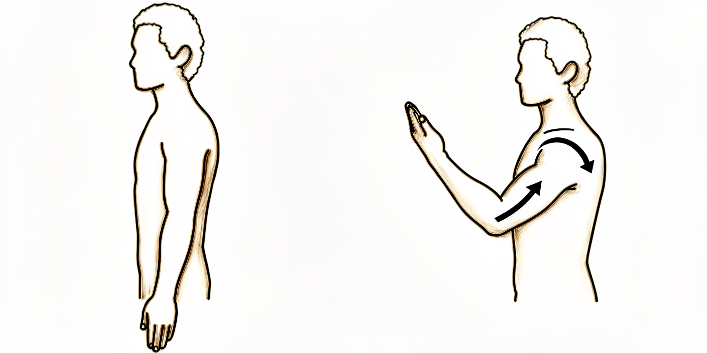
肘部屈伸(以舒适为度)
掌心向上,将肘部舒适地向上弯曲至肩部方向,然后缓慢地向下伸直至舒适范围。活动幅度没有限制——以舒适为指引,动作保持平稳。上臂始终自然垂放于身体一侧。
10次,3–4次/天
Kieran Hirpara 4.0
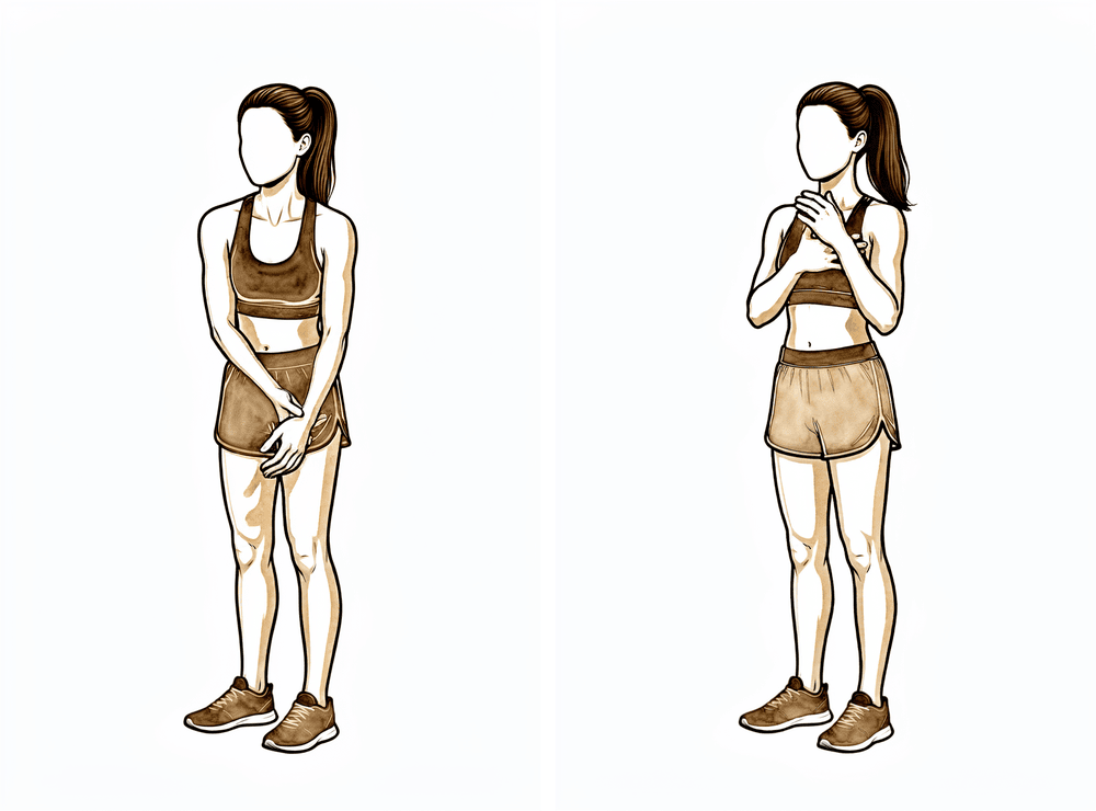
辅助肘关节屈曲
让手术侧手臂完全放松,然后用另一只手轻轻将肘部向上弯向肩部。您并非用手术侧手臂发力——工作由另一只手完成。这有助于在术后早期保持肘部灵活。
10次,3–4次/天
Kieran Hirpara 4.0
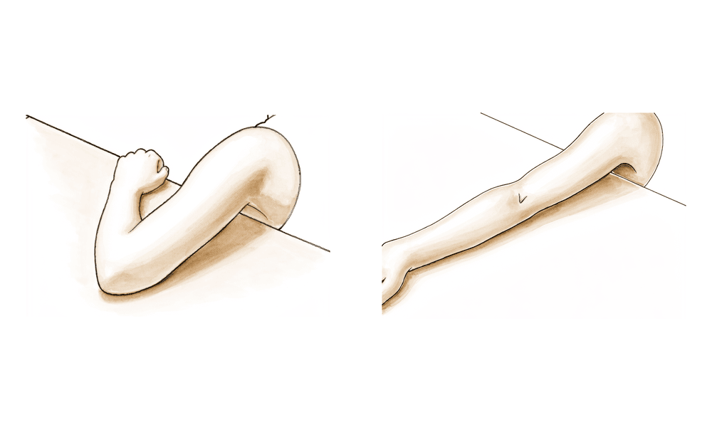
肘部伸直(以舒适为度)
缓慢地将肘关节伸直至舒适位置,然后再弯曲回去。在舒适度允许的范围内,可以完全伸直——无需限制或抑制伸直动作。动作应轻柔,避免任何突然或强力的动作。
10次,3–4次/天
Kieran Hirpara 4.0
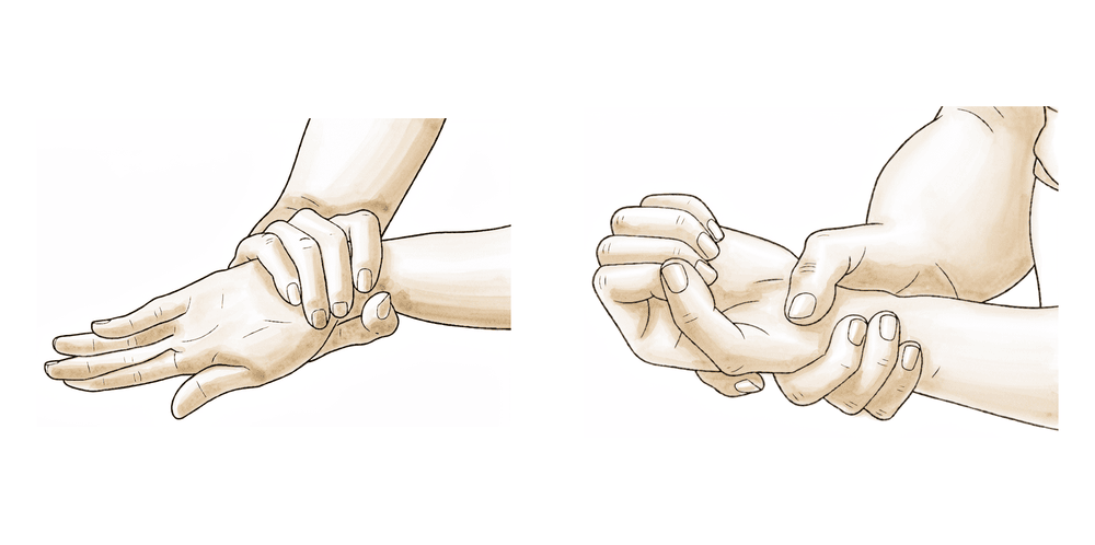
前臂旋转(掌心向上 / 掌心向下)
将肘部贴紧身体侧面,弯曲至约直角。轻轻将手掌向上转向天花板,保持片刻,然后向下转向地面。保持肘部贴紧,使动作来自前臂,而非肩部。
每个方向10次,每天3–4次
Kieran Hirpara 4.0
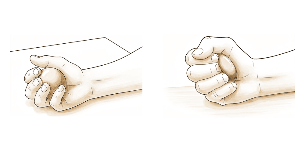
手、腕及抓握运动
从开始起就保持手和手指的活动。将手完全张开,然后轻轻握拳并松开。同时上下活动手腕。这有助于保持手部柔韧,并在肘部愈合期间防止僵硬和肿胀。
10–15 次,每日数次
Kieran Hirpara 4.0
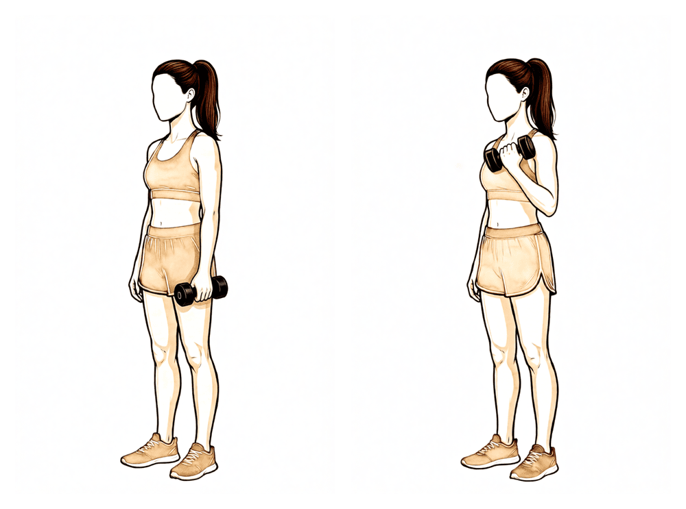
抗阻肱二头肌弯举(约4个月后)
仅在Hirpara医生或您的治疗师允许您开始负重训练(约4个月后)时开始。掌心向上,握住一个较轻的重量,缓慢将其卷起靠近肩部,然后有控制地放下。从非常轻的重量开始,并在接下来的几周内逐渐增加阻力。
10–15 次,待获批后逐步增加
Kieran Hirpara 4.0
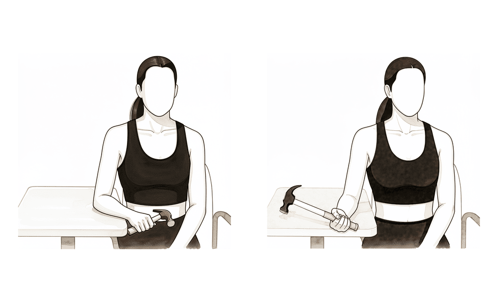
抗阻前臂旋转(约4个月起)
仅在获得开始负重训练的许可后(约4个月)方可开始。将肘部贴近身体,手持弹力带或轻重量的一端,手掌向上对抗轻阻力进行旋转,然后受控地回到起始位置。此动作旨在重建肱二头肌提供的旋后(扭转)肌力。从轻量开始,逐步增加强度。
10–15 次,待获批后逐步增加
这些是您在手册中用于在早期几周保持肘部、前臂和手部活动的练习。取下吊带以进行这些练习。仅在舒适的范围内活动,并按照Hirpara医生和治疗师的指导开始进行。两项强化练习将在后期引入:仅在您被确认可以开始负重(约4个月时)之后。
您的临床方案
本页其余部分为皮质骨纽扣远端肱二头肌腱修复术后的康复临床方案。本节内容应提供给您的物理治疗师或手部治疗师,且以下每个阶段均以通俗易懂的语言解释当前正在发生的情况。
指导原则是早期舒适活动与延迟负重:皮质骨纽扣固定结构足够坚固,允许从第一天起进行无限制活动,因此不使用铰链支具,也不使用伸肌阻挡装置。利用该结构的强度旨在预防僵硬,而非作为早期负重的依据;为保护腱-骨愈合并最大限度降低再断裂风险,刻意推迟抗阻强化训练和提举动作。
第一阶段 — 保护性舒适活动(第 0 周 → 第 6 周)
佩戴简易吊带以提供舒适感并提醒避免负重使用;进行锻炼和清洁时取下。目标是维持舒适、接近全范围的关节活动度,同时保护修复部位免受任何负荷。
致您的物理治疗师:
- 固定: 简易吊带佩戴 6 周,锻炼和清洁时取下。不使用铰链支具;不使用伸展阻挡器。
- 关节活动度: 从第 1 天起,在舒适范围内进行所有活动:主动和被动肘关节屈曲、伸展以及前臂旋转。无活动弧限制,无伸展阻挡。
- 目标: 维持舒适的全范围关节活动度;保护修复部位免受负荷;保持手、腕和肩部的活动性。
- 锻炼: 主动肘关节屈曲/伸展至舒适范围;辅助(被动)肘关节屈曲;肘部贴紧体侧进行前臂旋前/旋后的主动范围活动;手部、腕部和抓握运动;肩胛骨和肩部关节活动度。禁止进行抗阻肱二头肌或旋前肌群负荷训练。
- 注意事项: 禁止提重物、抓握或抗阻强化训练;避免在负荷下突然进行强制离心伸展;避免肩部过度伸展。
- 晋级标准: 伤口愈合,舒适地接近全范围关节活动度,第 6 周时不再使用吊带。
第二阶段 — 全范围活动,无负重(第6周 → 约4个月)
停止使用悬吊带。手臂可自由用于轻度日常任务,但禁止抗阻负重或提举:肌腱仍在向骨骼成熟附着,尽管活动是安全的,但此阶段对负荷的谨慎尤为关键。
致您的物理治疗师:
- 目标: 全对称、无痛的活动范围;手和手臂的正常轻度功能性使用。
- 活动范围/使用: 在舒适允许范围内进行全范围活动;日常轻度使用,无抗阻负重或提举。
- 练习: 继续活动范围训练;手部、腕部及握力;肩胛骨和肩部强化。目前仍禁止抗阻肱二头肌或旋后负荷。
- 进阶标准: 全无痛活动范围;伤口及修复处稳定 → 约4个月时开始分级负荷。
第三阶段 — 强化与分级负荷(约 4 → 6 个月)
开始负荷。引入肘关节屈曲和前臂旋后的轻度抗阻强化训练,并逐步向功能性需求,进而向工作或运动特异性需求过渡。
致您的物理治疗师:
- 目标: 重建屈曲和旋后力量;恢复工作和运动。
- 练习: 在约 4 个月时开始轻度抗阻强化和提举;逐步进阶(等长屈曲和抗阻旋后 → 功能性模式 → 工作和运动特异性负荷)。
- 进阶标准: 无痛的抗阻屈曲和旋后;力量接近健侧。
恢复工作与活动
在前6周内,请预期仅将手臂用于佩戴吊带保护下的轻度、舒适的日常任务。早期通常可以进行轻度的桌面工作或单手操作;较重或需要双手配合的体力工作需等待负重训练开始并逐步建立后再进行。
关键里程碑如下:
- 早期数月内禁止提重物及抗阻负重: 这是有意为之,旨在让肌腱在骨骼上愈合。
- 约4个月起进行轻度提重和强化训练: 从轻柔开始,每周逐步进阶。
- 约4个月起恢复非对抗性运动;约6个月起恢复无限制(对抗性)运动,前提是满足以下标准:无痛的全范围关节活动度、力量至少达到对侧的90–100%,且能够耐受您工作或运动的具体要求。
佩戴吊带期间严禁驾驶。一旦脱离吊带,且经复诊确认能够舒适且安全地控制车辆,方可恢复驾驶。请务必遵循Hirpara医生的具体建议,因为时间安排可能因工作和运动的要求而异。
方案结束后
本方案与本诊所的通用康复建议配合使用;请参阅术后疼痛管理和伤口护理。上述分阶段计划与关于肱二头肌远端肌腱修复术后康复的已发表证据相符,您的持续康复将由物理治疗师或手部治疗师根据您肘部的进展情况进行个体化指导。
Evidence & references
This is the clinical evidence summary written for health professionals. It is technical, and it lists the research this page was built from. You do not need to read it to understand your treatment or to make a decision about it.
Topic scope: Post-operative rehabilitation after surgical repair of a ruptured distal biceps tendon reattached to the radial tuberosity with cortical-button fixation. The protocol here reflects Dr Hirpara's practice — a sling-only, early-comfort-motion approach with deliberately delayed loading — set against the published spectrum of distal-biceps rehabilitation protocols.
Defining principle: Cortical-button fixation is the strongest available construct, and biomechanical and clinical evidence show it tolerates immediate motion safely. Dr Hirpara's stance is to spend that strength on early movement, not early loading: a simple sling for 6 weeks (no hinged brace, no extension block) with all motion to comfort from day 1 including extension, but with resisted strengthening and lifting held back until ~4 months to protect tendon-to-bone healing and minimise re-rupture, and unrestricted activity / return to sport at ~6 months on criterion-based clearance. This sits at the protective end of loading while matching the most current thinking on early motion for stiffness prevention.
Where this protocol sits in the evidence
Published distal-biceps protocols span a wide range, from rigid hinged-brace extension-block schemes to immediate unrestricted motion. Dr Hirpara's plan diverges from the "traditional" template in two deliberate ways:
- Immobilisation: A simple sling for comfort, not a hinged ROM brace, and no extension block. Motion is unrestricted to comfort from day 1. The mainstream BWH/MGB protocols instead use a posterior splint at 90° for 5–7 days, then a hinged brace with a 45°→30° extension block opened ~10°/week to full extension by ~6 weeks (or ~3 weeks in the accelerated variant). Dr Hirpara's approach is at the early-motion end and is justified by the strength of the cortical-button construct.
- Loading: Resisted strengthening and lifting are deferred to ~4 months. This is more conservative than the published loading timelines (accelerated resisted work at week 6; standard/MGB at weeks 8–10–12; light weights weeks 12–14). Crucially, this conservatism is a choice made despite having the construct that would permit earlier loading.
The endpoint — unrestricted activity / return to sport at ~6 months, criterion-based — is the single most commonly cited endpoint across protocols and matches the mainstream consensus.
Key evidence and controversies
-
Early/immediate motion is safe with modern fixation. (Moderate) Biomechanical work supports immediate motion: Bisson et al (AJSM 2007) found aggressive rehabilitation safe after the modified 2-incision approach, and Rose et al (KSSTA 2010) showed single-incision EndoButton/FiberWire repairs survive 2,000 immediate-motion cycles. Several series report no increased re-rupture with immediate post-operative motion. This underpins the sling-only, motion-to-comfort approach.
-
Cortical-button strength enables early motion. (Moderate) Cortical-button (± interference screw) fixation has the highest load-to-failure of the available constructs (Olsen JSES 2014; Spang JSES 2006; Lang OTSR 2018 — comparable functional outcomes across constructs but higher load-to-failure for cortical button). Spencer/Edwin (HAND 2008) argued EndoButton fixation strength may allow earlier ROM. This fixation strength is the explicit rationale for permitting immediate movement.
-
Mobilisation timing may not change outcome. (Moderate) A retrospective comparison found no clinically significant difference in failure, complications, ROM or patient-reported outcomes for early versus delayed mobilisation after primary distal biceps repair. This undercuts the urgency of accelerating loading and supports a measured progression.
-
Conservative rehab may lower re-rupture. (Consensus / survey) Rosenthal/Ting/Sher (JSES 2023), a survey of fellowship-trained elbow surgeons, suggests more conservative post-operative rehab may be associated with lower re-rupture risk — a direct counterweight to the accelerated-loading trend and the rationale for deferring loading to ~4 months. Phelps et al (JSES Int 2025, Level IV systematic review) found no consensus on the optimal return-to-sport protocol, with protocols ranging from immobilisation to immediate motion.
-
Tendon elongation in the mid window. (Moderate) Marshall et al (OJSM 2016, radiostereometric) showed the repaired tendon elongates mostly at 4–8 weeks post-op with minimal change at 8–16 weeks — a biomechanical argument for caution against aggressive loading in that mid window even when motion itself is safe.
-
Incision and complications. (Moderate) Grewal et al RCT (JHS 2010): no overall functional difference single vs double incision (flexion strength slightly greater with two-incision, more minor complications with single-incision). Amarasooriya systematic review (AJSM 2020): synostosis occurred only with double incision; fixation technique did not significantly affect re-rupture. Incision choice mainly drives complication-avoidance precautions, not the ROM timeline. Re-rupture rates overall are low (0–5.6%; Garon & Greenberg 2016).
Phased rehabilitation timeline (this protocol)
| Phase | Window | Sling / brace | ROM / use | Strengthening / loading | Criteria to progress |
|---|---|---|---|---|---|
| I — Protected comfort motion | Week 0 → 6 | Simple sling 6 wk, off for exercises/hygiene. No hinged brace, no extension block | All motion to comfort from day 1 — active + passive flexion, extension and forearm rotation. No arc restriction | None. No resisted biceps/supination loading; hand/wrist/grip and scapular/shoulder ROM maintained | Wound healed; comfortable near-full ROM; out of sling at 6 wk |
| II — Full motion, unloaded | Week 6 → ~4 mo | Sling off | Full symmetric pain-free ROM; light everyday use | None yet — light functional use without resisted loading or lifting | Full painless ROM; wound/repair settled → begin loading ~4 mo |
| III — Strengthening & graded loading | ~4 → 6 mo | — | Full ROM maintained | Start LIGHT resisted strengthening / lifting at ~4 mo; progress gradually (isotonic curls + resisted supination → functional → job/sport-specific) | Pain-free resisted flexion/supination; strength approaching the other side |
| Return to activity | ~6 mo | — | Unrestricted | Unrestricted activity / return to sport, criterion-based | Full painless ROM; strength ≥90–100% of the other side; tolerance of job/sport-specific demands |
One-line summary: simple sling 6 weeks with all motion to comfort from day 1 (no brace, no extension block) → full unloaded motion to ~4 months → light resisted loading from ~4 months → unrestricted activity / sport at ~6 months, criterion-based.
Evidence strength flags
- MODERATE (biomechanical + cohort): safety of immediate/early motion with cortical-button fixation (Bisson 2007; Rose 2010; Olsen 2014; Spang 2006; Lang 2018); no clinically significant difference early vs delayed mobilisation; low overall re-rupture rates.
- MODERATE (biomechanical): tendon elongation concentrated 4–8 weeks (Marshall 2016) — supports caution on mid-window loading.
- CONSENSUS / survey-level: more conservative rehab may lower re-rupture (Rosenthal 2023); no consensus on optimal return-to-sport protocol (Phelps 2025, Level IV). The specific phase timings of this protocol are expert/consensus-derived, not trial-derived.
Overall evidence strength: Moderate. Phased timelines rest on consistent institutional protocol consensus reinforced by biomechanical studies and retrospective cohorts; few prospective RCTs of the rehabilitation progression itself, and no consensus on the optimal return-to-sport protocol.
CITATIONS
RAG corpus (180,000+ Orthopaedic articles)
- Grewal R, Athwal GS, MacDermid JC, et al. Single vs. double incision technique for the repair of distal biceps tendon ruptures: a randomized clinical trial. J Hand Surg Am. 2010.
- Amarasooriya M, Bain GI, Roper T, et al. Complications after distal biceps tendon repair: a systematic review. Am J Sports Med. 2020.
- Keener JD. Controversies in the surgical treatment of distal biceps tendon ruptures: single versus double-incision repairs. J Shoulder Elbow Surg. 2011;20(2):S113–S125.
- Dunphy TR, Hudson J, Batech M, et al. Surgical treatment of distal biceps tendon ruptures: an analysis of complications in 784 surgical repairs. Am J Sports Med. 2017;45(13):3020–3029.
- Bisson LJ, Gurske-de Perio J, Weber AE, et al. Is it safe to perform aggressive rehabilitation after distal biceps tendon repair using the modified 2-incision approach? A biomechanical study. Am J Sports Med. 2007.
- Rose DM, Archibald JD, Sutter EG, et al. Biomechanical analysis suggests early rehabilitation is possible after single-incision EndoButton distal biceps repair with FiberWire. Knee Surg Sports Traumatol Arthrosc. 2010;19(6).
- Marshall NE, Keller RA, Okoroha K, et al. Radiostereometric evaluation of tendon elongation after distal biceps repair. Orthop J Sports Med. 2016.
- Phelps BM, Birnbrich A, Singer W, et al. Postoperative rehabilitation and return to sport criteria following distal biceps tendon rupture surgery. JSES Int. 2025. (Level IV systematic review: no consensus on optimal RTS rehabilitation.)
- Rosenthal R, Ting RS, Sher D. Management of distal biceps tendon ruptures: a survey of fellowship-trained subspecialist elbow surgeons. J Shoulder Elbow Surg. 2023;32(10).
- Olsen JR, Shields E, Williams RB, et al. A comparison of cortical button with interference screw versus suture anchor techniques for distal biceps brachii tendon repairs. J Shoulder Elbow Surg. 2014;23(11):1607–1611.
- Spang JT, Weinhold PS, Karas SG. A biomechanical comparison of EndoButton versus suture anchor repair of distal biceps tendon injuries. J Shoulder Elbow Surg. 2006.
- Lang NW, Bukaty A, Sturz GD, et al. Treatment of primary total distal biceps tendon rupture using cortical button, transosseous fixation and suture anchor: a single center experience. Orthop Traumatol Surg Res. 2018.
- Edwin ES (Spencer EE Jr), Tisdale A, Kostka K, Ivy RE. Is therapy necessary after distal biceps tendon repair? HAND. 2008;3(4).
- Rubinger L, Solow M, Johal H, et al. Return to work following a distal biceps repair: a systematic review of the literature. J Shoulder Elbow Surg. 2020;29(5):1002–1009.
- Ford SE, Andersen JS, Macknet DM, et al. Major complications after distal biceps tendon repairs: retrospective cohort analysis of 970 cases. J Shoulder Elbow Surg. 2018;27(10):1898–1906.
- Cuzzolin M, Secco D, Guerra E, et al. Operative versus nonoperative management for distal biceps brachii tendon lesions: a systematic review and meta-analysis. Orthop J Sports Med. 2021.
- Garon MT, Greenberg JA. Complications of distal biceps repair. Orthop Clin North Am. 2016. (Re-rupture 0–5.6%.)
Published rehabilitation protocols (web)
- Brigham & Women's Hospital — Distal Biceps Tendon Repair Rehabilitation Protocol (standard). https://www.brighamandwomens.org/assets/BWH/patients-and-families/pdfs/elbow---distal-biceps-repair-protocol.pdf
- Brigham & Women's Hospital — Distal Biceps Tendon Repair Accelerated Protocol. https://www.brighamandwomens.org/assets/BWH/patients-and-families/rehabilitation-services/pdfs/elbow-accelerated-distal-biceps-repair-protocol-bwh.pdf
- Mass General Brigham Sports Medicine — Rehabilitation Protocol for Distal Biceps Tendon Repair (rev. 10/2021). https://www.massgeneral.org/assets/mgh/pdf/orthopaedics/sports-medicine/physical-therapy/rehabilitation-protocol-for-distal-biceps-repair.pdf





