远端锁骨切除术(Mumford手术)
Patients › Shoulder
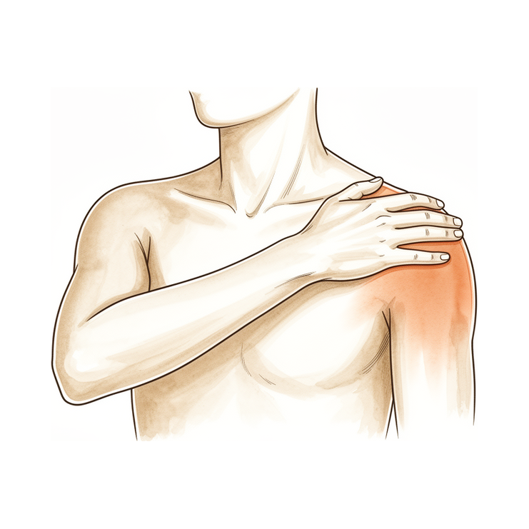
Mumford手术(远端锁骨切除术)通过切除锁骨末端磨损的小部分,以缓解肩锁关节(AC关节)引起的疼痛。以下是该微创手术的具体过程及术后恢复情况。
为何建议进行此手术
您的外科医生建议您进行锁骨远端切除术,也称为 Mumford 手术。该手术通过切除锁骨的外侧端,防止其与肩胛骨发生摩擦。您需要进行此手术的原因可能是存在持续性疼痛或磨损性关节炎,且非手术治疗未能改善症状。
此类手术通常适用于有陈旧性脱位或慢性疼痛病史,且从事重体力劳动或频繁抬臂的患者。主要目标是缓解您的疼痛并改善肩关节功能。虽然开放手术和关节镜手术均效果良好,但您的外科医生采用关节镜入路。该方法涉及小切口和摄像头,有助于您在获得相似长期疗效的同时,更快地恢复日常活动。
手术前
您需要在手术前禁食,并遵医嘱停用某些药物。请安排人员驾车送您回家,并穿着舒适的衣物。您可能需要接受X线检查、MRI、血液检查或麻醉评估,以评估您的健康状况并规划手术。您的外科医生将采用关节镜(钥匙孔)技术进行手术,通过两个或三个小切口将小型摄像头置入关节内。该方法有助于您更快恢复活动,同时避免留下大疤痕。请将您目前服用的所有药物清单带给您的就诊医生。
手术当天
您将抵达医院,并与您的外科医生及麻醉师会面。本手术采用全身麻醉联合区域神经阻滞进行。您在手术期间将完全处于睡眠状态,而神经阻滞(在苏醒前注射以麻木供应手臂的神经)可在术后最初 12 至 24 小时内提供镇痛效果。麻醉师将在手术前与您见面,并向您详细说明这两个部分。
随后,您将进入手术室,您的外科医生将采用关节镜入路进行手术。这需要两到三个小切口,并在关节内放置一台微型摄像头以引导操作。手术后,您将在复苏室苏醒,医疗团队将在麻木感消退期间监测您的舒适度。
手术内容
您的外科医生将采用关节镜技术进行此手术。他们会在您的肩部附近做两个或三个小切口,每个切口长约 1 厘米。通过这些开口,插入微型摄像头和专用工具以观察关节内部。这种方法使您的外科医生能够在不进行大切口的前提下,触及您锁骨的外侧端。
主要目标是切除锁骨外侧端的一小块骨头。您的外科医生将仔细切除该骨头,以防止其与肩胛骨摩擦。证据表明,切除约 5 毫米的骨头可确保骨头不再接触,而切除 2.5 毫米在许多情况下也取得了成功。您的外科医生将精确放置摄像头和工具,以避免损伤周围结构。
切除骨头后,小切口将被缝合。外科医生可能会使用可吸收缝线或医用胶来封闭皮肤。与较大的开放切口相比,采用这种关节镜方法您可以更快地恢复活动,同时获得相似的长期结果。该手术旨在通过消除关节内的摩擦源来缓解您的疼痛。
术后
您将在恢复区苏醒,您的医疗团队将为您管理疼痛。您的外科医生采用关节镜技术,进行两到三个小切口,并将微型摄像头置入关节内。您需要佩戴吊带,小切口处会有敷料覆盖。您可以立即开始轻柔地活动手指和手腕。大多数患者术后需住院一晚,但部分患者可在当天回家。术后24小时内,必须有人陪同您。预计您在4周内可恢复驾驶,6周内可恢复工作。
恢复
在术后的头几天,您的肩部可能会出现一些疼痛和肿胀。这是正常的,因为您的身体正在从小型钥匙孔切口处愈合。您的外科医生可能会建议使用冰袋和止痛药,以帮助您保持舒适。大多数人会发现,随着肿胀消退,不适感会逐渐减轻。
在休息期间,您将佩戴吊带以支撑手臂。您的物理治疗师将指导您进行温和的锻炼,以保持肩部活动而不造成过度牵拉。当您感觉准备好时,可以在家中进行轻微的日常生活活动,但应避免提重物或举手过顶。起初睡眠可能会比较困难;用枕头垫高身体通常有助于找到舒适的姿势。
随着活动能力恢复和肿胀消退,您会对肩部更有信心。您的外科医生和物理治疗师会明确告知您何时可以恢复驾驶、重返工作岗位或进行体育运动。您的个人恢复时间表可能与他人不同,因此请遵循他们针对您恢复情况给出的具体建议。
可能出现的问题
大多数患者恢复良好,但偶尔也会出现并发症。您的外科医生和医疗团队会密切监测您,以便尽早发现任何问题。
如果术后您的肩部仍然疼痛或感觉有摩擦感,可能是因为骨骼切除不足。有时骨骼会在同一位置重新生长。这会导致一种深层的酸痛,且无法通过简单的止痛药缓解。如果您有这种感觉,请告知您的外科医生,以便他们检查您的愈合情况。
如果您注意到锁骨与肩部连接处出现突然的锐痛或不稳定感,可能是因为切除的骨骼过多。这会使关节感觉松动或摇晃。请立即报告任何新的弹响或摩擦感。
在极少数情况下,锁骨或其下方的骨骼可能发生骨折。您可能会感到突然的断裂声或剧烈疼痛,导致无法活动手臂。这是一个需要紧急处理的严重问题。如果您怀疑发生骨折,请立即前往急诊科。
您的外科医生采用关节镜微创手术,通过两到三个小切口并将微型摄像头置入关节内。即使采用这种谨慎的手术方式,仍可能发生并发症。如需了解具体数据,请参阅本页的并发症表格,其中列出了典型的发生率。
何时联系我们
如果您出现发热、小钥匙孔切口处红肿加重或渗出,请联系您的外科医生。如果您感到突发剧烈疼痛、发现腿部肿胀且疼痛,或出现呼吸困难,请立即前往急诊就医。如果您手臂失去感觉或无法活动肩部,请立即联系我们。这些症状需要紧急评估以确保您的安全。
Evidence & references
Overview
- A well-performed distal clavicle excision will likely perform better than a poorly performed one, regardless of whether an open or arthroscopic approach is chosen [1].
- Patients undergoing arthroscopic distal clavicle excision through the direct approach can expect a faster return to activities compared with the open procedure while obtaining similar long-term outcomes [2].
- Arthroscopic distal clavicle resection has provided more 'good or excellent' results than has the open procedure, though this finding is comprised of low-level evidence [3].
- Simple excision of the outer end of the clavicle has yielded satisfactory results with no residual upward displacement disturbing patients [4].
- Patients with displacement greater than 100% of the thickness of the distal clavicle had poorer postoperative clinical outcomes [5].
- Incomplete excision and regrowth of the distal clavicle are the most common causes of revision surgery [6].
- Portal placement remains paramount in facilitating surgery and avoiding injury to adjacent extra-articular structures regardless of the technique chosen for distal clavicle resection [7].
- In appropriately selected patients, open or arthroscopic distal clavicle resection is necessary to relieve symptoms [8].
- Distal clavicle excision with 2.5 mm of bone was successful in many specimens, but a 5 mm resection guaranteed no bone-to-bone abutment [9].
- Arthroscopic and open distal clavicle excisions both provide significant pain reduction at 1 year with no significant difference in outcome measures between groups, except for VAS pain score improvement [10].
- Excision of the outer end of the clavicle is preferred for old dislocations, while open reduction and internal fixation are not recommended due to complications and poor functional results [15].
- Both the direct superior approach and the indirect subacromial approach to the arthroscopic distal clavicle resection result in successful clinical outcome with clinically insignificant difference at final follow-up [16].
Anatomy & Pathophysiology
- A precise, easy to use and low-cost non-invasive method able to draw and analyze the kinematics of the shoulder complex has not been developed yet [25].
- Normative kinematic values of scapulothoracic movements in the shoulder girdle have been provided [26].
- No reconstruction strategy completely restores the shoulder girdle to its preinjured state, although each technique restores different elements of joint kinematics [27].
- The trapezoid and conoid ligaments have unique functions in normal shoulder kinematics because of their anatomic attachments [28].
- Kinematic changes could be a potential source of pain and dysfunction in the shoulder with AC joint dislocation [29].
- Scapular and clavicular kinematics were affected in AC separation models [30].
- A comprehensive clinical approach emphasizing the evaluation of the extent of the anatomic injury and understanding its mechanical consequences regarding shoulder and arm function is key in the development of guidelines for developing operative or non-operative treatment protocols and for establishing outcomes of the treatment protocols [31].
- The inconsistency of AC joint testing parameters and the lack of thorough translation studies indicate a necessity for increased attention in the overall assessment of shoulder stability to close the gap in the foundational biomechanical research [32].
- Anatomically, the pectoralis minor tendon provides sufficient tissue length, excursion, and width [33].
- Biomechanically, the pectoralis minor tendon is as strong as the coracoacromial ligament [33].
- No significant biomechanical differences in displacement or stiffness were seen between the anatomical landmark technique and the coracoid-based landmarks technique for coracoclavicular stabilization [34].
- New surgical techniques continue to evolve as more biomechanical data emerge and kinematic understanding improves [35].
- Emerging concepts and strategies regarding horizontal and rotational instability and scapular biomechanics aim to lay the foundation for future studies aimed at improving treatment outcomes and patient management [36].
- Preliminary findings revealed no detectable differences between surgically reconstructed and uninjured sides in ACJ biomechanics, range of motion, and isometric strength [37].
- Nonoperatively treated shoulders showed increased internal rotation, upward rotation, and posterior tilting [37].
- Type I and II acromioclavicular joint disruptions impair long-term shoulder function in about half of patients 10 years after injury [40].
- At 150 to 200 N of loading, coracoacromial ligament excision and acromioplasty increase the rotator cuff force required to maintain normal glenohumeral biomechanics by 25% to 30% [41].
- Centre of pressure measurement detected sensorimotor functional deficits following surgical treatment of the shoulder joint in patients with confirmed successful clinical and functional outcomes [42].
Classification
- A well-performed distal clavicle excision will likely perform better than a poorly performed one, regardless of whether an open or arthroscopic approach is chosen [1].
- Patients undergoing an arthroscopic procedure specifically through the direct approach can expect a faster return to activities while obtaining similar long-term outcomes compared with the open procedure [2].
- Arthroscopic distal clavicle resection has provided more 'good or excellent' results than has the open procedure, but this finding is comprised of low-level evidence [3].
- Simple excision of the outer end of the clavicle has yielded satisfactory results with no residual upward displacement disturbing the patients [4].
- Patients with displacement greater than 100% of the thickness of the distal clavicle had poorer postoperative clinical outcomes [5].
- Incomplete excision and regrowth of the distal clavicle are the most common causes of revision [6].
- Portal placement remains paramount in both facilitating surgery and avoiding injury to adjacent extra-articular structures regardless of the technique chosen for distal clavicle resection [7].
- In appropriately selected patients, open or arthroscopic distal clavicle resection is necessary to relieve symptoms [8].
- Distal clavicle excision with 2.5 mm of bone was successful in many specimens, but a 5 mm resection guaranteed no bone-to-bone abutment [9].
- Arthroscopic and open distal clavicle excisions both provide significant pain reduction at 1 year with no significant difference in outcome measures between groups, except for VAS pain score improvement [10].
- Horizontal instability of the clavicle is evident with distal clavicle resection of greater than 10 mm [11].
- The new operative procedure combines resection arthroplasty with fixation of the clavicle in an anatomical position [12].
- A records review found that 10 of 894 (1.1%) rotator cuff repairs underwent subsequent distal clavicle resection [23].
- The cross-sectional A-frame morphology of the superior cortex of the distal clavicle provides a reproducible landmark that is eliminated approximately 1.0 cm medial to the distal, lateral end of the clavicle, which can be used intraoperatively to determine when adequate resection has been completed [24].
- Severe chronic symptomatic AC joint separations (Rockwood types III through V) can be repaired entirely by arthroscopy safely and effectively by transferring the coracoacromial ligament with a bone block in the distal clavicle [47].
Clinical Presentation
- A well-performed distal clavicle excision will likely perform better than a poorly performed one, regardless of whether an open or arthroscopic approach is chosen [1].
- Patients having an arthroscopic procedure, specifically through the direct approach, can expect a faster return to activities while obtaining similar long-term outcomes compared with the open procedure [2].
- Arthroscopic distal clavicle resection has provided more 'good or excellent' results than has the open procedure, but is comprised of low-level evidence [3].
- Simple excision of the outer end of the clavicle has yielded satisfactory results in patients with complete dislocation and subluxation of the acromioclavicular joint, with no residual upward displacement disturbing the patients [4].
- Patients with displacement greater than 100% of the thickness of the distal clavicle had poorer postoperative clinical outcomes [5].
- Incomplete excision and regrowth of the distal clavicle are the most common causes of revision surgery [6].
- Portal placement remains paramount in both facilitating surgery and avoiding injury to adjacent extra-articular structures regardless of the technique chosen for distal clavicle resection [7].
- In appropriately selected patients, open or arthroscopic distal clavicle resection is necessary to relieve symptoms [8].
- Although distal clavicle excision with 2.5 mm of bone was successful in many specimens, a 5 mm resection guaranteed no bone-to-bone abutment [9].
- Arthroscopic and open distal clavicle excisions both provide significant pain reduction at 1 year with no significant difference in outcome measures between groups, except for VAS pain score improvement [10].
- Horizontal instability of the clavicle is evident with distal clavicle resection of greater than 10 mm [11].
- Late loss of reduction was common, and clavicular resection reliably produced significant improvement in patients with persistent pain or posttraumatic arthritis [13].
- In carefully selected patients with isolated ACJ pathology, arthroscopic distal clavicle excision results in statistically and clinically significant improvements in range of motion and patient-reported outcome measures [14].
- Excision of the outer end of the clavicle is preferred for old dislocations, while open reduction and internal fixation are not recommended due to complications and poor functional results [15].
- Methods to diagnose both superior and posterior translation of the clavicle need further debate [17].
- Clinical examination and surgical treatment should address anatomic restoration of individual structures to optimize the mechanical capability of the claviscapular segment [18].
- For chronic symptomatic injuries, partial claviculectomy is believed to be the best procedure, offering negligible morbidity and rapid return to function [19].
- Operation should be considered only in thin patients with a prominent clavicle, those doing heavy work, or those whose work requires frequent shoulder abduction and flexion [20].
- Older patients and females were more likely to experience postoperative complications requiring reoperations, including revision ACJR, distal clavicle excision, and irrigation and debridement [21].
- Excellent clinical results were achieved with acromioclavicular joint reconstruction with coracoacromial ligament transfer using the docking technique, decreasing the risk of recurrent distal clavicle instability [46].
Investigations
- A well-performed distal clavicle excision will likely perform better than a poorly performed one, regardless of whether an open or arthroscopic approach is chosen [1].
- Patients having an arthroscopic procedure, specifically through the direct approach, can expect a faster return to activities while obtaining similar long-term outcomes compared with the open procedure [2].
- Arthroscopic distal clavicle resection has provided more 'good or excellent' results than has the open procedure, but is comprised of low-level evidence [3].
- Simple excision of the outer end of the clavicle has yielded satisfactory results in this group of patients, with no residual upward displacement disturbing the patients [4].
- Patients with displacement greater than 100% of the thickness of the distal clavicle had poorer postoperative clinical outcomes [5].
- Incomplete excision and regrowth of the distal clavicle are the most common causes of revision [6].
- Portal placement remains paramount in both facilitating surgery and avoiding injury to adjacent extra-articular structures regardless of the technique chosen for distal clavicle resection [7].
- In appropriately selected patients, open or arthroscopic distal clavicle resection is necessary to relieve symptoms [8].
- Distal clavicle excision with 2.5 mm of bone was successful in many specimens, but a 5 mm resection guaranteed no bone-to-bone abutment [9].
- Horizontal instability of the clavicle is evident with distal clavicle resection of greater than 10 mm [11].
- The new operative procedure combines resection arthroplasty with fixation of the clavicle in an anatomical position [12].
- In carefully selected patients with isolated ACJ pathology, arthroscopic distal clavicle excision results in statistically and clinically significant improvements in range of motion and patient-reported outcome measures [14].
- Methods to diagnose both superior and posterior translation of the clavicle need further debate [17].
- Clinical examination and surgical treatment should address anatomic restoration of individual structures to optimize the mechanical capability of the claviscapular segment [18].
- A 5-mm distal clavicle resection guaranteed no abutment but decreased joint stiffness [22].
- The cross-sectional A-frame morphology of the superior cortex of the distal clavicle provides a reproducible landmark that is eliminated approximately 1.0 cm medial to the distal, lateral end of the clavicle, which can be used intraoperatively to determine when adequate resection has been completed [24].
- Weighted stress radiographs significantly increased the measured elevation of the clavicle and the coracoclavicular distance compared to non-weighted views [54].
- There was no significant difference between open or arthroscopic distal clavicle excision (DCE) [55].
- Although radiological assessment showed a statistically significant immediate superior clavicular displacement after hardware removal following ACJ stabilization, with an increased incidence in the first year following stabilization, this may not negatively influence the results of ACJ stabilization in a clinically relevant way [56].
- Fifteen years postoperatively, good clinical results persisted and anatomic reduction was overall maintained, often with asymptomatic ossification of the coracoclavicular ligaments [57].
Treatment
- A well-performed distal clavicle excision will likely perform better than a poorly performed one, regardless of whether an open or arthroscopic approach is chosen [1].
- Patients undergoing arthroscopic distal clavicle excision via the direct approach can expect a faster return to activities compared with the open procedure while obtaining similar long-term outcomes [2].
- Arthroscopic distal clavicle resection has provided more 'good or excellent' results than the open procedure, though this is based on low-level evidence [3].
- Simple excision of the outer end of the clavicle has yielded satisfactory results with no residual upward displacement disturbing patients [4].
- Patients with displacement greater than 100% of the thickness of the distal clavicle had poorer postoperative clinical outcomes [5].
- Incomplete excision and regrowth of the distal clavicle are the most common causes of revision surgery [6].
- Portal placement remains paramount in facilitating surgery and avoiding injury to adjacent extra-articular structures regardless of the technique chosen for distal clavicle resection [7].
- In appropriately selected patients, open or arthroscopic distal clavicle resection is necessary to relieve symptoms [8].
- Distal clavicle excision with 2.5 mm of bone was successful in many specimens, but a 5 mm resection guaranteed no bone-to-bone abutment [9].
- Arthroscopic and open distal clavicle excisions both provide significant pain reduction at 1 year with no significant difference in outcome measures between groups, except for VAS pain score improvement [10].
- Horizontal instability of the clavicle is evident with distal clavicle resection of greater than 10 mm [11].
- Late loss of reduction was common, and clavicular resection reliably produced significant improvement in patients with persistent pain or posttraumatic arthritis [13].
- Excision of the outer end of the clavicle is preferred for old dislocations, while open reduction and internal fixation are not recommended due to complications and poor functional results [15].
- Both the direct superior approach and the indirect subacromial approach to arthroscopic distal clavicle resection result in successful clinical outcomes with clinically insignificant difference at final follow-up [16].
- A 5-mm distal clavicle resection guaranteed no abutment but decreased joint stiffness [22].
- Surgical treatment may offer early benefits in pain relief and coracoclavicular distance improvement but does not enhance long-term functional outcomes and is associated with higher specific complication rates [49].
- The slight increase in the in situ graft force only in the posterosuperior and posterior direction after distal clavicle excision suggests only a marginal protective role of the acromioclavicular articulation [50].
- A bone anchor system for distal fixation in the base of the coracoid process and a medialized hole in the clavicle restored anatomy best [52].
Complications
- A well-performed distal clavicle excision performs better than a poorly performed one, regardless of whether an open or arthroscopic approach is chosen [1].
- Incomplete excision and regrowth of the distal clavicle are the most common causes of revision surgery [6].
- Portal placement is paramount in facilitating surgery and avoiding injury to adjacent extra-articular structures [7].
- Distal clavicle excision with 2.5 mm of bone was successful in many specimens, but a 5 mm resection guaranteed no bone-to-bone abutment [9].
- Horizontal instability of the clavicle is evident with distal clavicle resection of greater than 10 mm [11].
- Patients with displacement greater than 100% of the thickness of the distal clavicle had poorer postoperative clinical outcomes [5].
- Older patients and females were more likely to experience postoperative complications requiring reoperations, including revision ACJR, distal clavicle excision, and irrigation and debridement [21].
- The incidence of complications in operative acromioclavicular joint separations in an active population was 1.35 per 100 person-years [59].
- Clavicle and coracoid fractures occurred in 1.9 out of 100 cases of operative acromioclavicular joint separations [59].
- Fracture of the distal clavicle or coracoid process after CC ligament repair or reconstruction is a rare but serious complication that can occur independent of bone tunnels created during the index procedure [62].
- Coracoclavicular ligament reconstruction is an effective surgical approach for decreasing the incidence of subacromial osteolysis [60].
- Excellent results can be obtained with coracoacromial ligament transfer using the docking technique, decreasing the risk of recurrent distal clavicle instability [61].
Recovery
- A well-performed distal clavicle excision will likely perform better than a poorly performed one, regardless of whether an open or arthroscopic approach is chosen [1].
- Patients undergoing an arthroscopic procedure, specifically through the direct approach, can expect a faster return to activities compared with the open procedure while obtaining similar long-term outcomes [2].
- Arthroscopic distal clavicle resection has provided more 'good or excellent' results than has the open procedure, though this is comprised of low-level evidence [3].
- Simple excision of the outer end of the clavicle has yielded satisfactory results with no residual upward displacement disturbing the patients [4].
- Patients with displacement greater than 100% of the thickness of the distal clavicle had poorer postoperative clinical outcomes [5].
- Incomplete excision and regrowth of the distal clavicle are the most common causes of revision [6].
- Arthroscopic and open distal clavicle excisions both provide significant pain reduction at 1 year with no significant difference in outcome measures between groups, except for VAS pain score improvement [10].
- Clavicular resection reliably produced significant improvement in patients with persistent pain or posttraumatic arthritis, although late loss of reduction was common [13].
- For chronic symptomatic injuries, partial claviculectomy is believed to be the best procedure, offering negligible morbidity and rapid return to function [19].
- Operation should be considered only in thin patients with a prominent clavicle, those doing heavy work, or those whose work requires frequent shoulder abduction and flexion [20].
- More than 90% of patients manage to return to driving within 4 weeks and to work within 6 weeks following arthroscopic subacromial decompression and acromio-clavicular joint excision [38].
- Late reconstruction of the ligaments in young patients with complete acromioclavicular separations can yield better results than excision of the lateral clavicle, allowing patients to return to strenuous sports or heavy labor [43].
- The described single-tunnel technique for coracoclavicular and acromioclavicular ligament reconstruction results in satisfactory objective and patient-reported outcomes and return to sports while avoiding coracoid and clavicle fractures [44].
- The anatomic reconstruction complex could withstand early rehabilitation, but the decrease in the structural properties and stiffness of the clavicle should be considered in optimizing the anatomic reconstruction technique [45].
- Satisfactory outcome depends upon restoring the stability of the clavicle as well as the acromioclavicular joint [53].
- The arthroscopic partial distal clavicle beveling procedure for nonincarcerated type IV AC separations resulted in a significant reduction in pain, improved daily function, and early return to sport [58].
Key Evidence
- [L5] A well-performed distal clavicle excision will likely perform better than a poorly performed one, regardless of whether an open or arthroscopic approach is chosen. [1] (10.1016/j.arthro.2018.03.004)
- [L3] Among patients undergoing distal clavicle excision for acromioclavicular joint pathology, those having an arthroscopic procedure, specifically through the direct approach, can expect a faster return to activities while obtaining similar long-term outcomes compared with the open procedure. [2] (10.1016/j.arthro.2009.12.007)
- [L3] Arthroscopic distal clavicle resection has provided more 'good or excellent' results than has the open procedure, but is comprised of low-level evidence. [3] (10.1097/blo.0b013e31802f5450)
- [L3] Patients with displacement greater than 100% of the thickness of the distal clavicle had poorer postoperative clinical outcomes. [5] (10.1186/s12891-025-09190-x)
- [L4] Incomplete excision and regrowth of the distal clavicle are the most common causes of revision. [6] (10.1016/j.arthro.2009.06.010)
- [Case_report] Regardless of the technique chosen for distal clavicle resection, portal placement remains paramount in both facilitating surgery and avoiding injury to adjacent extra-articular structures. [7] (10.1016/j.jse.2010.08.032)
- [L5] In appropriately selected patients, open or arthroscopic distal clavicle resection is necessary to relieve symptoms. [8] (10.5435/00124635-199905000-00004)
- [Abstract] Although distal clavicle excision with 2.5 mm of bone was successful in many specimens, a 5 mm resection guaranteed no bone-to-bone abutment. [9] (10.1016/j.jse.2007.02.105)
- [L1] Arthroscopic and open distal clavicle excisions both provide significant pain reduction at 1 year with no significant difference in outcome measures between groups, except for VAS pain score improvement. [10] (10.1016/j.jse.2006.10.006)
- [L4] Horizontal instability of the clavicle is evident with distal clavicle resection of greater than 10 mm. [11] (10.1016/j.xrrt.2021.05.003)
- [L4] The new operative procedure combines resection arthroplasty with fixation of the clavicle in an anatomical position. [12] (10.2106/00004623-197254060-00005)
- [L3] Late loss of reduction was common, and clavicular resection reliably produced significant improvement in patients with persistent pain or posttraumatic arthritis. [13] (10.2106/00004623-198769070-00013)
- [L4] In carefully selected patients with isolated ACJ pathology, arthroscopic distal clavicle excision results in statistically and clinically significant improvements in range of motion and patient-reported outcome measures. [14] (10.1016/j.jseint.2023.07.014)
- [L4] Excision of the outer end of the clavicle is preferred for old dislocations, while open reduction and internal fixation are not recommended due to complications and poor functional results. [15] (10.2106/00004623-196345080-00024)
- [L2] Both the direct superior approach and the indirect subacromial approach to the arthroscopic distal clavicle resection result in successful clinical outcome with clinically insignificant difference at final follow-up. [16] (10.1177/0363546506294855)
- [L4] Methods to diagnose both superior and posterior translation of the clavicle need further debate. [17] (10.1016/j.jseint.2019.11.006)
- [L5] Clinical examination and surgical treatment should address anatomic restoration of individual structures to optimize the mechanical capability of the claviscapular segment. [18] (10.5435/jaaos-d-24-00360)
- [L1] Operation should be considered only in thin patients with a prominent clavicle, those doing heavy work, or those whose work requires frequent shoulder abduction and flexion. [20] (10.2106/00004623-198668040-00011)
- [L4] Older patients and females were more likely to experience postoperative complications requiring reoperations, including revision ACJR, distal clavicle excision, and irrigation and debridement. [21] (10.1007/s00167-016-4206-y)
- [L5] A 5-mm distal clavicle resection guaranteed no abutment but decreased joint stiffness. [22] (10.1016/j.arthro.2007.07.004)
- [L3] This records review found that 10 of 894 (1.1%) rotator cuff repairs underwent subsequent distal clavicle resection. [23] (10.1177/2325967119844295)
- [L5] The cross-sectional A-frame morphology of the superior cortex of the distal clavicle provides a reproducible landmark that is eliminated approximately 1.0 cm medial to the distal, lateral end of the clavicle, which can be used intraoperatively to determine when adequate resection has been completed. [24] (10.1016/j.jse.2021.10.013)
- [L5] Despite technology innovations, a precise, easy to use and low-cost non-invasive method able to draw and analyze the kinematics of the shoulder complex has not been developed yet. [25] (10.1177/17585732221090226)
- [L5] This study provided normative kinematic values of scapulothoracic movements in the shoulder girdle. [26] (10.1016/j.jseint.2022.09.014)
- [L5] Although each technique was able to restore different elements of the joint kinematics, none of the strategies completely restored the shoulder girdle to its preinjured state. [27] (10.1177/03635465221095231)
- [L5] The trapezoid and conoid ligaments have unique functions in normal shoulder kinematics because of their anatomic attachments. [28] (10.1016/j.arthro.2009.12.031)
- [L5] The kinematic changes could be a potential source of pain and dysfunction in the shoulder with AC joint dislocation. [29] (10.1177/0363546512458571)
- [L5] Scapular and clavicular kinematics were affected in AC separation models. [30] (10.1016/j.jse.2013.01.004)
- [L5] A comprehensive clinical approach emphasizing the evaluation of the extent of the anatomic injury and understanding its mechanical consequences regarding shoulder and arm function is a key in the development of guidelines for developing operative or non-operative treatment protocols and for establishing outcomes of the treatment protocols. [31] (10.1177/17585732221122335)
- [L4] The inconsistency of AC joint testing parameters and the lack of thorough translation studies indicate a necessity for increased attention in the overall assessment of shoulder stability to close the gap in the foundational biomechanical research. [32] (10.1016/j.xrrt.2024.06.009)
- [L5] Anatomically, it provides sufficient tissue length, excursion, and width, and biomechanically, it is as strong as the coracoacromial ligament. [33] (10.1016/j.jse.2006.09.007)
- [L5] No significant biomechanical differences in displacement or stiffness were seen between the anatomical landmark technique and the coracoid-based landmarks technique. [34] (10.1177/23259671221132541)
- [L5] New surgical techniques continue to evolve as more biomechanical data emerge and kinematic understanding improves. [35] (10.5435/jaaos-d-16-00776)
- [L5] By exploring emerging concepts and strategies regarding horizontal and rotational instability and scapular biomechanics, the article aims to lay the foundation for future studies aimed at improving treatment outcomes and patient management. [36] (10.1016/j.jseint.2023.11.018)
- [L4] Preliminary findings revealed no detectable differences between surgically reconstructed and uninjured sides in ACJ biomechanics, range of motion, and isometric strength, while nonoperatively treated shoulders showed increased internal rotation, upward rotation, and posterior tilting. [37] (10.1177/23259671241274707)
- [L3] The results obtained in the present study suggest that more than 90% of the patients manage to return to driving within 4 weeks and to work within 6 weeks following arthroscopic subacromial decompression and acromio-clavicular joint excision. [38] (10.1111/j.1758-5740.2010.00048.x)
- [L4] Type I and II acromioclavicular joint disruptions impair long-term shoulder function in about half of patients 10 years after injury. [40] (10.1177/0363546508319047)
- [L5] At 150 to 200 N of loading, CAL excision and acromioplasty increase the rotator cuff force required to maintain normal glenohumeral biomechanics by 25% to 30%. [41] (10.1016/j.jse.2015.10.022)
- [L3] Centre of pressure measurement detected sensorimotor functional deficits following surgical treatment of the shoulder joint in patients with confirmed successful clinical and functional outcomes. [42] (10.1007/s00167-021-06751-0)
- [L4] Late reconstruction of the ligaments in young patients with complete acromioclavicular separations can yield better results than excision of the lateral clavicle, allowing patients to return to strenuous sports or heavy labor. [43] (10.2106/00004623-197658060-00008)
- [L4] The described technique results in satisfactory objective and patient-reported outcomes and return to sports while avoiding coracoid and clavicle fractures. [44] (10.1016/j.jse.2017.11.032)
- [L5] The low level of permanent elongation after cyclic loading suggests that the anatomic reconstruction complex could withstand early rehabilitation; however, the decrease in the structural properties and stiffness of the clavicle should be considered in optimizing the anatomic reconstruction technique. [45] (10.1177/0363546504264637)
- [L4] Excellent clinical results were achieved, decreasing the risk of recurrent distal clavicle instability. [46] (10.1186/1471-2474-10-6)
- [L4] Severe chronic symptomatic AC joint separations (Rockwood types III through V) can be repaired entirely by arthroscopy safely and effectively by transferring the coracoacromial ligament with a bone block in the distal clavicle. [47] (10.1016/j.arthro.2009.08.008)
- [L1] Surgical treatment may offer early benefits in pain relief and coracoclavicular distance improvement but does not enhance long-term functional outcomes and is associated with higher specific complication rates. [49] (10.1186/s12891-024-08100-x)
- [L5] The slight increase in the in situ graft force only in the posterosuperior and posterior direction after distal clavicle excision suggests only a marginal protective role of the acromioclavicular articulation. [50] (10.1177/0363546510374447)
- [L5] A bone anchor system for distal fixation in the base of the coracoid process and a medialized hole in the clavicle restored anatomy best. [52] (10.1007/s001670050182)
- [L4] Satisfactory outcome depends upon restoring the stability of the clavicle as well as the acromioclavicular joint. [53] (10.1111/j.1758-5740.2010.00102.x)
- [L4] Weighted stress radiographs significantly increased the measured elevation of the clavicle and the coracoclavicular distance compared to non-weighted views. [54] (10.1016/j.jseint.2023.06.011)
- [L4] There was no significant difference between open or arthroscopic distal clavicle excision (DCE). [55] (10.1177/17585732231157090)
- [L4] Although radiological assessment showed a statistically significant immediate superior clavicular displacement after this rarely required procedure, with an increased incidence in the first year following stabilization, this may not negatively influence the results of ACJ stabilization in a clinically relevant way. [56] (10.1007/s00167-022-06978-5)
- [L3] Fifteen years postoperatively, good clinical results persisted and anatomic reduction was overall maintained, often with asymptomatic ossification of the coracoclavicular ligaments. [57] (10.1177/03635465251355958)
- [L4] The arthroscopic partial distal clavicle beveling procedure for nonincarcerated type IV AC separations resulted in a significant reduction in pain, improved daily function, and early return to sport. [58] (10.1016/j.arthro.2016.06.013)
- [L3] This review demonstrated an incidence of 1.35 complications per 100 person-years, with clavicle and coracoid fractures occurring in 1.9 out of 100 cases. [59] (10.1177/2325967121s00330)
- [L1] The current analysis suggests coracoclavicular ligament reconstruction as an effective surgical approach for decreasing the incidence of subacromial osteolysis. [60] (10.1016/j.jse.2024.03.018)
- [Abstract] Excellent results can be obtained with this technique, decreasing the risk of recurrent distal clavicle instability. [61] (10.1016/j.jse.2007.02.104)
- [L4] Fracture of the distal clavicle or coracoid process after CC ligament repair or reconstruction is a rare but serious complication that can occur independent of bone tunnels created during the index procedure. [62] (10.1177/03635465211036713)
References
[1] Editorial Commentary: The “Mumford” & Sons: For Distal Clavicle Excisions, What Are Our Young Surgeons Doing, and How Well Are They Doing It?. Arthroscopy. 2018. DOI: 10.1016/j.arthro.2018.03.004 [2] Open Versus Arthroscopic Distal Clavicle Resection. Arthroscopy. 2010. DOI: 10.1016/j.arthro.2009.12.007 [3] Surgical Treatment of Symptomatic Acromioclavicular Joint Problems. Clinical Orthopaedics and Related Research. 2007. DOI: 10.1097/blo.0b013e31802f5450 [4] Complete Dislocation and Subluxation of the Acromioclavicular Joint: End Result in Seventy-three Cases.. The Journal of Bone and Joint Surgery. American Volume. 1961. [5] Predicting reduction loss risk after acromioclavicular joint dislocation treated with the endobutton device. BMC Musculoskeletal Disorders. 2025. DOI: 10.1186/s12891-025-09190-x [6] Open Versus Arthroscopic Acromioclavicular Joint Resection: A Retrospective Comparison Study. Arthroscopy. 2009. DOI: 10.1016/j.arthro.2009.06.010 [7] Acromioclavicular dislocation after arthroscopic distal clavicle resection: a case report. Journal of Shoulder and Elbow Surgery. 2011. DOI: 10.1016/j.jse.2010.08.032 [8] Painful Conditions of the Acromioclavicular Joint. Journal of the American Academy of Orthopaedic Surgeons. 1999. DOI: 10.5435/00124635-199905000-00004 [9] Arthroscopic Distal Clavicle Resection: A Biomechanical Analysis In A Cadaver Model. Journal of Shoulder and Elbow Surgery. 2007. DOI: 10.1016/j.jse.2007.02.105 [10] Arthroscopic versus open distal clavicle excision: Comparative results at six months and one year from a randomized, prospective clinical trial. Journal of Shoulder and Elbow Surgery. 2007. DOI: 10.1016/j.jse.2006.10.006 [11] The reverse coracoacromial ligament transfer for “horizontal” acromioclavicular joint instability. JSES Reviews, Reports, and Techniques. 2021. DOI: 10.1016/j.xrrt.2021.05.003 [12] Treatment of Acromioclavicular Injuries, Especially Complete Acromioclavicular Separation. The Journal of Bone & Joint Surgery. 1972. DOI: 10.2106/00004623-197254060-00005 [13] Dislocation of the acromioclavicular joint. An end-result study.. The Journal of Bone & Joint Surgery. 1987. DOI: 10.2106/00004623-198769070-00013 [14] Patient outcomes following arthroscopic distal clavicle excision: a prospective case series. JSES International. 2023. DOI: 10.1016/j.jseint.2023.07.014 [15] COMPLETE DISLOCATION OF THE ACROMIOCLAVICULAR JOINT. The Journal of Bone & Joint Surgery. 1963. DOI: 10.2106/00004623-196345080-00024 [16] Arthroscopic Distal Clavicle Resection in Athletes. The American Journal of Sports Medicine. 2007. DOI: 10.1177/0363546506294855 [17] Methods used to assess the severity of acromioclavicular joint separations in Japan: a survey. JSES International. 2020. DOI: 10.1016/j.jseint.2019.11.006 [18] Effect of Acromioclavicular Joint Injuries on the Acromioclavicular Joint Complex and Scapulohumeral Rhythm: A Functional and Mechanical Perspective. Journal of the American Academy of Orthopaedic Surgeons. 2025. DOI: 10.5435/jaaos-d-24-00360 [19] Acromioclavicular-Joint Injury: AN END-RESULT STUDY.. The Journal of Bone and Joint Surgery. American Volume. 1966. [20] Conservative or surgical treatment of acromioclavicular dislocation. A prospective, controlled, randomized study.. The Journal of Bone & Joint Surgery. 1986. DOI: 10.2106/00004623-198668040-00011 [21] Early complications of acromioclavicular joint reconstruction requiring reoperation. Knee Surgery, Sports Traumatology, Arthroscopy. 2016. DOI: 10.1007/s00167-016-4206-y [22] Arthroscopic Distal Clavicle Resection: A Biomechanical Analysis of Resection Length and Joint Compliance in a Cadaveric Model. Arthroscopy. 2007. DOI: 10.1016/j.arthro.2007.07.004 [23] Preoperative Factors Associated With Subsequent Distal Clavicle Resection After Rotator Cuff Repair. Orthopaedic Journal of Sports Medicine. 2019. DOI: 10.1177/2325967119844295 [24] Distal clavicle “A-frame” morphology: a reliable intraoperative guide for arthroscopic distal clavicle excision. Journal of Shoulder and Elbow Surgery. 2022. DOI: 10.1016/j.jse.2021.10.013 [25] Evaluation of the range of motion of scapulothoracic, acromioclavicular and sternoclavicular joints: State of the art. Shoulder & Elbow. 2022. DOI: 10.1177/17585732221090226 [26] Kinematic analysis of scapulothoracic movements in the shoulder girdle: a whole cadaver study. JSES International. 2023. DOI: 10.1016/j.jseint.2022.09.014 [27] Differences between Coracoclavicular, Acromioclavicular, or Combined Reconstruction Techniques on the Kinematics of the Shoulder Girdle. The American Journal of Sports Medicine. 2022. DOI: 10.1177/03635465221095231 [28] A Biomechanical Analysis of the Native Coracoclavicular Ligaments and Their Influence on a New Reconstruction Using a Coracoid Tunnel and Free Tendon Graft. Arthroscopy. 2010. DOI: 10.1016/j.arthro.2009.12.031 [29] The Function of the Acromioclavicular and Coracoclavicular Ligaments in Shoulder Motion. The American Journal of Sports Medicine. 2012. DOI: 10.1177/0363546512458571 [30] Acromioclavicular joint ligamentous system contributing to clavicular strut function: a cadaveric study. Journal of Shoulder and Elbow Surgery. 2013. DOI: 10.1016/j.jse.2013.01.004 [31] Acromioclavicular joint injuries revisited: Pathoanatomy, pathomechanics, and clinical presentation. Shoulder & Elbow. 2022. DOI: 10.1177/17585732221122335 [32] Acromioclavicular joint biomechanics: a systematic review. JSES Reviews, Reports, and Techniques. 2024. DOI: 10.1016/j.xrrt.2024.06.009 [33] Anatomy of the pectoralis minor tendon and its use in acromioclavicular joint reconstruction. Journal of Shoulder and Elbow Surgery. 2007. DOI: 10.1016/j.jse.2006.09.007 [34] Comparing the Anatomical Landmarks Versus the Coracoid-Based Landmarks Techniques for Coracoclavicular Stabilization After High-Grade Acromioclavicular Injury: A Biomechanical Study. Orthopaedic Journal of Sports Medicine. 2022. DOI: 10.1177/23259671221132541 [35] Challenges in Treating Acromioclavicular Separations: Current Concepts. Journal of the American Academy of Orthopaedic Surgeons. 2018. DOI: 10.5435/jaaos-d-16-00776 [36] Current trends in surgical treatment of the acromioclavicular joint injuries in 2023: a review of the literature. JSES International. 2024. DOI: 10.1016/j.jseint.2023.11.018 [37] Using Dynamic Stereo X-ray Imaging for In Vivo Acromioclavicular Joint Kinematics Assessment: A Preliminary Investigation. Orthopaedic Journal of Sports Medicine. 2024. DOI: 10.1177/23259671241274707 [38] Return to Work and Driving following Arthroscopic Subacromial Decompression and Acromioclavicular Joint Excision. Shoulder & Elbow. 2010. DOI: 10.1111/j.1758-5740.2010.00048.x [40] Long-Term Shoulder Function after Type I and II Acromioclavicular Joint Disruption. The American Journal of Sports Medicine. 2008. DOI: 10.1177/0363546508319047 [41] The effect of coracoacromial ligament excision and acromioplasty on the amount of rotator cuff force production necessary to restore intact glenohumeral biomechanics. Journal of Shoulder and Elbow Surgery. 2016. DOI: 10.1016/j.jse.2015.10.022 [42] Center of pressure (COP) measurement in patients with confirmed successful outcomes following shoulder surgery show significant sensorimotor deficits. Knee Surgery, Sports Traumatology, Arthroscopy. 2021. DOI: 10.1007/s00167-021-06751-0 [43] Late reconstruction of the ligaments following acromioclavicular separation. The Journal of Bone & Joint Surgery. 1976. DOI: 10.2106/00004623-197658060-00008 [44] Clinical outcomes of a single-tunnel technique for coracoclavicular and acromioclavicular ligament reconstruction. Journal of Shoulder and Elbow Surgery. 2018. DOI: 10.1016/j.jse.2017.11.032 [45] Biomechanical Rationale for Development of Anatomical Reconstructions of Coracoclavicular Ligaments after Complete Acromioclavicular Joint Dislocations. The American Journal of Sports Medicine. 2004. DOI: 10.1177/0363546504264637 [46] Acromioclavicular joint reconstruction with coracoacromial ligament transfer using the docking technique. BMC Musculoskeletal Disorders. 2009. DOI: 10.1186/1471-2474-10-6 [47] All‐Arthroscopic Weaver‐Dunn‐Chuinard Procedure With Double‐Button Fixation for Chronic Acromioclavicular Joint Dislocation. Arthroscopy. 2009. DOI: 10.1016/j.arthro.2009.08.008 [49] Comparative efficacy of operative versus conservative treatment for Rockwood type III acromioclavicular joint dislocation: a systematic review and meta-analysis of randomized controlled trials. BMC Musculoskeletal Disorders. 2024. DOI: 10.1186/s12891-024-08100-x [50] The Effect of Distal Clavicle Excision on in Situ Graft Forces in Coracoclavicular Ligament Reconstruction. The American Journal of Sports Medicine. 2010. DOI: 10.1177/0363546510374447 [52] Which stabilization technique corrects anatomy best in patients with AC‐separation?. Knee Surgery, Sports Traumatology, Arthroscopy. 1999. DOI: 10.1007/s001670050182 [53] Fracture Clavicle with Acromioclavicular Dislocation: A Complex Injury. Shoulder & Elbow. 2011. DOI: 10.1111/j.1758-5740.2010.00102.x [54] Position of scapula and clavicle in acute acromioclavicular joint dislocations: depressed scapula or elevated distal clavicle?. JSES International. 2023. DOI: 10.1016/j.jseint.2023.06.011 [55] A systematic review of the treatment of primary acromioclavicular joint osteoarthritis. Shoulder & Elbow. 2023. DOI: 10.1177/17585732231157090 [56] Low rate of substantial loss of reduction immediately after hardware removal following acromioclavicular joint stabilization using a suspensory fixation system. Knee Surgery, Sports Traumatology, Arthroscopy. 2022. DOI: 10.1007/s00167-022-06978-5 [57] Long-term Follow-up After Arthroscopically Assisted 2-Bundle Anatomic Reduction of Acute Acromioclavicular Joint Separations. The American Journal of Sports Medicine. 2025. DOI: 10.1177/03635465251355958 [58] Posterior Distal Clavicle Beveling for Chronic Nonincarcerated Type IV Acromioclavicular Separations: Surgical Technique and Early Clinical Outcomes. Arthroscopy. 2016. DOI: 10.1016/j.arthro.2016.06.013 [59] Characteristics and Complications of Operative Acromioclavicular Joint Separations in an Active Population (222). Orthopaedic Journal of Sports Medicine. 2021. DOI: 10.1177/2325967121s00330 [60] Subacromial osteolysis following hook plate fixation for acromioclavicular dislocation: a systematic review and meta-analysis. Journal of Shoulder and Elbow Surgery. 2024. DOI: 10.1016/j.jse.2024.03.018 [61] Ac Joint Reconstruction With Ca Ligament Transfer Using The Docking Technique. Journal of Shoulder and Elbow Surgery. 2007. DOI: 10.1016/j.jse.2007.02.104 [62] Coracoid or Clavicle Fractures Associated With Coracoclavicular Ligament Reconstruction. The American Journal of Sports Medicine. 2021. DOI: 10.1177/03635465211036713


