How your hand works Info Evidence
Last reviewed
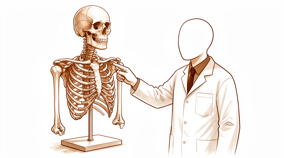
Hand anatomy – bones of the wrist, palm, and fingers – understanding the structure supports diagnosis.
The bones
Your hand is a marvel of engineering, designed for both gentle touch and powerful grip. It allows you to type, hold a coffee cup, and wave hello. Understanding its structure helps you make sense of your diagnosis.
The skeleton of your hand consists of twenty-seven bones. These are divided into three groups: the wrist, the palm, and the fingers.
In your wrist, eight small bones called carpals form two rows. You can feel the bony bumps on the sides of your wrist when you tilt it side to side. These carpals act like a flexible bridge between your forearm and hand.
Your palm contains five long bones called metacarpals. These are the bones you see when you make a fist. The knuckles you see on the back of your hand are the heads of these metacarpals.
Your fingers contain fourteen bones called phalanges. Each finger has three phalanges: proximal, middle, and distal. Your thumb is unique because it has only two phalanges. You can feel these bones along the length of your digits.
Think of your hand like a set of building blocks. The carpals are the base, the metacarpals are the pillars, and the phalanges are the caps. They stack together to create a structure that is both strong and dexterous.
The joints and how they move
Joints are where bones meet. They allow your hand to move in specific ways. Each joint type has a distinct range of motion.
The wrist joint is a condyloid joint. This means it allows bending forward and backward, as well as moving side to side. It does not allow significant rotation. This flexibility lets you type or play the piano.
The knuckles are also condyloid joints. They allow you to bend your fingers and spread them apart. However, they do not rotate. This stability is crucial for gripping objects firmly.
The joints in your fingers are hinge joints. They open and close like a door. This simple motion allows you to make a fist or point a finger. You cannot bend them sideways.
The base of your thumb features a saddle joint. This unique shape allows your thumb to move across your palm. It lets you touch your fingertips with your thumb. This movement, called opposition, is what makes human hands so versatile.
The joints between the wrist bones are plane joints. They allow small sliding movements. These subtle shifts help your wrist absorb shock and adapt to uneven surfaces.
Imagine your hand as a puppet. The hinge joints are the strings that pull fingers closed. The saddle joint is the pivot that lets the thumb dance. The condyloid joints provide the flexibility for fine motor skills.
The muscles, tendons and ligaments
Muscles generate the force for movement. In your hand, most muscles are located in your forearm. Their long tendons stretch down into your hand.
The flexor tendons run along the palm side of your wrist. They pull your fingers and thumb closed. When you grip a ball, these tendons are working hard.
The extensor tendons run along the back of your hand. They pull your fingers and thumb open. You can see these tendons stand up when you lift your hand with fingers extended.
Ligaments are tough bands of tissue that connect bone to bone. They stabilize your joints and prevent excessive movement. The volar plate is a key ligament in your finger joints. It prevents your fingers from bending backward too far.
The collateral ligaments on the sides of your finger joints keep them stable during side-to-side stress. Without them, your fingers would wobble and feel weak.
Think of your tendons like ropes on a ship. The muscles are the crew pulling the ropes. The ligaments are the anchors keeping the mast steady. Together, they create controlled, powerful movement.
The nerves
Nerves carry signals between your brain and your hand. They control muscle movement and provide sensation. Three main nerves serve your hand.
The median nerve runs through the center of your wrist. It provides sensation to your thumb, index, middle, and half of your ring finger. It also controls some small muscles in the base of your thumb. Compression here causes carpal tunnel syndrome.
The ulnar nerve runs along the inner side of your forearm. It provides sensation to your little finger and half of your ring finger. It controls many small muscles in your hand. This allows for fine finger movements and grip strength.
The radial nerve runs along the outer side of your forearm. It primarily controls the muscles that extend your wrist and fingers. It also provides sensation to the back of your hand. Damage here can cause "wrist drop."
Imagine your nerves as electrical wires. The median nerve powers the thumb and index finger. The ulnar nerve powers the pinky and ring finger. The radial nerve lifts your hand up. If one wire is cut, that part of your hand stops working correctly.
Evidence & references
title: "How your hand works" slug: anatomy region: hand audience: patient mesh_terms: [] article_count: 0 model_used: qwen3.5-35b-a3b-q8 generated_at: '2026-05-18T13:34:18+00:00' key_articles: [] synthesis_version: "v2" verifier_status: skipped


