Distal Clavicle Osteolysis Info Evidence
Last reviewed
What you're feeling
You likely feel pain right at the top of your shoulder where it meets your collarbone. This area is the acromioclavicular joint. The pain often feels sharp or aching. It may flare up after you lift heavy objects or do repetitive overhead work. You might notice the pain gets worse when you reach across your body to tuck in a shirt or fasten a bra behind your back.
Many patients find the pain is worse at night. Lying on the sore side can make it difficult to fall asleep. Waking up with stiffness is also common. Some people feel a grinding sensation or hear clicking sounds when they move their arm. If you have had previous shoulder surgery, this pain might be a sign of massive bone loss or a missed fracture.
Daily tasks become harder as the pain grows. Simple movements like reaching for a high shelf or carrying groceries can trigger sharp discomfort. Your surgeon may find that horizontal instability is present if too much bone was removed in the past. While some patients have no symptoms, others struggle with persistent pain or arthritis that does not go away. If you have had surgery before, you might feel that the joint is unstable or that the reduction has been lost over time.
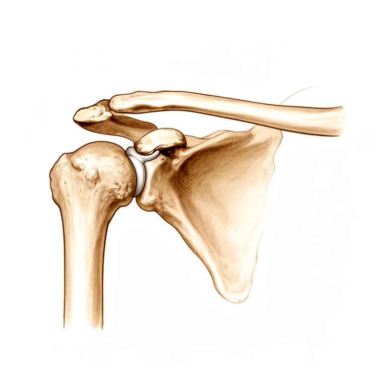
What's actually happening
In your shoulder, a small joint at the top of your collarbone is wearing down. This condition is called distal clavicle osteolysis. Think of the joint like a door hinge that has lost its smooth coating, or cartilage. Without this smooth layer, the bone ends rub against each other. This friction causes inflammation and pain, especially when you lift your arm or move your shoulder.
The tissues that usually hold this joint steady are like ropes made of tendon fibres. When these ropes stretch or tear, the joint becomes unstable. This instability changes how your shoulder moves, which can lead to more pain and trouble using your arm. Sometimes, a specific type of bacteria can cause this wear-and-tear, leading to bone loss in the end of the collarbone.
To fix this, your surgeon may remove the very end of the collarbone. This procedure is called distal clavicle resection. Removing this small piece of bone stops the rubbing and relieves the pain. Patients who have this done often feel better quickly and can return to their daily activities faster than with older open surgery methods. While the joint does not go back to being exactly like new, this surgery reliably improves pain and function for many people with persistent symptoms.
What we can do about it
Your journey usually starts with self-care and guided exercises. You will work with a physiotherapist to strengthen the muscles around your shoulder and improve how your joint moves. This approach aims to reduce pain and help you return to daily activities without surgery. You should give this conservative care a fair chance before considering more invasive steps.
If exercises alone do not provide enough relief, your surgeon may discuss medical options. These include pain medication and anti-inflammatory drugs to manage discomfort. In some cases, injections such as cortisone, hyaluronic acid, or PRP might be offered to calm inflammation in the joint. While these treatments can offer significant pain reduction, the evidence notes that results vary and do not guarantee a permanent fix for everyone.
Surgery is considered when persistent pain or posttraumatic arthritis continues despite these non-surgical efforts. In these situations, removing the outer end of the collarbone (distal clavicle resection) is often the best procedure to relieve symptoms. This operation offers negligible morbidity and allows for a rapid return to function, with patients typically seeing significant improvement in pain and shoulder stability.
When to see someone
See your GP if you have persistent shoulder pain that does not improve with rest. Ask for a specialist review if you feel weakness, instability, or if your shoulder locks or gives way. Contact your surgeon if symptoms interfere with your sleep or work. Seek help for sudden worsening of pain. Be aware that horizontal instability can occur if more than 10 mm of bone is removed. Clavicular tunnel widening was seen in 70% of patients after stabilization surgery. Incomplete removal of the bone end is a common reason for needing further surgery.
Evidence & references
title: "Distal Clavicle Osteolysis" slug: distal-clavicle-osteolysis region: shoulder audience: patient mesh_terms: ["Osteolysis, Essential", "Acromioclavicular Joint"] article_count: 751 model_used: qwen3.5-35b-a3b-q8 generated_at: '2026-05-18T14:17:47+00:00' key_articles: - title: "Propionibacterium acnes–mediated distal clavicular osteolysis: a case report" ref_num: 1 evidence_tier: case_report evidence_level: 5 doi: 10.1016/j.jse.2015.03.004 year: 2015 - title: "Open Versus Arthroscopic Distal Clavicle Resection" ref_num: 2 evidence_tier: paper evidence_level: 3 doi: 10.1016/j.arthro.2009.12.007 year: 2010 - title: "Gorham-Stout disease as a complication of posterior shoulder capsulorrhaphy" ref_num: 3 evidence_tier: paper evidence_level: 4 doi: 10.1016/j.jse.2012.05.024 year: 2012 - title: "Painful Conditions of the Acromioclavicular Joint" ref_num: 4 evidence_tier: paper evidence_level: 5 doi: 10.5435/00124635-199905000-00004 year: 1999 - title: "Dislocation of the acromioclavicular joint. An end-result study." ref_num: 5 evidence_tier: paper evidence_level: 3 doi: 10.2106/00004623-198769070-00013 year: 1987 - title: "Predicting reduction loss risk after acromioclavicular joint dislocation treated with the endobutton device" ref_num: 6 evidence_tier: paper evidence_level: 3 doi: 10.1186/s12891-025-09190-x year: 2025 - title: "Surgical Treatment of Symptomatic Acromioclavicular Joint Problems" ref_num: 8 evidence_tier: paper evidence_level: 3 doi: 10.1097/blo.0b013e31802f5450 year: 2007 - title: "Editorial Commentary: The “Mumford” & Sons: For Distal Clavicle Excisions, What Are Our Young Surgeons Doing, and How Well Are They Doing It?" ref_num: 9 evidence_tier: paper evidence_level: 5 doi: 10.1016/j.arthro.2018.03.004 year: 2018 - title: "Open Versus Arthroscopic Acromioclavicular Joint Resection: A Retrospective Comparison Study" ref_num: 10 evidence_tier: paper evidence_level: 4 doi: 10.1016/j.arthro.2009.06.010 year: 2009 - title: "The reverse coracoacromial ligament transfer for “horizontal” acromioclavicular joint instability" ref_num: 11 evidence_tier: paper evidence_level: 4 doi: 10.1016/j.xrrt.2021.05.003 year: 2021 - title: "Subacromial osteolysis following hook plate fixation for acromioclavicular dislocation: a systematic review and meta-analysis" ref_num: 12 evidence_tier: paper evidence_level: 1 doi: 10.1016/j.jse.2024.03.018 year: 2024 - title: "COMPLETE DISLOCATION OF THE ACROMIOCLAVICULAR JOINT" ref_num: 13 evidence_tier: paper evidence_level: 4 doi: 10.2106/00004623-196345080-00024 year: 1963 - title: "Methods used to assess the severity of acromioclavicular joint separations in Japan: a survey" ref_num: 15 evidence_tier: paper evidence_level: 4 doi: 10.1016/j.jseint.2019.11.006 year: 2020 - title: "Clavicular tunnel widening after coracoclavicular stabilization surgery: a systematic review and meta-analysis" ref_num: 16 evidence_tier: paper evidence_level: 1 doi: 10.1016/j.jse.2023.09.037 year: 2024 - title: "Arthroscopic versus open distal clavicle excision: Comparative results at six months and one year from a randomized, prospective clinical trial" ref_num: 17 evidence_tier: paper evidence_level: 1 doi: 10.1016/j.jse.2006.10.006 year: 2007 - title: "Low rate of substantial loss of reduction immediately after hardware removal following acromioclavicular joint stabilization using a suspensory fixation system" ref_num: 18 evidence_tier: paper evidence_level: 4 doi: 10.1007/s00167-022-06978-5 year: 2022 - title: "Long-term Follow-up After Arthroscopically Assisted 2-Bundle Anatomic Reduction of Acute Acromioclavicular Joint Separations" ref_num: 19 evidence_tier: paper evidence_level: 3 doi: 10.1177/03635465251355958 year: 2025 - title: "Acromioclavicular joint arthritis is not an indication for routine distal clavicle excision in arthroscopic rotator cuff repair" ref_num: 20 evidence_tier: paper evidence_level: 2 doi: 10.1007/s00167-020-06098-y year: 2020 - title: "Segmental clavicle fracture and acromio-clavicular joint disruption: an unusual case report" ref_num: 21 evidence_tier: case_report evidence_level: 4 doi: 10.1177/1758573214564496 year: 2014 - title: "A four-year-old neglected traumatic bipolar clavicular dislocation: a case report" ref_num: 22 evidence_tier: case_report evidence_level: 4 doi: 10.1016/j.xrrt.2021.03.007 year: 2021 - title: "Acromioclavicular joint reconstruction with coracoacromial ligament transfer using the docking technique" ref_num: 23 evidence_tier: paper evidence_level: 4 doi: 10.1186/1471-2474-10-6 year: 2009 - title: "Fracture Clavicle with Acromioclavicular Dislocation: A Complex Injury" ref_num: 24 evidence_tier: paper evidence_level: 4 doi: 10.1111/j.1758-5740.2010.00102.x year: 2011 - title: "Minimally Invasive Coracoclavicular Ligament Augmentation With a Flip Button/Polydioxanone Repair for Treatment of Total Acromioclavicular Joint Dislocation" ref_num: 25 evidence_tier: paper evidence_level: 4 doi: 10.1016/j.arthro.2006.12.015 year: 2007 - title: "Distal Clavicle Fracture as a Complication of Arthroscopic Distal Clavicle Resection" ref_num: 26 evidence_tier: paper evidence_level: 4 doi: 10.1016/j.arthro.2009.02.008 year: 2009 - title: "Outcome of total claviculectomy in six cases" ref_num: 27 evidence_tier: paper evidence_level: 4 doi: 10.1016/j.jse.2006.07.007 year: 2007 - title: "Evaluation of the range of motion of scapulothoracic, acromioclavicular and sternoclavicular joints: State of the art" ref_num: 29 evidence_tier: paper evidence_level: 5 doi: 10.1177/17585732221090226 year: 2022 - title: "Kinematic analysis of scapulothoracic movements in the shoulder girdle: a whole cadaver study" ref_num: 30 evidence_tier: paper evidence_level: 5 doi: 10.1016/j.jseint.2022.09.014 year: 2023 - title: "Differences between Coracoclavicular, Acromioclavicular, or Combined Reconstruction Techniques on the Kinematics of the Shoulder Girdle" ref_num: 31 evidence_tier: paper evidence_level: 5 doi: 10.1177/03635465221095231 year: 2022 - title: "A Biomechanical Analysis of the Native Coracoclavicular Ligaments and Their Influence on a New Reconstruction Using a Coracoid Tunnel and Free Tendon Graft" ref_num: 32 evidence_tier: paper evidence_level: 5 doi: 10.1016/j.arthro.2009.12.031 year: 2010 - title: "The Function of the Acromioclavicular and Coracoclavicular Ligaments in Shoulder Motion" ref_num: 33 evidence_tier: paper evidence_level: 5 doi: 10.1177/0363546512458571 year: 2012 - title: "Acromioclavicular joint ligamentous system contributing to clavicular strut function: a cadaveric study" ref_num: 34 evidence_tier: paper evidence_level: 5 doi: 10.1016/j.jse.2013.01.004 year: 2013 - title: "Acromioclavicular joint injuries revisited: Pathoanatomy, pathomechanics, and clinical presentation" ref_num: 35 evidence_tier: paper evidence_level: 5 doi: 10.1177/17585732221122335 year: 2022 - title: "Acromioclavicular joint biomechanics: a systematic review" ref_num: 36 evidence_tier: paper evidence_level: 4 doi: 10.1016/j.xrrt.2024.06.009 year: 2024 - title: "Anatomy of the pectoralis minor tendon and its use in acromioclavicular joint reconstruction" ref_num: 37 evidence_tier: paper evidence_level: 5 doi: 10.1016/j.jse.2006.09.007 year: 2007 - title: "Comparing the Anatomical Landmarks Versus the Coracoid-Based Landmarks Techniques for Coracoclavicular Stabilization After High-Grade Acromioclavicular Injury: A Biomechanical Study" ref_num: 38 evidence_tier: paper evidence_level: 5 doi: 10.1177/23259671221132541 year: 2022 - title: "Challenges in Treating Acromioclavicular Separations: Current Concepts" ref_num: 39 evidence_tier: paper evidence_level: 5 doi: 10.5435/jaaos-d-16-00776 year: 2018 - title: "Current trends in surgical treatment of the acromioclavicular joint injuries in 2023: a review of the literature" ref_num: 40 evidence_tier: paper evidence_level: 5 doi: 10.1016/j.jseint.2023.11.018 year: 2024 - title: "Using Dynamic Stereo X-ray Imaging for In Vivo Acromioclavicular Joint Kinematics Assessment: A Preliminary Investigation" ref_num: 41 evidence_tier: paper evidence_level: 4 doi: 10.1177/23259671241274707 year: 2024 - title: "Long-Term Shoulder Function after Type I and II Acromioclavicular Joint Disruption" ref_num: 43 evidence_tier: paper evidence_level: 4 doi: 10.1177/0363546508319047 year: 2008 - title: "The effect of coracoacromial ligament excision and acromioplasty on the amount of rotator cuff force production necessary to restore intact glenohumeral biomechanics" ref_num: 44 evidence_tier: paper evidence_level: 5 doi: 10.1016/j.jse.2015.10.022 year: 2016 - title: "Center of pressure (COP) measurement in patients with confirmed successful outcomes following shoulder surgery show significant sensorimotor deficits" ref_num: 45 evidence_tier: paper evidence_level: 3 doi: 10.1007/s00167-021-06751-0 year: 2021 - title: "Os acromiale may be a contraindication of the clavicle hook plate: case reports and literature review" ref_num: 46 evidence_tier: paper evidence_level: 4 doi: 10.1186/s12891-021-04841-1 year: 2021 - title: "Position of scapula and clavicle in acute acromioclavicular joint dislocations: depressed scapula or elevated distal clavicle?" ref_num: 47 evidence_tier: paper evidence_level: 4 doi: 10.1016/j.jseint.2023.06.011 year: 2023 - title: "All‐Arthroscopic Weaver‐Dunn‐Chuinard Procedure With Double‐Button Fixation for Chronic Acromioclavicular Joint Dislocation" ref_num: 48 evidence_tier: paper evidence_level: 4 doi: 10.1016/j.arthro.2009.08.008 year: 2009 - title: "Arthroscopic Versus Open Distal Clavicle Excision" ref_num: 49 evidence_tier: paper evidence_level: 3 doi: 10.1177/0363546511419633 year: 2011 - title: "ISAKOS Upper Extremity Committee Consensus Statement on the Need for Diversification of the Rockwood Classification for Acromioclavicular Joint Injuries" ref_num: 50 evidence_tier: paper evidence_level: 5 doi: 10.1016/j.arthro.2013.11.005 year: 2014 - title: "Comparison of the Hook Plate versus TightRope System in the Treatment of Acute Type III Acromioclavicular Dislocation" ref_num: 51 evidence_tier: paper evidence_level: 3 doi: 10.1155/2022/8706638 year: 2022 - title: "Managing acromio-clavicular joint pain: a scoping review" ref_num: 52 evidence_tier: paper evidence_level: 2 doi: 10.1177/1758573217700839 year: 2017 - title: "Bone Osteolysis Following Acromioclavicular Joint Reconstruction Using Synthetic Ligament (Surgilig™)" ref_num: 54 evidence_tier: paper evidence_level: 4 doi: 10.1111/sae.12035 year: 2014 - title: "A Prospective Evaluation of Untreated Acute Grade III Acromioclavicular Separations" ref_num: 56 evidence_tier: paper evidence_level: 2 doi: 10.1177/03635465010290060401 year: 2001 synthesis_version: "v2" verifier_status: skipped
Overview
- Distal clavicle resection combined with antibiotics halted osteolysis in a case of Propionibacterium acnes–mediated distal clavicular osteolysis, with the patient remaining symptom-free at 10 months post-surgery [1].
- Patients undergoing arthroscopic distal clavicle excision via the direct approach for acromioclavicular joint pathology can expect a faster return to activities compared with the open procedure, while obtaining similar long-term outcomes [2].
- Open or arthroscopic distal clavicle resection is necessary to relieve symptoms in appropriately selected patients [4].
- Late loss of reduction was common in acromioclavicular joint dislocations, but clavicular resection reliably produced significant improvement in patients with persistent pain or posttraumatic arthritis [5].
- Arthroscopic distal clavicle resection has provided more 'good or excellent' results than the open procedure, though this finding is comprised of low-level evidence [8].
- A well-performed distal clavicle excision will likely perform better than a poorly performed one, regardless of whether an open or arthroscopic approach is chosen [9].
- Excision of the outer end of the clavicle is preferred for old acromioclavicular joint dislocations, while open reduction and internal fixation are not recommended due to complications and poor functional results [13].
- For chronic symptomatic acromioclavicular joint injuries, partial claviculectomy is believed to be the best procedure, offering negligible morbidity and rapid return to function [14].
- Both arthroscopic and open distal clavicle excisions provide significant pain reduction at 1 year with no significant difference in outcome measures between groups, except for VAS pain score improvement [17].
- Routine distal clavicle excision is not absolutely necessary in patients with symptomatic acromioclavicular joint osteoarthritis undergoing arthroscopic rotator cuff repair [20].
- Total claviculectomy is a possible treatment option for chronic clavicular dislocation, yielding excellent outcomes and high patient satisfaction [22].
- Total claviculectomy yielded good results for patients with chronic osteitis and malignancy but unsatisfying results for those with chronic posttraumatic pain, despite full range of motion being regained in all cases [27].
Anatomy & Pathophysiology
- A precise, easy to use and low-cost non-invasive method able to draw and analyze the kinematics of the shoulder complex has not been developed yet [29].
- Normative kinematic values of scapulothoracic movements in the shoulder girdle have been provided [30].
- No reconstruction strategy completely restores the shoulder girdle to its preinjured state, although each technique restores different elements of joint kinematics [31].
- The trapezoid and conoid ligaments have unique functions in normal shoulder kinematics because of their anatomic attachments [32].
- Kinematic changes could be a potential source of pain and dysfunction in the shoulder with AC joint dislocation [33].
- Scapular and clavicular kinematics were affected in AC separation models [34].
- A comprehensive clinical approach emphasizing the evaluation of the extent of the anatomic injury and understanding its mechanical consequences regarding shoulder and arm function is a key in the development of guidelines for developing operative or non-operative treatment protocols and for establishing outcomes of the treatment protocols [35].
- The inconsistency of AC joint testing parameters and the lack of thorough translation studies indicate a necessity for increased attention in the overall assessment of shoulder stability to close the gap in the foundational biomechanical research [36].
- Anatomically, the pectoralis minor tendon provides sufficient tissue length, excursion, and width [37].
- Biomechanically, the pectoralis minor tendon is as strong as the coracoacromial ligament [37].
- No significant biomechanical differences in displacement or stiffness were seen between the anatomical landmark technique and the coracoid-based landmarks technique for coracoclavicular stabilization [38].
- New surgical techniques continue to evolve as more biomechanical data emerge and kinematic understanding improves [39].
- Emerging concepts and strategies regarding horizontal and rotational instability and scapular biomechanics aim to lay the foundation for future studies aimed at improving treatment outcomes and patient management [40].
- Preliminary findings revealed no detectable differences between surgically reconstructed and uninjured sides in ACJ biomechanics, range of motion, and isometric strength [41].
- Nonoperatively treated shoulders showed increased internal rotation, upward rotation, and posterior tilting [41].
- Type I and II acromioclavicular joint disruptions impair long-term shoulder function in about half of patients 10 years after injury [43].
- At 150 to 200 N of loading, CAL excision and acromioplasty increase the rotator cuff force required to maintain normal glenohumeral biomechanics by 25% to 30% [44].
- Centre of pressure measurement detected sensorimotor functional deficits following surgical treatment of the shoulder joint in patients with confirmed successful clinical and functional outcomes [45].
Classification
- The ISAKOS Upper Extremity Committee suggests adding grade IIIA and grade IIIB injuries to a modified Rockwood classification to distinguish between stable type III injuries and unstable grade III injuries with therapy-resistant scapular dysfunction and overriding clavicle [50].
- Methods to diagnose both superior and posterior translation of the clavicle need further debate [15].
- Patients with displacement greater than 100% of the thickness of the distal clavicle had poorer postoperative clinical outcomes [6].
- Horizontal instability of the clavicle is evident with distal clavicle resection of greater than 10 mm [11].
- Simple excision of the outer end of the clavicle has yielded satisfactory results in this group of patients, with no residual upward displacement disturbing the patients [7].
- Excision of the outer end of the clavicle is preferred for old dislocations, while open reduction and internal fixation are not recommended due to complications and poor functional results [13].
- Severe chronic symptomatic AC joint separations (Rockwood types III through V) can be repaired entirely by arthroscopy safely and effectively by transferring the coracoacromial ligament with a bone block in the distal clavicle [48].
- Incomplete excision and regrowth of the distal clavicle are the most common causes of revision [10].
- A well-performed distal clavicle excision will likely perform better than a poorly performed one, regardless of whether an open or arthroscopic approach is chosen [9].
- The combination of distal clavicle resection and antibiotics halted the osteolysis, and the patient has remained symptom free at 10 months after surgery [1].
- The case highlights the need to consider Gorham-Stout disease in patients presenting with massive osteolysis after shoulder surgery [3].
Clinical Presentation
- Distal clavicle osteolysis can be mediated by Propionibacterium acnes [1].
- Massive osteolysis may occur as a complication following shoulder surgery, such as posterior shoulder capsulorrhaphy [3].
- Segmental fractures of the clavicle are easily missed and may present with acromioclavicular joint disruption [21].
- Late loss of reduction is common in acromioclavicular joint dislocations [5].
- Patients with displacement greater than 100% of the thickness of the distal clavicle have poorer postoperative clinical outcomes [6].
- Horizontal instability of the clavicle is evident with distal clavicle resection of greater than 10 mm [11].
- Subacromial osteolysis following hook plate fixation for acromioclavicular dislocation has a relatively high and variable incidence [12].
- The primary factor influencing the reported incidence of subacromial osteolysis is the radiological assessment method [12].
- Clavicular tunnel widening was observed in 70% of patients at final follow-up after coracoclavicular stabilization surgery [16].
- Clavicular tunnel widening has a higher prevalence in chronic cases than in acute cases [16].
- Radiological assessment may show a statistically significant immediate superior clavicular displacement after hardware removal following acromioclavicular joint stabilization, with an increased incidence in the first year following stabilization [18].
- Methods to diagnose both superior and posterior translation of the clavicle need further debate [15].
- Distal clavicle fracture is a potential complication of misidentification of the AC joint and subsequent aggressive burring during shoulder arthroscopy [26].
Investigations
- Distal clavicle resection combined with antibiotics halted osteolysis in a case of Propionibacterium acnes–mediated distal clavicular osteolysis [1].
- Massive osteolysis after shoulder surgery requires consideration of Gorham-Stout disease as a diagnosis [3].
- Late loss of reduction was common in acromioclavicular joint dislocations, while clavicular resection reliably produced significant improvement in patients with persistent pain or posttraumatic arthritis [5].
- Patients with displacement greater than 100% of the thickness of the distal clavicle had poorer postoperative clinical outcomes after acromioclavicular joint dislocation treated with the endobutton device [6].
- Simple excision of the outer end of the clavicle yielded satisfactory results in patients with acromioclavicular joint dislocation and subluxation, with no residual upward displacement disturbing the patients [7].
- Horizontal instability of the clavicle is evident with distal clavicle resection of greater than 10 mm [11].
- Subacromial osteolysis following hook plate fixation for acromioclavicular dislocation has a relatively high and variable incidence, with the primary factor influencing the reported incidence being the radiological assessment method [12].
- Methods to diagnose both superior and posterior translation of the clavicle need further debate [15].
- Radiological assessment showed a statistically significant immediate superior clavicular displacement after hardware removal following acromioclavicular joint stabilization using a suspensory fixation system, with an increased incidence in the first year following stabilization [18].
- Weighted stress radiographs significantly increased the measured elevation of the clavicle and the coracoclavicular distance compared to non-weighted views in acute acromioclavicular joint dislocations [47].
- A high index of suspicion is needed to diagnose bone osteolysis following acromioclavicular joint reconstruction using synthetic ligament early before irretrievable bone loss occurs [54].
- Segmental fractures of the clavicle are easily missed [21].
Treatment
- The combination of distal clavicle resection and antibiotics halted osteolysis in a case of Propionibacterium acnes–mediated distal clavicular osteolysis, with the patient remaining symptom-free at 10 months post-surgery [1].
- Patients undergoing arthroscopic distal clavicle excision via the direct approach can expect a faster return to activities compared with open procedures while obtaining similar long-term outcomes [2].
- Open or arthroscopic distal clavicle resection is necessary to relieve symptoms in appropriately selected patients [4].
- Clavicular resection reliably produced significant improvement in patients with persistent pain or posttraumatic arthritis following late loss of reduction after acromioclavicular joint dislocation [5].
- Patients with displacement greater than 100% of the thickness of the distal clavicle had poorer postoperative clinical outcomes after acromioclavicular joint dislocation treated with the endobutton device [6].
- Simple excision of the outer end of the clavicle yielded satisfactory results in patients with acromioclavicular joint dislocation, with no residual upward displacement disturbing the patients [7].
- Arthroscopic distal clavicle resection has provided more 'good or excellent' results than the open procedure, though this finding is comprised of low-level evidence [8].
- A well-performed distal clavicle excision will likely perform better than a poorly performed one, regardless of whether an open or arthroscopic approach is chosen [9].
- Subacromial osteolysis following hook plate fixation for acromioclavicular dislocation has a relatively high and variable incidence, with the primary factor influencing the reported incidence being the radiological assessment method [12].
- Excision of the outer end of the clavicle is preferred for old acromioclavicular joint dislocations, while open reduction and internal fixation are not recommended due to complications and poor functional results [13].
- For chronic symptomatic acromioclavicular joint injuries, partial claviculectomy is believed to be the best procedure, offering negligible morbidity and rapid return to function [14].
- Both arthroscopic and open distal clavicle excisions provide significant pain reduction at 1 year with no significant difference in outcome measures between groups, except for VAS pain score improvement [17].
- Routine distal clavicle excision is not absolutely necessary in patients with symptomatic acromioclavicular joint osteoarthritis undergoing arthroscopic rotator cuff repair [20].
- Total claviculectomy is a possible treatment option for chronic clavicular dislocation, yielding excellent outcomes and high patient satisfaction [22].
- Acromioclavicular joint reconstruction with coracoacromial ligament transfer using the docking technique achieved excellent clinical results and decreased the risk of recurrent distal clavicle instability [23].
- Satisfactory outcomes for fracture clavicle with acromioclavicular dislocation depend upon restoring the stability of the clavicle as well as the acromioclavicular joint [24].
- Distal clavicle fracture is a potential complication of misidentification of the AC joint and subsequent aggressive burring during shoulder arthroscopy [26].
- Ipsilateral os acromiale may be a relative contraindication to the clavicle hook plate [46].
- Open and arthroscopic distal clavicle excision are both effective surgeries to treat recalcitrant acromioclavicular joint pain, providing similarly good to excellent results regarding patient satisfaction and shoulder function at intermediate-term follow-up [49].
- Less residual pain was found using the arthroscopic technique compared with the open procedure for distal clavicle excision [49].
- High-level studies on treatment modalities for acromioclavicular joint pain are limited [52].
Complications
- Distal clavicle osteolysis mediated by Propionibacterium acnes can be halted by the combination of distal clavicle resection and antibiotics, with patients remaining symptom-free at 10 months post-surgery [1].
- Gorham-Stout disease should be considered in patients presenting with massive osteolysis after shoulder surgery [3].
- Incomplete excision and regrowth of the distal clavicle are the most common causes of revision following distal clavicle resection [10].
- Subacromial osteolysis following hook plate fixation for acromioclavicular dislocation has a relatively high and variable incidence, with the primary factor influencing reported incidence being the radiological assessment method [12].
- Clavicular tunnel widening was observed in 70% of patients at final follow-up after coracoclavicular stabilization surgery, with a higher prevalence in chronic than in acute cases [16].
- Horizontal instability of the clavicle is evident with distal clavicle resection of greater than 10 mm [11].
- Patients with displacement greater than 100% of the thickness of the distal clavicle had poorer postoperative clinical outcomes after acromioclavicular joint dislocation treated with the endobutton device [6].
- Late loss of reduction was common in patients with acromioclavicular joint dislocation, while clavicular resection reliably produced significant improvement in patients with persistent pain or posttraumatic arthritis [5].
- Simple excision of the outer end of the clavicle has yielded satisfactory results in patients with acromioclavicular joint dislocation, with no residual upward displacement disturbing the patients [7].
- The minimally invasive TightRope system showed reduced risk of subacromial distal clavicle osteolysis compared to the hook plate in the treatment of acute type III acromioclavicular dislocation [51].
- Asymptomatic ossification of the coracoclavicular ligaments can occur 15 years postoperatively following anatomic reduction of acute acromioclavicular joint separations [19].
Recovery
- Patients undergoing arthroscopic distal clavicle excision via the direct approach can expect a faster return to activities compared with open procedures, while obtaining similar long-term outcomes [2].
- Late loss of reduction was common in patients with acromioclavicular joint dislocation, but clavicular resection reliably produced significant improvement in patients with persistent pain or posttraumatic arthritis [5].
- Simple excision of the outer end of the clavicle yielded satisfactory results in patients with acromioclavicular joint dislocation, with no residual upward displacement disturbing the patients [7].
- For chronic symptomatic acromioclavicular joint injuries, partial claviculectomy is believed to be the best procedure, offering negligible morbidity and rapid return to function [14].
- Incomplete excision and regrowth of the distal clavicle are the most common causes of revision after acromioclavicular joint resection [10].
- Total claviculectomy yielded good results for patients with chronic osteitis and malignancy but unsatisfying results for those with chronic posttraumatic pain, despite full range of motion being regained in all cases [27].
- A majority of patients with untreated acute grade III acromioclavicular separation will do well without any formal treatment, though a small percentage may require delayed surgical intervention [56].
- Patients with displacement greater than 100% of the thickness of the distal clavicle had poorer postoperative clinical outcomes following acromioclavicular joint dislocation treated with the endobutton device [6].
- Fifteen years postoperatively, good clinical results persisted and anatomic reduction was overall maintained after arthroscopically assisted 2-bundle anatomic reduction of acute acromioclavicular joint separations, often with asymptomatic ossification of the coracoclavicular ligaments [19].
- Radiological assessment showed a statistically significant immediate superior clavicular displacement after hardware removal following acromioclavicular joint stabilization using a suspensory fixation system, with an increased incidence in the first year following stabilization, though this may not negatively influence the results of acromioclavicular joint stabilization in a clinically relevant way [18].
- Clavicular tunnel widening was observed in 70% of patients at final follow-up after coracoclavicular stabilization surgery, with a higher prevalence in chronic than in acute cases [16].
- The short-term follow-up of 15 patients treated with minimally invasive coracoclavicular ligament augmentation using a flip button/polydioxanone repair revealed excellent radiologic and clinical results, with no subluxations or dislocations of the acromioclavicular joint noted [25].
- Satisfactory outcomes for fracture clavicle with acromioclavicular dislocation depend upon restoring the stability of the clavicle as well as the acromioclavicular joint [24].
- The combination of distal clavicle resection and antibiotics halted Propionibacterium acnes–mediated distal clavicular osteolysis, and the patient remained symptom free at 10 months after surgery [1].
Key Evidence
- [Case_report] The combination of distal clavicle resection and antibiotics halted the osteolysis, and the patient has remained symptom free at 10 months after surgery. (10.1016/j.jse.2015.03.004)
- [L3] Among patients undergoing distal clavicle excision for acromioclavicular joint pathology, those having an arthroscopic procedure, specifically through the direct approach, can expect a faster return to activities while obtaining similar long-term outcomes compared with the open procedure. (10.1016/j.arthro.2009.12.007)
- [L4] The case highlights the need to consider this diagnosis in patients presenting with massive osteolysis after shoulder surgery. (10.1016/j.jse.2012.05.024)
- [L5] In appropriately selected patients, open or arthroscopic distal clavicle resection is necessary to relieve symptoms. (10.5435/00124635-199905000-00004)
- [L3] Late loss of reduction was common, and clavicular resection reliably produced significant improvement in patients with persistent pain or posttraumatic arthritis. (10.2106/00004623-198769070-00013)
- [L3] Patients with displacement greater than 100% of the thickness of the distal clavicle had poorer postoperative clinical outcomes. (10.1186/s12891-025-09190-x)
- [L3] Arthroscopic distal clavicle resection has provided more 'good or excellent' results than has the open procedure, but is comprised of low-level evidence. (10.1097/blo.0b013e31802f5450)
- [L5] A well-performed distal clavicle excision will likely perform better than a poorly performed one, regardless of whether an open or arthroscopic approach is chosen. (10.1016/j.arthro.2018.03.004)
- [L4] Incomplete excision and regrowth of the distal clavicle are the most common causes of revision. (10.1016/j.arthro.2009.06.010)
- [L4] Horizontal instability of the clavicle is evident with distal clavicle resection of greater than 10 mm. (10.1016/j.xrrt.2021.05.003)
- [L1] Subacromial osteolysis has a relatively high and variable incidence, and the primary factor influencing the reported incidence is the radiological assessment method. (10.1016/j.jse.2024.03.018)
- [L4] Excision of the outer end of the clavicle is preferred for old dislocations, while open reduction and internal fixation are not recommended due to complications and poor functional results. (10.2106/00004623-196345080-00024)
- [L4] Methods to diagnose both superior and posterior translation of the clavicle need further debate. (10.1016/j.jseint.2019.11.006)
- [L1] Clavicular tunnel widening was observed in 70% of patients at final follow-up, with a higher prevalence in chronic than in acute cases. (10.1016/j.jse.2023.09.037)
- [L1] Arthroscopic and open distal clavicle excisions both provide significant pain reduction at 1 year with no significant difference in outcome measures between groups, except for VAS pain score improvement. (10.1016/j.jse.2006.10.006)
- [L4] Although radiological assessment showed a statistically significant immediate superior clavicular displacement after this rarely required procedure, with an increased incidence in the first year following stabilization, this may not negatively influence the results of ACJ stabilization in a clinically relevant way. (10.1007/s00167-022-06978-5)
- [L3] Fifteen years postoperatively, good clinical results persisted and anatomic reduction was overall maintained, often with asymptomatic ossification of the coracoclavicular ligaments. (10.1177/03635465251355958)
- [L2] Routine distal clavicle excision is not absolutely necessary, even in patients with symptomatic ACJ osteoarthritis. (10.1007/s00167-020-06098-y)
- [Case_report] The case highlights that segmental fractures of the clavicle are easily missed. (10.1177/1758573214564496)
- [Case_report] Total claviculectomy is a possible treatment option for chronic clavicular dislocation with excellent outcomes and high patient satisfaction. (10.1016/j.xrrt.2021.03.007)
- [L4] Excellent clinical results were achieved, decreasing the risk of recurrent distal clavicle instability. (10.1186/1471-2474-10-6)
- [L4] Satisfactory outcome depends upon restoring the stability of the clavicle as well as the acromioclavicular joint. (10.1111/j.1758-5740.2010.00102.x)
- [L4] The short-term follow-up of 15 recently operated patients reveals excellent radiologic and clinical results, with no subluxations or dislocations of the acromioclavicular joint noted. (10.1016/j.arthro.2006.12.015)
- [L4] Distal clavicle fracture is a potential complication of misidentification of the AC joint and subsequent aggressive burring during shoulder arthroscopy. (10.1016/j.arthro.2009.02.008)
- [L4] Total claviculectomy yielded good results for patients with chronic osteitis and malignancy but unsatisfying results for those with chronic posttraumatic pain, despite full range of motion being regained in all cases. (10.1016/j.jse.2006.07.007)
- [L5] Despite technology innovations, a precise, easy to use and low-cost non-invasive method able to draw and analyze the kinematics of the shoulder complex has not been developed yet. (10.1177/17585732221090226)
- [L5] This study provided normative kinematic values of scapulothoracic movements in the shoulder girdle. (10.1016/j.jseint.2022.09.014)
- [L5] Although each technique was able to restore different elements of the joint kinematics, none of the strategies completely restored the shoulder girdle to its preinjured state. (10.1177/03635465221095231)
- [L5] The trapezoid and conoid ligaments have unique functions in normal shoulder kinematics because of their anatomic attachments. (10.1016/j.arthro.2009.12.031)
- [L5] The kinematic changes could be a potential source of pain and dysfunction in the shoulder with AC joint dislocation. (10.1177/0363546512458571)
- [L5] Scapular and clavicular kinematics were affected in AC separation models. (10.1016/j.jse.2013.01.004)
- [L5] A comprehensive clinical approach emphasizing the evaluation of the extent of the anatomic injury and understanding its mechanical consequences regarding shoulder and arm function is a key in the development of guidelines for developing operative or non-operative treatment protocols and for establishing outcomes of the treatment protocols. (10.1177/17585732221122335)
- [L4] The inconsistency of AC joint testing parameters and the lack of thorough translation studies indicate a necessity for increased attention in the overall assessment of shoulder stability to close the gap in the foundational biomechanical research. (10.1016/j.xrrt.2024.06.009)
- [L5] Anatomically, it provides sufficient tissue length, excursion, and width, and biomechanically, it is as strong as the coracoacromial ligament. (10.1016/j.jse.2006.09.007)
- [L5] No significant biomechanical differences in displacement or stiffness were seen between the anatomical landmark technique and the coracoid-based landmarks technique. (10.1177/23259671221132541)
- [L5] New surgical techniques continue to evolve as more biomechanical data emerge and kinematic understanding improves. (10.5435/jaaos-d-16-00776)
- [L5] By exploring emerging concepts and strategies regarding horizontal and rotational instability and scapular biomechanics, the article aims to lay the foundation for future studies aimed at improving treatment outcomes and patient management. (10.1016/j.jseint.2023.11.018)
- [L4] Preliminary findings revealed no detectable differences between surgically reconstructed and uninjured sides in ACJ biomechanics, range of motion, and isometric strength, while nonoperatively treated shoulders showed increased internal rotation, upward rotation, and posterior tilting. (10.1177/23259671241274707)
- [L4] Type I and II acromioclavicular joint disruptions impair long-term shoulder function in about half of patients 10 years after injury. (10.1177/0363546508319047)
- [L5] At 150 to 200 N of loading, CAL excision and acromioplasty increase the rotator cuff force required to maintain normal glenohumeral biomechanics by 25% to 30%. (10.1016/j.jse.2015.10.022)
- [L3] Centre of pressure measurement detected sensorimotor functional deficits following surgical treatment of the shoulder joint in patients with confirmed successful clinical and functional outcomes. (10.1007/s00167-021-06751-0)
- [L4] Ipsilateral os acromiale may be a relative contraindication to the clavicle hook plate. (10.1186/s12891-021-04841-1)
- [L4] Weighted stress radiographs significantly increased the measured elevation of the clavicle and the coracoclavicular distance compared to non-weighted views. (10.1016/j.jseint.2023.06.011)
- [L4] Severe chronic symptomatic AC joint separations (Rockwood types III through V) can be repaired entirely by arthroscopy safely and effectively by transferring the coracoacromial ligament with a bone block in the distal clavicle. (10.1016/j.arthro.2009.08.008)
- [L3] Open and arthroscopic distal clavicle excision are both effective surgeries to treat recalcitrant acromioclavicular joint pain, providing similarly good to excellent results regarding patient satisfaction and shoulder function at intermediate-term follow-up, though less residual pain was found using the arthroscopic technique. (10.1177/0363546511419633)
- [L5] The ISAKOS Upper Extremity Committee suggests adding grade IIIA and grade IIIB injuries to a modified Rockwood classification to distinguish between stable type III injuries and unstable grade III injuries with therapy-resistant scapular dysfunction and overriding clavicle. (10.1016/j.arthro.2013.11.005)
- [L3] However, the minimally invasive TightRope system showed further benefits such as reduced reoperation for implant removal and reduced risk of subacromial distal clavicle osteolysis. (10.1155/2022/8706638)
- [L2] High-level studies on treatment modalities for acromio-clavicular joint pain are limited. (10.1177/1758573217700839)
- [L4] A high index of suspicion is needed to diagnose such complications early before irretrievable bone loss to osteolysis. (10.1111/sae.12035)
- [L2] A majority of patients with untreated acute grade III acromioclavicular separation will do well without any formal treatment, though a small percentage may require delayed surgical intervention. (10.1177/03635465010290060401)
References
[1] Propionibacterium acnes–mediated distal clavicular osteolysis: a case report. Journal of Shoulder and Elbow Surgery. 2015. DOI: 10.1016/j.jse.2015.03.004 [2] Open Versus Arthroscopic Distal Clavicle Resection. Arthroscopy. 2010. DOI: 10.1016/j.arthro.2009.12.007 [3] Gorham-Stout disease as a complication of posterior shoulder capsulorrhaphy. Journal of Shoulder and Elbow Surgery. 2012. DOI: 10.1016/j.jse.2012.05.024 [4] Painful Conditions of the Acromioclavicular Joint. Journal of the American Academy of Orthopaedic Surgeons. 1999. DOI: 10.5435/00124635-199905000-00004 [5] Dislocation of the acromioclavicular joint. An end-result study.. The Journal of Bone & Joint Surgery. 1987. DOI: 10.2106/00004623-198769070-00013 [6] Predicting reduction loss risk after acromioclavicular joint dislocation treated with the endobutton device. BMC Musculoskeletal Disorders. 2025. DOI: 10.1186/s12891-025-09190-x [7] Complete Dislocation and Subluxation of the Acromioclavicular Joint: End Result in Seventy-three Cases.. The Journal of Bone and Joint Surgery. American Volume. 1961. [8] Surgical Treatment of Symptomatic Acromioclavicular Joint Problems. Clinical Orthopaedics and Related Research. 2007. DOI: 10.1097/blo.0b013e31802f5450 [9] Editorial Commentary: The “Mumford” & Sons: For Distal Clavicle Excisions, What Are Our Young Surgeons Doing, and How Well Are They Doing It?. Arthroscopy. 2018. DOI: 10.1016/j.arthro.2018.03.004 [10] Open Versus Arthroscopic Acromioclavicular Joint Resection: A Retrospective Comparison Study. Arthroscopy. 2009. DOI: 10.1016/j.arthro.2009.06.010 [11] The reverse coracoacromial ligament transfer for “horizontal” acromioclavicular joint instability. JSES Reviews, Reports, and Techniques. 2021. DOI: 10.1016/j.xrrt.2021.05.003 [12] Subacromial osteolysis following hook plate fixation for acromioclavicular dislocation: a systematic review and meta-analysis. Journal of Shoulder and Elbow Surgery. 2024. DOI: 10.1016/j.jse.2024.03.018 [13] COMPLETE DISLOCATION OF THE ACROMIOCLAVICULAR JOINT. The Journal of Bone & Joint Surgery. 1963. DOI: 10.2106/00004623-196345080-00024 [14] Acromioclavicular-Joint Injury: AN END-RESULT STUDY.. The Journal of Bone and Joint Surgery. American Volume. 1966. [15] Methods used to assess the severity of acromioclavicular joint separations in Japan: a survey. JSES International. 2020. DOI: 10.1016/j.jseint.2019.11.006 [16] Clavicular tunnel widening after coracoclavicular stabilization surgery: a systematic review and meta-analysis. Journal of Shoulder and Elbow Surgery. 2024. DOI: 10.1016/j.jse.2023.09.037 [17] Arthroscopic versus open distal clavicle excision: Comparative results at six months and one year from a randomized, prospective clinical trial. Journal of Shoulder and Elbow Surgery. 2007. DOI: 10.1016/j.jse.2006.10.006 [18] Low rate of substantial loss of reduction immediately after hardware removal following acromioclavicular joint stabilization using a suspensory fixation system. Knee Surgery, Sports Traumatology, Arthroscopy. 2022. DOI: 10.1007/s00167-022-06978-5 [19] Long-term Follow-up After Arthroscopically Assisted 2-Bundle Anatomic Reduction of Acute Acromioclavicular Joint Separations. The American Journal of Sports Medicine. 2025. DOI: 10.1177/03635465251355958 [20] Acromioclavicular joint arthritis is not an indication for routine distal clavicle excision in arthroscopic rotator cuff repair. Knee Surgery, Sports Traumatology, Arthroscopy. 2020. DOI: 10.1007/s00167-020-06098-y [21] Segmental clavicle fracture and acromio-clavicular joint disruption: an unusual case report. Shoulder & Elbow. 2014. DOI: 10.1177/1758573214564496 [22] A four-year-old neglected traumatic bipolar clavicular dislocation: a case report. JSES Reviews, Reports, and Techniques. 2021. DOI: 10.1016/j.xrrt.2021.03.007 [23] Acromioclavicular joint reconstruction with coracoacromial ligament transfer using the docking technique. BMC Musculoskeletal Disorders. 2009. DOI: 10.1186/1471-2474-10-6 [24] Fracture Clavicle with Acromioclavicular Dislocation: A Complex Injury. Shoulder & Elbow. 2011. DOI: 10.1111/j.1758-5740.2010.00102.x [25] Minimally Invasive Coracoclavicular Ligament Augmentation With a Flip Button/Polydioxanone Repair for Treatment of Total Acromioclavicular Joint Dislocation. Arthroscopy. 2007. DOI: 10.1016/j.arthro.2006.12.015 [26] Distal Clavicle Fracture as a Complication of Arthroscopic Distal Clavicle Resection. Arthroscopy. 2009. DOI: 10.1016/j.arthro.2009.02.008 [27] Outcome of total claviculectomy in six cases. Journal of Shoulder and Elbow Surgery. 2007. DOI: 10.1016/j.jse.2006.07.007 [29] Evaluation of the range of motion of scapulothoracic, acromioclavicular and sternoclavicular joints: State of the art. Shoulder & Elbow. 2022. DOI: 10.1177/17585732221090226 [30] Kinematic analysis of scapulothoracic movements in the shoulder girdle: a whole cadaver study. JSES International. 2023. DOI: 10.1016/j.jseint.2022.09.014 [31] Differences between Coracoclavicular, Acromioclavicular, or Combined Reconstruction Techniques on the Kinematics of the Shoulder Girdle. The American Journal of Sports Medicine. 2022. DOI: 10.1177/03635465221095231 [32] A Biomechanical Analysis of the Native Coracoclavicular Ligaments and Their Influence on a New Reconstruction Using a Coracoid Tunnel and Free Tendon Graft. Arthroscopy. 2010. DOI: 10.1016/j.arthro.2009.12.031 [33] The Function of the Acromioclavicular and Coracoclavicular Ligaments in Shoulder Motion. The American Journal of Sports Medicine. 2012. DOI: 10.1177/0363546512458571 [34] Acromioclavicular joint ligamentous system contributing to clavicular strut function: a cadaveric study. Journal of Shoulder and Elbow Surgery. 2013. DOI: 10.1016/j.jse.2013.01.004 [35] Acromioclavicular joint injuries revisited: Pathoanatomy, pathomechanics, and clinical presentation. Shoulder & Elbow. 2022. DOI: 10.1177/17585732221122335 [36] Acromioclavicular joint biomechanics: a systematic review. JSES Reviews, Reports, and Techniques. 2024. DOI: 10.1016/j.xrrt.2024.06.009 [37] Anatomy of the pectoralis minor tendon and its use in acromioclavicular joint reconstruction. Journal of Shoulder and Elbow Surgery. 2007. DOI: 10.1016/j.jse.2006.09.007 [38] Comparing the Anatomical Landmarks Versus the Coracoid-Based Landmarks Techniques for Coracoclavicular Stabilization After High-Grade Acromioclavicular Injury: A Biomechanical Study. Orthopaedic Journal of Sports Medicine. 2022. DOI: 10.1177/23259671221132541 [39] Challenges in Treating Acromioclavicular Separations: Current Concepts. Journal of the American Academy of Orthopaedic Surgeons. 2018. DOI: 10.5435/jaaos-d-16-00776 [40] Current trends in surgical treatment of the acromioclavicular joint injuries in 2023: a review of the literature. JSES International. 2024. DOI: 10.1016/j.jseint.2023.11.018 [41] Using Dynamic Stereo X-ray Imaging for In Vivo Acromioclavicular Joint Kinematics Assessment: A Preliminary Investigation. Orthopaedic Journal of Sports Medicine. 2024. DOI: 10.1177/23259671241274707 [43] Long-Term Shoulder Function after Type I and II Acromioclavicular Joint Disruption. The American Journal of Sports Medicine. 2008. DOI: 10.1177/0363546508319047 [44] The effect of coracoacromial ligament excision and acromioplasty on the amount of rotator cuff force production necessary to restore intact glenohumeral biomechanics. Journal of Shoulder and Elbow Surgery. 2016. DOI: 10.1016/j.jse.2015.10.022 [45] Center of pressure (COP) measurement in patients with confirmed successful outcomes following shoulder surgery show significant sensorimotor deficits. Knee Surgery, Sports Traumatology, Arthroscopy. 2021. DOI: 10.1007/s00167-021-06751-0 [46] Os acromiale may be a contraindication of the clavicle hook plate: case reports and literature review. BMC Musculoskeletal Disorders. 2021. DOI: 10.1186/s12891-021-04841-1 [47] Position of scapula and clavicle in acute acromioclavicular joint dislocations: depressed scapula or elevated distal clavicle?. JSES International. 2023. DOI: 10.1016/j.jseint.2023.06.011 [48] All‐Arthroscopic Weaver‐Dunn‐Chuinard Procedure With Double‐Button Fixation for Chronic Acromioclavicular Joint Dislocation. Arthroscopy. 2009. DOI: 10.1016/j.arthro.2009.08.008 [49] Arthroscopic Versus Open Distal Clavicle Excision. The American Journal of Sports Medicine. 2011. DOI: 10.1177/0363546511419633 [50] ISAKOS Upper Extremity Committee Consensus Statement on the Need for Diversification of the Rockwood Classification for Acromioclavicular Joint Injuries. Arthroscopy. 2014. DOI: 10.1016/j.arthro.2013.11.005 [51] Comparison of the Hook Plate versus TightRope System in the Treatment of Acute Type III Acromioclavicular Dislocation. Advances in Orthopedics. 2022. DOI: 10.1155/2022/8706638 [52] Managing acromio-clavicular joint pain: a scoping review. Shoulder & Elbow. 2017. DOI: 10.1177/1758573217700839 [54] Bone Osteolysis Following Acromioclavicular Joint Reconstruction Using Synthetic Ligament (Surgilig™). Shoulder & Elbow. 2014. DOI: 10.1111/sae.12035 [56] A Prospective Evaluation of Untreated Acute Grade III Acromioclavicular Separations. The American Journal of Sports Medicine. 2001. DOI: 10.1177/03635465010290060401


