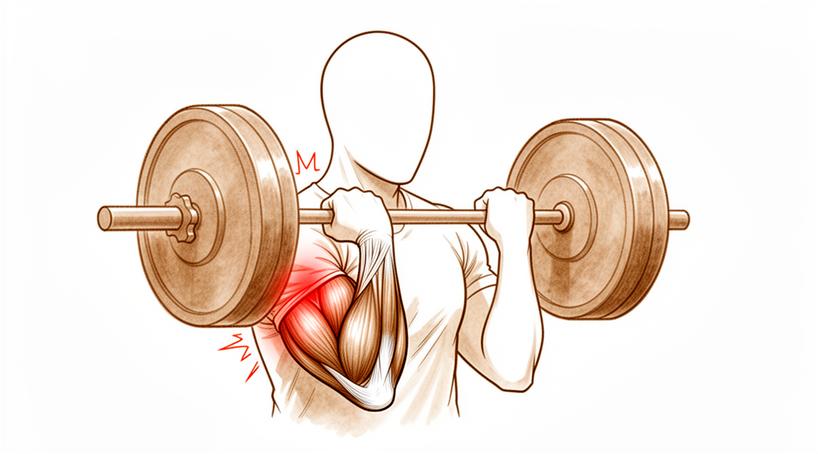
Ruptura do Bíceps Distal
Patients › Elbow
Distal biceps rupture causes sudden elbow pain, bruising, and weakness—often needing surgical repair.
O que você está sentindo
Você pode notar uma dor súbita e aguda na parte frontal do seu braço, perto do cotovelo. Isso geralmente ocorre quando você levanta algo pesado ou sobrecarrega o braço. Muitas pessoas descrevem a sensação como se tivessem sido chutadas ou atingidas nesse local. Você também pode ver ou sentir um inchaço no braço, às vezes chamado de "músculo Popeye". Isso acontece porque o tendão se descolou do osso.
Seu braço pode parecer fraco quando você tenta dobrar o cotovelo ou virar a palma da mão para cima. Tarefas simples podem se tornar difíceis. Você pode ter dificuldade para levantar uma sacola de compras, abrir um pote ou usar uma chave de fenda. Alcançar as costas para fechar um sutiã ou guardar uma camisa pode parecer desconfortável ou doloroso. Algumas pessoas têm dificuldade para dormir do lado afetado porque a pressão agrava a área dolorida.
A dor frequentemente piora após a atividade. Pode estar pior ao acordar pela manhã se você dormiu com o braço dobrado. O repouso geralmente ajuda a aliviar a dor, mas a fraqueza pode persistir. Se você tiver uma ruptura parcial, os sintomas podem ser menos graves do que em uma ruptura completa, mas ainda assim afetam sua rotina diária. As mulheres têm mais probabilidade de ter rupturas parciais do que completas.
Se você tiver uma ruptura isolada da cabeça curta do bíceps, pode sentir uma lacuna ou sulco no músculo. Isso é raro, mas requer atenção precoce se houver fraqueza considerável. Seu cirurgião verificará esses sinais para decidir o melhor caminho a seguir. A maioria das pessoas com rupturas do bíceps distal apresenta padrões semelhantes de dor e fraqueza, independentemente de a lesão ser aguda ou crônica. A boa notícia é que o reparo cirúrgico fornece resultados consistentemente bons para os resultados avaliados pelos pacientes. Você pode esperar excelentes resultados funcionais em mais de 1 ano após o reparo, seja usando parafusos bioabsorvíveis ou não absorvíveis.
O que está realmente acontecendo
O músculo bíceps termina no cotovelo como um tendão espesso e forte. Pense neste tendão como uma corda de alta resistência que conecta o músculo do braço ao osso do antebraço. Sua principal função é ajudar a flexionar o cotovelo e supinar a palma da mão. Quando este tendão se rompe ou se rompe completamente, essa conexão é interrompida. O músculo já não consegue puxar o osso de forma eficaz, o que explica a fraqueza súbita e a dor.
Esta lesão frequentemente ocorre quando você tenta levantar algo pesado ou segurar um objeto que está caindo com o braço esticado. A força exercida coloca estresse excessivo no tendão, fazendo com que ele se rompa. Você pode ouvir um estalo ou sentir uma ruptura aguda. Após a lesão, o músculo pode se contrair no braço, criando um inchaço visível. Isso acontece porque o tendão já não mantém o músculo em sua posição normal próxima ao cotovelo.
O cotovelo é uma articulação de dobradiça complexa. Ele depende dos ossos, ligamentos e tendões trabalhando juntos para garantir estabilidade. Quando o tendão do bíceps é danificado, isso pode afetar a forma como o cotovelo se move e suporta carga. O seu cirurgião avaliará o rompimento para determinar se é completo ou parcial. Um rompimento parcial significa que algumas fibras ainda estão conectadas, enquanto um rompimento completo significa que o tendão se desprendeu totalmente.
A reparação do tendão restaura essa ligação crucial entre o músculo e o osso. As técnicas cirúrgicas modernas permitem que o seu cirurgião reative o tendão com segurança ao osso do antebraço. Seja usando âncoras absorvíveis ou não absorvíveis, o objetivo é reposicionar o tendão no seu local original. Isso permite que o tecido cicatrize e recupere a força.
O tempo é importante para a sua recuperação. A maioria dos problemas após a cirurgia resulta de esperar demasiado para operar ou de usar uma incisão grande que perturba os tecidos circundantes. Se esperar mais de 21 dias, o tendão pode cicatrizar em uma posição encurtada, dificultando a reparação. No entanto, mesmo as reparações tardias podem produzir excelentes resultados funcionais. O seu cirurgião escolherá a melhor abordagem para minimizar complicações e ajudá-lo a retornar às atividades normais com segurança.
O que podemos fazer a respeito
Você pode começar descansando o braço e evitando levantar pesos ou movimentos de torção que sobrecarreguem a parte frontal do cotovelo. Seu cirurgião pode recomendar um período de fisioterapia para ajudar a reduzir o inchaço e restaurar suavemente a mobilidade. O objetivo é manter a articulação flexível enquanto o tendão lesionado se recupera. Você deve dar uma chance justa a essa abordagem conservadora antes de considerar medidas mais invasivas.
Se você sentir dor, seu cirurgião pode sugerir analgésicos de venda livre ou medicamentos anti-inflamatórios para ajudá-lo a controlar o desconforto. Esses medicamentos não curam o tendão, mas podem tornar as atividades diárias mais suportáveis durante a recuperação. Em alguns casos, injeções, como cortisona, podem ser usadas para acalmar a inflamação. No entanto, esses tratamentos não reparam o tendão rompido em si. Seus efeitos são temporários e não constituem uma solução a longo prazo para uma ruptura completa.
A cirurgia é considerada quando o tratamento não cirúrgico não alivia seus sintomas ou quando você precisa recuperar a força total para o trabalho ou esportes. Seu cirurgião discutirá o melhor momento para o reparo, observando que os resultados são excelentes mesmo que o reparo seja adiado por mais de 21 dias. A operação envolve reanexar o tendão rompido ao osso para restaurar a força do braço. Essa abordagem fornece resultados consistentemente bons em termos de resultados avaliados pelo paciente e força. Para rupturas parciais que não melhoram com o repouso, a cirurgia também é uma opção viável. Em casos raros em que o tendão não pode ser reanexado diretamente, seu cirurgião pode usar um enxerto para reconstruir a área. A taxa geral de complicações graves decorrentes da cirurgia é baixa, independentemente da técnica utilizada.
O que esperar
Você pode esperar resultados excelentes a longo prazo após a cirurgia para reparar o tendão do bíceps rompido. Seja seu cirurgião utiliza parafusos absorvíveis que se dissolvem ou parafusos não absorvíveis que permanecem no local, seus resultados funcionais após mais de 1 ano são excelentes. A maioria dos pacientes relata resultados consistentemente bons com base em como se sentem e funcionam no dia a dia. Mesmo que você tenha uma ruptura parcial que não cicatrizou com repouso, o reparo cirúrgico continua sendo uma opção viável após a falha do tratamento não cirúrgico.
O tempo desempenha um papel significativo na sua experiência de recuperação. Se você esperar mais de 21 dias para realizar o reparo, ainda pode esperar resultados funcionais semelhantes aos tratados imediatamente. No entanto, o reparo tardio está associado a uma alta taxa de complicações iniciais. A maioria dos problemas decorrentes da cirurgia é atribuída principalmente a esse atraso no momento da intervenção. Eles também estão secundariamente ligados à necessidade de exposição extensa da parte frontal do braço durante o procedimento. Se a sua ruptura for crônica ou recorrente, a reconstrução usando tecido doador (aloinjerto) é segura e eficaz. Essa abordagem resulta em excelentes resultados relatados pelos pacientes e amplitude de movimento, sem complicações significativas nos grupos estudados.
Complicações graves são raras. A taxa de complicações principais após o reparo do tendão do bíceps distal é de 4,6%. No geral, um em cada 20 pacientes terá uma complicação principal, e um em cada cinco pacientes terá uma complicação menor. A lesão nervosa é o problema mais comum que você pode enfrentar. Apesar desses riscos, a taxa geral de complicações graves permanece baixa, independentemente da abordagem cirúrgica. O nível de treinamento do seu cirurgião não altera significativamente esses perfis de risco em comparação com grandes estudos.
Se você optar por não realizar a cirurgia, ou se a ruptura for manejada de forma conservadora, os resultados são geralmente inferiores aos do reparo cirúrgico. A cirurgia oferece a melhor chance de restaurar a força e a função. Embora os reparos crônicos possam ter uma taxa de complicações imediatas ligeiramente maior do que os agudos, os resultados funcionais a longo prazo são comparáveis. Você deve antecipar um período de ajuste. As semanas iniciais podem envolver desconforto e movimento limitado, mas o objetivo é o retorno ao uso normal. A maior parte da morbidade decorre de atrasos ou de exposição cirúrgica extensa, portanto, seguir as orientações do seu cirurgião sobre o momento e os cuidados pós-operatórios é crucial para o melhor resultado possível.
Quando procurar ajuda médica
Consulte o seu médico de família se tiver dor persistente que não melhora com o repouso. Solicite uma avaliação especializada se notar fraqueza ou instabilidade no braço. Procure atendimento se o cotovelo bloquear ou ceder. Entre em contato com o seu médico se os sintomas interferirem no sono ou no trabalho. Procure ajuda para qualquer piora súbita da dor. Estes sinais podem indicar uma rotura do tendão do bíceps distal. Recomenda-se intervenção precoce se sentir uma laceração no músculo ou fraqueza considerável. Adiar a reparação pode aumentar o risco de complicações. O seu cirurgião irá examiná-lo para determinar o melhor curso de ação.
Evidence & references
Overview
- Clinical and functional outcomes at more than 1 year after distal biceps tendon repair are excellent for patients treated with bioabsorbable screws [1].
- Clinical and functional outcomes at more than 1 year after distal biceps tendon repair are excellent for patients treated with nonabsorbable screws [1].
- Selective disruption of the short head of the biceps distal tendon may be effectively treated with anatomic repair when diagnosed appropriately [2].
- Patients have excellent overall outcomes after distal biceps tendon rupture repair [3].
- Functional outcomes for chronic distal biceps ruptures are comparable to those seen in acute distal biceps ruptures, despite a potentially slightly higher immediate complication rate [4].
- Patients treated with delayed distal biceps tendon repair (>21 days) can expect similar functional outcomes to those treated acutely, despite a high rate of initial complications [5].
- Most morbidity from repair of the distal biceps tendon is attributed primarily to a delay in the timing of the repair [6].
- Most morbidity from repair of the distal biceps tendon is attributed secondarily to an extensive anterior exposure [6].
- Bioabsorbable interference screw fixation of distal biceps ruptures through a single anterior incision is a safe and successful technique [7].
- Surgical repair of distal biceps ruptures provides consistently good results in terms of patient-scored outcomes [8].
- The surgical repair of distal biceps tendon ruptures has an overall low rate of serious complications, regardless of approach or technique [9].
- Surgical treatment of partial distal biceps tendon tears is a viable option after failed nonsurgical treatment [11].
- Utilizing a double-incision technique with a combination of cortical button and interference screw fixation helps determine the best method of fixation and approach to minimize postoperative complications in patients with distal biceps ruptures [14].
- Two-incision distal biceps repairs demonstrate a high degree of patient satisfaction [21].
- Two-incision distal biceps repairs demonstrate a low complication rate [21].
Anatomy & Pathophysiology
- The elbow's stability and complex kinematics are accounted for by a combination of bony articulation and soft-tissue stabilizers [29].
- Musculoskeletal ultrasonography provides a dynamic, functional assessment of elbow structures, allowing visualization of pathology under stress and motion [30].
- The medial elbow joint space increases under a valgus load [31].
- The medial elbow joint space decreases when a maximal grip contraction is performed [31].
- A low carrying angle is measured in elite tennis players just before ball impact during the forehand, suggesting a dynamic varus instant accommodation moving towards full extension [32].
- The decrease in the carrying angle is a consequence of an increase in elbow flexion position dictated by the transition from a closed to open, semi-open stances [32].
- Determining which structures need to be repaired is important to avoid complications that could lead to elbow instability [33].
- Ulnar collateral ligament reconstruction using a suspension button fixation technique reliably restored elbow kinematics to the intact state [34].
- Limiting the number of innings pitched is likely the best way to reduce elbow injury in youth pitchers because biomechanical variables correlated with peak valgus torque are not easily modifiable [35].
- The medial elbow joint space was significantly reduced under 60-N valgus stress plus 50% MVC compared to 60-N valgus stress alone [37].
- No significant relationships between adaptations in shoulder strength or ROM were related to chronic structural adaptations of the elbow in professional baseball pitchers [38].
- The spin move is a simple maneuver that can improve exposure of the coronoid process regardless of the degree of elbow instability [39].
- Reattachment of the flexor and extensor tendons at the epicondyle should be considered in the development of improved repair techniques for stabilizers of the elbow [40].
- An internal joint stabilizer with a standardized treatment protocol could maintain concentric reduction while allowing early functional motion and improve clinical outcomes for patients with complex persistent elbow instability [41].
- Activation of lateral forearm muscles decreased elbow varus angulation throughout flexion [42].
- In patients treated with surgery for snapping triceps syndrome, it is crucial to make sure full resolution of the snapping by examining all dislocating structures during passive elbow motion and/or myoelectrical stimulation to achieve excellent results [43].
- Pre-operative evaluations in elbow stiffness should identify involved articular and periarticular tissues and determine whether articular surfaces and osteoarticular congruence are preserved [44].
- Elbow vascularized composite allotransplantation may be technically feasible on the basis of its consistent vascular anatomy and a proposed surgical technique [45].
- Elbow valgus stress might correlate with ball velocity at 75% partial valgus stress pitch [46].
- Physical examination of the elbow is a critical component in formulating an accurate diagnosis [47].
Classification
- Distal biceps tendon tears in women present differently than in men [10].
- The incidence of women sustaining a distal biceps tendon tear is 3.2% [12].
- Partial tears are statistically more common than complete ruptures in women [12].
- Partial ruptures of the distal biceps brachii tendon represent a spectrum of patterns with varying involvement of the long head (LH) and short head (SH) tendons [16].
- Selective disruption of the short head of the biceps distal tendon may be effectively treated with anatomic repair when diagnosed appropriately [2].
Clinical Presentation
- Distal biceps tendon tears in women present differently than in men [10].
- The incidence of women sustaining a distal biceps tendon tear is 3.2% [12].
- Partial tears are statistically more common than complete ruptures in women [12].
- Isolated rupture of the short head of the biceps brachii is a rare injury [15].
- Early intervention for isolated short head rupture is recommended if a sulcus or gap in the musculotendinous unit is palpable or considerable weakness is present [15].
- Partial ruptures of the distal biceps brachii tendon represent a spectrum of patterns with varying involvement of the long head (LH) and short head (SH) tendons [16].
- The Hook test has imperfect validity for diagnosing distal biceps tendon ruptures and should not be used alone [17].
- The prevalence of distal biceps tendon signal changes on MRI in asymptomatic patients is very low [18].
- Patients with a smaller radioulnar window have a higher risk of rupturing the distal biceps tendon [19].
Investigations
- The Hook test has imperfect validity and should not be used alone to diagnose distal biceps tendon ruptures [17].
- The prevalence of distal biceps tendon signal changes on MRI in asymptomatic patients is very low [18].
- Patients with a smaller radioulnar window have a higher risk of rupturing the distal biceps tendon [19].
- Distal biceps tendon tears in women present differently than in men [10].
- Isolated rupture of the short head of the biceps brachii is a rare injury [15].
- Early intervention is recommended for isolated rupture of the short head of the biceps brachii if a sulcus or gap in the musculotendinous unit is palpable or considerable weakness is present [15].
Treatment
Operative Repair
- Clinical and functional outcomes at more than 1 year after distal biceps tendon repair are excellent for both bioabsorbable and nonabsorbable screw fixation groups [1].
- Selective disruption of the short head of the biceps distal tendon may be effectively treated with anatomic repair when diagnosed appropriately [2].
- Patients have excellent overall outcomes after distal biceps tendon rupture repair [3].
- Functional outcomes in chronic distal biceps rupture repairs are comparable to those in acute distal biceps rupture populations, despite a potentially slightly higher immediate complication rate [4].
- Patients treated with distal biceps tendon repair after a delay of more than 21 days can expect similar functional outcomes to those treated acutely, despite a high rate of initial complications [5].
- Most morbidity from repair of the distal biceps tendon is attributed primarily to a delay in the timing of the repair and secondarily to an extensive anterior exposure [6].
- Bioabsorbable interference screw fixation of distal biceps ruptures through a single anterior incision is a safe and successful technique [7].
- Surgical repair of distal biceps ruptures provides consistently good results in terms of patient-scored outcomes [8].
- The surgical repair of distal biceps tendon ruptures has an overall low rate of serious complications, regardless of approach or technique [9].
- Surgical treatment of partial distal biceps tendon tears is a viable option after failed nonsurgical treatment [11].
- Reinsertion of the distal biceps tendon on the brachialis tendon is a safe and effective procedure with a low complication rate, providing satisfactory subjective and objective results at long-term follow-up [13].
- Utilizing a double-incision technique with a combination of cortical button and interference screw fixation helps determine the best method of fixation and approach with the goal of minimizing postoperative complications [14].
- Distal biceps reconstruction using an Achilles tendon allograft, transosseous EndoButton, and Pulvertaft weave with tendon wrap technique is recommended for patients with retracted, irreparable distal biceps ruptures [26].
- A two-incision repair with suture anchor attachment is a safe and effective method for treatment of distal biceps tendon ruptures [36].
- Repair of distal biceps ruptures using an Endobutton fixation results in a nearly normal return of strength and function, which is significantly better than in those managed nonoperatively [50].
Non-Operative Management
- Incomplete distal triceps tears with active elbow extension against resistance are managed nonsurgically [52].
Complications
- Distal biceps tendon repair has an overall low rate of serious complications, regardless of approach or technique [9].
- The major complication rate after distal biceps tendon repair is 4.6% [23].
- One in five patients will have a minor complication after surgery on the distal biceps tendon [24].
- One in twenty patients will have a major complication after surgery on the distal biceps tendon [24].
- Nerve injury is the most common complication after distal biceps tendon repair [22].
- Most morbidity from repair of the distal biceps tendon is attributed primarily to a delay in the timing of the repair [6].
- Most morbidity from repair of the distal biceps tendon is attributed secondarily to an extensive anterior exposure [6].
- Chronic distal biceps rupture repair may have a slightly higher immediate complication rate compared to acute repair [4].
- Delayed repair of distal biceps tendon ruptures (>21 days) is associated with a high rate of initial complications [5].
- Allograft reconstruction of distal biceps injuries for chronic and recurrent tears results in no significant complications among the studied cohort [20].
- Distal biceps repairs using the two-incision technique demonstrate a low complication rate [21].
- Reinsertion of the distal biceps tendon on the brachialis tendon is associated with a low complication rate [13].
- Complication rates after distal biceps tendon repair performed by newly trained surgeons are similar to those previously reported in large cohort studies [22].
Recovery
- Clinical and functional outcomes at more than 1 year after distal biceps tendon repair are excellent for patients treated with bioabsorbable screws [1].
- Clinical and functional outcomes at more than 1 year after distal biceps tendon repair are excellent for patients treated with nonabsorbable screws [1].
- Selective disruption of the short head of the biceps distal tendon may be effectively treated with anatomic repair when diagnosed appropriately [2].
- Patients have excellent overall outcomes after distal biceps tendon rupture repair [3].
- Functional outcomes for chronic distal biceps ruptures are comparable to those seen in acute distal biceps ruptures, despite a potentially slightly higher immediate complication rate [4].
- Patients treated with distal biceps tendon repair after a delay of more than 21 days can expect similar functional outcomes to those treated acutely, despite a high rate of initial complications [5].
- Most morbidity from repair of the distal biceps tendon is attributed primarily to a delay in the timing of the repair [6].
- Most morbidity from repair of the distal biceps tendon is attributed secondarily to an extensive anterior exposure [6].
- Surgical repair of distal biceps ruptures provides consistently good results in terms of patient-scored outcomes [8].
- Surgical treatment of partial distal biceps tendon tears is a viable option after failed nonsurgical treatment [11].
- Reinsertion of the distal biceps tendon on the brachialis tendon is a safe and effective procedure with a low complication rate, providing satisfactory subjective and objective results at long-term follow-up [13].
- Allograft reconstruction of distal biceps injuries for chronic and recurrent tears results in excellent patient-reported outcomes and range of motion [20].
- Allograft reconstruction of distal biceps injuries for chronic and recurrent tears results in no significant complications among the studied cohort [20].
- Complication rates after distal biceps tendon repair performed by newly trained surgeons are similar to those previously reported in large cohort studies [22].
- Nerve injury is the most common complication after distal biceps tendon repair performed by newly trained surgeons [22].
- One in 5 patients will have a minor complication after surgery on the distal biceps tendon [24].
- One in 20 patients will have a major complication after surgery on the distal biceps tendon [24].
- Patients with biceps tendon repair etiology had a trend toward greater motion improvement than that of patients with a traumatic etiology regarding anconeus interposition for proximal radioulnar synostosis [25].
- Nearly all complete distal biceps ruptures can be effectively reapproximated to the radial tuberosity within the first 6 weeks of injury using single-incision repair with suture anchors [28].
- Primary repair of chronic distal biceps tendon tears greater than 6 weeks from injury demonstrated excellent patient-reported outcome measures (PROMs) and elbow range of motion (ROM) [48].
- Delayed reconstruction of irreparable distal biceps tendon ruptures with semitendinosus autograft produces similar strength, range of motion, and complication rates compared with delayed primary repair [55].
- Delayed reconstruction of irreparable distal biceps tendon ruptures with semitendinosus autograft produces slightly worse functional outcome scores compared with delayed primary repair [55].
Key Evidence
- [L3] Clinical and functional outcome at more than 1 year after distal biceps tendon repair was excellent in both groups. [1] (10.1016/j.jse.2015.12.007)
- [L4] When diagnosed appropriately, selective disruption of the short head of the biceps distal tendon may be effectively treated with anatomic repair. [2] (10.1016/j.jse.2016.09.050)
- [L3] Overall, patients have excellent outcome after distal biceps tendon rupture repair. [3] (10.1007/s00402-018-3018-6)
- [L4] Although there may be a slightly higher immediate complication rate, the functional outcomes remain comparable with those seen in the patient population with acute distal biceps. [4] (10.1177/23259671211065772)
- [L3] Despite a high rate of initial complications, patients treated with distal biceps tendon repair after a delay (>21 days) can expect similar functional outcomes to those treated acutely. [5] (10.1016/j.jse.2017.02.025)
- [L4] Most morbidity from repair of the distal biceps tendon can be attributed primarily to a delay in the timing of the repair and secondarily to an extensive anterior exposure. [6] (10.2106/00004623-200011000-00010)
- [Paper] This is a safe and successful technique for the management of distal biceps tendon ruptures. [7] (10.1007/s00402-009-0974-x)
- [L4] Surgical repair of distal biceps ruptures provides consistently good results in terms of patient-scored outcomes. [8] (10.1016/j.injury.2012.10.029)
- [L3] The surgical repair of distal biceps tendon ruptures has an overall low rate of serious complications, regardless of approach or technique. [9] (10.1177/0363546517720200)
- [L4] Distal biceps tendon tears in women present differently than in men. [10] (10.1016/j.jse.2010.01.015)
- [L4] Surgical treatment of partial distal biceps tendon tears is a viable option after failed nonsurgical treatment. [11] (10.1016/j.jhsa.2010.04.024)
- [L4] The incidence of women sustaining a distal biceps tendon tear is 3.2%, with partial tears being statistically more common than complete ruptures. [12] (10.1016/j.jse.2014.02.006)
- [L4] Reinsertion of the distal biceps tendon on the brachialis tendon can be considered a safe and effective procedure with low complication rate, providing satisfactory subjective and objective results at long-term follow-up. [13] (10.1007/s00167-008-0705-9)
- [L4] This will help surgeons determine the best method of fixation and approach with the goal of minimizing postoperative complications in patients with distal biceps ruptures. [14] (10.1177/17585732241312212)
- [L4] Isolated rupture of the short head of the biceps brachii is a rare injury and early intervention is recommended if a sulcus or gap in musculotendinous unit is palpable or considerable weakness is present. [15] (10.1007/s00167-010-1108-2)
- [L4] Partial ruptures of the distal biceps brachii tendon represent a spectrum of patterns with varying involvement of the LH and SH tendons. [16] (10.1016/j.jse.2020.04.021)
- [L2] The authors caution against using the Hook test alone to diagnose distal biceps tendon ruptures due to its imperfect validity. [17] (10.1016/j.jhsa.2023.07.004)
- [L3] The prevalence of distal biceps tendon signal changes on MRI in asymptomatic patients is very low. [18] (10.1016/j.jhsa.2022.01.020)
- [L3] Therefore, patients with a smaller radioulnar window have a higher risk of rupturing the distal biceps tendon. [19] (10.1016/j.jse.2023.09.020)
- [L5] Allograft reconstruction of distal biceps injuries for chronic and recurrent tears results in excellent patient-reported outcomes and range of motion, with no significant complications among our cohort. [20] (10.1016/j.xrrt.2025.100661)
- [L3] In this large cohort of 2-incision distal biceps repairs, we found a high degree of patient satisfaction and a low complication rate. [21] (10.1016/j.jse.2014.12.032)
- [L4] Complication rates after distal biceps tendon repair performed by newly trained surgeons were similar to those previously reported in large cohort studies, with nerve injury as the most common complication. [22] (10.1016/j.jse.2022.09.014)
- [L2] This is the largest analysis of complications after distal biceps repair, indicating a major complication rate of 4.6%. [23] (10.1177/0363546519899933)
- [L4] Patients should be counseled that 1 in 5 patients will have a minor complication and 1 in 20 patients will have a major complication after surgery on the distal biceps tendon. [24] (10.1016/j.jse.2016.02.032)
- [L3] Patients with biceps tendon repair etiology had a trend toward greater motion improvement than that of patients with a traumatic etiology. [25] (10.1016/j.jse.2014.07.011)
- [L4] This technique is recommended for patients who have a retracted, irreparable distal biceps rupture. [26] (10.1016/j.jse.2016.01.014)
- [L4] The study demonstrates that nearly all complete distal biceps ruptures can be effectively reapproximated to the radial tuberosity within the first 6 weeks of injury. [28] (10.1016/j.jse.2006.03.002)
- [L5] This article discusses the basic anatomy of the elbow and the biomechanics of this joint, noting that a combination of bony articulation and soft-tissue stabilizers accounts for the elbow's stability and complex kinematics. [29] (10.1016/j.csm.2004.06.008)
- [L5] Musculoskeletal ultrasonography provides a dynamic, functional assessment of elbow structures, allowing visualization of pathology under stress and motion. [30] (10.5435/jaaos-d-20-00935)
- [L4] Medial elbow joint space increases under a valgus load and then decreases when a maximal grip contraction is performed. [31] (10.1177/0363546518755149)
- [L4] The observed decrease in the carrying angle is a consequence of an increase in elbow flexion position dictated by the transition from a closed to open, semi‐open stances. [32] (10.1002/ksa.12016)
- [L3] It is important to determine which structures need to be repaired to avoid complications that could lead to elbow instability. [33] (10.1016/j.jse.2019.07.006)
- [L5] Ulnar collateral ligament reconstruction using a suspension button fixation technique reliably restored elbow kinematics to the intact state. [34] (10.1177/0363546509350109)
- [L5] Given that the biomechanical variables correlated with peak valgus torque are not easily modifiable, limiting the number of innings pitched is likely the best way to reduce elbow injury in youth pitchers. [35] (10.1016/j.jse.2004.01.013)
- [L4] Based on our data, we believe that a two-incision repair with suture anchor attachment is a safe and effective method for treatment of distal biceps tendon ruptures. [36] (10.1007/s001670050134)
- [L5] The medial elbow joint space was significantly reduced under 60-N valgus stress plus 50% MVC compared to 60-N valgus stress alone. [37] (10.1016/j.jse.2022.03.027)
- [L3] However, no significant relationships between adaptations in shoulder strength or ROM were related to chronic structural adaptations of the elbow. [38] (10.1177/03635465251317509)
- [L5] The spin move is a simple maneuver that can improve exposure of the coronoid process regardless of the degree of elbow instability. [39] (10.1016/j.jse.2022.11.020)
- [L5] Thus, it should be considered in the development of improved repair techniques for stabilizers of the elbow. [40] (10.1186/s12891-018-2341-y)
- [L4] An internal joint stabilizer with a standardized treatment protocol could maintain concentric reduction while allowing early functional motion and improve clinical outcomes for patients with complex persistent elbow instability. [41] (10.1097/corr.0000000000002159)
- [L5] In addition, activation of lateral forearm muscles decreased elbow varus angulation throughout flexion. [42] (10.1016/j.jhsg.2024.11.006)
- [L4] In patients treated with surgery, it is crucial to make sure full resolution of the snapping by examining all dislocating structures during passive elbow motion and/or myoelectrical stimulation to achieve excellent results. [43] (10.1016/j.xrrt.2025.08.017)
- [L5] Pre-operative evaluations in elbow stiffness should identify involved articular and periarticular tissues and determine whether articular surfaces and osteoarticular congruence are preserved. [44] (10.1016/j.jisako.2023.10.009)
- [L5] Elbow VCA may be technically feasible on the basis of its consistent vascular anatomy and our proposed surgical technique. [45] (10.1016/j.jse.2017.04.014)
- [L5] Elbow valgus stress might correlate with ball velocity at 75% partial valgus stress pitch. [46] (10.1016/j.jse.2022.08.009)
- [L5] Physical examination of the elbow is a critical component in formulating an accurate diagnosis. [47] (10.5435/jaaos-d-16-00622)
- [L4] Primary repair of chronic distal biceps tendon tears greater than 6 weeks from injury demonstrated excellent PROMs and elbow ROM. [48] (10.1177/15589447221107691)
- [L3] Repair of distal biceps ruptures using an Endobutton fixation results in nearly normal return of strength and function, which is significantly better than in those managed nonoperatively. [50] (10.1016/j.jse.2015.10.008)
- [L5] Incomplete tears with active elbow extension against resistance are managed nonsurgically. [52] (10.5435/00124635-201001000-00005)
- [L3] Delayed reconstruction of irreparable distal biceps tendon ruptures with semitendinosus autograft produces similar strength, range of motion, and complication rates but slightly worse functional outcome scores compared with delayed primary repair. [55] (10.1016/j.jse.2019.01.006)
References
[1] Distal biceps tendon repair: comparison of clinical and radiological outcome between bioabsorbable and nonabsorbable screws. Journal of Shoulder and Elbow Surgery. 2016. DOI: 10.1016/j.jse.2015.12.007 [2] Rupture of the short head component of a bifurcated distal biceps tendon. Journal of Shoulder and Elbow Surgery. 2017. DOI: 10.1016/j.jse.2016.09.050 [3] Functional outcome in patients who underwent distal biceps tendon repair. Archives of Orthopaedic and Trauma Surgery. 2018. DOI: 10.1007/s00402-018-3018-6 [4] Surgical Results of Chronic Distal Biceps Ruptures: A Systematic Review. Orthopaedic Journal of Sports Medicine. 2022. DOI: 10.1177/23259671211065772 [5] Delayed repair of distal biceps tendon ruptures is successful: a case-control study. Journal of Shoulder and Elbow Surgery. 2017. DOI: 10.1016/j.jse.2017.02.025 [6] Complications of Repair of the Distal Biceps Tendon with the Modified Two-Incision Technique†. The Journal of Bone and Joint Surgery-American Volume. 2000. DOI: 10.2106/00004623-200011000-00010 [7] Bioabsorbable interference screw fixation of distal biceps ruptures through a single anterior incision: a single-surgeon case series and review of the literature. Archives of Orthopaedic and Trauma Surgery. 2009. DOI: 10.1007/s00402-009-0974-x [8] Distal biceps tendon rupture: Current concepts. Injury. 2013. DOI: 10.1016/j.injury.2012.10.029 [9] Surgical Treatment of Distal Biceps Tendon Ruptures: An Analysis of Complications in 784 Surgical Repairs. The American Journal of Sports Medicine. 2017. DOI: 10.1177/0363546517720200 [10] Distal biceps tendon tears in women. Journal of Shoulder and Elbow Surgery. 2010. DOI: 10.1016/j.jse.2010.01.015 [11] Surgical Treatment of Partial Distal Biceps Tendon Ruptures. The Journal of Hand Surgery. 2010. DOI: 10.1016/j.jhsa.2010.04.024 [12] Re-rupture rate of primarily repaired distal biceps tendon injuries. Journal of Shoulder and Elbow Surgery. 2014. DOI: 10.1016/j.jse.2014.02.006 [13] Surgical repair of the distal biceps brachii tendon: clinical and isokinetic long‐term follow‐up. Knee Surgery, Sports Traumatology, Arthroscopy. 2009. DOI: 10.1007/s00167-008-0705-9 [14] Outcomes of utilizing double-incision technique with combination of cortical button and interference screw fixation for distal biceps rupture: A case series. Shoulder & Elbow. 2025. DOI: 10.1177/17585732241312212 [15] Traumatic isolated closed rupture of the short head of the biceps brachii in military paratrooper. Knee Surgery, Sports Traumatology, Arthroscopy. 2010. DOI: 10.1007/s00167-010-1108-2 [16] Partial rupture of the distal biceps brachii tendon: a magnetic resonance imaging analysis. Journal of Shoulder and Elbow Surgery. 2020. DOI: 10.1016/j.jse.2020.04.021 [17] Reliability and Validity of the Hook Test for Diagnosis of Distal Biceps Tendon Ruptures. The Journal of Hand Surgery. 2023. DOI: 10.1016/j.jhsa.2023.07.004 [18] Evaluation of MRI Signal Changes of the Distal Biceps Tendon in Asymptomatic Patients. The Journal of Hand Surgery. 2022. DOI: 10.1016/j.jhsa.2022.01.020 [19] Smaller radioulnar window is associated with a distal biceps tendon rupture in patients with limited forearm rotation: a 3-dimensional computed tomography comparison study of proximal impingement caused by radial tuberosity hypertrophy—a single-center case series. Journal of Shoulder and Elbow Surgery. 2024. DOI: 10.1016/j.jse.2023.09.020 [20] Allograft reconstruction of distal biceps ruptures: surgical technique and analysis of outcomes in chronic tears and failed repairs. JSES Reviews, Reports, and Techniques. 2026. DOI: 10.1016/j.xrrt.2025.100661 [21] Endobutton versus transosseous suture repair of distal biceps rupture using the two-incision technique: a comparison series. Journal of Shoulder and Elbow Surgery. 2015. DOI: 10.1016/j.jse.2014.12.032 [22] Trends and complications of distal biceps tendon repair among American Board of Orthopaedic Surgery part II oral examination candidates. Journal of Shoulder and Elbow Surgery. 2023. DOI: 10.1016/j.jse.2022.09.014 [23] Complications After Distal Biceps Tendon Repair: A Systematic Review. The American Journal of Sports Medicine. 2020. DOI: 10.1177/0363546519899933 [24] Factors associated with adverse events after distal biceps tendon repair or reconstruction. Journal of Shoulder and Elbow Surgery. 2016. DOI: 10.1016/j.jse.2016.02.032 [25] Outcomes of anconeus interposition for proximal radioulnar synostosis. Journal of Shoulder and Elbow Surgery. 2014. DOI: 10.1016/j.jse.2014.07.011 [26] Distal biceps reconstruction using an Achilles tendon allograft, transosseous EndoButton, and Pulvertaft weave with tendon wrap technique for retracted, irreparable distal biceps ruptures. Journal of Shoulder and Elbow Surgery. 2016. DOI: 10.1016/j.jse.2016.01.014 [28] Single-incision repair of acute distal biceps ruptures by use of suture anchors. Journal of Shoulder and Elbow Surgery. 2007. DOI: 10.1016/j.jse.2006.03.002 [29] Elbow anatomy and structural biomechanics. Clinics in Sports Medicine. 2004. DOI: 10.1016/j.csm.2004.06.008 [30] Ultrasound Examination Techniques for Elbow Injuries in Overhead Athletes. Journal of the American Academy of Orthopaedic Surgeons. 2020. DOI: 10.5435/jaaos-d-20-00935 [31] Medial Elbow Joint Space Increases With Valgus Stress and Decreases When Cued to Perform A Maximal Grip Contraction. The American Journal of Sports Medicine. 2018. DOI: 10.1177/0363546518755149 [32] A low carrying angle is measured in elite tennis players just before ball impact during the forehand, suggesting a dynamic varus instant accommodation moving towards full extension. Knee Surgery, Sports Traumatology, Arthroscopy. 2024. DOI: 10.1002/ksa.12016 [33] Isolated displaced type II partial articular radial head fracture: correlation of preoperative imaging with intraoperative findings of lateral ulnar collateral ligament tear. Journal of Shoulder and Elbow Surgery. 2020. DOI: 10.1016/j.jse.2019.07.006 [34] Revision Ulnar Collateral Ligament Reconstruction Using a Suspension Button Fixation Technique. The American Journal of Sports Medicine. 2009. DOI: 10.1177/0363546509350109 [35] Valgus torque in youth baseball pitchers: a biomechanical study. Journal of Shoulder and Elbow Surgery. 2004. DOI: 10.1016/j.jse.2004.01.013 [36] Repair of distal biceps tendon rupture with suture anchors. Knee Surgery, Sports Traumatology, Arthroscopy. 1999. DOI: 10.1007/s001670050134 [37] Changes in medial elbow joint space with differences in contraction strength of flexor-pronator muscle under elbow valgus stress. Journal of Shoulder and Elbow Surgery. 2022. DOI: 10.1016/j.jse.2022.03.027 [38] Chronic Structural Adaptations of the Shoulder and Elbow Are Correlated in Professional Baseball Pitchers. The American Journal of Sports Medicine. 2025. DOI: 10.1177/03635465251317509 [39] The spin move to facilitate antegrade coronoid fixation in terrible triad injuries. Journal of Shoulder and Elbow Surgery. 2023. DOI: 10.1016/j.jse.2022.11.020 [40] Reattachment of the flexor and extensor tendons at the epicondyle in elbow instability: a biomechanical comparison of techniques. BMC Musculoskeletal Disorders. 2018. DOI: 10.1186/s12891-018-2341-y [41] Does an Internal Joint Stabilizer and Standardized Protocol Prevent Recurrent Instability in Complex Persistent Elbow Instability?. Clinical Orthopaedics & Related Research. 2022. DOI: 10.1097/corr.0000000000002159 [42] The Effect of Arm Abduction and Forearm Muscle Activation on Kinematics During Elbow Flexion. Journal of Hand Surgery Global Online. 2025. DOI: 10.1016/j.jhsg.2024.11.006 [43] Snapping triceps syndrome: a review of the literature and proposed operative treatment algorithm. JSES Reviews, Reports, and Techniques. 2026. DOI: 10.1016/j.xrrt.2025.08.017 [44] Elbow stiffness: Arthritis and heterotopic ossification. Journal of ISAKOS. 2024. DOI: 10.1016/j.jisako.2023.10.009 [45] Elbow vascularized composite allotransplantation—surgical anatomy and technique. Journal of Shoulder and Elbow Surgery. 2017. DOI: 10.1016/j.jse.2017.04.014 [46] Effect of quantitative partial valgus stress during baseball pitching on ball velocity and subjective pitch-effort. Journal of Shoulder and Elbow Surgery. 2023. DOI: 10.1016/j.jse.2022.08.009 [47] Comprehensive Review of the Elbow Physical Examination. Journal of the American Academy of Orthopaedic Surgeons. 2018. DOI: 10.5435/jaaos-d-16-00622 [48] Primary Repair of Chronic Distal Biceps Tendon Tears. HAND. 2022. DOI: 10.1177/15589447221107691 [50] A comparison of nonoperative vs. Endobutton repair of distal biceps ruptures. Journal of Shoulder and Elbow Surgery. 2016. DOI: 10.1016/j.jse.2015.10.008 [52] Distal Triceps Rupture. American Academy of Orthopaedic Surgeon. 2010. DOI: 10.5435/00124635-201001000-00005 [55] Management of chronic distal biceps tendon ruptures: primary repair vs. semitendinosus autograft reconstruction. Journal of Shoulder and Elbow Surgery*. 2019. DOI: 10.1016/j.jse.2019.01.006


