Vỡ gân cơ nhị đầu xa
Patients › Elbow
Distal biceps rupture causes sudden elbow pain, bruising, and weakness—often needing surgical repair.
Những gì bạn đang cảm thấy
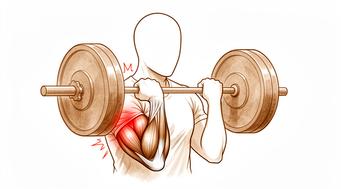
Bạn có thể nhận thấy một cơn đau nhói đột ngột ở phía trước cánh tay trên gần khuỷu tay. Điều này thường xảy ra khi bạn nâng vật nặng hoặc căng cơ cánh tay. Nhiều người mô tả cảm giác như bị đá hoặc đập vào vị trí đó. Bạn cũng có thể nhìn thấy hoặc cảm thấy một khối phình lên ở cánh tay trên, đôi khi được gọi là "cơ Popeye". Điều này xảy ra do gân bị kéo rời khỏi xương.
Cánh tay của bạn có thể cảm thấy yếu khi bạn cố gắng gập khuỷu tay hoặc xoay lòng bàn tay lên trên. Các hoạt động đơn giản có thể trở nên khó khăn. Bạn có thể gặp khó khăn khi nâng túi mua sắm, mở lọ hoặc sử dụng tua vít. Việc với tay ra sau lưng để cài áo ngực hoặc nhét áo vào quần có thể cảm thấy bất tiện hoặc gây đau. Một số người thấy khó ngủ ở bên bị ảnh hưởng vì áp lực làm trầm trọng thêm vùng đau.
Cơn đau thường bùng phát sau khi vận động. Cơn đau có thể nặng hơn khi bạn thức dậy vào buổi sáng nếu bạn đã ngủ với cánh tay ở tư thế gập. Nghỉ ngơi thường giúp làm dịu cơn đau, nhưng tình trạng yếu cơ có thể kéo dài. Nếu bạn bị rách một phần, các triệu chứng có thể ít nghiêm trọng hơn so với đứt hoàn toàn, nhưng chúng vẫn ảnh hưởng đến sinh hoạt hàng ngày của bạn. Phụ nữ có khả năng bị rách một phần hơn là đứt hoàn toàn.
Nếu bạn bị rách cô lập phần đầu ngắn của cơ nhị đầu, bạn có thể cảm thấy một khe hở hoặc rãnh trong cơ. Điều này hiếm gặp, nhưng cần được chú ý sớm nếu bạn có tình trạng yếu cơ đáng kể. Bác sĩ phẫu thuật sẽ kiểm tra các dấu hiệu này để quyết định hướng điều trị tốt nhất. Hầu hết mọi người bị đứt cơ nhị đầu ở đoạn xa đều trải qua các mô hình đau và yếu tương tự, bất kể chấn thương là cấp tính hay mạn tính. Tin tốt là việc sửa chữa bằng phẫu thuật mang lại kết quả nhất quán và tốt đẹp dựa trên các kết quả đánh giá do bệnh nhân tự báo cáo. Bạn có thể mong đợi kết quả chức năng xuất sắc sau hơn 1 năm kể từ khi sửa chữa, bất kể việc sử dụng vít có thể hấp thu sinh học hay không hấp thu.
Những gì thực sự đang xảy ra
Cơ nhị đầu của bạn kết thúc tại khuỷu tay dưới dạng một gân dày và chắc khỏe. Hãy tưởng tượng gân này như một sợi dây cáp chịu lực cao, nối cơ ở cánh tay trên với xương cẳng tay. Chức năng chính của nó là giúp bạn gập khuỷu tay và xoay lòng bàn tay hướng lên trên. Khi gân này bị rách hoặc đứt, sự kết nối đó bị phá vỡ. Cơ không còn có thể kéo xương một cách hiệu quả, đó là lý do tại sao bạn cảm thấy yếu đột ngột và đau đớn.
Chấn thương này thường xảy ra khi bạn cố nâng vật nặng hoặc đỡ một vật đang rơi với cánh tay duỗi thẳng. Lực tác động gây quá tải cho gân, khiến nó bị đứt. Bạn có thể nghe thấy tiếng bốp hoặc cảm thấy như bị xé mạnh. Sau chấn thương, cơ có thể co cụm lại ở cánh tay trên, tạo thành một khối u nổi rõ. Điều này xảy ra vì gân không còn giữ cơ ở vị trí bình thường gần khuỷu tay nữa.
Khuỷu tay là một khớp bản lề phức tạp. Nó dựa vào sự phối hợp của xương, dây chằng và gân để duy trì sự ổn định. Khi gân nhị đầu bị tổn thương, nó có thể ảnh hưởng đến cách khuỷu tay cử động và chịu lực. Bác sĩ phẫu thuật sẽ xem xét vết rách để xác định xem đó là rách toàn phần hay một phần. Rách một phần có nghĩa là một số sợi gân vẫn còn bám dính, trong khi rách toàn phần có nghĩa là gân đã bị tách hoàn toàn.
Việc sửa chữa gân khôi phục lại mối liên kết quan trọng giữa cơ và xương. Các kỹ thuật phẫu thuật hiện đại cho phép bác sĩ phẫu thuật tái gắn gân chắc chắn vào xương cẳng tay. Dù sử dụng chằng hấp thu hay không hấp thu, mục tiêu là đưa gân trở lại vị trí ban đầu. Điều này cho phép mô lành lại và khôi phục sức mạnh.
Thời điểm can thiệp rất quan trọng đối với quá trình hồi phục của bạn. Hầu hết các vấn đề sau phẫu thuật đều xuất phát từ việc chờ đợi quá lâu để phẫu thuật hoặc sử dụng vết rạch lớn làm tổn thương các mô xung quanh. Nếu bạn chờ đợi hơn 21 ngày, gân có thể bị sẹo ở vị trí co ngắn, khiến việc sửa chữa trở nên khó khăn hơn. Tuy nhiên, ngay cả các ca sửa chữa muộn vẫn có thể mang lại kết quả chức năng xuất sắc. Bác sĩ phẫu thuật sẽ lựa chọn phương pháp tiếp cận tốt nhất để giảm thiểu biến chứng và giúp bạn quay lại các hoạt động bình thường một cách an toàn.
Những gì chúng tôi có thể làm về vấn đề này
Bạn có thể bắt đầu bằng cách nghỉ ngơi cánh tay và tránh nâng vật nặng hoặc các chuyển động xoay gây căng thẳng cho phần trước khuỷu tay. Bác sĩ phẫu thuật của bạn có thể khuyên bạn nên thực hiện một giai đoạn vật lý trị liệu để giúp giảm sưng và khôi phục nhẹ nhàng tầm vận động. Mục tiêu là duy trì tính linh hoạt của khớp trong khi gân bị tổn thương ổn định. Bạn nên dành cho phương pháp bảo tồn này một cơ hội hợp lý để phát huy tác dụng trước khi xem xét các bước xâm lấn hơn.
Nếu bạn gặp phải tình trạng đau đớn, bác sĩ phẫu thuật của bạn có thể đề nghị sử dụng thuốc giảm đau không kê đơn hoặc thuốc chống viêm để giúp bạn kiểm soát sự khó chịu. Những loại thuốc này không chữa lành gân, nhưng chúng có thể giúp các hoạt động hàng ngày dễ chịu hơn trong quá trình hồi phục của bạn. Trong một số trường hợp, các loại tiêm như cortisone có thể được sử dụng để làm dịu tình trạng viêm. Tuy nhiên, những phương pháp điều trị này không sửa chữa chính gân bị rách. Tác dụng của chúng chỉ là tạm thời và chúng không phải là giải pháp lâu dài cho tình trạng rách hoàn toàn.
Phẫu thuật được xem xét khi việc chăm sóc không phẫu thuật không làm giảm các triệu chứng của bạn hoặc khi bạn cần khôi phục lại sức mạnh toàn diện để làm việc hoặc tham gia thể thao. Bác sĩ phẫu thuật của bạn sẽ thảo luận về thời điểm tốt nhất để sửa chữa, lưu ý rằng kết quả điều trị rất tốt ngay cả khi việc sửa chữa bị trì hoãn hơn 21 ngày. Phẫu thuật liên quan đến việc nối lại gân bị rách vào xương để khôi phục sức mạnh cho cánh tay của bạn. Phương pháp này mang lại kết quả nhất quán và tốt xét về kết quả được bệnh nhân đánh giá và sức mạnh. Đối với các vết rách một phần không cải thiện sau khi nghỉ ngơi, phẫu thuật cũng là một lựa chọn khả thi. Trong những trường hợp hiếm hoi mà gân không thể được nối lại trực tiếp, bác sĩ phẫu thuật của bạn có thể sử dụng một mảnh ghép để tái tạo khu vực đó. Tỷ lệ biến chứng nghiêm trọng từ phẫu thuật nói chung là thấp, bất kể kỹ thuật được sử dụng.
Những gì bạn có thể mong đợi
Bạn có thể mong đợi kết quả dài hạn xuất sắc sau phẫu thuật sửa chữa gân cơ nhị đầu bị rách. Dù bác sĩ phẫu thuật của bạn sử dụng vít có thể hấp thụ (tan rã) hay vít không hấp thụ (giữ nguyên tại chỗ), kết quả chức năng sau hơn 1 năm đều rất tốt. Hầu hết bệnh nhân báo cáo kết quả tốt một cách nhất quán dựa trên cảm giác và khả năng hoạt động của họ trong cuộc sống hàng ngày. Ngay cả khi bạn có một vết rách một phần không lành lại sau khi nghỉ ngơi, việc sửa chữa bằng phẫu thuật vẫn là một lựa chọn khả thi sau khi điều trị không phẫu thuật thất bại.
Thời gian đóng một vai trò quan trọng trong trải nghiệm hồi phục của bạn. Nếu bạn chờ hơn 21 ngày để thực hiện sửa chữa, bạn vẫn có thể mong đợi các kết quả chức năng tương tự như những trường hợp được điều trị ngay lập tức. Tuy nhiên, việc sửa chữa chậm trễ có liên quan đến tỷ lệ biến chứng ban đầu cao. Hầu hết các vấn đề phát sinh từ phẫu thuật chủ yếu được quy cho sự chậm trễ về thời gian này. Chúng cũng liên quan thứ yếu đến nhu cầu tiếp cận rộng rãi phía trước cánh tay trong quá trình phẫu thuật. Nếu vết rách của bạn là mạn tính hoặc tái phát, việc tái tạo bằng mô hiến tặng (allograft) là an toàn và hiệu quả. Phương pháp này mang lại kết quả báo cáo từ bệnh nhân và tầm vận động xuất sắc, không có biến chứng đáng kể trong các nhóm nghiên cứu.
Các biến chứng nghiêm trọng là hiếm gặp. Tỷ lệ biến chứng chính sau sửa chữa gân cơ nhị đầu xa là 4,6%. Nhìn chung, một trong 20 bệnh nhân sẽ gặp phải một biến chứng chính, và một trong năm bệnh nhân sẽ gặp phải một biến chứng nhẹ. Tổn thương thần kinh là vấn đề phổ biến nhất mà bạn có thể gặp phải. Mặc dù có những rủi ro này, tỷ lệ biến chứng nghiêm trọng nói chung vẫn thấp bất kể phương pháp phẫu thuật nào. Mức độ đào tạo của bác sĩ phẫu thuật của bạn không làm thay đổi đáng kể các hồ sơ rủi ro này so với các nghiên cứu quy mô lớn.
Nếu bạn chọn không phẫu thuật, hoặc nếu vết rách được quản lý bảo tồn, kết quả nói chung là kém hơn so với sửa chữa bằng phẫu thuật. Phẫu thuật cung cấp cơ hội tốt nhất để khôi phục sức mạnh và chức năng. Trong khi các sửa chữa mạn tính có thể có tỷ lệ biến chứng tức thời cao hơn một chút so với các trường hợp cấp tính, kết quả chức năng dài hạn là tương đương. Bạn nên chuẩn bị cho một giai đoạn thích nghi. Những tuần đầu tiên có thể bao gồm khó chịu và hạn chế vận động, nhưng mục tiêu là quay lại sử dụng bình thường. Hầu hết các vấn đề sức khỏe phát sinh từ sự chậm trễ hoặc tiếp cận phẫu thuật rộng rãi, vì vậy việc tuân thủ lời khuyên của bác sĩ phẫu thuật về thời gian và chăm sóc sau phẫu thuật là rất quan trọng để có kết quả tốt nhất có thể.
Khi nào cần gặp bác sĩ
Hãy gặp bác sĩ đa khoa nếu bạn có tình trạng đau dai dẳng không cải thiện khi nghỉ ngơi. Hãy yêu cầu được bác sĩ chuyên khoa thăm khám nếu bạn nhận thấy yếu hoặc mất vững ở cánh tay. Hãy tìm kiếm sự chăm sóc y tế nếu khuỷu tay của bạn bị khóa hoặc đột ngột mất sức. Hãy liên hệ với bác sĩ nếu các triệu chứng ảnh hưởng đến giấc ngủ hoặc công việc của bạn. Hãy tìm sự trợ giúp nếu có bất kỳ sự xấu đi đột ngột nào của cơn đau. Những dấu hiệu này có thể cho thấy một đứt gân cơ nhị đầu xa. Can thiệp sớm được khuyến nghị nếu bạn cảm thấy có một khe hở ở cơ hoặc có tình trạng yếu đáng kể. Việc trì hoãn sửa chữa có thể làm tăng nguy cơ biến chứng. Bác sĩ phẫu thuật của bạn sẽ thăm khám để xác định phương hướng điều trị tốt nhất.
Evidence & references
Overview
- Clinical and functional outcomes at more than 1 year after distal biceps tendon repair are excellent for patients treated with bioabsorbable screws [1].
- Clinical and functional outcomes at more than 1 year after distal biceps tendon repair are excellent for patients treated with nonabsorbable screws [1].
- Selective disruption of the short head of the biceps distal tendon may be effectively treated with anatomic repair when diagnosed appropriately [2].
- Patients have excellent overall outcomes after distal biceps tendon rupture repair [3].
- Functional outcomes for chronic distal biceps ruptures are comparable to those seen in acute distal biceps ruptures, despite a potentially slightly higher immediate complication rate [4].
- Patients treated with delayed distal biceps tendon repair (>21 days) can expect similar functional outcomes to those treated acutely, despite a high rate of initial complications [5].
- Most morbidity from repair of the distal biceps tendon is attributed primarily to a delay in the timing of the repair [6].
- Most morbidity from repair of the distal biceps tendon is attributed secondarily to an extensive anterior exposure [6].
- Bioabsorbable interference screw fixation of distal biceps ruptures through a single anterior incision is a safe and successful technique [7].
- Surgical repair of distal biceps ruptures provides consistently good results in terms of patient-scored outcomes [8].
- The surgical repair of distal biceps tendon ruptures has an overall low rate of serious complications, regardless of approach or technique [9].
- Surgical treatment of partial distal biceps tendon tears is a viable option after failed nonsurgical treatment [11].
- Utilizing a double-incision technique with a combination of cortical button and interference screw fixation helps determine the best method of fixation and approach to minimize postoperative complications in patients with distal biceps ruptures [14].
- Two-incision distal biceps repairs demonstrate a high degree of patient satisfaction [21].
- Two-incision distal biceps repairs demonstrate a low complication rate [21].
Anatomy & Pathophysiology
- The elbow's stability and complex kinematics are accounted for by a combination of bony articulation and soft-tissue stabilizers [29].
- Musculoskeletal ultrasonography provides a dynamic, functional assessment of elbow structures, allowing visualization of pathology under stress and motion [30].
- The medial elbow joint space increases under a valgus load [31].
- The medial elbow joint space decreases when a maximal grip contraction is performed [31].
- A low carrying angle is measured in elite tennis players just before ball impact during the forehand, suggesting a dynamic varus instant accommodation moving towards full extension [32].
- The decrease in the carrying angle is a consequence of an increase in elbow flexion position dictated by the transition from a closed to open, semi-open stances [32].
- Determining which structures need to be repaired is important to avoid complications that could lead to elbow instability [33].
- Ulnar collateral ligament reconstruction using a suspension button fixation technique reliably restored elbow kinematics to the intact state [34].
- Limiting the number of innings pitched is likely the best way to reduce elbow injury in youth pitchers because biomechanical variables correlated with peak valgus torque are not easily modifiable [35].
- The medial elbow joint space was significantly reduced under 60-N valgus stress plus 50% MVC compared to 60-N valgus stress alone [37].
- No significant relationships between adaptations in shoulder strength or ROM were related to chronic structural adaptations of the elbow in professional baseball pitchers [38].
- The spin move is a simple maneuver that can improve exposure of the coronoid process regardless of the degree of elbow instability [39].
- Reattachment of the flexor and extensor tendons at the epicondyle should be considered in the development of improved repair techniques for stabilizers of the elbow [40].
- An internal joint stabilizer with a standardized treatment protocol could maintain concentric reduction while allowing early functional motion and improve clinical outcomes for patients with complex persistent elbow instability [41].
- Activation of lateral forearm muscles decreased elbow varus angulation throughout flexion [42].
- In patients treated with surgery for snapping triceps syndrome, it is crucial to make sure full resolution of the snapping by examining all dislocating structures during passive elbow motion and/or myoelectrical stimulation to achieve excellent results [43].
- Pre-operative evaluations in elbow stiffness should identify involved articular and periarticular tissues and determine whether articular surfaces and osteoarticular congruence are preserved [44].
- Elbow vascularized composite allotransplantation may be technically feasible on the basis of its consistent vascular anatomy and a proposed surgical technique [45].
- Elbow valgus stress might correlate with ball velocity at 75% partial valgus stress pitch [46].
- Physical examination of the elbow is a critical component in formulating an accurate diagnosis [47].
Classification
- Distal biceps tendon tears in women present differently than in men [10].
- The incidence of women sustaining a distal biceps tendon tear is 3.2% [12].
- Partial tears are statistically more common than complete ruptures in women [12].
- Partial ruptures of the distal biceps brachii tendon represent a spectrum of patterns with varying involvement of the long head (LH) and short head (SH) tendons [16].
- Selective disruption of the short head of the biceps distal tendon may be effectively treated with anatomic repair when diagnosed appropriately [2].
Clinical Presentation
- Distal biceps tendon tears in women present differently than in men [10].
- The incidence of women sustaining a distal biceps tendon tear is 3.2% [12].
- Partial tears are statistically more common than complete ruptures in women [12].
- Isolated rupture of the short head of the biceps brachii is a rare injury [15].
- Early intervention for isolated short head rupture is recommended if a sulcus or gap in the musculotendinous unit is palpable or considerable weakness is present [15].
- Partial ruptures of the distal biceps brachii tendon represent a spectrum of patterns with varying involvement of the long head (LH) and short head (SH) tendons [16].
- The Hook test has imperfect validity for diagnosing distal biceps tendon ruptures and should not be used alone [17].
- The prevalence of distal biceps tendon signal changes on MRI in asymptomatic patients is very low [18].
- Patients with a smaller radioulnar window have a higher risk of rupturing the distal biceps tendon [19].
Investigations
- The Hook test has imperfect validity and should not be used alone to diagnose distal biceps tendon ruptures [17].
- The prevalence of distal biceps tendon signal changes on MRI in asymptomatic patients is very low [18].
- Patients with a smaller radioulnar window have a higher risk of rupturing the distal biceps tendon [19].
- Distal biceps tendon tears in women present differently than in men [10].
- Isolated rupture of the short head of the biceps brachii is a rare injury [15].
- Early intervention is recommended for isolated rupture of the short head of the biceps brachii if a sulcus or gap in the musculotendinous unit is palpable or considerable weakness is present [15].
Treatment
Operative Repair
- Clinical and functional outcomes at more than 1 year after distal biceps tendon repair are excellent for both bioabsorbable and nonabsorbable screw fixation groups [1].
- Selective disruption of the short head of the biceps distal tendon may be effectively treated with anatomic repair when diagnosed appropriately [2].
- Patients have excellent overall outcomes after distal biceps tendon rupture repair [3].
- Functional outcomes in chronic distal biceps rupture repairs are comparable to those in acute distal biceps rupture populations, despite a potentially slightly higher immediate complication rate [4].
- Patients treated with distal biceps tendon repair after a delay of more than 21 days can expect similar functional outcomes to those treated acutely, despite a high rate of initial complications [5].
- Most morbidity from repair of the distal biceps tendon is attributed primarily to a delay in the timing of the repair and secondarily to an extensive anterior exposure [6].
- Bioabsorbable interference screw fixation of distal biceps ruptures through a single anterior incision is a safe and successful technique [7].
- Surgical repair of distal biceps ruptures provides consistently good results in terms of patient-scored outcomes [8].
- The surgical repair of distal biceps tendon ruptures has an overall low rate of serious complications, regardless of approach or technique [9].
- Surgical treatment of partial distal biceps tendon tears is a viable option after failed nonsurgical treatment [11].
- Reinsertion of the distal biceps tendon on the brachialis tendon is a safe and effective procedure with a low complication rate, providing satisfactory subjective and objective results at long-term follow-up [13].
- Utilizing a double-incision technique with a combination of cortical button and interference screw fixation helps determine the best method of fixation and approach with the goal of minimizing postoperative complications [14].
- Distal biceps reconstruction using an Achilles tendon allograft, transosseous EndoButton, and Pulvertaft weave with tendon wrap technique is recommended for patients with retracted, irreparable distal biceps ruptures [26].
- A two-incision repair with suture anchor attachment is a safe and effective method for treatment of distal biceps tendon ruptures [36].
- Repair of distal biceps ruptures using an Endobutton fixation results in a nearly normal return of strength and function, which is significantly better than in those managed nonoperatively [50].
Non-Operative Management
- Incomplete distal triceps tears with active elbow extension against resistance are managed nonsurgically [52].
Complications
- Distal biceps tendon repair has an overall low rate of serious complications, regardless of approach or technique [9].
- The major complication rate after distal biceps tendon repair is 4.6% [23].
- One in five patients will have a minor complication after surgery on the distal biceps tendon [24].
- One in twenty patients will have a major complication after surgery on the distal biceps tendon [24].
- Nerve injury is the most common complication after distal biceps tendon repair [22].
- Most morbidity from repair of the distal biceps tendon is attributed primarily to a delay in the timing of the repair [6].
- Most morbidity from repair of the distal biceps tendon is attributed secondarily to an extensive anterior exposure [6].
- Chronic distal biceps rupture repair may have a slightly higher immediate complication rate compared to acute repair [4].
- Delayed repair of distal biceps tendon ruptures (>21 days) is associated with a high rate of initial complications [5].
- Allograft reconstruction of distal biceps injuries for chronic and recurrent tears results in no significant complications among the studied cohort [20].
- Distal biceps repairs using the two-incision technique demonstrate a low complication rate [21].
- Reinsertion of the distal biceps tendon on the brachialis tendon is associated with a low complication rate [13].
- Complication rates after distal biceps tendon repair performed by newly trained surgeons are similar to those previously reported in large cohort studies [22].
Recovery
- Clinical and functional outcomes at more than 1 year after distal biceps tendon repair are excellent for patients treated with bioabsorbable screws [1].
- Clinical and functional outcomes at more than 1 year after distal biceps tendon repair are excellent for patients treated with nonabsorbable screws [1].
- Selective disruption of the short head of the biceps distal tendon may be effectively treated with anatomic repair when diagnosed appropriately [2].
- Patients have excellent overall outcomes after distal biceps tendon rupture repair [3].
- Functional outcomes for chronic distal biceps ruptures are comparable to those seen in acute distal biceps ruptures, despite a potentially slightly higher immediate complication rate [4].
- Patients treated with distal biceps tendon repair after a delay of more than 21 days can expect similar functional outcomes to those treated acutely, despite a high rate of initial complications [5].
- Most morbidity from repair of the distal biceps tendon is attributed primarily to a delay in the timing of the repair [6].
- Most morbidity from repair of the distal biceps tendon is attributed secondarily to an extensive anterior exposure [6].
- Surgical repair of distal biceps ruptures provides consistently good results in terms of patient-scored outcomes [8].
- Surgical treatment of partial distal biceps tendon tears is a viable option after failed nonsurgical treatment [11].
- Reinsertion of the distal biceps tendon on the brachialis tendon is a safe and effective procedure with a low complication rate, providing satisfactory subjective and objective results at long-term follow-up [13].
- Allograft reconstruction of distal biceps injuries for chronic and recurrent tears results in excellent patient-reported outcomes and range of motion [20].
- Allograft reconstruction of distal biceps injuries for chronic and recurrent tears results in no significant complications among the studied cohort [20].
- Complication rates after distal biceps tendon repair performed by newly trained surgeons are similar to those previously reported in large cohort studies [22].
- Nerve injury is the most common complication after distal biceps tendon repair performed by newly trained surgeons [22].
- One in 5 patients will have a minor complication after surgery on the distal biceps tendon [24].
- One in 20 patients will have a major complication after surgery on the distal biceps tendon [24].
- Patients with biceps tendon repair etiology had a trend toward greater motion improvement than that of patients with a traumatic etiology regarding anconeus interposition for proximal radioulnar synostosis [25].
- Nearly all complete distal biceps ruptures can be effectively reapproximated to the radial tuberosity within the first 6 weeks of injury using single-incision repair with suture anchors [28].
- Primary repair of chronic distal biceps tendon tears greater than 6 weeks from injury demonstrated excellent patient-reported outcome measures (PROMs) and elbow range of motion (ROM) [48].
- Delayed reconstruction of irreparable distal biceps tendon ruptures with semitendinosus autograft produces similar strength, range of motion, and complication rates compared with delayed primary repair [55].
- Delayed reconstruction of irreparable distal biceps tendon ruptures with semitendinosus autograft produces slightly worse functional outcome scores compared with delayed primary repair [55].
Key Evidence
- [L3] Clinical and functional outcome at more than 1 year after distal biceps tendon repair was excellent in both groups. [1] (10.1016/j.jse.2015.12.007)
- [L4] When diagnosed appropriately, selective disruption of the short head of the biceps distal tendon may be effectively treated with anatomic repair. [2] (10.1016/j.jse.2016.09.050)
- [L3] Overall, patients have excellent outcome after distal biceps tendon rupture repair. [3] (10.1007/s00402-018-3018-6)
- [L4] Although there may be a slightly higher immediate complication rate, the functional outcomes remain comparable with those seen in the patient population with acute distal biceps. [4] (10.1177/23259671211065772)
- [L3] Despite a high rate of initial complications, patients treated with distal biceps tendon repair after a delay (>21 days) can expect similar functional outcomes to those treated acutely. [5] (10.1016/j.jse.2017.02.025)
- [L4] Most morbidity from repair of the distal biceps tendon can be attributed primarily to a delay in the timing of the repair and secondarily to an extensive anterior exposure. [6] (10.2106/00004623-200011000-00010)
- [Paper] This is a safe and successful technique for the management of distal biceps tendon ruptures. [7] (10.1007/s00402-009-0974-x)
- [L4] Surgical repair of distal biceps ruptures provides consistently good results in terms of patient-scored outcomes. [8] (10.1016/j.injury.2012.10.029)
- [L3] The surgical repair of distal biceps tendon ruptures has an overall low rate of serious complications, regardless of approach or technique. [9] (10.1177/0363546517720200)
- [L4] Distal biceps tendon tears in women present differently than in men. [10] (10.1016/j.jse.2010.01.015)
- [L4] Surgical treatment of partial distal biceps tendon tears is a viable option after failed nonsurgical treatment. [11] (10.1016/j.jhsa.2010.04.024)
- [L4] The incidence of women sustaining a distal biceps tendon tear is 3.2%, with partial tears being statistically more common than complete ruptures. [12] (10.1016/j.jse.2014.02.006)
- [L4] Reinsertion of the distal biceps tendon on the brachialis tendon can be considered a safe and effective procedure with low complication rate, providing satisfactory subjective and objective results at long-term follow-up. [13] (10.1007/s00167-008-0705-9)
- [L4] This will help surgeons determine the best method of fixation and approach with the goal of minimizing postoperative complications in patients with distal biceps ruptures. [14] (10.1177/17585732241312212)
- [L4] Isolated rupture of the short head of the biceps brachii is a rare injury and early intervention is recommended if a sulcus or gap in musculotendinous unit is palpable or considerable weakness is present. [15] (10.1007/s00167-010-1108-2)
- [L4] Partial ruptures of the distal biceps brachii tendon represent a spectrum of patterns with varying involvement of the LH and SH tendons. [16] (10.1016/j.jse.2020.04.021)
- [L2] The authors caution against using the Hook test alone to diagnose distal biceps tendon ruptures due to its imperfect validity. [17] (10.1016/j.jhsa.2023.07.004)
- [L3] The prevalence of distal biceps tendon signal changes on MRI in asymptomatic patients is very low. [18] (10.1016/j.jhsa.2022.01.020)
- [L3] Therefore, patients with a smaller radioulnar window have a higher risk of rupturing the distal biceps tendon. [19] (10.1016/j.jse.2023.09.020)
- [L5] Allograft reconstruction of distal biceps injuries for chronic and recurrent tears results in excellent patient-reported outcomes and range of motion, with no significant complications among our cohort. [20] (10.1016/j.xrrt.2025.100661)
- [L3] In this large cohort of 2-incision distal biceps repairs, we found a high degree of patient satisfaction and a low complication rate. [21] (10.1016/j.jse.2014.12.032)
- [L4] Complication rates after distal biceps tendon repair performed by newly trained surgeons were similar to those previously reported in large cohort studies, with nerve injury as the most common complication. [22] (10.1016/j.jse.2022.09.014)
- [L2] This is the largest analysis of complications after distal biceps repair, indicating a major complication rate of 4.6%. [23] (10.1177/0363546519899933)
- [L4] Patients should be counseled that 1 in 5 patients will have a minor complication and 1 in 20 patients will have a major complication after surgery on the distal biceps tendon. [24] (10.1016/j.jse.2016.02.032)
- [L3] Patients with biceps tendon repair etiology had a trend toward greater motion improvement than that of patients with a traumatic etiology. [25] (10.1016/j.jse.2014.07.011)
- [L4] This technique is recommended for patients who have a retracted, irreparable distal biceps rupture. [26] (10.1016/j.jse.2016.01.014)
- [L4] The study demonstrates that nearly all complete distal biceps ruptures can be effectively reapproximated to the radial tuberosity within the first 6 weeks of injury. [28] (10.1016/j.jse.2006.03.002)
- [L5] This article discusses the basic anatomy of the elbow and the biomechanics of this joint, noting that a combination of bony articulation and soft-tissue stabilizers accounts for the elbow's stability and complex kinematics. [29] (10.1016/j.csm.2004.06.008)
- [L5] Musculoskeletal ultrasonography provides a dynamic, functional assessment of elbow structures, allowing visualization of pathology under stress and motion. [30] (10.5435/jaaos-d-20-00935)
- [L4] Medial elbow joint space increases under a valgus load and then decreases when a maximal grip contraction is performed. [31] (10.1177/0363546518755149)
- [L4] The observed decrease in the carrying angle is a consequence of an increase in elbow flexion position dictated by the transition from a closed to open, semi‐open stances. [32] (10.1002/ksa.12016)
- [L3] It is important to determine which structures need to be repaired to avoid complications that could lead to elbow instability. [33] (10.1016/j.jse.2019.07.006)
- [L5] Ulnar collateral ligament reconstruction using a suspension button fixation technique reliably restored elbow kinematics to the intact state. [34] (10.1177/0363546509350109)
- [L5] Given that the biomechanical variables correlated with peak valgus torque are not easily modifiable, limiting the number of innings pitched is likely the best way to reduce elbow injury in youth pitchers. [35] (10.1016/j.jse.2004.01.013)
- [L4] Based on our data, we believe that a two-incision repair with suture anchor attachment is a safe and effective method for treatment of distal biceps tendon ruptures. [36] (10.1007/s001670050134)
- [L5] The medial elbow joint space was significantly reduced under 60-N valgus stress plus 50% MVC compared to 60-N valgus stress alone. [37] (10.1016/j.jse.2022.03.027)
- [L3] However, no significant relationships between adaptations in shoulder strength or ROM were related to chronic structural adaptations of the elbow. [38] (10.1177/03635465251317509)
- [L5] The spin move is a simple maneuver that can improve exposure of the coronoid process regardless of the degree of elbow instability. [39] (10.1016/j.jse.2022.11.020)
- [L5] Thus, it should be considered in the development of improved repair techniques for stabilizers of the elbow. [40] (10.1186/s12891-018-2341-y)
- [L4] An internal joint stabilizer with a standardized treatment protocol could maintain concentric reduction while allowing early functional motion and improve clinical outcomes for patients with complex persistent elbow instability. [41] (10.1097/corr.0000000000002159)
- [L5] In addition, activation of lateral forearm muscles decreased elbow varus angulation throughout flexion. [42] (10.1016/j.jhsg.2024.11.006)
- [L4] In patients treated with surgery, it is crucial to make sure full resolution of the snapping by examining all dislocating structures during passive elbow motion and/or myoelectrical stimulation to achieve excellent results. [43] (10.1016/j.xrrt.2025.08.017)
- [L5] Pre-operative evaluations in elbow stiffness should identify involved articular and periarticular tissues and determine whether articular surfaces and osteoarticular congruence are preserved. [44] (10.1016/j.jisako.2023.10.009)
- [L5] Elbow VCA may be technically feasible on the basis of its consistent vascular anatomy and our proposed surgical technique. [45] (10.1016/j.jse.2017.04.014)
- [L5] Elbow valgus stress might correlate with ball velocity at 75% partial valgus stress pitch. [46] (10.1016/j.jse.2022.08.009)
- [L5] Physical examination of the elbow is a critical component in formulating an accurate diagnosis. [47] (10.5435/jaaos-d-16-00622)
- [L4] Primary repair of chronic distal biceps tendon tears greater than 6 weeks from injury demonstrated excellent PROMs and elbow ROM. [48] (10.1177/15589447221107691)
- [L3] Repair of distal biceps ruptures using an Endobutton fixation results in nearly normal return of strength and function, which is significantly better than in those managed nonoperatively. [50] (10.1016/j.jse.2015.10.008)
- [L5] Incomplete tears with active elbow extension against resistance are managed nonsurgically. [52] (10.5435/00124635-201001000-00005)
- [L3] Delayed reconstruction of irreparable distal biceps tendon ruptures with semitendinosus autograft produces similar strength, range of motion, and complication rates but slightly worse functional outcome scores compared with delayed primary repair. [55] (10.1016/j.jse.2019.01.006)
References
[1] Distal biceps tendon repair: comparison of clinical and radiological outcome between bioabsorbable and nonabsorbable screws. Journal of Shoulder and Elbow Surgery. 2016. DOI: 10.1016/j.jse.2015.12.007 [2] Rupture of the short head component of a bifurcated distal biceps tendon. Journal of Shoulder and Elbow Surgery. 2017. DOI: 10.1016/j.jse.2016.09.050 [3] Functional outcome in patients who underwent distal biceps tendon repair. Archives of Orthopaedic and Trauma Surgery. 2018. DOI: 10.1007/s00402-018-3018-6 [4] Surgical Results of Chronic Distal Biceps Ruptures: A Systematic Review. Orthopaedic Journal of Sports Medicine. 2022. DOI: 10.1177/23259671211065772 [5] Delayed repair of distal biceps tendon ruptures is successful: a case-control study. Journal of Shoulder and Elbow Surgery. 2017. DOI: 10.1016/j.jse.2017.02.025 [6] Complications of Repair of the Distal Biceps Tendon with the Modified Two-Incision Technique†. The Journal of Bone and Joint Surgery-American Volume. 2000. DOI: 10.2106/00004623-200011000-00010 [7] Bioabsorbable interference screw fixation of distal biceps ruptures through a single anterior incision: a single-surgeon case series and review of the literature. Archives of Orthopaedic and Trauma Surgery. 2009. DOI: 10.1007/s00402-009-0974-x [8] Distal biceps tendon rupture: Current concepts. Injury. 2013. DOI: 10.1016/j.injury.2012.10.029 [9] Surgical Treatment of Distal Biceps Tendon Ruptures: An Analysis of Complications in 784 Surgical Repairs. The American Journal of Sports Medicine. 2017. DOI: 10.1177/0363546517720200 [10] Distal biceps tendon tears in women. Journal of Shoulder and Elbow Surgery. 2010. DOI: 10.1016/j.jse.2010.01.015 [11] Surgical Treatment of Partial Distal Biceps Tendon Ruptures. The Journal of Hand Surgery. 2010. DOI: 10.1016/j.jhsa.2010.04.024 [12] Re-rupture rate of primarily repaired distal biceps tendon injuries. Journal of Shoulder and Elbow Surgery. 2014. DOI: 10.1016/j.jse.2014.02.006 [13] Surgical repair of the distal biceps brachii tendon: clinical and isokinetic long‐term follow‐up. Knee Surgery, Sports Traumatology, Arthroscopy. 2009. DOI: 10.1007/s00167-008-0705-9 [14] Outcomes of utilizing double-incision technique with combination of cortical button and interference screw fixation for distal biceps rupture: A case series. Shoulder & Elbow. 2025. DOI: 10.1177/17585732241312212 [15] Traumatic isolated closed rupture of the short head of the biceps brachii in military paratrooper. Knee Surgery, Sports Traumatology, Arthroscopy. 2010. DOI: 10.1007/s00167-010-1108-2 [16] Partial rupture of the distal biceps brachii tendon: a magnetic resonance imaging analysis. Journal of Shoulder and Elbow Surgery. 2020. DOI: 10.1016/j.jse.2020.04.021 [17] Reliability and Validity of the Hook Test for Diagnosis of Distal Biceps Tendon Ruptures. The Journal of Hand Surgery. 2023. DOI: 10.1016/j.jhsa.2023.07.004 [18] Evaluation of MRI Signal Changes of the Distal Biceps Tendon in Asymptomatic Patients. The Journal of Hand Surgery. 2022. DOI: 10.1016/j.jhsa.2022.01.020 [19] Smaller radioulnar window is associated with a distal biceps tendon rupture in patients with limited forearm rotation: a 3-dimensional computed tomography comparison study of proximal impingement caused by radial tuberosity hypertrophy—a single-center case series. Journal of Shoulder and Elbow Surgery. 2024. DOI: 10.1016/j.jse.2023.09.020 [20] Allograft reconstruction of distal biceps ruptures: surgical technique and analysis of outcomes in chronic tears and failed repairs. JSES Reviews, Reports, and Techniques. 2026. DOI: 10.1016/j.xrrt.2025.100661 [21] Endobutton versus transosseous suture repair of distal biceps rupture using the two-incision technique: a comparison series. Journal of Shoulder and Elbow Surgery. 2015. DOI: 10.1016/j.jse.2014.12.032 [22] Trends and complications of distal biceps tendon repair among American Board of Orthopaedic Surgery part II oral examination candidates. Journal of Shoulder and Elbow Surgery. 2023. DOI: 10.1016/j.jse.2022.09.014 [23] Complications After Distal Biceps Tendon Repair: A Systematic Review. The American Journal of Sports Medicine. 2020. DOI: 10.1177/0363546519899933 [24] Factors associated with adverse events after distal biceps tendon repair or reconstruction. Journal of Shoulder and Elbow Surgery. 2016. DOI: 10.1016/j.jse.2016.02.032 [25] Outcomes of anconeus interposition for proximal radioulnar synostosis. Journal of Shoulder and Elbow Surgery. 2014. DOI: 10.1016/j.jse.2014.07.011 [26] Distal biceps reconstruction using an Achilles tendon allograft, transosseous EndoButton, and Pulvertaft weave with tendon wrap technique for retracted, irreparable distal biceps ruptures. Journal of Shoulder and Elbow Surgery. 2016. DOI: 10.1016/j.jse.2016.01.014 [28] Single-incision repair of acute distal biceps ruptures by use of suture anchors. Journal of Shoulder and Elbow Surgery. 2007. DOI: 10.1016/j.jse.2006.03.002 [29] Elbow anatomy and structural biomechanics. Clinics in Sports Medicine. 2004. DOI: 10.1016/j.csm.2004.06.008 [30] Ultrasound Examination Techniques for Elbow Injuries in Overhead Athletes. Journal of the American Academy of Orthopaedic Surgeons. 2020. DOI: 10.5435/jaaos-d-20-00935 [31] Medial Elbow Joint Space Increases With Valgus Stress and Decreases When Cued to Perform A Maximal Grip Contraction. The American Journal of Sports Medicine. 2018. DOI: 10.1177/0363546518755149 [32] A low carrying angle is measured in elite tennis players just before ball impact during the forehand, suggesting a dynamic varus instant accommodation moving towards full extension. Knee Surgery, Sports Traumatology, Arthroscopy. 2024. DOI: 10.1002/ksa.12016 [33] Isolated displaced type II partial articular radial head fracture: correlation of preoperative imaging with intraoperative findings of lateral ulnar collateral ligament tear. Journal of Shoulder and Elbow Surgery. 2020. DOI: 10.1016/j.jse.2019.07.006 [34] Revision Ulnar Collateral Ligament Reconstruction Using a Suspension Button Fixation Technique. The American Journal of Sports Medicine. 2009. DOI: 10.1177/0363546509350109 [35] Valgus torque in youth baseball pitchers: a biomechanical study. Journal of Shoulder and Elbow Surgery. 2004. DOI: 10.1016/j.jse.2004.01.013 [36] Repair of distal biceps tendon rupture with suture anchors. Knee Surgery, Sports Traumatology, Arthroscopy. 1999. DOI: 10.1007/s001670050134 [37] Changes in medial elbow joint space with differences in contraction strength of flexor-pronator muscle under elbow valgus stress. Journal of Shoulder and Elbow Surgery. 2022. DOI: 10.1016/j.jse.2022.03.027 [38] Chronic Structural Adaptations of the Shoulder and Elbow Are Correlated in Professional Baseball Pitchers. The American Journal of Sports Medicine. 2025. DOI: 10.1177/03635465251317509 [39] The spin move to facilitate antegrade coronoid fixation in terrible triad injuries. Journal of Shoulder and Elbow Surgery. 2023. DOI: 10.1016/j.jse.2022.11.020 [40] Reattachment of the flexor and extensor tendons at the epicondyle in elbow instability: a biomechanical comparison of techniques. BMC Musculoskeletal Disorders. 2018. DOI: 10.1186/s12891-018-2341-y [41] Does an Internal Joint Stabilizer and Standardized Protocol Prevent Recurrent Instability in Complex Persistent Elbow Instability?. Clinical Orthopaedics & Related Research. 2022. DOI: 10.1097/corr.0000000000002159 [42] The Effect of Arm Abduction and Forearm Muscle Activation on Kinematics During Elbow Flexion. Journal of Hand Surgery Global Online. 2025. DOI: 10.1016/j.jhsg.2024.11.006 [43] Snapping triceps syndrome: a review of the literature and proposed operative treatment algorithm. JSES Reviews, Reports, and Techniques. 2026. DOI: 10.1016/j.xrrt.2025.08.017 [44] Elbow stiffness: Arthritis and heterotopic ossification. Journal of ISAKOS. 2024. DOI: 10.1016/j.jisako.2023.10.009 [45] Elbow vascularized composite allotransplantation—surgical anatomy and technique. Journal of Shoulder and Elbow Surgery. 2017. DOI: 10.1016/j.jse.2017.04.014 [46] Effect of quantitative partial valgus stress during baseball pitching on ball velocity and subjective pitch-effort. Journal of Shoulder and Elbow Surgery. 2023. DOI: 10.1016/j.jse.2022.08.009 [47] Comprehensive Review of the Elbow Physical Examination. Journal of the American Academy of Orthopaedic Surgeons. 2018. DOI: 10.5435/jaaos-d-16-00622 [48] Primary Repair of Chronic Distal Biceps Tendon Tears. HAND. 2022. DOI: 10.1177/15589447221107691 [50] A comparison of nonoperative vs. Endobutton repair of distal biceps ruptures. Journal of Shoulder and Elbow Surgery. 2016. DOI: 10.1016/j.jse.2015.10.008 [52] Distal Triceps Rupture. American Academy of Orthopaedic Surgeon. 2010. DOI: 10.5435/00124635-201001000-00005 [55] Management of chronic distal biceps tendon ruptures: primary repair vs. semitendinosus autograft reconstruction. Journal of Shoulder and Elbow Surgery*. 2019. DOI: 10.1016/j.jse.2019.01.006


