Distal Biceps Rupture Info Evidence
Last reviewed
Distal biceps rupture causes sudden elbow pain, bruising, and weakness—often needing surgical repair.
What you're feeling
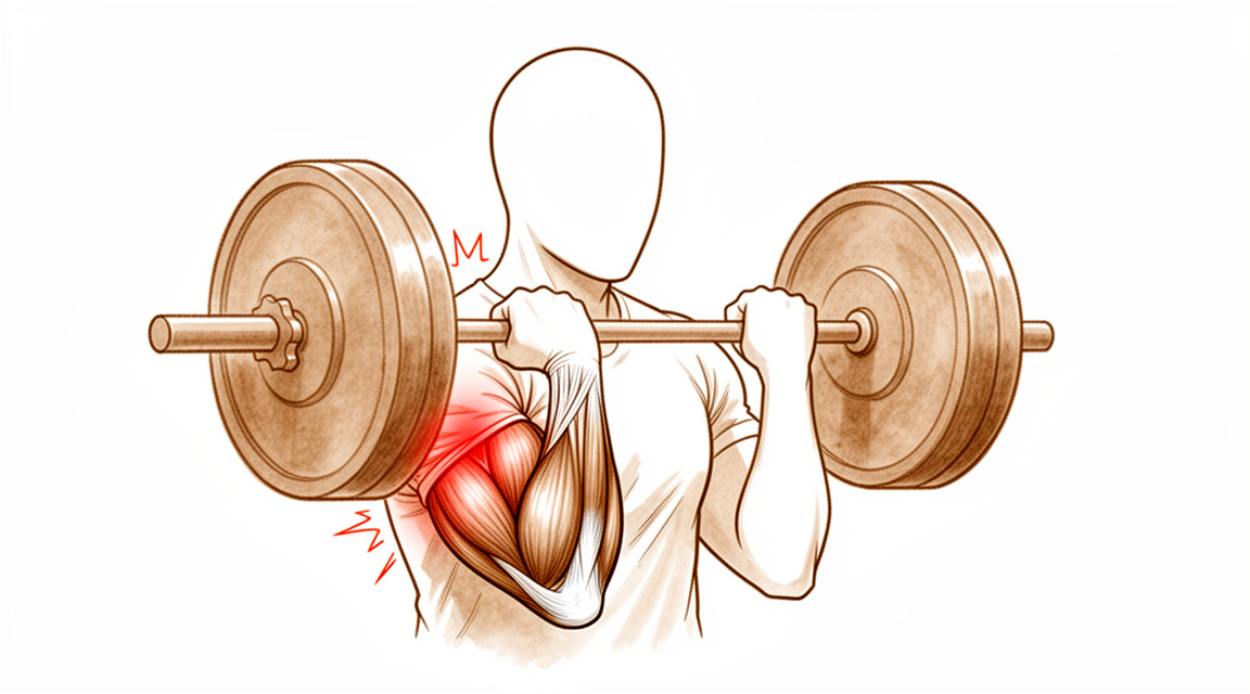
You may notice a sudden, sharp pain in the front of your upper arm near the elbow. This often happens when you lift something heavy or strain your arm. Many people describe it as feeling like they were kicked or hit in that spot. You might also see or feel a bulge in your upper arm, sometimes called a "Popeye muscle." This happens because the tendon has pulled away from the bone.
Your arm may feel weak when you try to bend your elbow or turn your palm upward. Simple tasks can become difficult. You might struggle to lift a grocery bag, open a jar, or use a screwdriver. Reaching behind your back to fasten a bra or tucking in a shirt may feel awkward or painful. Some people find it hard to sleep on the affected side because the pressure aggravates the sore area.
Pain often flares up after activity. It may be worse when you wake up in the morning if you slept with your arm bent. Rest usually helps calm the ache, but the weakness can linger. If you have a partial tear, the symptoms might be less severe than a complete rupture, but they still affect your daily routine. Women are more likely to have partial tears than complete ones.
If you have an isolated tear of the short head of the biceps, you might feel a gap or sulcus in the muscle. This is rare, but it requires early attention if you have considerable weakness. Your surgeon will check for these signs to decide on the best path forward. Most people with distal biceps tears experience similar patterns of pain and weakness, regardless of whether the injury is acute or chronic. The good news is that surgical repair provides consistently good results for patient-scored outcomes. You can expect excellent functional outcomes at more than 1 year after repair, whether using bioabsorbable or nonabsorbable screws.
What's actually happening
Your biceps muscle ends at your elbow as a thick, strong tendon. Think of this tendon like a heavy-duty rope that connects your upper arm muscle to your forearm bone. Its main job is to help you bend your elbow and turn your palm upward. When this tendon tears or ruptures, that connection is broken. The muscle can no longer pull on the bone effectively, which is why you feel sudden weakness and pain.
This injury often happens when you try to lift something heavy or catch a falling object with your arm straight. The force puts too much stress on the tendon, causing it to snap. You might hear a pop or feel a sharp tear. After the injury, the muscle may bunch up in your upper arm, creating a visible bulge. This happens because the tendon is no longer holding the muscle in its normal position near the elbow.
The elbow is a complex hinge joint. It relies on bones, ligaments, and tendons working together for stability. When the biceps tendon is damaged, it can affect how your elbow moves and bears weight. Your surgeon will look at the tear to see if it is complete or partial. A partial tear means some fibers are still attached, while a complete tear means the tendon has fully detached.
Repairing the tendon restores that crucial link between muscle and bone. Modern surgical techniques allow your surgeon to reattach the tendon securely to the forearm bone. Whether using absorbable or non-absorbable anchors, the goal is to bring the tendon back to its original spot. This allows the tissue to heal and regain strength.
Timing matters for your recovery. Most problems after surgery come from waiting too long to operate or using a large incision that disturbs surrounding tissues. If you wait more than 21 days, the tendon may scar in a shortened position, making repair harder. However, even delayed repairs can still yield excellent functional outcomes. Your surgeon will choose the best approach to minimize complications and help you return to normal activities safely.
What we can do about it
You can start by resting your arm and avoiding heavy lifting or twisting motions that strain the front of your elbow. Your surgeon may recommend a period of physiotherapy to help reduce swelling and gently restore movement. The goal is to keep the joint flexible while the injured tendon settles. You should give this conservative approach a fair chance to work before considering more invasive steps.
If you experience pain, your surgeon may suggest over-the-counter pain medication or anti-inflammatory drugs to help you manage discomfort. These medicines do not heal the tendon, but they can make daily activities more bearable while you recover. In some cases, injections such as cortisone might be used to calm inflammation. However, these treatments do not repair the torn tendon itself. Their effects are temporary, and they are not a long-term solution for a complete rupture.
Surgery is considered when non-surgical care does not relieve your symptoms or when you need to regain full strength for work or sports. Your surgeon will discuss the best timing for repair, noting that outcomes are excellent even if the repair is delayed by more than 21 days. The operation involves reattaching the torn tendon to the bone to restore your arm’s power. This approach provides consistently good results in terms of patient-scored outcomes and strength. For partial tears that do not improve with rest, surgery is also a viable option. In rare cases where the tendon cannot be reattached directly, your surgeon may use a graft to reconstruct the area. The overall rate of serious complications from surgery is low, regardless of the technique used.
What to expect
You can expect excellent long-term results after surgery to repair your torn biceps tendon. Whether your surgeon uses absorbable screws that dissolve or nonabsorbable screws that stay in place, your functional outcomes at more than 1 year are excellent. Most patients report consistently good results based on how they feel and function in daily life. Even if you have a partial tear that did not heal with rest, surgical repair remains a viable option after nonsurgical treatment fails.
Timing plays a significant role in your recovery experience. If you wait more than 21 days to have the repair, you can still expect functional outcomes similar to those treated immediately. However, delayed repair is associated with a high rate of initial complications. Most issues from the surgery are primarily attributed to this delay in timing. They are also secondarily linked to the need for extensive exposure of the front of the arm during the procedure. If your tear is chronic or recurrent, reconstruction using donor tissue (allograft) is safe and effective. This approach results in excellent patient-reported outcomes and range of motion with no significant complications in studied groups.
Serious complications are rare. The major complication rate after distal biceps tendon repair is 4.6%. Overall, one in 20 patients will have a major complication, and one in five patients will have a minor complication. Nerve injury is the most common issue you might face. Despite these risks, the overall rate of serious complications remains low regardless of the surgical approach. Your surgeon’s level of training does not significantly change these risk profiles compared to large studies.
If you choose not to have surgery, or if the tear is managed conservatively, outcomes are generally inferior to surgical repair. Surgery provides the best chance for restoring strength and function. While chronic repairs may have a slightly higher immediate complication rate than acute ones, the long-term functional results are comparable. You should anticipate a period of adjustment. The initial weeks may involve discomfort and limited movement, but the goal is a return to normal use. Most morbidity stems from delays or extensive surgical exposure, so following your surgeon’s advice on timing and aftercare is crucial for the best possible outcome.
When to see someone
See your GP if you have persistent pain that does not improve with rest. Ask for a specialist review if you notice weakness or instability in your arm. Seek care if your elbow locks or gives way. Contact your doctor if symptoms interfere with your sleep or work. Get help for any sudden worsening of pain. These signs may indicate a distal biceps tendon tear. Early intervention is recommended if you feel a gap in the muscle or have considerable weakness. Delaying repair can increase the risk of complications. Your surgeon will examine you to determine the best course of action.
Evidence & references
title: "Distal Biceps Rupture" slug: distal-biceps-rupture region: elbow audience: patient mesh_terms: ["Rupture", "Elbow Joint", "Elbow", "Elbow Injuries", "Arm Injuries", "Tendons", "Suture Anchors", "Muscle, Skeletal"] article_count: 356 model_used: Qwen3.6-35B-A3B-Q8_0.gguf generated_at: '2026-06-13T09:42:53+00:00' key_articles: - title: "Distal biceps tendon repair: comparison of clinical and radiological outcome between bioabsorbable and nonabsorbable screws" ref_num: 1 evidence_tier: paper evidence_level: 3 doi: 10.1016/j.jse.2015.12.007 year: 2016 - title: "Rupture of the short head component of a bifurcated distal biceps tendon" ref_num: 2 evidence_tier: paper evidence_level: 4 doi: 10.1016/j.jse.2016.09.050 year: 2017 - title: "Functional outcome in patients who underwent distal biceps tendon repair" ref_num: 3 evidence_tier: paper evidence_level: 3 doi: 10.1007/s00402-018-3018-6 year: 2018 - title: "Surgical Results of Chronic Distal Biceps Ruptures: A Systematic Review" ref_num: 4 evidence_tier: paper evidence_level: 4 doi: 10.1177/23259671211065772 year: 2022 - title: "Delayed repair of distal biceps tendon ruptures is successful: a case-control study" ref_num: 5 evidence_tier: paper evidence_level: 3 doi: 10.1016/j.jse.2017.02.025 year: 2017 - title: "Complications of Repair of the Distal Biceps Tendon with the Modified Two-Incision Technique*†" ref_num: 6 evidence_tier: paper evidence_level: 4 doi: 10.2106/00004623-200011000-00010 year: 2000 - title: "Bioabsorbable interference screw fixation of distal biceps ruptures through a single anterior incision: a single-surgeon case series and review of the literature" ref_num: 7 evidence_tier: paper doi: 10.1007/s00402-009-0974-x year: 2009 - title: "Distal biceps tendon rupture: Current concepts" ref_num: 8 evidence_tier: paper evidence_level: 4 doi: 10.1016/j.injury.2012.10.029 year: 2013 - title: "Surgical Treatment of Distal Biceps Tendon Ruptures: An Analysis of Complications in 784 Surgical Repairs" ref_num: 9 evidence_tier: paper evidence_level: 3 doi: 10.1177/0363546517720200 year: 2017 - title: "Distal biceps tendon tears in women" ref_num: 10 evidence_tier: paper evidence_level: 4 doi: 10.1016/j.jse.2010.01.015 year: 2010 - title: "Surgical Treatment of Partial Distal Biceps Tendon Ruptures" ref_num: 11 evidence_tier: paper evidence_level: 4 doi: 10.1016/j.jhsa.2010.04.024 year: 2010 - title: "Re-rupture rate of primarily repaired distal biceps tendon injuries" ref_num: 12 evidence_tier: paper evidence_level: 4 doi: 10.1016/j.jse.2014.02.006 year: 2014 - title: "Surgical repair of the distal biceps brachii tendon: clinical and isokinetic long‐term follow‐up" ref_num: 13 evidence_tier: paper evidence_level: 4 doi: 10.1007/s00167-008-0705-9 year: 2009 - title: "Outcomes of utilizing double-incision technique with combination of cortical button and interference screw fixation for distal biceps rupture: A case series" ref_num: 14 evidence_tier: paper evidence_level: 4 doi: 10.1177/17585732241312212 year: 2025 - title: "Traumatic isolated closed rupture of the short head of the biceps brachii in military paratrooper" ref_num: 15 evidence_tier: paper evidence_level: 4 doi: 10.1007/s00167-010-1108-2 year: 2010 - title: "Partial rupture of the distal biceps brachii tendon: a magnetic resonance imaging analysis" ref_num: 16 evidence_tier: paper evidence_level: 4 doi: 10.1016/j.jse.2020.04.021 year: 2020 - title: "Reliability and Validity of the Hook Test for Diagnosis of Distal Biceps Tendon Ruptures" ref_num: 17 evidence_tier: paper evidence_level: 2 doi: 10.1016/j.jhsa.2023.07.004 year: 2023 - title: "Evaluation of MRI Signal Changes of the Distal Biceps Tendon in Asymptomatic Patients" ref_num: 18 evidence_tier: paper evidence_level: 3 doi: 10.1016/j.jhsa.2022.01.020 year: 2022 - title: "Smaller radioulnar window is associated with a distal biceps tendon rupture in patients with limited forearm rotation: a 3-dimensional computed tomography comparison study of proximal impingement caused by radial tuberosity hypertrophy—a single-center case series" ref_num: 19 evidence_tier: paper evidence_level: 3 doi: 10.1016/j.jse.2023.09.020 year: 2024 - title: "Allograft reconstruction of distal biceps ruptures: surgical technique and analysis of outcomes in chronic tears and failed repairs" ref_num: 20 evidence_tier: paper evidence_level: 5 doi: 10.1016/j.xrrt.2025.100661 year: 2026 - title: "Endobutton versus transosseous suture repair of distal biceps rupture using the two-incision technique: a comparison series" ref_num: 21 evidence_tier: paper evidence_level: 3 doi: 10.1016/j.jse.2014.12.032 year: 2015 - title: "Trends and complications of distal biceps tendon repair among American Board of Orthopaedic Surgery part II oral examination candidates" ref_num: 22 evidence_tier: paper evidence_level: 4 doi: 10.1016/j.jse.2022.09.014 year: 2023 - title: "Complications After Distal Biceps Tendon Repair: A Systematic Review" ref_num: 23 evidence_tier: paper evidence_level: 2 doi: 10.1177/0363546519899933 year: 2020 - title: "Factors associated with adverse events after distal biceps tendon repair or reconstruction" ref_num: 24 evidence_tier: paper evidence_level: 4 doi: 10.1016/j.jse.2016.02.032 year: 2016 - title: "Outcomes of anconeus interposition for proximal radioulnar synostosis" ref_num: 25 evidence_tier: paper evidence_level: 3 doi: 10.1016/j.jse.2014.07.011 year: 2014 - title: "Distal biceps reconstruction using an Achilles tendon allograft, transosseous EndoButton, and Pulvertaft weave with tendon wrap technique for retracted, irreparable distal biceps ruptures" ref_num: 26 evidence_tier: paper evidence_level: 4 doi: 10.1016/j.jse.2016.01.014 year: 2016 - title: "Single-incision repair of acute distal biceps ruptures by use of suture anchors" ref_num: 28 evidence_tier: paper evidence_level: 4 doi: 10.1016/j.jse.2006.03.002 year: 2007 - title: "Elbow anatomy and structural biomechanics" ref_num: 29 evidence_tier: paper evidence_level: 5 doi: 10.1016/j.csm.2004.06.008 year: 2004 - title: "Ultrasound Examination Techniques for Elbow Injuries in Overhead Athletes" ref_num: 30 evidence_tier: paper evidence_level: 5 doi: 10.5435/jaaos-d-20-00935 year: 2020 - title: "Medial Elbow Joint Space Increases With Valgus Stress and Decreases When Cued to Perform A Maximal Grip Contraction" ref_num: 31 evidence_tier: paper evidence_level: 4 doi: 10.1177/0363546518755149 year: 2018 - title: "A low carrying angle is measured in elite tennis players just before ball impact during the forehand, suggesting a dynamic varus instant accommodation moving towards full extension" ref_num: 32 evidence_tier: paper evidence_level: 4 doi: 10.1002/ksa.12016 year: 2024 - title: "Isolated displaced type II partial articular radial head fracture: correlation of preoperative imaging with intraoperative findings of lateral ulnar collateral ligament tear" ref_num: 33 evidence_tier: paper evidence_level: 3 doi: 10.1016/j.jse.2019.07.006 year: 2020 - title: "Revision Ulnar Collateral Ligament Reconstruction Using a Suspension Button Fixation Technique" ref_num: 34 evidence_tier: paper evidence_level: 5 doi: 10.1177/0363546509350109 year: 2009 - title: "Valgus torque in youth baseball pitchers: a biomechanical study" ref_num: 35 evidence_tier: paper evidence_level: 5 doi: 10.1016/j.jse.2004.01.013 year: 2004 - title: "Repair of distal biceps tendon rupture with suture anchors" ref_num: 36 evidence_tier: paper evidence_level: 4 doi: 10.1007/s001670050134 year: 1999 - title: "Changes in medial elbow joint space with differences in contraction strength of flexor-pronator muscle under elbow valgus stress" ref_num: 37 evidence_tier: paper evidence_level: 5 doi: 10.1016/j.jse.2022.03.027 year: 2022 - title: "Chronic Structural Adaptations of the Shoulder and Elbow Are Correlated in Professional Baseball Pitchers" ref_num: 38 evidence_tier: paper evidence_level: 3 doi: 10.1177/03635465251317509 year: 2025 - title: "The spin move to facilitate antegrade coronoid fixation in terrible triad injuries" ref_num: 39 evidence_tier: paper evidence_level: 5 doi: 10.1016/j.jse.2022.11.020 year: 2023 - title: "Reattachment of the flexor and extensor tendons at the epicondyle in elbow instability: a biomechanical comparison of techniques" ref_num: 40 evidence_tier: paper evidence_level: 5 doi: 10.1186/s12891-018-2341-y year: 2018 - title: "Does an Internal Joint Stabilizer and Standardized Protocol Prevent Recurrent Instability in Complex Persistent Elbow Instability?" ref_num: 41 evidence_tier: paper evidence_level: 4 doi: 10.1097/corr.0000000000002159 year: 2022 - title: "The Effect of Arm Abduction and Forearm Muscle Activation on Kinematics During Elbow Flexion" ref_num: 42 evidence_tier: paper evidence_level: 5 doi: 10.1016/j.jhsg.2024.11.006 year: 2025 - title: "Snapping triceps syndrome: a review of the literature and proposed operative treatment algorithm" ref_num: 43 evidence_tier: paper evidence_level: 4 doi: 10.1016/j.xrrt.2025.08.017 year: 2026 - title: "Elbow stiffness: Arthritis and heterotopic ossification" ref_num: 44 evidence_tier: paper evidence_level: 5 doi: 10.1016/j.jisako.2023.10.009 year: 2024 - title: "Elbow vascularized composite allotransplantation—surgical anatomy and technique" ref_num: 45 evidence_tier: paper evidence_level: 5 doi: 10.1016/j.jse.2017.04.014 year: 2017 - title: "Effect of quantitative partial valgus stress during baseball pitching on ball velocity and subjective pitch-effort" ref_num: 46 evidence_tier: paper evidence_level: 5 doi: 10.1016/j.jse.2022.08.009 year: 2023 - title: "Comprehensive Review of the Elbow Physical Examination" ref_num: 47 evidence_tier: paper evidence_level: 5 doi: 10.5435/jaaos-d-16-00622 year: 2018 - title: "Primary Repair of Chronic Distal Biceps Tendon Tears" ref_num: 48 evidence_tier: paper evidence_level: 4 doi: 10.1177/15589447221107691 year: 2022 - title: "A comparison of nonoperative vs. Endobutton repair of distal biceps ruptures" ref_num: 50 evidence_tier: paper evidence_level: 3 doi: 10.1016/j.jse.2015.10.008 year: 2016 - title: "Distal Triceps Rupture" ref_num: 52 evidence_tier: paper evidence_level: 5 doi: 10.5435/00124635-201001000-00005 year: 2010 - title: "Management of chronic distal biceps tendon ruptures: primary repair vs. semitendinosus autograft reconstruction" ref_num: 55 evidence_tier: paper evidence_level: 3 doi: 10.1016/j.jse.2019.01.006 year: 2019 synthesis_version: "v2" verifier_status: skipped
Overview
- Clinical and functional outcomes at more than 1 year after distal biceps tendon repair are excellent for patients treated with bioabsorbable screws [1].
- Clinical and functional outcomes at more than 1 year after distal biceps tendon repair are excellent for patients treated with nonabsorbable screws [1].
- Selective disruption of the short head of the biceps distal tendon may be effectively treated with anatomic repair when diagnosed appropriately [2].
- Patients have excellent overall outcomes after distal biceps tendon rupture repair [3].
- Functional outcomes for chronic distal biceps ruptures are comparable to those seen in acute distal biceps ruptures, despite a potentially slightly higher immediate complication rate [4].
- Patients treated with delayed distal biceps tendon repair (>21 days) can expect similar functional outcomes to those treated acutely, despite a high rate of initial complications [5].
- Most morbidity from repair of the distal biceps tendon is attributed primarily to a delay in the timing of the repair [6].
- Most morbidity from repair of the distal biceps tendon is attributed secondarily to an extensive anterior exposure [6].
- Bioabsorbable interference screw fixation of distal biceps ruptures through a single anterior incision is a safe and successful technique [7].
- Surgical repair of distal biceps ruptures provides consistently good results in terms of patient-scored outcomes [8].
- The surgical repair of distal biceps tendon ruptures has an overall low rate of serious complications, regardless of approach or technique [9].
- Surgical treatment of partial distal biceps tendon tears is a viable option after failed nonsurgical treatment [11].
- Utilizing a double-incision technique with a combination of cortical button and interference screw fixation helps determine the best method of fixation and approach to minimize postoperative complications in patients with distal biceps ruptures [14].
- Two-incision distal biceps repairs demonstrate a high degree of patient satisfaction [21].
- Two-incision distal biceps repairs demonstrate a low complication rate [21].
Anatomy & Pathophysiology
- The elbow's stability and complex kinematics are accounted for by a combination of bony articulation and soft-tissue stabilizers [29].
- Musculoskeletal ultrasonography provides a dynamic, functional assessment of elbow structures, allowing visualization of pathology under stress and motion [30].
- The medial elbow joint space increases under a valgus load [31].
- The medial elbow joint space decreases when a maximal grip contraction is performed [31].
- A low carrying angle is measured in elite tennis players just before ball impact during the forehand, suggesting a dynamic varus instant accommodation moving towards full extension [32].
- The decrease in the carrying angle is a consequence of an increase in elbow flexion position dictated by the transition from a closed to open, semi-open stances [32].
- Determining which structures need to be repaired is important to avoid complications that could lead to elbow instability [33].
- Ulnar collateral ligament reconstruction using a suspension button fixation technique reliably restored elbow kinematics to the intact state [34].
- Limiting the number of innings pitched is likely the best way to reduce elbow injury in youth pitchers because biomechanical variables correlated with peak valgus torque are not easily modifiable [35].
- The medial elbow joint space was significantly reduced under 60-N valgus stress plus 50% MVC compared to 60-N valgus stress alone [37].
- No significant relationships between adaptations in shoulder strength or ROM were related to chronic structural adaptations of the elbow in professional baseball pitchers [38].
- The spin move is a simple maneuver that can improve exposure of the coronoid process regardless of the degree of elbow instability [39].
- Reattachment of the flexor and extensor tendons at the epicondyle should be considered in the development of improved repair techniques for stabilizers of the elbow [40].
- An internal joint stabilizer with a standardized treatment protocol could maintain concentric reduction while allowing early functional motion and improve clinical outcomes for patients with complex persistent elbow instability [41].
- Activation of lateral forearm muscles decreased elbow varus angulation throughout flexion [42].
- In patients treated with surgery for snapping triceps syndrome, it is crucial to make sure full resolution of the snapping by examining all dislocating structures during passive elbow motion and/or myoelectrical stimulation to achieve excellent results [43].
- Pre-operative evaluations in elbow stiffness should identify involved articular and periarticular tissues and determine whether articular surfaces and osteoarticular congruence are preserved [44].
- Elbow vascularized composite allotransplantation may be technically feasible on the basis of its consistent vascular anatomy and a proposed surgical technique [45].
- Elbow valgus stress might correlate with ball velocity at 75% partial valgus stress pitch [46].
- Physical examination of the elbow is a critical component in formulating an accurate diagnosis [47].
Classification
- Distal biceps tendon tears in women present differently than in men [10].
- The incidence of women sustaining a distal biceps tendon tear is 3.2% [12].
- Partial tears are statistically more common than complete ruptures in women [12].
- Partial ruptures of the distal biceps brachii tendon represent a spectrum of patterns with varying involvement of the long head (LH) and short head (SH) tendons [16].
- Selective disruption of the short head of the biceps distal tendon may be effectively treated with anatomic repair when diagnosed appropriately [2].
Clinical Presentation
- Distal biceps tendon tears in women present differently than in men [10].
- The incidence of women sustaining a distal biceps tendon tear is 3.2% [12].
- Partial tears are statistically more common than complete ruptures in women [12].
- Isolated rupture of the short head of the biceps brachii is a rare injury [15].
- Early intervention for isolated short head rupture is recommended if a sulcus or gap in the musculotendinous unit is palpable or considerable weakness is present [15].
- Partial ruptures of the distal biceps brachii tendon represent a spectrum of patterns with varying involvement of the long head (LH) and short head (SH) tendons [16].
- The Hook test has imperfect validity for diagnosing distal biceps tendon ruptures and should not be used alone [17].
- The prevalence of distal biceps tendon signal changes on MRI in asymptomatic patients is very low [18].
- Patients with a smaller radioulnar window have a higher risk of rupturing the distal biceps tendon [19].
Investigations
- The Hook test has imperfect validity and should not be used alone to diagnose distal biceps tendon ruptures [17].
- The prevalence of distal biceps tendon signal changes on MRI in asymptomatic patients is very low [18].
- Patients with a smaller radioulnar window have a higher risk of rupturing the distal biceps tendon [19].
- Distal biceps tendon tears in women present differently than in men [10].
- Isolated rupture of the short head of the biceps brachii is a rare injury [15].
- Early intervention is recommended for isolated rupture of the short head of the biceps brachii if a sulcus or gap in the musculotendinous unit is palpable or considerable weakness is present [15].
Treatment
Operative Repair
- Clinical and functional outcomes at more than 1 year after distal biceps tendon repair are excellent for both bioabsorbable and nonabsorbable screw fixation groups [1].
- Selective disruption of the short head of the biceps distal tendon may be effectively treated with anatomic repair when diagnosed appropriately [2].
- Patients have excellent overall outcomes after distal biceps tendon rupture repair [3].
- Functional outcomes in chronic distal biceps rupture repairs are comparable to those in acute distal biceps rupture populations, despite a potentially slightly higher immediate complication rate [4].
- Patients treated with distal biceps tendon repair after a delay of more than 21 days can expect similar functional outcomes to those treated acutely, despite a high rate of initial complications [5].
- Most morbidity from repair of the distal biceps tendon is attributed primarily to a delay in the timing of the repair and secondarily to an extensive anterior exposure [6].
- Bioabsorbable interference screw fixation of distal biceps ruptures through a single anterior incision is a safe and successful technique [7].
- Surgical repair of distal biceps ruptures provides consistently good results in terms of patient-scored outcomes [8].
- The surgical repair of distal biceps tendon ruptures has an overall low rate of serious complications, regardless of approach or technique [9].
- Surgical treatment of partial distal biceps tendon tears is a viable option after failed nonsurgical treatment [11].
- Reinsertion of the distal biceps tendon on the brachialis tendon is a safe and effective procedure with a low complication rate, providing satisfactory subjective and objective results at long-term follow-up [13].
- Utilizing a double-incision technique with a combination of cortical button and interference screw fixation helps determine the best method of fixation and approach with the goal of minimizing postoperative complications [14].
- Distal biceps reconstruction using an Achilles tendon allograft, transosseous EndoButton, and Pulvertaft weave with tendon wrap technique is recommended for patients with retracted, irreparable distal biceps ruptures [26].
- A two-incision repair with suture anchor attachment is a safe and effective method for treatment of distal biceps tendon ruptures [36].
- Repair of distal biceps ruptures using an Endobutton fixation results in a nearly normal return of strength and function, which is significantly better than in those managed nonoperatively [50].
Non-Operative Management
- Incomplete distal triceps tears with active elbow extension against resistance are managed nonsurgically [52].
Complications
- Distal biceps tendon repair has an overall low rate of serious complications, regardless of approach or technique [9].
- The major complication rate after distal biceps tendon repair is 4.6% [23].
- One in five patients will have a minor complication after surgery on the distal biceps tendon [24].
- One in twenty patients will have a major complication after surgery on the distal biceps tendon [24].
- Nerve injury is the most common complication after distal biceps tendon repair [22].
- Most morbidity from repair of the distal biceps tendon is attributed primarily to a delay in the timing of the repair [6].
- Most morbidity from repair of the distal biceps tendon is attributed secondarily to an extensive anterior exposure [6].
- Chronic distal biceps rupture repair may have a slightly higher immediate complication rate compared to acute repair [4].
- Delayed repair of distal biceps tendon ruptures (>21 days) is associated with a high rate of initial complications [5].
- Allograft reconstruction of distal biceps injuries for chronic and recurrent tears results in no significant complications among the studied cohort [20].
- Distal biceps repairs using the two-incision technique demonstrate a low complication rate [21].
- Reinsertion of the distal biceps tendon on the brachialis tendon is associated with a low complication rate [13].
- Complication rates after distal biceps tendon repair performed by newly trained surgeons are similar to those previously reported in large cohort studies [22].
Recovery
- Clinical and functional outcomes at more than 1 year after distal biceps tendon repair are excellent for patients treated with bioabsorbable screws [1].
- Clinical and functional outcomes at more than 1 year after distal biceps tendon repair are excellent for patients treated with nonabsorbable screws [1].
- Selective disruption of the short head of the biceps distal tendon may be effectively treated with anatomic repair when diagnosed appropriately [2].
- Patients have excellent overall outcomes after distal biceps tendon rupture repair [3].
- Functional outcomes for chronic distal biceps ruptures are comparable to those seen in acute distal biceps ruptures, despite a potentially slightly higher immediate complication rate [4].
- Patients treated with distal biceps tendon repair after a delay of more than 21 days can expect similar functional outcomes to those treated acutely, despite a high rate of initial complications [5].
- Most morbidity from repair of the distal biceps tendon is attributed primarily to a delay in the timing of the repair [6].
- Most morbidity from repair of the distal biceps tendon is attributed secondarily to an extensive anterior exposure [6].
- Surgical repair of distal biceps ruptures provides consistently good results in terms of patient-scored outcomes [8].
- Surgical treatment of partial distal biceps tendon tears is a viable option after failed nonsurgical treatment [11].
- Reinsertion of the distal biceps tendon on the brachialis tendon is a safe and effective procedure with a low complication rate, providing satisfactory subjective and objective results at long-term follow-up [13].
- Allograft reconstruction of distal biceps injuries for chronic and recurrent tears results in excellent patient-reported outcomes and range of motion [20].
- Allograft reconstruction of distal biceps injuries for chronic and recurrent tears results in no significant complications among the studied cohort [20].
- Complication rates after distal biceps tendon repair performed by newly trained surgeons are similar to those previously reported in large cohort studies [22].
- Nerve injury is the most common complication after distal biceps tendon repair performed by newly trained surgeons [22].
- One in 5 patients will have a minor complication after surgery on the distal biceps tendon [24].
- One in 20 patients will have a major complication after surgery on the distal biceps tendon [24].
- Patients with biceps tendon repair etiology had a trend toward greater motion improvement than that of patients with a traumatic etiology regarding anconeus interposition for proximal radioulnar synostosis [25].
- Nearly all complete distal biceps ruptures can be effectively reapproximated to the radial tuberosity within the first 6 weeks of injury using single-incision repair with suture anchors [28].
- Primary repair of chronic distal biceps tendon tears greater than 6 weeks from injury demonstrated excellent patient-reported outcome measures (PROMs) and elbow range of motion (ROM) [48].
- Delayed reconstruction of irreparable distal biceps tendon ruptures with semitendinosus autograft produces similar strength, range of motion, and complication rates compared with delayed primary repair [55].
- Delayed reconstruction of irreparable distal biceps tendon ruptures with semitendinosus autograft produces slightly worse functional outcome scores compared with delayed primary repair [55].
Key Evidence
- [L3] Clinical and functional outcome at more than 1 year after distal biceps tendon repair was excellent in both groups. (10.1016/j.jse.2015.12.007)
- [L4] When diagnosed appropriately, selective disruption of the short head of the biceps distal tendon may be effectively treated with anatomic repair. (10.1016/j.jse.2016.09.050)
- [L3] Overall, patients have excellent outcome after distal biceps tendon rupture repair. (10.1007/s00402-018-3018-6)
- [L4] Although there may be a slightly higher immediate complication rate, the functional outcomes remain comparable with those seen in the patient population with acute distal biceps. (10.1177/23259671211065772)
- [L3] Despite a high rate of initial complications, patients treated with distal biceps tendon repair after a delay (>21 days) can expect similar functional outcomes to those treated acutely. (10.1016/j.jse.2017.02.025)
- [L4] Most morbidity from repair of the distal biceps tendon can be attributed primarily to a delay in the timing of the repair and secondarily to an extensive anterior exposure. (10.2106/00004623-200011000-00010)
- [Paper] This is a safe and successful technique for the management of distal biceps tendon ruptures. (10.1007/s00402-009-0974-x)
- [L4] Surgical repair of distal biceps ruptures provides consistently good results in terms of patient-scored outcomes. (10.1016/j.injury.2012.10.029)
- [L3] The surgical repair of distal biceps tendon ruptures has an overall low rate of serious complications, regardless of approach or technique. (10.1177/0363546517720200)
- [L4] Distal biceps tendon tears in women present differently than in men. (10.1016/j.jse.2010.01.015)
- [L4] Surgical treatment of partial distal biceps tendon tears is a viable option after failed nonsurgical treatment. (10.1016/j.jhsa.2010.04.024)
- [L4] The incidence of women sustaining a distal biceps tendon tear is 3.2%, with partial tears being statistically more common than complete ruptures. (10.1016/j.jse.2014.02.006)
- [L4] Reinsertion of the distal biceps tendon on the brachialis tendon can be considered a safe and effective procedure with low complication rate, providing satisfactory subjective and objective results at long-term follow-up. (10.1007/s00167-008-0705-9)
- [L4] This will help surgeons determine the best method of fixation and approach with the goal of minimizing postoperative complications in patients with distal biceps ruptures. (10.1177/17585732241312212)
- [L4] Isolated rupture of the short head of the biceps brachii is a rare injury and early intervention is recommended if a sulcus or gap in musculotendinous unit is palpable or considerable weakness is present. (10.1007/s00167-010-1108-2)
- [L4] Partial ruptures of the distal biceps brachii tendon represent a spectrum of patterns with varying involvement of the LH and SH tendons. (10.1016/j.jse.2020.04.021)
- [L2] The authors caution against using the Hook test alone to diagnose distal biceps tendon ruptures due to its imperfect validity. (10.1016/j.jhsa.2023.07.004)
- [L3] The prevalence of distal biceps tendon signal changes on MRI in asymptomatic patients is very low. (10.1016/j.jhsa.2022.01.020)
- [L3] Therefore, patients with a smaller radioulnar window have a higher risk of rupturing the distal biceps tendon. (10.1016/j.jse.2023.09.020)
- [L5] Allograft reconstruction of distal biceps injuries for chronic and recurrent tears results in excellent patient-reported outcomes and range of motion, with no significant complications among our cohort. (10.1016/j.xrrt.2025.100661)
- [L3] In this large cohort of 2-incision distal biceps repairs, we found a high degree of patient satisfaction and a low complication rate. (10.1016/j.jse.2014.12.032)
- [L4] Complication rates after distal biceps tendon repair performed by newly trained surgeons were similar to those previously reported in large cohort studies, with nerve injury as the most common complication. (10.1016/j.jse.2022.09.014)
- [L2] This is the largest analysis of complications after distal biceps repair, indicating a major complication rate of 4.6%. (10.1177/0363546519899933)
- [L4] Patients should be counseled that 1 in 5 patients will have a minor complication and 1 in 20 patients will have a major complication after surgery on the distal biceps tendon. (10.1016/j.jse.2016.02.032)
- [L3] Patients with biceps tendon repair etiology had a trend toward greater motion improvement than that of patients with a traumatic etiology. (10.1016/j.jse.2014.07.011)
- [L4] This technique is recommended for patients who have a retracted, irreparable distal biceps rupture. (10.1016/j.jse.2016.01.014)
- [L4] The study demonstrates that nearly all complete distal biceps ruptures can be effectively reapproximated to the radial tuberosity within the first 6 weeks of injury. (10.1016/j.jse.2006.03.002)
- [L5] This article discusses the basic anatomy of the elbow and the biomechanics of this joint, noting that a combination of bony articulation and soft-tissue stabilizers accounts for the elbow's stability and complex kinematics. (10.1016/j.csm.2004.06.008)
- [L5] Musculoskeletal ultrasonography provides a dynamic, functional assessment of elbow structures, allowing visualization of pathology under stress and motion. (10.5435/jaaos-d-20-00935)
- [L4] Medial elbow joint space increases under a valgus load and then decreases when a maximal grip contraction is performed. (10.1177/0363546518755149)
- [L4] The observed decrease in the carrying angle is a consequence of an increase in elbow flexion position dictated by the transition from a closed to open, semi‐open stances. (10.1002/ksa.12016)
- [L3] It is important to determine which structures need to be repaired to avoid complications that could lead to elbow instability. (10.1016/j.jse.2019.07.006)
- [L5] Ulnar collateral ligament reconstruction using a suspension button fixation technique reliably restored elbow kinematics to the intact state. (10.1177/0363546509350109)
- [L5] Given that the biomechanical variables correlated with peak valgus torque are not easily modifiable, limiting the number of innings pitched is likely the best way to reduce elbow injury in youth pitchers. (10.1016/j.jse.2004.01.013)
- [L4] Based on our data, we believe that a two-incision repair with suture anchor attachment is a safe and effective method for treatment of distal biceps tendon ruptures. (10.1007/s001670050134)
- [L5] The medial elbow joint space was significantly reduced under 60-N valgus stress plus 50% MVC compared to 60-N valgus stress alone. (10.1016/j.jse.2022.03.027)
- [L3] However, no significant relationships between adaptations in shoulder strength or ROM were related to chronic structural adaptations of the elbow. (10.1177/03635465251317509)
- [L5] The spin move is a simple maneuver that can improve exposure of the coronoid process regardless of the degree of elbow instability. (10.1016/j.jse.2022.11.020)
- [L5] Thus, it should be considered in the development of improved repair techniques for stabilizers of the elbow. (10.1186/s12891-018-2341-y)
- [L4] An internal joint stabilizer with a standardized treatment protocol could maintain concentric reduction while allowing early functional motion and improve clinical outcomes for patients with complex persistent elbow instability. (10.1097/corr.0000000000002159)
- [L5] In addition, activation of lateral forearm muscles decreased elbow varus angulation throughout flexion. (10.1016/j.jhsg.2024.11.006)
- [L4] In patients treated with surgery, it is crucial to make sure full resolution of the snapping by examining all dislocating structures during passive elbow motion and/or myoelectrical stimulation to achieve excellent results. (10.1016/j.xrrt.2025.08.017)
- [L5] Pre-operative evaluations in elbow stiffness should identify involved articular and periarticular tissues and determine whether articular surfaces and osteoarticular congruence are preserved. (10.1016/j.jisako.2023.10.009)
- [L5] Elbow VCA may be technically feasible on the basis of its consistent vascular anatomy and our proposed surgical technique. (10.1016/j.jse.2017.04.014)
- [L5] Elbow valgus stress might correlate with ball velocity at 75% partial valgus stress pitch. (10.1016/j.jse.2022.08.009)
- [L5] Physical examination of the elbow is a critical component in formulating an accurate diagnosis. (10.5435/jaaos-d-16-00622)
- [L4] Primary repair of chronic distal biceps tendon tears greater than 6 weeks from injury demonstrated excellent PROMs and elbow ROM. (10.1177/15589447221107691)
- [L3] Repair of distal biceps ruptures using an Endobutton fixation results in nearly normal return of strength and function, which is significantly better than in those managed nonoperatively. (10.1016/j.jse.2015.10.008)
- [L5] Incomplete tears with active elbow extension against resistance are managed nonsurgically. (10.5435/00124635-201001000-00005)
- [L3] Delayed reconstruction of irreparable distal biceps tendon ruptures with semitendinosus autograft produces similar strength, range of motion, and complication rates but slightly worse functional outcome scores compared with delayed primary repair. (10.1016/j.jse.2019.01.006)
References
[1] Distal biceps tendon repair: comparison of clinical and radiological outcome between bioabsorbable and nonabsorbable screws. Journal of Shoulder and Elbow Surgery. 2016. DOI: 10.1016/j.jse.2015.12.007 [2] Rupture of the short head component of a bifurcated distal biceps tendon. Journal of Shoulder and Elbow Surgery. 2017. DOI: 10.1016/j.jse.2016.09.050 [3] Functional outcome in patients who underwent distal biceps tendon repair. Archives of Orthopaedic and Trauma Surgery. 2018. DOI: 10.1007/s00402-018-3018-6 [4] Surgical Results of Chronic Distal Biceps Ruptures: A Systematic Review. Orthopaedic Journal of Sports Medicine. 2022. DOI: 10.1177/23259671211065772 [5] Delayed repair of distal biceps tendon ruptures is successful: a case-control study. Journal of Shoulder and Elbow Surgery. 2017. DOI: 10.1016/j.jse.2017.02.025 [6] Complications of Repair of the Distal Biceps Tendon with the Modified Two-Incision Technique†. The Journal of Bone and Joint Surgery-American Volume. 2000. DOI: 10.2106/00004623-200011000-00010 [7] Bioabsorbable interference screw fixation of distal biceps ruptures through a single anterior incision: a single-surgeon case series and review of the literature. Archives of Orthopaedic and Trauma Surgery. 2009. DOI: 10.1007/s00402-009-0974-x [8] Distal biceps tendon rupture: Current concepts. Injury. 2013. DOI: 10.1016/j.injury.2012.10.029 [9] Surgical Treatment of Distal Biceps Tendon Ruptures: An Analysis of Complications in 784 Surgical Repairs. The American Journal of Sports Medicine. 2017. DOI: 10.1177/0363546517720200 [10] Distal biceps tendon tears in women. Journal of Shoulder and Elbow Surgery. 2010. DOI: 10.1016/j.jse.2010.01.015 [11] Surgical Treatment of Partial Distal Biceps Tendon Ruptures. The Journal of Hand Surgery. 2010. DOI: 10.1016/j.jhsa.2010.04.024 [12] Re-rupture rate of primarily repaired distal biceps tendon injuries. Journal of Shoulder and Elbow Surgery. 2014. DOI: 10.1016/j.jse.2014.02.006 [13] Surgical repair of the distal biceps brachii tendon: clinical and isokinetic long‐term follow‐up. Knee Surgery, Sports Traumatology, Arthroscopy. 2009. DOI: 10.1007/s00167-008-0705-9 [14] Outcomes of utilizing double-incision technique with combination of cortical button and interference screw fixation for distal biceps rupture: A case series. Shoulder & Elbow. 2025. DOI: 10.1177/17585732241312212 [15] Traumatic isolated closed rupture of the short head of the biceps brachii in military paratrooper. Knee Surgery, Sports Traumatology, Arthroscopy. 2010. DOI: 10.1007/s00167-010-1108-2 [16] Partial rupture of the distal biceps brachii tendon: a magnetic resonance imaging analysis. Journal of Shoulder and Elbow Surgery. 2020. DOI: 10.1016/j.jse.2020.04.021 [17] Reliability and Validity of the Hook Test for Diagnosis of Distal Biceps Tendon Ruptures. The Journal of Hand Surgery. 2023. DOI: 10.1016/j.jhsa.2023.07.004 [18] Evaluation of MRI Signal Changes of the Distal Biceps Tendon in Asymptomatic Patients. The Journal of Hand Surgery. 2022. DOI: 10.1016/j.jhsa.2022.01.020 [19] Smaller radioulnar window is associated with a distal biceps tendon rupture in patients with limited forearm rotation: a 3-dimensional computed tomography comparison study of proximal impingement caused by radial tuberosity hypertrophy—a single-center case series. Journal of Shoulder and Elbow Surgery. 2024. DOI: 10.1016/j.jse.2023.09.020 [20] Allograft reconstruction of distal biceps ruptures: surgical technique and analysis of outcomes in chronic tears and failed repairs. JSES Reviews, Reports, and Techniques. 2026. DOI: 10.1016/j.xrrt.2025.100661 [21] Endobutton versus transosseous suture repair of distal biceps rupture using the two-incision technique: a comparison series. Journal of Shoulder and Elbow Surgery. 2015. DOI: 10.1016/j.jse.2014.12.032 [22] Trends and complications of distal biceps tendon repair among American Board of Orthopaedic Surgery part II oral examination candidates. Journal of Shoulder and Elbow Surgery. 2023. DOI: 10.1016/j.jse.2022.09.014 [23] Complications After Distal Biceps Tendon Repair: A Systematic Review. The American Journal of Sports Medicine. 2020. DOI: 10.1177/0363546519899933 [24] Factors associated with adverse events after distal biceps tendon repair or reconstruction. Journal of Shoulder and Elbow Surgery. 2016. DOI: 10.1016/j.jse.2016.02.032 [25] Outcomes of anconeus interposition for proximal radioulnar synostosis. Journal of Shoulder and Elbow Surgery. 2014. DOI: 10.1016/j.jse.2014.07.011 [26] Distal biceps reconstruction using an Achilles tendon allograft, transosseous EndoButton, and Pulvertaft weave with tendon wrap technique for retracted, irreparable distal biceps ruptures. Journal of Shoulder and Elbow Surgery. 2016. DOI: 10.1016/j.jse.2016.01.014 [28] Single-incision repair of acute distal biceps ruptures by use of suture anchors. Journal of Shoulder and Elbow Surgery. 2007. DOI: 10.1016/j.jse.2006.03.002 [29] Elbow anatomy and structural biomechanics. Clinics in Sports Medicine. 2004. DOI: 10.1016/j.csm.2004.06.008 [30] Ultrasound Examination Techniques for Elbow Injuries in Overhead Athletes. Journal of the American Academy of Orthopaedic Surgeons. 2020. DOI: 10.5435/jaaos-d-20-00935 [31] Medial Elbow Joint Space Increases With Valgus Stress and Decreases When Cued to Perform A Maximal Grip Contraction. The American Journal of Sports Medicine. 2018. DOI: 10.1177/0363546518755149 [32] A low carrying angle is measured in elite tennis players just before ball impact during the forehand, suggesting a dynamic varus instant accommodation moving towards full extension. Knee Surgery, Sports Traumatology, Arthroscopy. 2024. DOI: 10.1002/ksa.12016 [33] Isolated displaced type II partial articular radial head fracture: correlation of preoperative imaging with intraoperative findings of lateral ulnar collateral ligament tear. Journal of Shoulder and Elbow Surgery. 2020. DOI: 10.1016/j.jse.2019.07.006 [34] Revision Ulnar Collateral Ligament Reconstruction Using a Suspension Button Fixation Technique. The American Journal of Sports Medicine. 2009. DOI: 10.1177/0363546509350109 [35] Valgus torque in youth baseball pitchers: a biomechanical study. Journal of Shoulder and Elbow Surgery. 2004. DOI: 10.1016/j.jse.2004.01.013 [36] Repair of distal biceps tendon rupture with suture anchors. Knee Surgery, Sports Traumatology, Arthroscopy. 1999. DOI: 10.1007/s001670050134 [37] Changes in medial elbow joint space with differences in contraction strength of flexor-pronator muscle under elbow valgus stress. Journal of Shoulder and Elbow Surgery. 2022. DOI: 10.1016/j.jse.2022.03.027 [38] Chronic Structural Adaptations of the Shoulder and Elbow Are Correlated in Professional Baseball Pitchers. The American Journal of Sports Medicine. 2025. DOI: 10.1177/03635465251317509 [39] The spin move to facilitate antegrade coronoid fixation in terrible triad injuries. Journal of Shoulder and Elbow Surgery. 2023. DOI: 10.1016/j.jse.2022.11.020 [40] Reattachment of the flexor and extensor tendons at the epicondyle in elbow instability: a biomechanical comparison of techniques. BMC Musculoskeletal Disorders. 2018. DOI: 10.1186/s12891-018-2341-y [41] Does an Internal Joint Stabilizer and Standardized Protocol Prevent Recurrent Instability in Complex Persistent Elbow Instability?. Clinical Orthopaedics & Related Research. 2022. DOI: 10.1097/corr.0000000000002159 [42] The Effect of Arm Abduction and Forearm Muscle Activation on Kinematics During Elbow Flexion. Journal of Hand Surgery Global Online. 2025. DOI: 10.1016/j.jhsg.2024.11.006 [43] Snapping triceps syndrome: a review of the literature and proposed operative treatment algorithm. JSES Reviews, Reports, and Techniques. 2026. DOI: 10.1016/j.xrrt.2025.08.017 [44] Elbow stiffness: Arthritis and heterotopic ossification. Journal of ISAKOS. 2024. DOI: 10.1016/j.jisako.2023.10.009 [45] Elbow vascularized composite allotransplantation—surgical anatomy and technique. Journal of Shoulder and Elbow Surgery. 2017. DOI: 10.1016/j.jse.2017.04.014 [46] Effect of quantitative partial valgus stress during baseball pitching on ball velocity and subjective pitch-effort. Journal of Shoulder and Elbow Surgery. 2023. DOI: 10.1016/j.jse.2022.08.009 [47] Comprehensive Review of the Elbow Physical Examination. Journal of the American Academy of Orthopaedic Surgeons. 2018. DOI: 10.5435/jaaos-d-16-00622 [48] Primary Repair of Chronic Distal Biceps Tendon Tears. HAND. 2022. DOI: 10.1177/15589447221107691 [50] A comparison of nonoperative vs. Endobutton repair of distal biceps ruptures. Journal of Shoulder and Elbow Surgery. 2016. DOI: 10.1016/j.jse.2015.10.008 [52] Distal Triceps Rupture. American Academy of Orthopaedic Surgeon. 2010. DOI: 10.5435/00124635-201001000-00005 [55] Management of chronic distal biceps tendon ruptures: primary repair vs. semitendinosus autograft reconstruction. Journal of Shoulder and Elbow Surgery*. 2019. DOI: 10.1016/j.jse.2019.01.006


