肱二头肌远端断裂
Patients › Elbow
Distal biceps rupture causes sudden elbow pain, bruising, and weakness—often needing surgical repair.
您的感受
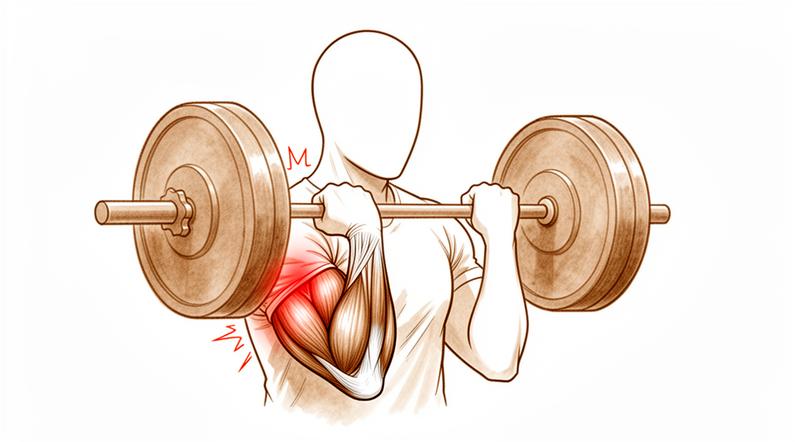
您可能会在上臂靠近肘部的前方感到突然的锐痛。这通常发生在您提起重物或手臂用力过猛时。许多人形容这种感觉像是被踢到或击中该部位。您可能还会看到或摸到上臂出现隆起,有时被称为“波派肌”(Popeye muscle)。这是因为肌腱已从骨骼上剥离。
当您尝试弯曲肘部或翻转手掌向上时,手臂可能会感到无力。简单的日常任务可能会变得困难。您可能难以提起购物袋、打开罐子或使用螺丝刀。将手伸到背后扣内衣或把衬衫塞进裤子里可能会感到别扭或疼痛。有些人发现难以侧卧睡觉,因为压力会加重疼痛区域。
疼痛通常在活动后加剧。如果您睡觉时手臂弯曲,早晨醒来时疼痛可能会更严重。休息通常有助于缓解酸痛,但无力感可能会持续存在。如果是部分撕裂,症状可能比完全断裂轻,但仍会影响您的日常生活。女性发生部分撕裂而非完全断裂的可能性更大。
如果肱二头肌短头发生孤立性撕裂,您可能会在肌肉中感觉到间隙或凹陷。这种情况很少见,但如果伴有明显无力,则需要尽早处理。您的外科医生会检查这些体征,以决定最佳的治疗方案。无论损伤是急性还是慢性,大多数远端肱二头肌撕裂患者都会经历类似的疼痛和无力模式。好消息是,手术修复在患者评分的结局指标上始终能提供良好的结果。无论使用可吸收螺钉还是不可吸收螺钉,您在修复后1年以上都能获得优异的功能结局。
实际发生了什么
您的肱二头肌在肘部以一条厚实且强韧的肌腱结束。可以将这条肌腱想象成一根重型绳索,它将上臂肌肉与前臂骨骼连接起来。它的主要功能是帮助您弯曲肘部并使手掌向上翻转。当这条肌腱撕裂或断裂时,这种连接就会中断。肌肉无法再有效地牵拉骨骼,这就是您会感到突然无力和疼痛的原因。
这种损伤通常发生在您试图用伸直的手臂提起重物或接住掉落物体时。施加的力量使肌腱承受了过大的应力,导致其断裂。您可能会听到“啪”的一声或感到剧烈的撕裂感。受伤后,肌肉可能会在上臂处堆积,形成可见的隆起。这是因为肌腱不再将肌肉固定在靠近肘部的正常位置。
肘部是一个复杂的铰链关节。它依靠骨骼、韧带和肌腱协同工作来维持稳定性。当肱二头肌肌腱受损时,会影响肘部的活动和承重功能。您的外科医生会检查撕裂情况,以确定是完全撕裂还是部分撕裂。部分撕裂意味着部分纤维仍然附着,而完全撕裂意味着肌腱已完全脱离。
修复肌腱可以恢复肌肉与骨骼之间至关重要的连接。现代外科技术允许您的外科医生将肌腱牢固地重新附着到前臂骨骼上。无论使用可吸收还是不可吸收锚钉,目标都是将肌腱恢复到其原始位置。这有助于组织愈合并恢复力量。
时机对您的康复至关重要。术后大多数问题源于手术延迟或使用了会干扰周围组织的大切口。如果等待超过21天,肌腱可能会在缩短的位置形成瘢痕,使修复变得更加困难。然而,即使是延迟修复也能获得良好的功能结果。您的外科医生将选择最佳方案以最大限度地减少并发症,并帮助您安全地恢复正常活动。
我们能采取的措施
您可以从休息手臂开始,避免提重物或扭转动作,以免加重肘前侧的牵拉。您的外科医生可能会建议进行一段时间的物理治疗,以帮助减轻肿胀并温和地恢复关节活动度。目标是在受损肌腱愈合的过程中保持关节的灵活性。在考虑更侵入性的治疗之前,您应给予这种保守治疗足够的时间来发挥作用。
如果您感到疼痛,您的外科医生可能会建议服用非处方止痛药或抗炎药,以帮助缓解不适。这些药物并不能治愈肌腱,但可以在您恢复期间使日常活动更容易忍受。在某些情况下,可能会使用皮质类固醇注射来减轻炎症。然而,这些治疗并不能修复撕裂的肌腱本身。其效果是暂时的,并非完全断裂的长期解决方案。
当非手术治疗无法缓解您的症状,或者您需要恢复工作或运动所需的完全力量时,就会考虑手术。您的外科医生会讨论最佳的修复时机,并指出即使修复延迟超过21天,预后依然良好。手术涉及将撕裂的肌腱重新附着到骨骼上,以恢复手臂的力量。这种方法在患者评分的预后和力量方面均能提供一致的良好结果。对于休息后未改善的部分撕裂,手术也是一个可行的选择。在极少数肌腱无法直接重新附着的病例中,您的外科医生可能会使用移植物来重建该区域。无论采用何种技术,手术严重并发症的总体发生率均较低。
预期情况
修复断裂的二头肌腱手术后,您可以期待获得优异的长期结果。无论您的外科医生使用的是可吸收螺钉(可自行溶解)还是不可吸收螺钉(保留在体内),术后1年以上的功能预后均十分理想。大多数患者根据其在日常生活中的感受和身体功能,报告结果持续良好。即使您存在未通过休息愈合的部分撕裂,在非手术治疗失败后,手术修复仍然是一个可行的选择。
时机在您的康复过程中起着重要作用。如果您等待超过21天再进行修复,您仍然可以期待获得与立即治疗相似的功能预后。然而,延迟修复与较高的初始并发症发生率相关。大多数手术问题主要归因于这种时机上的延迟。其次,这些问题也与手术过程中需要对前臂进行广泛暴露有关。如果您的撕裂是慢性的或复发的,使用供体组织(同种异体移植物)进行重建是安全且有效的。这种方法在研究组中产生了优异的患者报告结果和活动范围,且无显著并发症。
严重并发症罕见。远端二头肌腱修复术后的主要并发症发生率为4.6%。总体而言,每20名患者中就有1名会出现主要并发症,每5名患者中就有1名会出现次要并发症。神经损伤是您可能面临的最常见问题。尽管存在这些风险,无论采用何种手术入路,严重并发症的总体发生率仍然很低。与大型研究相比,外科医生的培训水平并不会显著改变这些风险特征。
如果您选择不进行手术,或以保守方式处理撕裂,其预后通常不如手术修复。手术为恢复力量和功能提供了最佳机会。虽然慢性修复的即时并发症率可能略高于急性修复,但长期功能结果相当。您应预期会经历一段适应期。最初几周可能会涉及不适和活动受限,但目标是恢复正常使用。大多数并发症源于延误或广泛的手术暴露,因此遵循外科医生关于时机和术后护理的建议对于获得最佳结果至关重要。
何时就医
若休息后疼痛持续不缓解,请咨询全科医生。若发现手臂无力或不稳,请要求专科医生评估。若肘关节出现交锁或打软腿,请及时就医。若症状影响睡眠或工作,请联系医生。若疼痛突然加重,请寻求帮助。这些体征可能提示肱二头肌远端肌腱撕裂。若感觉肌肉处有凹陷或存在明显无力,建议尽早干预。延迟修复可能增加并发症风险。您的外科医生将对您进行检查,以确定最佳治疗方案。
Evidence & references
Overview
- Clinical and functional outcomes at more than 1 year after distal biceps tendon repair are excellent for patients treated with bioabsorbable screws [1].
- Clinical and functional outcomes at more than 1 year after distal biceps tendon repair are excellent for patients treated with nonabsorbable screws [1].
- Selective disruption of the short head of the biceps distal tendon may be effectively treated with anatomic repair when diagnosed appropriately [2].
- Patients have excellent overall outcomes after distal biceps tendon rupture repair [3].
- Functional outcomes for chronic distal biceps ruptures are comparable to those seen in acute distal biceps ruptures, despite a potentially slightly higher immediate complication rate [4].
- Patients treated with delayed distal biceps tendon repair (>21 days) can expect similar functional outcomes to those treated acutely, despite a high rate of initial complications [5].
- Most morbidity from repair of the distal biceps tendon is attributed primarily to a delay in the timing of the repair [6].
- Most morbidity from repair of the distal biceps tendon is attributed secondarily to an extensive anterior exposure [6].
- Bioabsorbable interference screw fixation of distal biceps ruptures through a single anterior incision is a safe and successful technique [7].
- Surgical repair of distal biceps ruptures provides consistently good results in terms of patient-scored outcomes [8].
- The surgical repair of distal biceps tendon ruptures has an overall low rate of serious complications, regardless of approach or technique [9].
- Surgical treatment of partial distal biceps tendon tears is a viable option after failed nonsurgical treatment [11].
- Utilizing a double-incision technique with a combination of cortical button and interference screw fixation helps determine the best method of fixation and approach to minimize postoperative complications in patients with distal biceps ruptures [14].
- Two-incision distal biceps repairs demonstrate a high degree of patient satisfaction [21].
- Two-incision distal biceps repairs demonstrate a low complication rate [21].
Anatomy & Pathophysiology
- The elbow's stability and complex kinematics are accounted for by a combination of bony articulation and soft-tissue stabilizers [29].
- Musculoskeletal ultrasonography provides a dynamic, functional assessment of elbow structures, allowing visualization of pathology under stress and motion [30].
- The medial elbow joint space increases under a valgus load [31].
- The medial elbow joint space decreases when a maximal grip contraction is performed [31].
- A low carrying angle is measured in elite tennis players just before ball impact during the forehand, suggesting a dynamic varus instant accommodation moving towards full extension [32].
- The decrease in the carrying angle is a consequence of an increase in elbow flexion position dictated by the transition from a closed to open, semi-open stances [32].
- Determining which structures need to be repaired is important to avoid complications that could lead to elbow instability [33].
- Ulnar collateral ligament reconstruction using a suspension button fixation technique reliably restored elbow kinematics to the intact state [34].
- Limiting the number of innings pitched is likely the best way to reduce elbow injury in youth pitchers because biomechanical variables correlated with peak valgus torque are not easily modifiable [35].
- The medial elbow joint space was significantly reduced under 60-N valgus stress plus 50% MVC compared to 60-N valgus stress alone [37].
- No significant relationships between adaptations in shoulder strength or ROM were related to chronic structural adaptations of the elbow in professional baseball pitchers [38].
- The spin move is a simple maneuver that can improve exposure of the coronoid process regardless of the degree of elbow instability [39].
- Reattachment of the flexor and extensor tendons at the epicondyle should be considered in the development of improved repair techniques for stabilizers of the elbow [40].
- An internal joint stabilizer with a standardized treatment protocol could maintain concentric reduction while allowing early functional motion and improve clinical outcomes for patients with complex persistent elbow instability [41].
- Activation of lateral forearm muscles decreased elbow varus angulation throughout flexion [42].
- In patients treated with surgery for snapping triceps syndrome, it is crucial to make sure full resolution of the snapping by examining all dislocating structures during passive elbow motion and/or myoelectrical stimulation to achieve excellent results [43].
- Pre-operative evaluations in elbow stiffness should identify involved articular and periarticular tissues and determine whether articular surfaces and osteoarticular congruence are preserved [44].
- Elbow vascularized composite allotransplantation may be technically feasible on the basis of its consistent vascular anatomy and a proposed surgical technique [45].
- Elbow valgus stress might correlate with ball velocity at 75% partial valgus stress pitch [46].
- Physical examination of the elbow is a critical component in formulating an accurate diagnosis [47].
Classification
- Distal biceps tendon tears in women present differently than in men [10].
- The incidence of women sustaining a distal biceps tendon tear is 3.2% [12].
- Partial tears are statistically more common than complete ruptures in women [12].
- Partial ruptures of the distal biceps brachii tendon represent a spectrum of patterns with varying involvement of the long head (LH) and short head (SH) tendons [16].
- Selective disruption of the short head of the biceps distal tendon may be effectively treated with anatomic repair when diagnosed appropriately [2].
Clinical Presentation
- Distal biceps tendon tears in women present differently than in men [10].
- The incidence of women sustaining a distal biceps tendon tear is 3.2% [12].
- Partial tears are statistically more common than complete ruptures in women [12].
- Isolated rupture of the short head of the biceps brachii is a rare injury [15].
- Early intervention for isolated short head rupture is recommended if a sulcus or gap in the musculotendinous unit is palpable or considerable weakness is present [15].
- Partial ruptures of the distal biceps brachii tendon represent a spectrum of patterns with varying involvement of the long head (LH) and short head (SH) tendons [16].
- The Hook test has imperfect validity for diagnosing distal biceps tendon ruptures and should not be used alone [17].
- The prevalence of distal biceps tendon signal changes on MRI in asymptomatic patients is very low [18].
- Patients with a smaller radioulnar window have a higher risk of rupturing the distal biceps tendon [19].
Investigations
- The Hook test has imperfect validity and should not be used alone to diagnose distal biceps tendon ruptures [17].
- The prevalence of distal biceps tendon signal changes on MRI in asymptomatic patients is very low [18].
- Patients with a smaller radioulnar window have a higher risk of rupturing the distal biceps tendon [19].
- Distal biceps tendon tears in women present differently than in men [10].
- Isolated rupture of the short head of the biceps brachii is a rare injury [15].
- Early intervention is recommended for isolated rupture of the short head of the biceps brachii if a sulcus or gap in the musculotendinous unit is palpable or considerable weakness is present [15].
Treatment
Operative Repair
- Clinical and functional outcomes at more than 1 year after distal biceps tendon repair are excellent for both bioabsorbable and nonabsorbable screw fixation groups [1].
- Selective disruption of the short head of the biceps distal tendon may be effectively treated with anatomic repair when diagnosed appropriately [2].
- Patients have excellent overall outcomes after distal biceps tendon rupture repair [3].
- Functional outcomes in chronic distal biceps rupture repairs are comparable to those in acute distal biceps rupture populations, despite a potentially slightly higher immediate complication rate [4].
- Patients treated with distal biceps tendon repair after a delay of more than 21 days can expect similar functional outcomes to those treated acutely, despite a high rate of initial complications [5].
- Most morbidity from repair of the distal biceps tendon is attributed primarily to a delay in the timing of the repair and secondarily to an extensive anterior exposure [6].
- Bioabsorbable interference screw fixation of distal biceps ruptures through a single anterior incision is a safe and successful technique [7].
- Surgical repair of distal biceps ruptures provides consistently good results in terms of patient-scored outcomes [8].
- The surgical repair of distal biceps tendon ruptures has an overall low rate of serious complications, regardless of approach or technique [9].
- Surgical treatment of partial distal biceps tendon tears is a viable option after failed nonsurgical treatment [11].
- Reinsertion of the distal biceps tendon on the brachialis tendon is a safe and effective procedure with a low complication rate, providing satisfactory subjective and objective results at long-term follow-up [13].
- Utilizing a double-incision technique with a combination of cortical button and interference screw fixation helps determine the best method of fixation and approach with the goal of minimizing postoperative complications [14].
- Distal biceps reconstruction using an Achilles tendon allograft, transosseous EndoButton, and Pulvertaft weave with tendon wrap technique is recommended for patients with retracted, irreparable distal biceps ruptures [26].
- A two-incision repair with suture anchor attachment is a safe and effective method for treatment of distal biceps tendon ruptures [36].
- Repair of distal biceps ruptures using an Endobutton fixation results in a nearly normal return of strength and function, which is significantly better than in those managed nonoperatively [50].
Non-Operative Management
- Incomplete distal triceps tears with active elbow extension against resistance are managed nonsurgically [52].
Complications
- Distal biceps tendon repair has an overall low rate of serious complications, regardless of approach or technique [9].
- The major complication rate after distal biceps tendon repair is 4.6% [23].
- One in five patients will have a minor complication after surgery on the distal biceps tendon [24].
- One in twenty patients will have a major complication after surgery on the distal biceps tendon [24].
- Nerve injury is the most common complication after distal biceps tendon repair [22].
- Most morbidity from repair of the distal biceps tendon is attributed primarily to a delay in the timing of the repair [6].
- Most morbidity from repair of the distal biceps tendon is attributed secondarily to an extensive anterior exposure [6].
- Chronic distal biceps rupture repair may have a slightly higher immediate complication rate compared to acute repair [4].
- Delayed repair of distal biceps tendon ruptures (>21 days) is associated with a high rate of initial complications [5].
- Allograft reconstruction of distal biceps injuries for chronic and recurrent tears results in no significant complications among the studied cohort [20].
- Distal biceps repairs using the two-incision technique demonstrate a low complication rate [21].
- Reinsertion of the distal biceps tendon on the brachialis tendon is associated with a low complication rate [13].
- Complication rates after distal biceps tendon repair performed by newly trained surgeons are similar to those previously reported in large cohort studies [22].
Recovery
- Clinical and functional outcomes at more than 1 year after distal biceps tendon repair are excellent for patients treated with bioabsorbable screws [1].
- Clinical and functional outcomes at more than 1 year after distal biceps tendon repair are excellent for patients treated with nonabsorbable screws [1].
- Selective disruption of the short head of the biceps distal tendon may be effectively treated with anatomic repair when diagnosed appropriately [2].
- Patients have excellent overall outcomes after distal biceps tendon rupture repair [3].
- Functional outcomes for chronic distal biceps ruptures are comparable to those seen in acute distal biceps ruptures, despite a potentially slightly higher immediate complication rate [4].
- Patients treated with distal biceps tendon repair after a delay of more than 21 days can expect similar functional outcomes to those treated acutely, despite a high rate of initial complications [5].
- Most morbidity from repair of the distal biceps tendon is attributed primarily to a delay in the timing of the repair [6].
- Most morbidity from repair of the distal biceps tendon is attributed secondarily to an extensive anterior exposure [6].
- Surgical repair of distal biceps ruptures provides consistently good results in terms of patient-scored outcomes [8].
- Surgical treatment of partial distal biceps tendon tears is a viable option after failed nonsurgical treatment [11].
- Reinsertion of the distal biceps tendon on the brachialis tendon is a safe and effective procedure with a low complication rate, providing satisfactory subjective and objective results at long-term follow-up [13].
- Allograft reconstruction of distal biceps injuries for chronic and recurrent tears results in excellent patient-reported outcomes and range of motion [20].
- Allograft reconstruction of distal biceps injuries for chronic and recurrent tears results in no significant complications among the studied cohort [20].
- Complication rates after distal biceps tendon repair performed by newly trained surgeons are similar to those previously reported in large cohort studies [22].
- Nerve injury is the most common complication after distal biceps tendon repair performed by newly trained surgeons [22].
- One in 5 patients will have a minor complication after surgery on the distal biceps tendon [24].
- One in 20 patients will have a major complication after surgery on the distal biceps tendon [24].
- Patients with biceps tendon repair etiology had a trend toward greater motion improvement than that of patients with a traumatic etiology regarding anconeus interposition for proximal radioulnar synostosis [25].
- Nearly all complete distal biceps ruptures can be effectively reapproximated to the radial tuberosity within the first 6 weeks of injury using single-incision repair with suture anchors [28].
- Primary repair of chronic distal biceps tendon tears greater than 6 weeks from injury demonstrated excellent patient-reported outcome measures (PROMs) and elbow range of motion (ROM) [48].
- Delayed reconstruction of irreparable distal biceps tendon ruptures with semitendinosus autograft produces similar strength, range of motion, and complication rates compared with delayed primary repair [55].
- Delayed reconstruction of irreparable distal biceps tendon ruptures with semitendinosus autograft produces slightly worse functional outcome scores compared with delayed primary repair [55].
Key Evidence
- [L3] Clinical and functional outcome at more than 1 year after distal biceps tendon repair was excellent in both groups. [1] (10.1016/j.jse.2015.12.007)
- [L4] When diagnosed appropriately, selective disruption of the short head of the biceps distal tendon may be effectively treated with anatomic repair. [2] (10.1016/j.jse.2016.09.050)
- [L3] Overall, patients have excellent outcome after distal biceps tendon rupture repair. [3] (10.1007/s00402-018-3018-6)
- [L4] Although there may be a slightly higher immediate complication rate, the functional outcomes remain comparable with those seen in the patient population with acute distal biceps. [4] (10.1177/23259671211065772)
- [L3] Despite a high rate of initial complications, patients treated with distal biceps tendon repair after a delay (>21 days) can expect similar functional outcomes to those treated acutely. [5] (10.1016/j.jse.2017.02.025)
- [L4] Most morbidity from repair of the distal biceps tendon can be attributed primarily to a delay in the timing of the repair and secondarily to an extensive anterior exposure. [6] (10.2106/00004623-200011000-00010)
- [Paper] This is a safe and successful technique for the management of distal biceps tendon ruptures. [7] (10.1007/s00402-009-0974-x)
- [L4] Surgical repair of distal biceps ruptures provides consistently good results in terms of patient-scored outcomes. [8] (10.1016/j.injury.2012.10.029)
- [L3] The surgical repair of distal biceps tendon ruptures has an overall low rate of serious complications, regardless of approach or technique. [9] (10.1177/0363546517720200)
- [L4] Distal biceps tendon tears in women present differently than in men. [10] (10.1016/j.jse.2010.01.015)
- [L4] Surgical treatment of partial distal biceps tendon tears is a viable option after failed nonsurgical treatment. [11] (10.1016/j.jhsa.2010.04.024)
- [L4] The incidence of women sustaining a distal biceps tendon tear is 3.2%, with partial tears being statistically more common than complete ruptures. [12] (10.1016/j.jse.2014.02.006)
- [L4] Reinsertion of the distal biceps tendon on the brachialis tendon can be considered a safe and effective procedure with low complication rate, providing satisfactory subjective and objective results at long-term follow-up. [13] (10.1007/s00167-008-0705-9)
- [L4] This will help surgeons determine the best method of fixation and approach with the goal of minimizing postoperative complications in patients with distal biceps ruptures. [14] (10.1177/17585732241312212)
- [L4] Isolated rupture of the short head of the biceps brachii is a rare injury and early intervention is recommended if a sulcus or gap in musculotendinous unit is palpable or considerable weakness is present. [15] (10.1007/s00167-010-1108-2)
- [L4] Partial ruptures of the distal biceps brachii tendon represent a spectrum of patterns with varying involvement of the LH and SH tendons. [16] (10.1016/j.jse.2020.04.021)
- [L2] The authors caution against using the Hook test alone to diagnose distal biceps tendon ruptures due to its imperfect validity. [17] (10.1016/j.jhsa.2023.07.004)
- [L3] The prevalence of distal biceps tendon signal changes on MRI in asymptomatic patients is very low. [18] (10.1016/j.jhsa.2022.01.020)
- [L3] Therefore, patients with a smaller radioulnar window have a higher risk of rupturing the distal biceps tendon. [19] (10.1016/j.jse.2023.09.020)
- [L5] Allograft reconstruction of distal biceps injuries for chronic and recurrent tears results in excellent patient-reported outcomes and range of motion, with no significant complications among our cohort. [20] (10.1016/j.xrrt.2025.100661)
- [L3] In this large cohort of 2-incision distal biceps repairs, we found a high degree of patient satisfaction and a low complication rate. [21] (10.1016/j.jse.2014.12.032)
- [L4] Complication rates after distal biceps tendon repair performed by newly trained surgeons were similar to those previously reported in large cohort studies, with nerve injury as the most common complication. [22] (10.1016/j.jse.2022.09.014)
- [L2] This is the largest analysis of complications after distal biceps repair, indicating a major complication rate of 4.6%. [23] (10.1177/0363546519899933)
- [L4] Patients should be counseled that 1 in 5 patients will have a minor complication and 1 in 20 patients will have a major complication after surgery on the distal biceps tendon. [24] (10.1016/j.jse.2016.02.032)
- [L3] Patients with biceps tendon repair etiology had a trend toward greater motion improvement than that of patients with a traumatic etiology. [25] (10.1016/j.jse.2014.07.011)
- [L4] This technique is recommended for patients who have a retracted, irreparable distal biceps rupture. [26] (10.1016/j.jse.2016.01.014)
- [L4] The study demonstrates that nearly all complete distal biceps ruptures can be effectively reapproximated to the radial tuberosity within the first 6 weeks of injury. [28] (10.1016/j.jse.2006.03.002)
- [L5] This article discusses the basic anatomy of the elbow and the biomechanics of this joint, noting that a combination of bony articulation and soft-tissue stabilizers accounts for the elbow's stability and complex kinematics. [29] (10.1016/j.csm.2004.06.008)
- [L5] Musculoskeletal ultrasonography provides a dynamic, functional assessment of elbow structures, allowing visualization of pathology under stress and motion. [30] (10.5435/jaaos-d-20-00935)
- [L4] Medial elbow joint space increases under a valgus load and then decreases when a maximal grip contraction is performed. [31] (10.1177/0363546518755149)
- [L4] The observed decrease in the carrying angle is a consequence of an increase in elbow flexion position dictated by the transition from a closed to open, semi‐open stances. [32] (10.1002/ksa.12016)
- [L3] It is important to determine which structures need to be repaired to avoid complications that could lead to elbow instability. [33] (10.1016/j.jse.2019.07.006)
- [L5] Ulnar collateral ligament reconstruction using a suspension button fixation technique reliably restored elbow kinematics to the intact state. [34] (10.1177/0363546509350109)
- [L5] Given that the biomechanical variables correlated with peak valgus torque are not easily modifiable, limiting the number of innings pitched is likely the best way to reduce elbow injury in youth pitchers. [35] (10.1016/j.jse.2004.01.013)
- [L4] Based on our data, we believe that a two-incision repair with suture anchor attachment is a safe and effective method for treatment of distal biceps tendon ruptures. [36] (10.1007/s001670050134)
- [L5] The medial elbow joint space was significantly reduced under 60-N valgus stress plus 50% MVC compared to 60-N valgus stress alone. [37] (10.1016/j.jse.2022.03.027)
- [L3] However, no significant relationships between adaptations in shoulder strength or ROM were related to chronic structural adaptations of the elbow. [38] (10.1177/03635465251317509)
- [L5] The spin move is a simple maneuver that can improve exposure of the coronoid process regardless of the degree of elbow instability. [39] (10.1016/j.jse.2022.11.020)
- [L5] Thus, it should be considered in the development of improved repair techniques for stabilizers of the elbow. [40] (10.1186/s12891-018-2341-y)
- [L4] An internal joint stabilizer with a standardized treatment protocol could maintain concentric reduction while allowing early functional motion and improve clinical outcomes for patients with complex persistent elbow instability. [41] (10.1097/corr.0000000000002159)
- [L5] In addition, activation of lateral forearm muscles decreased elbow varus angulation throughout flexion. [42] (10.1016/j.jhsg.2024.11.006)
- [L4] In patients treated with surgery, it is crucial to make sure full resolution of the snapping by examining all dislocating structures during passive elbow motion and/or myoelectrical stimulation to achieve excellent results. [43] (10.1016/j.xrrt.2025.08.017)
- [L5] Pre-operative evaluations in elbow stiffness should identify involved articular and periarticular tissues and determine whether articular surfaces and osteoarticular congruence are preserved. [44] (10.1016/j.jisako.2023.10.009)
- [L5] Elbow VCA may be technically feasible on the basis of its consistent vascular anatomy and our proposed surgical technique. [45] (10.1016/j.jse.2017.04.014)
- [L5] Elbow valgus stress might correlate with ball velocity at 75% partial valgus stress pitch. [46] (10.1016/j.jse.2022.08.009)
- [L5] Physical examination of the elbow is a critical component in formulating an accurate diagnosis. [47] (10.5435/jaaos-d-16-00622)
- [L4] Primary repair of chronic distal biceps tendon tears greater than 6 weeks from injury demonstrated excellent PROMs and elbow ROM. [48] (10.1177/15589447221107691)
- [L3] Repair of distal biceps ruptures using an Endobutton fixation results in nearly normal return of strength and function, which is significantly better than in those managed nonoperatively. [50] (10.1016/j.jse.2015.10.008)
- [L5] Incomplete tears with active elbow extension against resistance are managed nonsurgically. [52] (10.5435/00124635-201001000-00005)
- [L3] Delayed reconstruction of irreparable distal biceps tendon ruptures with semitendinosus autograft produces similar strength, range of motion, and complication rates but slightly worse functional outcome scores compared with delayed primary repair. [55] (10.1016/j.jse.2019.01.006)
References
[1] Distal biceps tendon repair: comparison of clinical and radiological outcome between bioabsorbable and nonabsorbable screws. Journal of Shoulder and Elbow Surgery. 2016. DOI: 10.1016/j.jse.2015.12.007 [2] Rupture of the short head component of a bifurcated distal biceps tendon. Journal of Shoulder and Elbow Surgery. 2017. DOI: 10.1016/j.jse.2016.09.050 [3] Functional outcome in patients who underwent distal biceps tendon repair. Archives of Orthopaedic and Trauma Surgery. 2018. DOI: 10.1007/s00402-018-3018-6 [4] Surgical Results of Chronic Distal Biceps Ruptures: A Systematic Review. Orthopaedic Journal of Sports Medicine. 2022. DOI: 10.1177/23259671211065772 [5] Delayed repair of distal biceps tendon ruptures is successful: a case-control study. Journal of Shoulder and Elbow Surgery. 2017. DOI: 10.1016/j.jse.2017.02.025 [6] Complications of Repair of the Distal Biceps Tendon with the Modified Two-Incision Technique†. The Journal of Bone and Joint Surgery-American Volume. 2000. DOI: 10.2106/00004623-200011000-00010 [7] Bioabsorbable interference screw fixation of distal biceps ruptures through a single anterior incision: a single-surgeon case series and review of the literature. Archives of Orthopaedic and Trauma Surgery. 2009. DOI: 10.1007/s00402-009-0974-x [8] Distal biceps tendon rupture: Current concepts. Injury. 2013. DOI: 10.1016/j.injury.2012.10.029 [9] Surgical Treatment of Distal Biceps Tendon Ruptures: An Analysis of Complications in 784 Surgical Repairs. The American Journal of Sports Medicine. 2017. DOI: 10.1177/0363546517720200 [10] Distal biceps tendon tears in women. Journal of Shoulder and Elbow Surgery. 2010. DOI: 10.1016/j.jse.2010.01.015 [11] Surgical Treatment of Partial Distal Biceps Tendon Ruptures. The Journal of Hand Surgery. 2010. DOI: 10.1016/j.jhsa.2010.04.024 [12] Re-rupture rate of primarily repaired distal biceps tendon injuries. Journal of Shoulder and Elbow Surgery. 2014. DOI: 10.1016/j.jse.2014.02.006 [13] Surgical repair of the distal biceps brachii tendon: clinical and isokinetic long‐term follow‐up. Knee Surgery, Sports Traumatology, Arthroscopy. 2009. DOI: 10.1007/s00167-008-0705-9 [14] Outcomes of utilizing double-incision technique with combination of cortical button and interference screw fixation for distal biceps rupture: A case series. Shoulder & Elbow. 2025. DOI: 10.1177/17585732241312212 [15] Traumatic isolated closed rupture of the short head of the biceps brachii in military paratrooper. Knee Surgery, Sports Traumatology, Arthroscopy. 2010. DOI: 10.1007/s00167-010-1108-2 [16] Partial rupture of the distal biceps brachii tendon: a magnetic resonance imaging analysis. Journal of Shoulder and Elbow Surgery. 2020. DOI: 10.1016/j.jse.2020.04.021 [17] Reliability and Validity of the Hook Test for Diagnosis of Distal Biceps Tendon Ruptures. The Journal of Hand Surgery. 2023. DOI: 10.1016/j.jhsa.2023.07.004 [18] Evaluation of MRI Signal Changes of the Distal Biceps Tendon in Asymptomatic Patients. The Journal of Hand Surgery. 2022. DOI: 10.1016/j.jhsa.2022.01.020 [19] Smaller radioulnar window is associated with a distal biceps tendon rupture in patients with limited forearm rotation: a 3-dimensional computed tomography comparison study of proximal impingement caused by radial tuberosity hypertrophy—a single-center case series. Journal of Shoulder and Elbow Surgery. 2024. DOI: 10.1016/j.jse.2023.09.020 [20] Allograft reconstruction of distal biceps ruptures: surgical technique and analysis of outcomes in chronic tears and failed repairs. JSES Reviews, Reports, and Techniques. 2026. DOI: 10.1016/j.xrrt.2025.100661 [21] Endobutton versus transosseous suture repair of distal biceps rupture using the two-incision technique: a comparison series. Journal of Shoulder and Elbow Surgery. 2015. DOI: 10.1016/j.jse.2014.12.032 [22] Trends and complications of distal biceps tendon repair among American Board of Orthopaedic Surgery part II oral examination candidates. Journal of Shoulder and Elbow Surgery. 2023. DOI: 10.1016/j.jse.2022.09.014 [23] Complications After Distal Biceps Tendon Repair: A Systematic Review. The American Journal of Sports Medicine. 2020. DOI: 10.1177/0363546519899933 [24] Factors associated with adverse events after distal biceps tendon repair or reconstruction. Journal of Shoulder and Elbow Surgery. 2016. DOI: 10.1016/j.jse.2016.02.032 [25] Outcomes of anconeus interposition for proximal radioulnar synostosis. Journal of Shoulder and Elbow Surgery. 2014. DOI: 10.1016/j.jse.2014.07.011 [26] Distal biceps reconstruction using an Achilles tendon allograft, transosseous EndoButton, and Pulvertaft weave with tendon wrap technique for retracted, irreparable distal biceps ruptures. Journal of Shoulder and Elbow Surgery. 2016. DOI: 10.1016/j.jse.2016.01.014 [28] Single-incision repair of acute distal biceps ruptures by use of suture anchors. Journal of Shoulder and Elbow Surgery. 2007. DOI: 10.1016/j.jse.2006.03.002 [29] Elbow anatomy and structural biomechanics. Clinics in Sports Medicine. 2004. DOI: 10.1016/j.csm.2004.06.008 [30] Ultrasound Examination Techniques for Elbow Injuries in Overhead Athletes. Journal of the American Academy of Orthopaedic Surgeons. 2020. DOI: 10.5435/jaaos-d-20-00935 [31] Medial Elbow Joint Space Increases With Valgus Stress and Decreases When Cued to Perform A Maximal Grip Contraction. The American Journal of Sports Medicine. 2018. DOI: 10.1177/0363546518755149 [32] A low carrying angle is measured in elite tennis players just before ball impact during the forehand, suggesting a dynamic varus instant accommodation moving towards full extension. Knee Surgery, Sports Traumatology, Arthroscopy. 2024. DOI: 10.1002/ksa.12016 [33] Isolated displaced type II partial articular radial head fracture: correlation of preoperative imaging with intraoperative findings of lateral ulnar collateral ligament tear. Journal of Shoulder and Elbow Surgery. 2020. DOI: 10.1016/j.jse.2019.07.006 [34] Revision Ulnar Collateral Ligament Reconstruction Using a Suspension Button Fixation Technique. The American Journal of Sports Medicine. 2009. DOI: 10.1177/0363546509350109 [35] Valgus torque in youth baseball pitchers: a biomechanical study. Journal of Shoulder and Elbow Surgery. 2004. DOI: 10.1016/j.jse.2004.01.013 [36] Repair of distal biceps tendon rupture with suture anchors. Knee Surgery, Sports Traumatology, Arthroscopy. 1999. DOI: 10.1007/s001670050134 [37] Changes in medial elbow joint space with differences in contraction strength of flexor-pronator muscle under elbow valgus stress. Journal of Shoulder and Elbow Surgery. 2022. DOI: 10.1016/j.jse.2022.03.027 [38] Chronic Structural Adaptations of the Shoulder and Elbow Are Correlated in Professional Baseball Pitchers. The American Journal of Sports Medicine. 2025. DOI: 10.1177/03635465251317509 [39] The spin move to facilitate antegrade coronoid fixation in terrible triad injuries. Journal of Shoulder and Elbow Surgery. 2023. DOI: 10.1016/j.jse.2022.11.020 [40] Reattachment of the flexor and extensor tendons at the epicondyle in elbow instability: a biomechanical comparison of techniques. BMC Musculoskeletal Disorders. 2018. DOI: 10.1186/s12891-018-2341-y [41] Does an Internal Joint Stabilizer and Standardized Protocol Prevent Recurrent Instability in Complex Persistent Elbow Instability?. Clinical Orthopaedics & Related Research. 2022. DOI: 10.1097/corr.0000000000002159 [42] The Effect of Arm Abduction and Forearm Muscle Activation on Kinematics During Elbow Flexion. Journal of Hand Surgery Global Online. 2025. DOI: 10.1016/j.jhsg.2024.11.006 [43] Snapping triceps syndrome: a review of the literature and proposed operative treatment algorithm. JSES Reviews, Reports, and Techniques. 2026. DOI: 10.1016/j.xrrt.2025.08.017 [44] Elbow stiffness: Arthritis and heterotopic ossification. Journal of ISAKOS. 2024. DOI: 10.1016/j.jisako.2023.10.009 [45] Elbow vascularized composite allotransplantation—surgical anatomy and technique. Journal of Shoulder and Elbow Surgery. 2017. DOI: 10.1016/j.jse.2017.04.014 [46] Effect of quantitative partial valgus stress during baseball pitching on ball velocity and subjective pitch-effort. Journal of Shoulder and Elbow Surgery. 2023. DOI: 10.1016/j.jse.2022.08.009 [47] Comprehensive Review of the Elbow Physical Examination. Journal of the American Academy of Orthopaedic Surgeons. 2018. DOI: 10.5435/jaaos-d-16-00622 [48] Primary Repair of Chronic Distal Biceps Tendon Tears. HAND. 2022. DOI: 10.1177/15589447221107691 [50] A comparison of nonoperative vs. Endobutton repair of distal biceps ruptures. Journal of Shoulder and Elbow Surgery. 2016. DOI: 10.1016/j.jse.2015.10.008 [52] Distal Triceps Rupture. American Academy of Orthopaedic Surgeon. 2010. DOI: 10.5435/00124635-201001000-00005 [55] Management of chronic distal biceps tendon ruptures: primary repair vs. semitendinosus autograft reconstruction. Journal of Shoulder and Elbow Surgery*. 2019. DOI: 10.1016/j.jse.2019.01.006


