Deformidade em botão
Patients › Hand
Extensor central-slip injury at the middle finger joint; early splinting prevents the zigzag deformity, established cases need surgery.
O que você está sentindo
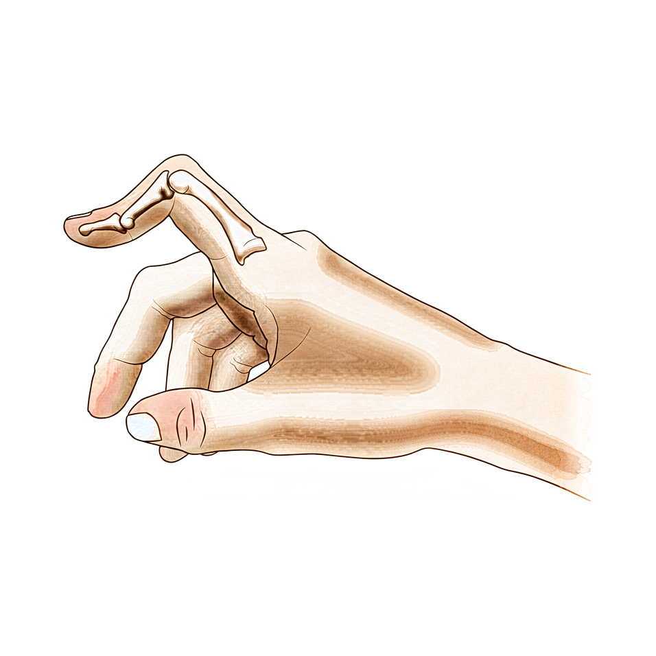
Você pode notar que a articulação do seu dedo médio se curva para dentro, enquanto a articulação da ponta se projeta para fora. Essa forma específica é chamada de deformidade em botão de flor (boutonniere). Isso ocorre quando os tendões na parte superior do seu dedo saem do lugar. Você pode perceber essa mudança se desenvolver lentamente ao longo do tempo, ou pode aparecer subitamente após uma lesão.
A dor geralmente se concentra em torno da articulação média do seu dedo. O desconforto pode parecer uma dor profunda ou uma ardência aguda quando você move o dedo. Você pode perceber que dobrar o dedo completamente causa mais dor do que mantê-lo reto. Atividades que exigem preensão ou pinçamento podem se tornar difíceis. Tarefas simples como abotoar uma camisa, girar uma maçaneta ou digitar podem parecer desconfortáveis ou dolorosas.
Seu dedo pode parecer rígido, especialmente pela manhã. Você pode notar inchaço ao redor da articulação média. Essa rigidez pode dificultar o fechamento da mão em punho. Se você tem artrite reumatoide, a deformidade pode progredir de maneira diferente do que se tivesse sido causada por trauma. Sem artrite ou lesão, cerca de 13% das pessoas apresentam essa condição.
A ponta do seu dedo também pode parecer instável. Em alguns casos, a articulação da ponta se curva para baixo mais do que o normal. Isso pode dificultar o apoio do dedo plano sobre uma mesa. Você pode ter dificuldade para empurrar portas pesadas ou levantar objetos leves com essa mão. Dormir de lado pode pressionar o dedo deformado, causando desconforto.
É importante distinguir essa deformidade verdadeira de uma lesão de aparência semelhante chamada pseudoboutonniere. As duas condições exigem cuidados diferentes. Seu cirurgião observará atentamente como o seu dedo se move para diferenciá-las. Compreender exatamente o que você está sentindo ajuda a orientar o melhor plano de tratamento para você.
O que está realmente acontecendo
A articulação do seu dedo é uma dobradiça complexa composta por ossos, tendões e uma capa protetora chamada cápsula articular. Em um dedo saudável, um tendão central percorre a parte média do dedo para ajudá-lo a estender. Esse tendão atua como a corda principal que abre o seu dedo.
Na deformidade em botão de camisa, esse tendão central está danificado ou enfraquecido. Pode estar rompido devido a trauma ou esticado por condições como a artrite reumatoide. Quando esse suporte central falha, o equilíbrio das forças no seu dedo muda. Os tendões laterais, que normalmente ajudam a flexionar o dedo, começam a puxar com força excessiva.
Pense nisso como um zíper que saiu do trilho. As peças ainda estão lá, mas não estão mais deslizando suavemente juntas. Como o tendão central não está mantendo as coisas no lugar, os tendões laterais deslizam para os lados. Isso faz com que a articulação média do seu dedo se flexione para dentro, enquanto a articulação da ponta pode se projetar para fora.
Essa mudança na tensão é o motivo pelo qual o seu dedo parece torto e sente-se rígido. A cápsula articular, que normalmente mantém a articulação estável, torna-se tensa e desequilibrada. Com o tempo, os tecidos se adaptam a essa nova posição incorreta. É por isso que a deformidade pode persistir mesmo após você tentar tratamentos conservadores, como o uso de talas ou fisioterapia.
O fator mais importante nesse problema é a alteração nos seus tendões e estruturas relacionadas. Essas mudanças ocorrem precocemente, razão pela qual o diagnóstico preciso é fundamental. Seu cirurgião precisa distinguir entre uma verdadeira deformidade em botão de camisa e um problema semelhante chamado lesão pseudobotão de camisa. O caminho do tratamento depende inteiramente de fazer essa distinção corretamente.
Se o dano for detectado precocemente, seu cirurgião pode focar na restauração da posição do tendão central. Em alguns casos, pode ser utilizada uma pequena porção de outro tendão para reconstruir o deslizamento central. Esse novo enxerto imita a função do tendão original enquanto mantém os outros movimentos do dedo intactos. O objetivo é manter a articulação centralizada e estável para que você possa movê-la novamente.
No entanto, se a deformidade estiver presente há muito tempo, os tecidos podem ter sofrido alterações permanentes. Nesses casos crônicos, os reparos simples podem não funcionar. A história natural dessa condição frequentemente leva a problemas persistentes, especialmente se houver artrite reumatoide envolvida. Os resultados a longo prazo para a reconstrução de tecidos moles nesses casos podem ser imprevisíveis. Às vezes, é necessário um procedimento definitivo de salvamento para corrigir a forma e a função do seu dedo.
O que podemos fazer a respeito
Iniciamos com o tratamento não cirúrgico para restaurar o movimento. Você tentará o uso de gesso seriado para obter extensão adequada. Isso é seguido pelo uso de órtese de flexão em movimento relativo por três meses. Seu cirurgião também pode recomendar fisioterapia. Um a dois graus de melhora na amplitude de movimento podem ser alcançados com o tratamento não operatório. No entanto, a deformidade pode persistir mesmo após o manejo conservador dedicado. Utilizamos órteses de flexão em movimento relativo para aumentar a flexão ativa da articulação interfalângica distal. Isso ajuda a melhorar a extensão da articulação da falange média. Você deve dar tempo a essa abordagem para funcionar antes de considerar a cirurgia.
O manejo médico foca no conforto e na inflamação. Se a sua deformidade estiver associada à artrite reumatoide, abordamos a atividade da doença subjacente. Medicamentos analgésicos e anti-inflamatórios ajudam a controlar o desconforto. Injeções podem ser oferecidas para reduzir o inchaço na articulação. Esses tratamentos visam manter a articulação móvel e sem dor enquanto você realiza a terapia. O objetivo é estabilizar a condição e melhorar sua função diária sem uma operação.
A cirurgia é considerada quando o tratamento conservativo atingiu seu limite. Diferenciamos uma verdadeira deformidade em botãonière de uma lesão pseudoboutonnière antes de decidir. Essa distinção é crítica para determinar o manejo clínico. Um resultado operatório bem-sucedido depende de um exame pré-operatório completo, do estadiamento correto da deformidade e do momento adequado do tratamento. Se a reconstrução de tecidos moles for necessária, entendemos que os resultados a longo prazo podem ser pouco confiáveis. A deformidade recorrente ou persistente é melhor tratada com um procedimento de salvamento. Em alguns casos, um enxerto tendinoso em forma de Y fornece resultados bons ou excelentes. Seu cirurgião determinará a etiologia verdadeira antes da intervenção cirúrgica. Isso garante que o procedimento escolhido corresponda à sua anatomia e necessidades específicas.
O que esperar
Seu prognóstico depende fortemente de se tratar de uma deformidade verdadeira ou de uma lesão semelhante chamada pseudoboutonniere. Seu cirurgião deve confirmar o diagnóstico primeiro, pois o caminho do tratamento muda completamente com base nessa distinção. Se você tiver artrite reumatoide, os resultados a longo prazo da reparação de tecidos moles são frequentemente pouco confiáveis. Nesses casos, uma deformidade persistente ou recorrente pode exigir um procedimento de salvamento mais tarde.
Para a maioria das pessoas sem artrite reumatoide, a condição nem sempre se resolve espontaneamente. O tratamento não operatório pode melhorar sua amplitude de movimento em um a dois graus. No entanto, a deformidade visível pode persistir mesmo após você concluir o manejo conservador dedicado. Se o cuidado conservador não for suficiente, a cirurgia oferece uma opção sólida. Um enxerto tendinoso em forma de Y fornece resultados bons ou excelentes em 16 dos 18 pacientes em séries relatadas. O sucesso também depende de seu cirurgião realizar um exame completo, estadiar a deformidade corretamente e escolher o momento certo para a intervenção.
A recuperação é um processo gradual. Se você começar com o cuidado não operatório, poderá usar gesso seriado para endireitar o dedo, seguido por três meses de uso de órtese de flexão em movimento relativo. Essa abordagem produz resultados semelhantes a outros métodos para casos crônicos e geralmente é tentada antes de considerar a cirurgia. Você deve esperar usar uma órtese por vários meses para manter a extensão e melhorar a flexão.
Se a cirurgia for necessária, o objetivo é restaurar a função e o alinhamento. Você precisará seguir as instruções específicas do seu cirurgião de perto. A história natural dessa deformidade pode variar, mas o manejo precoce e preciso leva aos melhores resultados. Seja paciente com o processo de cicatrização. Leva tempo para que os tendões e as articulações se adaptem às suas novas posições. Seu cirurgião o guiará por cada etapa para garantir o melhor retorno possível às suas atividades diárias.
Quando procurar ajuda
Consulte o seu médico de família se notar uma curvatura na articulação média do dedo que não se endireita. Solicite uma avaliação especializada se tiver dor persistente que não melhora com o repouso. Procure atendimento se sentir fraqueza ou instabilidade no dedo. Consulte um médico se o dedo travar ou ceder durante o uso. Contacte o seu cirurgião se os sintomas interferirem no seu sono ou trabalho. A piora súbita da deformidade também exige atenção imediata. O diagnóstico preciso é fundamental para um tratamento adequado. Diferenciar uma deformidade verdadeira de uma lesão semelhante ajuda a determinar o cuidado correto. A avaliação precoce garante o melhor resultado para a função da sua mão.
Evidence & references
Overview
- Differentiating a true boutonniere deformity from a pseudoboutonniere injury is critical in determining clinical management [2].
- An understanding of the anatomy, clinical presentation, treatment options, and expected outcomes is crucial for optimal treatment of posttraumatic boutonniere and swan neck deformities [4].
- The natural history of the boutonnière deformity in rheumatoid arthritis is outlined, and a simple method of repair is described [3].
- The prevalence of boutonnière deformity without rheumatoid arthritis or trauma is approximately 13% [5].
- One to two grades of ROM improvement can be achieved with nonoperative treatment, although deformity can persist even after dedicated conservative management [8].
- Similar results occurred for chronic boutonniere deformity using serial casting for adequate extension followed by 3 months of RMF orthotic use, which should be attempted prior to surgical intervention [1].
- Long-term results following soft tissue reconstruction for boutonniere deformity in rheumatoid arthritis are unreliable, and recurrent or persistent deformity is best treated with a salvage procedure [9].
- A successful operative result for swan-neck and boutonniere deformities in the rheumatoid hand depends on complete preoperative examination, correct staging of the deformity, and proper timing of treatment [10].
- The Y-shaped tendon graft can be a useful procedure for the correction of chronic boutonniere deformity, providing good or excellent results in 16 of 18 patients in one series [6].
- Detachment of up to two-thirds of the phalangeal length was effective in reducing extensor lag of the DIP joint and did not cause any boutonniere deformity in a cadaveric model of fractional Fowler tenotomy for chronic mallet finger [7].
Anatomy & Pathophysiology
- Boutonnière deformity can persist even after dedicated conservative management [8].
- One to two grades of range of motion improvement can be achieved with nonoperative treatment of Boutonnière deformity [8].
- Accurate diagnosis and treatment of finger metacarpophalangeal joint injuries begins with an understanding of all potential diagnoses [15].
- Hand surgery and hand therapy practice interventions, including use of relative motion flexion orthoses for management of non-surgical and surgical extensor mechanism injuries, may benefit from an in-depth look at extensor mechanism zone III and IV anatomy and biomechanics [19].
- The most important factor in the development of finger deformities is the changes occurring in the tendons and related structures, especially in early stages [21].
- Reconstruction of the extensor central slip using a distally based flexor digitorum superficialis slip provides a robust repair that anatomically mimics the extensor central slip while maintaining the function of the donor FDS tendon [24].
- The main goals of any treatment of a proximal interphalangeal joint complication are maintaining concentric reduction of the joint, restoring joint stability, and facilitating early range-of-motion exercises [33].
Classification
- Differentiating a true boutonniere deformity from a pseudoboutonniere injury is critical in determining clinical management [2].
- The natural history of the boutonnière deformity in rheumatoid arthritis is outlined [3].
- The prevalence of boutonnière deformity without rheumatoid arthritis or trauma is approximately 13% [5].
- A modified Terrono classification for Type 1 thumb deformity in rheumatoid arthritis could detect advanced deformity earlier and was more strongly correlated with hand function [17].
Clinical Presentation
- Differentiating a true boutonniere deformity from a pseudoboutonniere injury is critical in determining clinical management [2].
- An understanding of the clinical presentation is crucial for optimal treatment of posttraumatic boutonnière and swan neck deformities [4].
- Accurate diagnosis of finger metacarpophalangeal joint injuries begins with an understanding of all potential diagnoses [15].
- The natural history of the boutonnière deformity in rheumatoid arthritis is outlined in historical literature [3].
- The prevalence of boutonnière deformity without rheumatoid arthritis or trauma is approximately 13% [5].
- The swan neck deformity can progress significantly with time due to increasing distal interphalangeal joint flexion contracture [14].
Investigations
- Differentiating a true boutonniere deformity from a pseudoboutonniere injury is critical in determining clinical management [2].
- An understanding of the anatomy, clinical presentation, treatment options, and expected outcomes is crucial for optimal treatment of posttraumatic boutonnière and swan neck deformities [4].
- Accurate diagnosis and treatment of finger metacarpophalangeal joint injuries begins with an understanding of all potential diagnoses [15].
- It is necessary to determine the true etiology before surgical intervention [12].
- A successful operative result depends on complete preoperative examination, correct staging of the deformity, and proper timing of treatment [10].
- Cortical breaks were commonly visualized in MCP and PIP joints with HR-pQCT and microCT [37].
Treatment
- Serial casting for adequate extension followed by 3 months of relative motion flexion (RMF) orthotic use should be attempted prior to surgical intervention for chronic boutonniere deformity [1].
- Differentiating a true boutonniere deformity from a pseudoboutonniere injury is critical in determining clinical management [2].
- A simple method of repair is described for the boutonnière deformity in rheumatoid arthritis [3].
- Understanding the anatomy, clinical presentation, treatment options, and expected outcomes is crucial for optimal treatment of posttraumatic boutonnière and swan neck deformities [4].
- The prevalence of boutonnière deformity without rheumatoid arthritis or trauma is approximately 13% [5].
- The Y-shaped tendon graft is a useful procedure for the correction of chronic boutonniere deformity, providing good or excellent results in 16 of 18 patients in a reported series [6].
- Detachment of up to two-thirds of the phalangeal length is effective in reducing extensor lag of the DIP joint and does not cause any boutonniere deformity in a cadaveric model [7].
- One to two grades of ROM improvement can be achieved with nonoperative treatment, although deformity can persist even after dedicated conservative management [8].
- Long-term results following soft tissue reconstruction for boutonniere deformity in rheumatoid arthritis are unreliable, and recurrent or persistent deformity is best treated with a salvage procedure [9].
- A successful operative result for swan-neck and boutonniere deformities in the rheumatoid hand depends on complete preoperative examination, correct staging of the deformity, and proper timing of treatment [10].
- Metacarpophalangeal joint arthroplasty improves function and deformity and achieves nearly uniform patient satisfaction in rheumatoid arthritis [11].
- One technique does not treat all finger deformities uniformly, highlighting the need to determine the true etiology before surgical intervention [12].
- The use of relative motion flexion orthoses (RMFO) is effective in increasing active distal interphalangeal joint flexion and improving PIP extension in patients with Burton stage 1 chronic boutonniere deformity [13].
Complications
- Differentiating a true boutonniere deformity from a pseudoboutonniere injury is critical in determining clinical management [2].
- The prevalence of boutonniere deformity without rheumatoid arthritis or trauma is approximately 13% [5].
- Detachment of up to two-thirds of the phalangeal length was effective in reducing extensor lag of the DIP joint and did not cause any boutonniere deformity in a cadaveric model [7].
- Long-term results following soft tissue reconstruction for boutonniere finger deformity in rheumatoid arthritis are unreliable [9].
- Recurrent or persistent deformity is best treated with a salvage procedure [9].
- A successful operative result depends on complete preoperative examination, correct staging of the deformity, and proper timing of treatment [10].
- One technique does not treat all deformities uniformly, highlighting the need to determine the true etiology before surgical intervention [12].
- Swan neck deformity can progress significantly with time due to increasing DIPJ flexion contracture [14].
Recovery
- Serial casting for adequate extension followed by 3 months of relative motion flexion (RMF) orthotic use yields similar results for chronic boutonniere deformity and should be attempted prior to surgical intervention [1].
- One to two grades of range of motion (ROM) improvement can be achieved with nonoperative treatment, although deformity can persist even after dedicated conservative management [8].
- The Y-shaped tendon graft is a useful procedure for the correction of chronic boutonniere deformity, providing good or excellent results in 16 of 18 patients in a reported series [6].
- The use of relative motion flexion orthoses (RMFO) is effective in increasing active distal interphalangeal joint flexion and improving proximal interphalangeal (PIP) extension in patients with Burton stage 1 chronic boutonniere deformity [13].
- Long-term results following soft tissue reconstruction for boutonniere deformity in rheumatoid arthritis are unreliable, and recurrent or persistent deformity is best treated with a salvage procedure [9].
- A successful operative result for boutonniere deformity depends on complete preoperative examination, correct staging of the deformity, and proper timing of treatment [10].
Key Evidence
- [L4] Similar results occurred for chronic boutonniere deformity using serial casting for adequate extension followed by 3 months of RMF orthotic use, which should be attempted prior to surgical intervention. [1] (10.1016/j.jht.2023.02.005)
- [L5] Differentiating a true boutonniere deformity from a pseudoboutonniere injury is critical in determining clinical management. [2] (10.1016/j.jhsa.2022.10.019)
- [L4] The natural history of the boutonnière deformity in rheumatoid arthritis is outlined, and a simple method of repair is described. [3] (10.2106/00004623-196951070-00009)
- [L5] An understanding of the anatomy, clinical presentation, treatment options, and expected outcomes is crucial for optimal treatment of posttraumatic boutonnière and swan neck deformities. [4] (10.5435/jaaos-d-14-00272)
- [L3] The prevalence of boutonnière deformity without rheumatoid arthritis or trauma is approximately 13%. [5] (10.1177/1753193417704610)
- [L4] The Y-shaped tendon graft can be a useful procedure for the correction of chronic boutonniere deformity; in our patient series, this provided good or excellent results in 16 of 18 patients. [6] (10.1016/j.jhsa.2021.01.003)
- [L5] Detachment of up to two-thirds of the phalangeal length was effective in reducing extensor lag of the DIP joint and did not cause any boutonniere deformity in this cadaveric model. [7] (10.1016/j.jhsa.2012.07.039)
- [L3] One to two grades of ROM improvement can be achieved, although deformity can persist even after dedicated conservative management. [8] (10.1016/j.jht.2025.02.013)
- [L5] Long-term results following soft tissue reconstruction are unreliable, and recurrent or persistent deformity is best treated with a salvage procedure. [9] (10.1016/j.jhsa.2011.05.029)
- [L5] A successful operative result depends on complete preoperative examination, correct staging of the deformity, and proper timing of treatment. [10] (10.5435/00124635-199903000-00002)
- [L5] Follow-up studies show that this surgery improves function and deformity and achieves nearly uniform patient satisfaction. [11] (10.5435/00124635-200305000-00005)
- [L5] It emphasizes that one technique does not treat all deformities uniformly and highlights the need to determine the true etiology before surgical intervention. [12] (10.1016/j.jhsa.2022.07.008)
- [L4] The use of RMFO is effective in increasing active distal interphalangeal joint flexion and improving PIP extension in patients with Burton stage 1 chronic boutonniere deformity. [13] (10.1016/j.jhsa.2022.08.007)
- [L5] The swan neck deformity in this individual progressed significantly with time because of increasing DIPJ flexion contracture. [14] (10.1016/j.jht.2009.11.005)
- [L5] Accurate diagnosis and treatment of finger metacarpophalangeal joint injuries in athletes begins with an understanding of all potential diagnoses, allowing for safe and early return to play. [15] (10.5435/jaaos-d-21-01031)
- [L3] The modified classification could detect advanced deformity earlier and was more strongly correlated with hand function. [17] (10.1177/1753193419886719)
- [L5] Hand surgery and hand therapy practice interventions, including use of RMF orthoses for management of non-surgical and surgical EM injuries may benefit from an in-depth look at the EM zone III and IV anatomy and biomechanics. [19] (10.1016/j.jht.2023.01.002)
- [L4] The most important factor in the development of finger deformities is the changes occurring in the tendons and related structures, especially in early stages. [21] (10.2106/00004623-195739030-00006)
- [L4] The modified technique provides a robust repair that anatomically mimics the extensor central slip yet maintains the function of the donor FDS tendon. [24] (10.1016/j.jhsa.2009.01.025)
- [L5] The main goals of any treatment of a PIP joint complication are maintaining concentric reduction of the joint, restoring joint stability, and facilitating early range-of-motion exercises. [33] (10.1016/j.hcl.2017.12.014)
- [L4] Cortical breaks were commonly visualized in MCP and PIP joints with HR-pQCT and microCT. [37] (10.1186/s12891-016-1148-y)
References
[1] The relative motion concept in acute and chronic boutonniere deformity: Invited commentary. Journal of Hand Therapy. 2023. DOI: 10.1016/j.jht.2023.02.005 [2] Boutonniere Versus Pseudoboutonniere Deformities: Pathoanatomy, Diagnosis, and Treatment. The Journal of Hand Surgery. 2023. DOI: 10.1016/j.jhsa.2022.10.019 [3] Correction of the Rheumatoid Boutonnière Deformity. The Journal of Bone & Joint Surgery. 1969. DOI: 10.2106/00004623-196951070-00009 [4] Posttraumatic Boutonnière and Swan Neck Deformities. Journal of the American Academy of Orthopaedic Surgeons. 2015. DOI: 10.5435/jaaos-d-14-00272 [5] Thumb boutonnière deformity without rheumatoid arthritis or trauma. Journal of Hand Surgery (European Volume). 2017. DOI: 10.1177/1753193417704610 [6] Y-Shaped Tendon Graft—A Technique in the Reconstruction of Posttraumatic Chronic Boutonniere Deformity. The Journal of Hand Surgery. 2021. DOI: 10.1016/j.jhsa.2021.01.003 [7] Fractional Fowler Tenotomy for Chronic Mallet Finger: A Cadaveric Biomechanical Study. The Journal of Hand Surgery. 2012. DOI: 10.1016/j.jhsa.2012.07.039 [8] Nonoperative treatment of the Boutonniere deformity: Is there a difference in outcomes?. Journal of Hand Therapy. 2025. DOI: 10.1016/j.jht.2025.02.013 [9] Treatment of Boutonniere Finger Deformity in Rheumatoid Arthritis. The Journal of Hand Surgery. 2011. DOI: 10.1016/j.jhsa.2011.05.029 [10] Operative Correction of Swan-Neck and Boutonniere Deformities in the Rheumatoid Hand. Journal of the American Academy of Orthopaedic Surgeons. 1999. DOI: 10.5435/00124635-199903000-00002 [11] Metacarpophalangeal Joint Arthroplasty in Rheumatoid Arthritis. Journal of the American Academy of Orthopaedic Surgeons. 2003. DOI: 10.5435/00124635-200305000-00005 [12] Clarification of Extensor Tenotomy for Finger Deformities. The Journal of Hand Surgery. 2022. DOI: 10.1016/j.jhsa.2022.07.008 [13] The Use of Relative Motion Flexion Orthoses for Chronic Boutonniere Deformity. The Journal of Hand Surgery. 2024. DOI: 10.1016/j.jhsa.2022.08.007 [14] Swan Neck Deformity after Distal Interphalangeal Joint Flexion Contractures: A Biomechanical Analysis. Journal of Hand Therapy. 2010. DOI: 10.1016/j.jht.2009.11.005 [15] Finger Metacarpophalangeal Joint Injuries in Athletes: Evaluation, Diagnosis, Treatment, and Return to Play. Journal of the American Academy of Orthopaedic Surgeons. 2023. DOI: 10.5435/jaaos-d-21-01031 [17] A modified Terrono classification for Type 1 thumb deformity in rheumatoid arthritis: a cross-sectional analysis. Journal of Hand Surgery (European Volume). 2019. DOI: 10.1177/1753193419886719 [19] An in-depth look at zone III and IV anatomy of the finger extensor mechanism and some clinical implications for use of the relative motion flexion orthosis. Journal of Hand Therapy. 2023. DOI: 10.1016/j.jht.2023.01.002 [21] Finger Deformities Caused by Rheumatoid Arthritis. The Journal of Bone & Joint Surgery. 1957. DOI: 10.2106/00004623-195739030-00006 [24] Reconstruction of the Extensor Central Slip Using a Distally Based Flexor Digitorum Superficialis Slip. The Journal of Hand Surgery. 2009. DOI: 10.1016/j.jhsa.2009.01.025 [33] Complications of Proximal Interphalangeal Joint Injuries. Hand Clinics. 2018. DOI: 10.1016/j.hcl.2017.12.014 [37] Visual detection of cortical breaks in hand joints: reliability and validity of high-resolution peripheral quantitative CT compared to microCT. BMC Musculoskeletal Disorders. 2016. DOI: 10.1186/s12891-016-1148-y


