பூட்டோனியர் கோளாறு
Patients › Hand
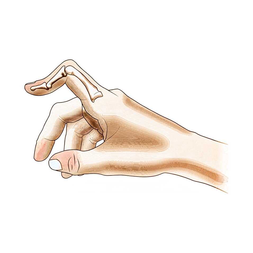
Extensor central-slip injury at the middle finger joint; early splinting prevents the zigzag deformity, established cases need surgery.
நீங்கள் என்ன உணர்கிறீர்கள்
உங்கள் நடுத்தர விரல் மூட்டு உள்நோக்கி வளைந்திருப்பதை நீங்கள் கவனிக்கலாம், அதே நேரத்தில் முனை மூட்டு வெளியே நிற்கிறது. இந்த குறிப்பிட்ட வடிவம் ஒரு பூட்டோனியர் சிதைவு என்று அழைக்கப்படுகிறது. உங்கள் விரலின் மேல் உள்ள தசைகள் இடத்திலிருந்து விலகிச் செல்லும்போது இது நிகழ்கிறது. இந்த மாற்றம் காலப்போக்கில் மெதுவாக வளர்ச்சியடைவதை நீங்கள் காணலாம், அல்லது காயத்திற்குப் பிறகு திடீரென்று தோன்றலாம்.
உங்கள் விரலின் நடுத்தர மூட்டு சுற்றி வலி பெரும்பாலும் மையமாக உள்ளது. நீங்கள் விரலை நகர்த்தும் போது அசௌகரியம் ஒரு ஆழமான வலி அல்லது கூர்மையான குத்தலாக உணரப்படலாம். விரலை முழுமையாக வளைப்பது அதை நேராக வைத்திருப்பதை விட அதிக வலியை ஏற்படுத்துகிறது என்பதை நீங்கள் காணலாம். பிடியுதல் அல்லது பிடுங்குதல் தேவைப்படும் செயல்பாடுகள் கடினமாகிவிடும். சட்டை பொத்தான் போடுவது, கதவு கைப்பிடியை திருப்புவது அல்லது தட்டச்சு செய்வது போன்ற எளிய பணிகள் சங்கடமாகவோ அல்லது வேதனையாகவோ இருக்கலாம்.
உங்கள் விரல் இறுக்கமாக உணரலாம், குறிப்பாக காலையில். நடுத்தர மூட்டு சுற்றி வீக்கத்தை நீங்கள் கவனிக்கலாம். இந்த கடினத்தன்மை ஒரு குத்து செய்ய கடினமாக இருக்கும். உங்களுக்கு ரியூமடோய்டு மூட்டுவலி இருந்தால், காயம் ஏற்பட்டால் மாறுபடும். மூட்டுவலி அல்லது காயம் இல்லாமல், சுமார் 13% பேர் இந்த நிலையை அனுபவிக்கிறார்கள்.
உங்கள் விரல் நுனியும் நிலையற்றதாக உணரலாம். சில சந்தர்ப்பங்களில், விரல் முனை மூட்டு வழக்கத்தை விட அதிகமாக கீழே வளைகிறது. இது உங்கள் விரலை ஒரு மேசையில் தட்டையாக வைப்பதை கடினமாக்கும். கனமான கதவுகளைத் தள்ளுவதற்கு அல்லது இலகுவான பொருட்களை அந்தக் கையால் தூக்குவதற்கு நீங்கள் போராடலாம். உங்கள் பக்கத்தில் தூங்குவது சிதைந்த விரலை அழுத்தி, அச om கரியத்தை ஏற்படுத்தும்.
உங்கள் மருத்துவர் உங்கள் விரல்கள் எவ்வாறு நகர்கின்றன என்பதை உன்னிப்பாகப் பார்த்து அவற்றை வேறுபடுத்துவார். நீங்கள் என்ன உணர்கிறீர்கள் என்பதை சரியாகப் புரிந்துகொள்வது உங்களுக்கு சிறந்த சிகிச்சைத் திட்டத்தை வழிநடத்த உதவுகிறது.
உண்மையில் என்ன நடக்கிறது
உங்கள் விரல் மூட்டு எலும்புகள், தசைகள், மற்றும் கூட்டு காப்ஸ்யூல் என்று அழைக்கப்படும் ஒரு பாதுகாப்பு சட்டை ஆகியவற்றால் ஆன ஒரு சிக்கலான மடிப்பு ஆகும். ஒரு ஆரோக்கியமான விரலில், ஒரு மத்திய தசை உங்கள் விரலின் நடுப்பகுதியில் நேராக இயங்குகிறது. இது உங்கள் விரலை நேராக இழுக்க உதவுகிறது. இந்த தசை உங்கள் விரலைத் திறக்கும் முக்கிய கயிறு போல செயல்படுகிறது.
பவுட்டோனியர் கோளாறில், அந்த மைய நரம்பு சேதமடைந்து அல்லது பலவீனமடைகிறது. அது அதிர்ச்சியால் கிழிந்திருக்கலாம் அல்லது ருமாடோய்டு மூட்டுவலி போன்ற நிலைகளால் நீட்டப்படலாம். இந்த மைய ஆதரவு தோல்வியுற்றால், உங்கள் விரலில் உள்ள சக்தியின் சமநிலை மாறுகிறது. விரலை வளைக்க வழக்கமாக உதவும் பக்க நரம்புகள், மிகவும் கடினமாக இழுக்கத் தொடங்குகின்றன.
உங்கள் விரலின் நடுத்தர மூட்டு உள்நோக்கி வளைந்து, முனை மூட்டு வெளியே தள்ளப்படலாம். உங்கள் விரலின் நடுத்தர மூட்டு உள்நோக்கி வளைந்து, உங்கள் விரலின் நடுத்தர மூட்டு உள்நோக்கி வளைந்து, உங்கள் விரலின் முனை மூட்டு வெளியே தள்ளப்படலாம்.
உங்கள் விரல் வளைந்ததாகவும், இறுக்கமாகவும் இருப்பதற்கான காரணம் இந்த பதட்ட மாற்றம் தான். பொதுவாக மூட்டு நிலையானதாக வைத்திருக்கும் மூட்டுக் காப்ஸ்யூல் இறுக்கமாகவும், சமநிலையற்றதாகவும் மாறும். காலப்போக்கில், திசுக்கள் இந்த புதிய, தவறான நிலைக்குத் தழுவுகின்றன. இதனால்தான் நீங்கள் அடுக்கு அல்லது சிகிச்சை போன்ற பழக்கவழக்கமான சிகிச்சைகளை முயற்சித்த பிறகும், கோளாறு நீடிக்கும்.
இந்த பிரச்சனையில் மிக முக்கியமான காரணி உங்கள் தசைகள் மற்றும் தொடர்புடைய கட்டமைப்புகளில் ஏற்படும் மாற்றங்கள் ஆகும். இந்த மாற்றங்கள் ஆரம்பத்தில் நிகழ்கின்றன, அதனால்தான் துல்லியமான நோயறிதல் முக்கியமானது. உங்கள் அறுவை சிகிச்சை நிபுணர் ஒரு உண்மையான பூட்டோனியர் சிதைவு மற்றும் போலி பூட்டோனியர் காயம் என்று அழைக்கப்படும் இதேபோன்ற பிரச்சினைக்கு இடையே வேறுபடுத்த வேண்டும். சிகிச்சை பாதை இந்த வேறுபாட்டை சரியாகப் பெறுவதைப் பொறுத்தது.
இந்த புதிய அறுவை சிகிச்சை, மற்ற விரல்களின் இயக்கங்களை அப்படியே வைத்திருக்கும் அதே வேளையில், அசல் இடுப்பு செயல்பாட்டைப் பின்பற்றுகிறது. கூட்டு மையமாகவும் நிலையானதாகவும் இருப்பதை நோக்கமாகக் கொண்டது, இதனால் நீங்கள் அதை மீண்டும் நகர்த்த முடியும்.
எவ்வாறாயினும், சிதைவு நீண்ட காலமாக இருந்தால், திசுக்கள் நிரந்தரமாக மாறியிருக்கலாம். இந்த நாள்பட்ட சந்தர்ப்பங்களில், எளிய பழுதுபார்ப்புகள் வேலை செய்யாமல் போகலாம். இந்த நிலைமையின் இயற்கையான வரலாறு பெரும்பாலும் நீடித்த பிரச்சினைகளுக்கு வழிவகுக்கிறது, குறிப்பாக ரியூமாடோய்டு மூட்டுவலி சம்பந்தப்பட்டால். இந்த சந்தர்ப்பங்களில் மென்மையான திசு புனரமைப்பிற்கான நீண்டகால முடிவுகள் நம்பகமானதாக இருக்காது. சில நேரங்களில், உங்கள் விரலின் வடிவத்தையும் செயல்பாட்டையும் சரிசெய்ய இன்னும் உறுதியான மீட்பு நடைமுறை தேவைப்படுகிறது.
நாம் என்ன செய்ய முடியும்
இயக்கத்தை மீட்டெடுப்பதற்காக அறுவை சிகிச்சை அல்லாத பராமரிப்புடன் தொடங்குகிறோம். போதுமான நீட்டிப்புக்காக தொடர்ச்சியான வார்ப்பை முயற்சிப்பீர்கள். இதைத் தொடர்ந்து மூன்று மாத உறவினர் இயக்கம் வளைவு ஆர்த்தெசிக் பயன்பாடு உள்ளது. உங்கள் அறுவை சிகிச்சையாளர் உடலியல் சிகிச்சையையும் பரிந்துரைக்கலாம். அறுவை சிகிச்சையின்றி ஒன்று முதல் இரண்டு கிரேடுகள் வரையிலான இயக்கம் முன்னேற்றத்தை அடைய முடியும். இருப்பினும், அறுவை சிகிச்சைக்குப் பிறகும் சிதைவு நீடிக்கும். செயலில் உள்ள தூர இடைமுனை கூட்டு வளைவை அதிகரிக்க உறவினர் இயக்கம் வளைவு ஆர்த்தோஸ்களைப் பயன்படுத்துகிறோம். இது உங்கள் நடுத்தர விரல் கூட்டு நீட்டினை மேம்படுத்த உதவுகிறது. அறுவை சிகிச்சையை கருத்தில் கொள்வதற்கு முன்பு இந்த அணுகுமுறை வேலை செய்ய நேரம் கொடுக்க வேண்டும்.
மருத்துவ மேலாண்மை ஆறுதல் மற்றும் அழற்சியில் கவனம் செலுத்துகிறது. உங்கள் சிதைவு ருமாடோய்டு மூட்டு அழற்சியுடன் தொடர்புடையதாக இருந்தால், நாங்கள் அடிப்படை நோய் செயல்பாட்டை நிர்வகிக்கிறோம். வலி மருந்துகள் மற்றும் அழற்சி எதிர்ப்பு மருந்துகள் அச om கரியத்தை நிர்வகிக்க உதவுகின்றன. மூட்டு வீக்கத்தைக் குறைக்க ஊசிகள் வழங்கப்படலாம். இந்த சிகிச்சைகள் மூட்டு மொபைல் மற்றும் வலி இல்லாததாக வைத்திருப்பதை நோக்கமாகக் கொண்டுள்ளன. சிகிச்சைக்கு உட்படுத்தும் போது நோக்கம் நிலைமையை உறுதிப்படுத்துவதும், அறுவை சிகிச்சை இல்லாமல் உங்கள் அன்றாட செயல்பாட்டை மேம்படுத்துவதும் ஆகும்.
கன்சர்வேடிவ் பராமரிப்பு அதன் வரம்பை எட்டியபோது அறுவை சிகிச்சை கருதப்படுகிறது. ஒரு உண்மையான பூட்டோனியர் சிதைவை ஒரு போலி பூட்டோனியர் காயத்திலிருந்து நாங்கள் வேறுபடுத்துகிறோம். மருத்துவ நிர்வாகத்தை தீர்மானிப்பதில் இந்த வேறுபாடு முக்கியமானது. ஒரு வெற்றிகரமான அறுவை சிகிச்சை முடிவு முழுமையான அறுவை சிகிச்சைக்கு முந்தைய பரிசோதனை, சிதைவின் சரியான நிலை மற்றும் சிகிச்சையின் சரியான நேரத்தைப் பொறுத்தது. மென்மையான திசு புனரமைப்பு தேவைப்பட்டால், நீண்ட கால முடிவுகள் நம்பகமானதாக இருக்காது என்பதை நாங்கள் புரிந்துகொள்கிறோம். மீண்டும் நிகழும் அல்லது நீடித்த சிதைவு ஒரு மீட்பு நடைமுறையுடன் சிறப்பாக சிகிச்சையளிக்கப்படுகிறது. சில சந்தர்ப்பங்களில், ஒரு Y- வடிவ இடுப்பு மாற்று அறுவை சிகிச்சை நல்ல அல்லது சிறந்த முடிவுகளை வழங்குகிறது. உங்கள் அறுவை சிகிச்சை தலையீட்டிற்கு முன் உண்மையான காரணியை உங்கள் அறுவை சிகிச்சை நிர்வகிப்பவர் தீர்மானிப்பார். இது உங்கள் குறிப்பிட்ட உடற்கூறியல் மற்றும் தேவைகளுக்கு பொருந்தும் செயல்முறையைத் தேர்ந்தெடுவதை உறுதி செய்கிறது.
எதிர்பார்ப்பது என்ன
உங்கள் முன்னோக்கு இது ஒரு உண்மையான சிதைவு அல்லது pseudoboutonniere என்று அழைக்கப்படும் இதே போன்ற காயம் என்பதைப் பொறுத்தது. உங்கள் அறுவை சிகிச்சையாளர் முதலில் நோயறிதலை உறுதிப்படுத்த வேண்டும், ஏனென்றால் அந்த வேறுபாட்டின் அடிப்படையில் சிகிச்சை பாதை முற்றிலும் மாறுகிறது. உங்களுக்கு ருமாடோய்டு மூட்டுவலி இருந்தால், மென்மையான திசு சரிசெய்தலில் இருந்து நீண்டகால முடிவுகள் பெரும்பாலும் நம்பகமானவை அல்ல. இந்த சந்தர்ப்பங்களில், தொடர்ச்சியான அல்லது தொடர்ச்சியான சிதைவு பின்னர் மீட்பு நடைமுறை தேவைப்படலாம்.
ருமாடோய்டு மூட்டுவலி இல்லாத பெரும்பாலான மக்களுக்கு, இந்த நிலை எப்போதும் தானாகவே குணமடையாது. அறுவை சிகிச்சையற்ற சிகிச்சை உங்கள் இயக்க வரம்பை ஒன்று முதல் இரண்டு தரங்களாக மேம்படுத்தலாம். இருப்பினும், நீங்கள் பிரத்யேக கன்சர்வேடிவ் மேலாண்மையை முடித்த பிறகும் தெரியும் சிதைவு நீடிக்கும். கன்சர்வேடிவ் பராமரிப்பு போதுமானதாக இல்லாவிட்டால், அறுவை சிகிச்சை ஒரு வலுவான விருப்பத்தை வழங்குகிறது. Y வடிவ இடுப்பு மாற்றுதல் அறிக்கையிடப்பட்ட தொடரில் 18 நோயாளிகளில் 16 பேரில் நல்ல அல்லது சிறந்த முடிவுகளை வழங்குகிறது. உங்கள் அறுவை சிகிச்சையாளர் ஒரு முழுமையான பரிசோதனையை மேற்கொள்வதையும், சிதைவை சரியாக நிலைநிறுத்துவதையும், தலையீட்டிற்கான சரியான நேரத்தைத் தேர்ந்தெடுப்பதையும் நம்பியுள்ளது.
குணமடைதல் என்பது ஒரு படிப்படியான செயல்முறையாகும். நீங்கள் அறுவை சிகிச்சையுடன் தொடங்கினால், விரலை நேராகச் செய்ய தொடர்ச்சியான வார்ப்பைப் பயன்படுத்தலாம், அதைத் தொடர்ந்து மூன்று மாதங்கள் உறவினர் இயக்கம் வளைவு ஆர்த்தோசிஸ் பயன்பாடு. இந்த அணுகுமுறை நாள்பட்ட நோய்களுக்கான பிற முறைகளுக்கு ஒத்த முடிவுகளை அளிக்கிறது மற்றும் பொதுவாக அறுவை சிகிச்சையை பரிசீலிப்பதற்கு முன்பு முயற்சிக்கப்படுகிறது. நீட்டிப்பைப் பராமரிக்கவும் வளைவை மேம்படுத்தவும் பல மாதங்கள் ஆர்த்தோசிஸ் அணிய வேண்டும் என்று நீங்கள் எதிர்பார்க்க வேண்டும்.
அறுவை சிகிச்சை தேவைப்பட்டால், செயல்பாடு மற்றும் சீரமைப்பை மீட்டெடுப்பதே குறிக்கோள். உங்கள் அறுவை சிகிச்சையாளரின் குறிப்பிட்ட வழிமுறைகளை நீங்கள் நெருக்கமாகப் பின்பற்ற வேண்டும். இந்த சிதைவின் இயற்கையான வரலாறு மாறுபடலாம், ஆனால் ஆரம்ப மற்றும் துல்லியமான மேலாண்மை சிறந்த முடிவுகளுக்கு வழிவகுக்கிறது. குணப்படுத்தும் செயல்முறையில் பொறுமையாக இருங்கள். இரைப்பைகள் மற்றும் மூட்டுகள் அவற்றின் புதிய நிலைகளுக்கு ஏற்ப நேரம் எடுக்கும். உங்கள் தினசரி நடவடிக்கைகளுக்கு சிறந்த முறையில் திரும்புவதை உறுதிசெய்ய உங்கள் அறுவை சிகிச்சையாளர் ஒவ்வொரு கட்டத்திலும் உங்களுக்கு வழிகாட்டுவார்.
யாரையாவது எப்போது பார்க்க வேண்டும்
உங்கள் விரலின் நடுத்தர மூட்டுவில் வளைவு இருப்பதை நீங்கள் கவனித்தால் உங்கள் GP ஐப் பார்க்கவும். ஓய்வில் முன்னேறாத தொடர்ச்சியான வலி இருந்தால் ஒரு நிபுணர் மதிப்பாய்வைக் கேளுங்கள். விரலில் பலவீனம் அல்லது நிலையற்ற தன்மையை நீங்கள் உணர்ந்தால் கவனிப்பை நாடுங்கள். விரல் பூட்டப்பட்டால் அல்லது பயன்பாட்டின் போது இடமளித்தால் ஒரு மருத்துவரிடம் செல்லுங்கள். உங்கள் தூக்கத்திலோ அல்லது வேலையிலோ அறிகுறிகள் தலையிட்டால் உங்கள் அறுவை சிகிச்சையாளரைத் தொடர்பு கொள்ளுங்கள். சிதைவின் திடீர் மோசமடைதல் உடனடி கவனம் தேவை. சரியான சிகிச்சைக்கு துல்லியமான நோயறிதல் முக்கியமானது. இதேபோன்ற காயத்திலிருந்து உண்மையான சிதைவை வேறுபடுத்துவது சரியான கவனிப்பைத் தீர்மானிக்க உதவுகிறது. ஆரம்ப மதிப்பீடு உங்கள் கை செயல்பாட்டிற்கான சிறந்த முடிவை உறுதி செய்கிறது.
Evidence & references
Overview
- Differentiating a true boutonniere deformity from a pseudoboutonniere injury is critical in determining clinical management [2].
- An understanding of the anatomy, clinical presentation, treatment options, and expected outcomes is crucial for optimal treatment of posttraumatic boutonniere and swan neck deformities [4].
- The natural history of the boutonnière deformity in rheumatoid arthritis is outlined, and a simple method of repair is described [3].
- The prevalence of boutonnière deformity without rheumatoid arthritis or trauma is approximately 13% [5].
- One to two grades of ROM improvement can be achieved with nonoperative treatment, although deformity can persist even after dedicated conservative management [8].
- Similar results occurred for chronic boutonniere deformity using serial casting for adequate extension followed by 3 months of RMF orthotic use, which should be attempted prior to surgical intervention [1].
- Long-term results following soft tissue reconstruction for boutonniere deformity in rheumatoid arthritis are unreliable, and recurrent or persistent deformity is best treated with a salvage procedure [9].
- A successful operative result for swan-neck and boutonniere deformities in the rheumatoid hand depends on complete preoperative examination, correct staging of the deformity, and proper timing of treatment [10].
- The Y-shaped tendon graft can be a useful procedure for the correction of chronic boutonniere deformity, providing good or excellent results in 16 of 18 patients in one series [6].
- Detachment of up to two-thirds of the phalangeal length was effective in reducing extensor lag of the DIP joint and did not cause any boutonniere deformity in a cadaveric model of fractional Fowler tenotomy for chronic mallet finger [7].
Anatomy & Pathophysiology
- Boutonnière deformity can persist even after dedicated conservative management [8].
- One to two grades of range of motion improvement can be achieved with nonoperative treatment of Boutonnière deformity [8].
- Accurate diagnosis and treatment of finger metacarpophalangeal joint injuries begins with an understanding of all potential diagnoses [15].
- Hand surgery and hand therapy practice interventions, including use of relative motion flexion orthoses for management of non-surgical and surgical extensor mechanism injuries, may benefit from an in-depth look at extensor mechanism zone III and IV anatomy and biomechanics [19].
- The most important factor in the development of finger deformities is the changes occurring in the tendons and related structures, especially in early stages [21].
- Reconstruction of the extensor central slip using a distally based flexor digitorum superficialis slip provides a robust repair that anatomically mimics the extensor central slip while maintaining the function of the donor FDS tendon [24].
- The main goals of any treatment of a proximal interphalangeal joint complication are maintaining concentric reduction of the joint, restoring joint stability, and facilitating early range-of-motion exercises [33].
Classification
- Differentiating a true boutonniere deformity from a pseudoboutonniere injury is critical in determining clinical management [2].
- The natural history of the boutonnière deformity in rheumatoid arthritis is outlined [3].
- The prevalence of boutonnière deformity without rheumatoid arthritis or trauma is approximately 13% [5].
- A modified Terrono classification for Type 1 thumb deformity in rheumatoid arthritis could detect advanced deformity earlier and was more strongly correlated with hand function [17].
Clinical Presentation
- Differentiating a true boutonniere deformity from a pseudoboutonniere injury is critical in determining clinical management [2].
- An understanding of the clinical presentation is crucial for optimal treatment of posttraumatic boutonnière and swan neck deformities [4].
- Accurate diagnosis of finger metacarpophalangeal joint injuries begins with an understanding of all potential diagnoses [15].
- The natural history of the boutonnière deformity in rheumatoid arthritis is outlined in historical literature [3].
- The prevalence of boutonnière deformity without rheumatoid arthritis or trauma is approximately 13% [5].
- The swan neck deformity can progress significantly with time due to increasing distal interphalangeal joint flexion contracture [14].
Investigations
- Differentiating a true boutonniere deformity from a pseudoboutonniere injury is critical in determining clinical management [2].
- An understanding of the anatomy, clinical presentation, treatment options, and expected outcomes is crucial for optimal treatment of posttraumatic boutonnière and swan neck deformities [4].
- Accurate diagnosis and treatment of finger metacarpophalangeal joint injuries begins with an understanding of all potential diagnoses [15].
- It is necessary to determine the true etiology before surgical intervention [12].
- A successful operative result depends on complete preoperative examination, correct staging of the deformity, and proper timing of treatment [10].
- Cortical breaks were commonly visualized in MCP and PIP joints with HR-pQCT and microCT [37].
Treatment
- Serial casting for adequate extension followed by 3 months of relative motion flexion (RMF) orthotic use should be attempted prior to surgical intervention for chronic boutonniere deformity [1].
- Differentiating a true boutonniere deformity from a pseudoboutonniere injury is critical in determining clinical management [2].
- A simple method of repair is described for the boutonnière deformity in rheumatoid arthritis [3].
- Understanding the anatomy, clinical presentation, treatment options, and expected outcomes is crucial for optimal treatment of posttraumatic boutonnière and swan neck deformities [4].
- The prevalence of boutonnière deformity without rheumatoid arthritis or trauma is approximately 13% [5].
- The Y-shaped tendon graft is a useful procedure for the correction of chronic boutonniere deformity, providing good or excellent results in 16 of 18 patients in a reported series [6].
- Detachment of up to two-thirds of the phalangeal length is effective in reducing extensor lag of the DIP joint and does not cause any boutonniere deformity in a cadaveric model [7].
- One to two grades of ROM improvement can be achieved with nonoperative treatment, although deformity can persist even after dedicated conservative management [8].
- Long-term results following soft tissue reconstruction for boutonniere deformity in rheumatoid arthritis are unreliable, and recurrent or persistent deformity is best treated with a salvage procedure [9].
- A successful operative result for swan-neck and boutonniere deformities in the rheumatoid hand depends on complete preoperative examination, correct staging of the deformity, and proper timing of treatment [10].
- Metacarpophalangeal joint arthroplasty improves function and deformity and achieves nearly uniform patient satisfaction in rheumatoid arthritis [11].
- One technique does not treat all finger deformities uniformly, highlighting the need to determine the true etiology before surgical intervention [12].
- The use of relative motion flexion orthoses (RMFO) is effective in increasing active distal interphalangeal joint flexion and improving PIP extension in patients with Burton stage 1 chronic boutonniere deformity [13].
Complications
- Differentiating a true boutonniere deformity from a pseudoboutonniere injury is critical in determining clinical management [2].
- The prevalence of boutonniere deformity without rheumatoid arthritis or trauma is approximately 13% [5].
- Detachment of up to two-thirds of the phalangeal length was effective in reducing extensor lag of the DIP joint and did not cause any boutonniere deformity in a cadaveric model [7].
- Long-term results following soft tissue reconstruction for boutonniere finger deformity in rheumatoid arthritis are unreliable [9].
- Recurrent or persistent deformity is best treated with a salvage procedure [9].
- A successful operative result depends on complete preoperative examination, correct staging of the deformity, and proper timing of treatment [10].
- One technique does not treat all deformities uniformly, highlighting the need to determine the true etiology before surgical intervention [12].
- Swan neck deformity can progress significantly with time due to increasing DIPJ flexion contracture [14].
Recovery
- Serial casting for adequate extension followed by 3 months of relative motion flexion (RMF) orthotic use yields similar results for chronic boutonniere deformity and should be attempted prior to surgical intervention [1].
- One to two grades of range of motion (ROM) improvement can be achieved with nonoperative treatment, although deformity can persist even after dedicated conservative management [8].
- The Y-shaped tendon graft is a useful procedure for the correction of chronic boutonniere deformity, providing good or excellent results in 16 of 18 patients in a reported series [6].
- The use of relative motion flexion orthoses (RMFO) is effective in increasing active distal interphalangeal joint flexion and improving proximal interphalangeal (PIP) extension in patients with Burton stage 1 chronic boutonniere deformity [13].
- Long-term results following soft tissue reconstruction for boutonniere deformity in rheumatoid arthritis are unreliable, and recurrent or persistent deformity is best treated with a salvage procedure [9].
- A successful operative result for boutonniere deformity depends on complete preoperative examination, correct staging of the deformity, and proper timing of treatment [10].
Key Evidence
- [L4] Similar results occurred for chronic boutonniere deformity using serial casting for adequate extension followed by 3 months of RMF orthotic use, which should be attempted prior to surgical intervention. [1] (10.1016/j.jht.2023.02.005)
- [L5] Differentiating a true boutonniere deformity from a pseudoboutonniere injury is critical in determining clinical management. [2] (10.1016/j.jhsa.2022.10.019)
- [L4] The natural history of the boutonnière deformity in rheumatoid arthritis is outlined, and a simple method of repair is described. [3] (10.2106/00004623-196951070-00009)
- [L5] An understanding of the anatomy, clinical presentation, treatment options, and expected outcomes is crucial for optimal treatment of posttraumatic boutonnière and swan neck deformities. [4] (10.5435/jaaos-d-14-00272)
- [L3] The prevalence of boutonnière deformity without rheumatoid arthritis or trauma is approximately 13%. [5] (10.1177/1753193417704610)
- [L4] The Y-shaped tendon graft can be a useful procedure for the correction of chronic boutonniere deformity; in our patient series, this provided good or excellent results in 16 of 18 patients. [6] (10.1016/j.jhsa.2021.01.003)
- [L5] Detachment of up to two-thirds of the phalangeal length was effective in reducing extensor lag of the DIP joint and did not cause any boutonniere deformity in this cadaveric model. [7] (10.1016/j.jhsa.2012.07.039)
- [L3] One to two grades of ROM improvement can be achieved, although deformity can persist even after dedicated conservative management. [8] (10.1016/j.jht.2025.02.013)
- [L5] Long-term results following soft tissue reconstruction are unreliable, and recurrent or persistent deformity is best treated with a salvage procedure. [9] (10.1016/j.jhsa.2011.05.029)
- [L5] A successful operative result depends on complete preoperative examination, correct staging of the deformity, and proper timing of treatment. [10] (10.5435/00124635-199903000-00002)
- [L5] Follow-up studies show that this surgery improves function and deformity and achieves nearly uniform patient satisfaction. [11] (10.5435/00124635-200305000-00005)
- [L5] It emphasizes that one technique does not treat all deformities uniformly and highlights the need to determine the true etiology before surgical intervention. [12] (10.1016/j.jhsa.2022.07.008)
- [L4] The use of RMFO is effective in increasing active distal interphalangeal joint flexion and improving PIP extension in patients with Burton stage 1 chronic boutonniere deformity. [13] (10.1016/j.jhsa.2022.08.007)
- [L5] The swan neck deformity in this individual progressed significantly with time because of increasing DIPJ flexion contracture. [14] (10.1016/j.jht.2009.11.005)
- [L5] Accurate diagnosis and treatment of finger metacarpophalangeal joint injuries in athletes begins with an understanding of all potential diagnoses, allowing for safe and early return to play. [15] (10.5435/jaaos-d-21-01031)
- [L3] The modified classification could detect advanced deformity earlier and was more strongly correlated with hand function. [17] (10.1177/1753193419886719)
- [L5] Hand surgery and hand therapy practice interventions, including use of RMF orthoses for management of non-surgical and surgical EM injuries may benefit from an in-depth look at the EM zone III and IV anatomy and biomechanics. [19] (10.1016/j.jht.2023.01.002)
- [L4] The most important factor in the development of finger deformities is the changes occurring in the tendons and related structures, especially in early stages. [21] (10.2106/00004623-195739030-00006)
- [L4] The modified technique provides a robust repair that anatomically mimics the extensor central slip yet maintains the function of the donor FDS tendon. [24] (10.1016/j.jhsa.2009.01.025)
- [L5] The main goals of any treatment of a PIP joint complication are maintaining concentric reduction of the joint, restoring joint stability, and facilitating early range-of-motion exercises. [33] (10.1016/j.hcl.2017.12.014)
- [L4] Cortical breaks were commonly visualized in MCP and PIP joints with HR-pQCT and microCT. [37] (10.1186/s12891-016-1148-y)
References
[1] The relative motion concept in acute and chronic boutonniere deformity: Invited commentary. Journal of Hand Therapy. 2023. DOI: 10.1016/j.jht.2023.02.005 [2] Boutonniere Versus Pseudoboutonniere Deformities: Pathoanatomy, Diagnosis, and Treatment. The Journal of Hand Surgery. 2023. DOI: 10.1016/j.jhsa.2022.10.019 [3] Correction of the Rheumatoid Boutonnière Deformity. The Journal of Bone & Joint Surgery. 1969. DOI: 10.2106/00004623-196951070-00009 [4] Posttraumatic Boutonnière and Swan Neck Deformities. Journal of the American Academy of Orthopaedic Surgeons. 2015. DOI: 10.5435/jaaos-d-14-00272 [5] Thumb boutonnière deformity without rheumatoid arthritis or trauma. Journal of Hand Surgery (European Volume). 2017. DOI: 10.1177/1753193417704610 [6] Y-Shaped Tendon Graft—A Technique in the Reconstruction of Posttraumatic Chronic Boutonniere Deformity. The Journal of Hand Surgery. 2021. DOI: 10.1016/j.jhsa.2021.01.003 [7] Fractional Fowler Tenotomy for Chronic Mallet Finger: A Cadaveric Biomechanical Study. The Journal of Hand Surgery. 2012. DOI: 10.1016/j.jhsa.2012.07.039 [8] Nonoperative treatment of the Boutonniere deformity: Is there a difference in outcomes?. Journal of Hand Therapy. 2025. DOI: 10.1016/j.jht.2025.02.013 [9] Treatment of Boutonniere Finger Deformity in Rheumatoid Arthritis. The Journal of Hand Surgery. 2011. DOI: 10.1016/j.jhsa.2011.05.029 [10] Operative Correction of Swan-Neck and Boutonniere Deformities in the Rheumatoid Hand. Journal of the American Academy of Orthopaedic Surgeons. 1999. DOI: 10.5435/00124635-199903000-00002 [11] Metacarpophalangeal Joint Arthroplasty in Rheumatoid Arthritis. Journal of the American Academy of Orthopaedic Surgeons. 2003. DOI: 10.5435/00124635-200305000-00005 [12] Clarification of Extensor Tenotomy for Finger Deformities. The Journal of Hand Surgery. 2022. DOI: 10.1016/j.jhsa.2022.07.008 [13] The Use of Relative Motion Flexion Orthoses for Chronic Boutonniere Deformity. The Journal of Hand Surgery. 2024. DOI: 10.1016/j.jhsa.2022.08.007 [14] Swan Neck Deformity after Distal Interphalangeal Joint Flexion Contractures: A Biomechanical Analysis. Journal of Hand Therapy. 2010. DOI: 10.1016/j.jht.2009.11.005 [15] Finger Metacarpophalangeal Joint Injuries in Athletes: Evaluation, Diagnosis, Treatment, and Return to Play. Journal of the American Academy of Orthopaedic Surgeons. 2023. DOI: 10.5435/jaaos-d-21-01031 [17] A modified Terrono classification for Type 1 thumb deformity in rheumatoid arthritis: a cross-sectional analysis. Journal of Hand Surgery (European Volume). 2019. DOI: 10.1177/1753193419886719 [19] An in-depth look at zone III and IV anatomy of the finger extensor mechanism and some clinical implications for use of the relative motion flexion orthosis. Journal of Hand Therapy. 2023. DOI: 10.1016/j.jht.2023.01.002 [21] Finger Deformities Caused by Rheumatoid Arthritis. The Journal of Bone & Joint Surgery. 1957. DOI: 10.2106/00004623-195739030-00006 [24] Reconstruction of the Extensor Central Slip Using a Distally Based Flexor Digitorum Superficialis Slip. The Journal of Hand Surgery. 2009. DOI: 10.1016/j.jhsa.2009.01.025 [33] Complications of Proximal Interphalangeal Joint Injuries. Hand Clinics. 2018. DOI: 10.1016/j.hcl.2017.12.014 [37] Visual detection of cortical breaks in hand joints: reliability and validity of high-resolution peripheral quantitative CT compared to microCT. BMC Musculoskeletal Disorders. 2016. DOI: 10.1186/s12891-016-1148-y


