纽扣畸形
Patients › Hand
Extensor central-slip injury at the middle finger joint; early splinting prevents the zigzag deformity, established cases need surgery.
您的感受
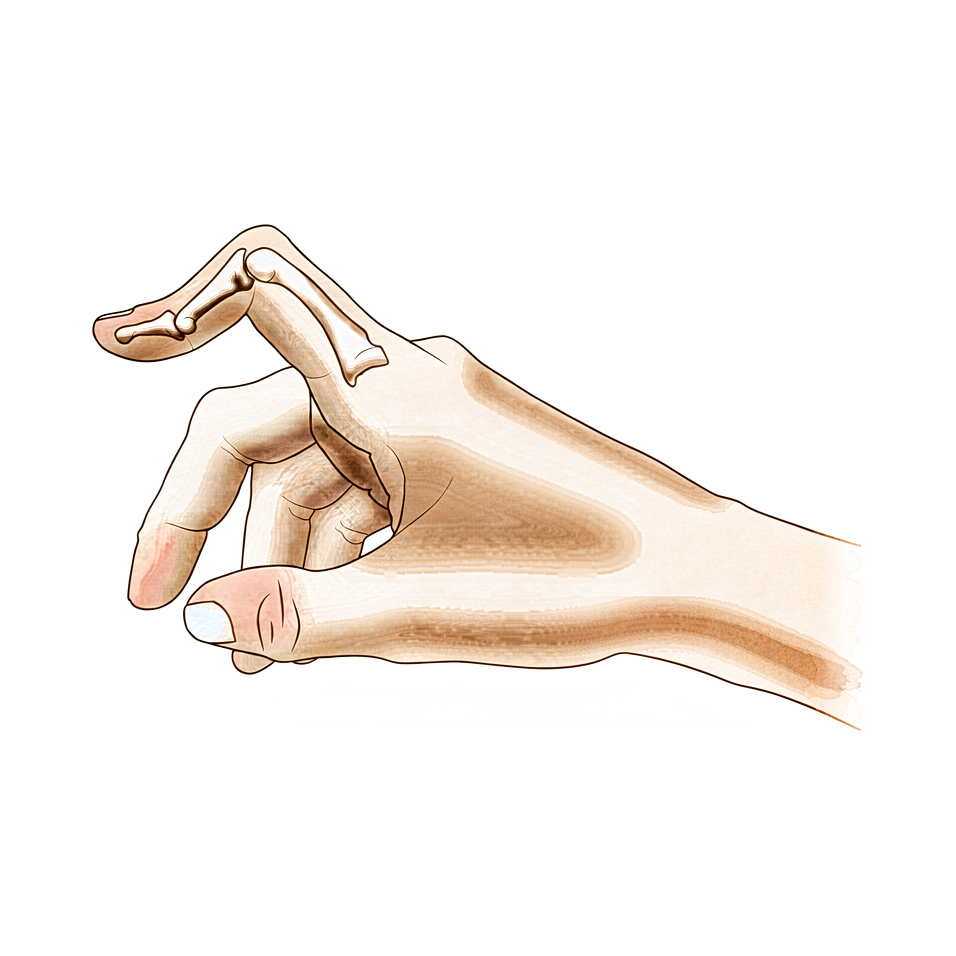
您可能会注意到中指近端指间关节向内侧弯曲,而远端指间关节向外侧伸展。这种特定的形态称为纽扣花样畸形(boutonniere deformity)。当手指背侧的肌腱发生错位时,就会出现这种情况。这种形态变化可能是逐渐发展的,也可能在受伤后突然出现。
疼痛通常集中在手指的中节指间关节。不适感可能表现为深部酸痛,或在活动手指时出现刺痛。您可能会发现,完全弯曲手指比保持伸直状态引起更剧烈的疼痛。需要抓握或捏取的活动会变得困难。简单的日常任务,如扣衬衫纽扣、转动门把手或打字,可能会感到别扭或疼痛。
您的手指可能会感到僵硬,尤其是在早晨。您可能会注意到中节指间关节周围出现肿胀。这种僵硬会使握拳变得困难。如果您患有类风湿关节炎,畸形的进展方式可能与创伤引起的不同。在没有关节炎或外伤的情况下,约 13% 的人会出现这种情况。
您的手指末端也可能感到不稳定。在某些情况下,远端指间关节会比平时更向下弯曲。这会使您将手指平放在桌面上变得困难。您可能会发现难以用这只手推开沉重的门或提起轻物。侧卧睡觉时,身体可能会压迫变形的指头,引起不适。
区分这种真正的畸形与外观相似的另一种损伤——假性纽扣花样畸形(pseudoboutonniere)非常重要。这两种情况需要不同的治疗。您的外科医生会仔细观察您手指的活动情况以进行鉴别。准确了解您的症状有助于为您制定最佳的治疗方案。
实际发生了什么
您的手指关节是一个由骨骼、肌腱和保护性套筒(称为关节囊)组成的复杂铰链结构。在健康的手指中,中央肌腱沿手指正中垂直下行,协助手指伸直。这根肌腱就像拉动手指张开的主绳索。
在纽扣畸形(Boutonnière deformity)中,这根中央肌腱受损或减弱。它可能因创伤而撕裂,或因类风湿关节炎等疾病而被拉长。当这一中央支撑失效时,手指内的力量平衡发生改变。通常协助弯曲手指的侧方肌腱开始过度牵拉。
这就像一条脱轨的拉链。部件仍然存在,但不再平滑地滑动。由于中央肌腱无法将结构固定到位,侧方肌腱向两侧滑移。这导致手指的中节指间关节向内弯曲,而远端指间关节可能向外突出。
这种张力的改变导致手指看起来弯曲且感觉僵硬。通常保持关节稳定的关节囊变得紧张且失衡。随着时间的推移,组织会适应这种新的错误位置。这就是为什么即使尝试夹板固定或治疗等保守治疗,畸形仍可能持续存在。
该问题中最重要的因素是肌腱及相关结构的变化。这些变化发生得很早,因此准确诊断至关重要。您的外科医生需要区分真正的纽扣畸形和一种类似的问题,称为假性纽扣畸形(pseudoboutonniere injury)。治疗路径完全取决于能否正确做出这一区分。
如果损伤在早期被发现,您的外科医生可能会专注于恢复中央肌腱的位置。在某些情况下,他们可能会使用取自另一根肌腱的一小块组织来重建中央腱束。这种新移植物模拟原始肌腱的功能,同时保持其他手指活动的完整性。目标是保持关节居中且稳定,以便您能够再次活动它。
然而,如果畸形已存在很长时间,组织可能发生永久性改变。在这些慢性病例中,简单修复可能无效。该疾病的自然病程通常导致持续性问题,尤其是当涉及类风湿关节炎时。在这些情况下,软组织重建的长期结果可能不可靠。有时,需要进行更确定的挽救性手术来矫正手指的形状和功能。
我们能采取的措施
我们从非手术治疗开始,以恢复关节活动度。您将尝试系列石膏固定以获得充分的伸展。随后需佩戴相对运动屈曲支具三个月。您的外科医生也可能建议进行物理治疗。通过非手术治疗可实现一到两个等级的活动度改善。然而,即使经过系统的保守治疗,畸形仍可能持续存在。我们使用相对运动屈曲支具来增加远端指间关节的主动屈曲。这有助于改善您中指关节的伸展。在考虑手术之前,必须给予这种方法足够的时间来发挥作用。
药物治疗侧重于缓解不适和控制炎症。如果您的畸形与类风湿关节炎有关,我们将针对潜在的疾病活动进行治疗。止痛药和非甾体抗炎药有助于缓解不适。可能会提供注射治疗以减少关节肿胀。这些治疗旨在您在接受理疗期间保持关节活动自如且无痛。目标是在不进行手术的情况下稳定病情并改善您的日常功能。
当保守治疗达到极限时,会考虑手术治疗。在做出决定之前,我们会区分真性纽扣花样畸形与假性纽扣花样损伤。这一区别对于确定临床管理方案至关重要。成功的手术结果取决于完善的术前检查、畸形的正确分期以及适当的治疗时机。如果需要软组织重建,我们了解长期结果可能不可靠。复发性或持续性畸形最好通过补救性手术进行治疗。在某些情况下,Y形肌腱移植物可提供良好或极佳的效果。您的外科医生将在手术干预前确定真正的病因。这确保所选择的手术方案符合您的特定解剖结构和需求。
预期情况
您的预后很大程度上取决于这是真正的畸形还是称为假性纽扣孔畸形的类似损伤。您的外科医生必须先确诊,因为治疗路径完全取决于这一区分。如果您患有类风湿关节炎,软组织修复的长期结果通常不可靠。在这种情况下,持续或复发的畸形可能需要在后期进行挽救性手术。
对于大多数没有类风湿关节炎的患者,该病症并不总能自行消退。非手术治疗可以将您的活动度提高一到两个等级。然而,即使完成专门的保守治疗,可见的畸形仍可能持续存在。如果保守治疗不足,手术是一个强有力的选择。据报道,在系列病例中,Y形肌腱移植在18例患者中有16例取得了良好或优秀的结果。成功还取决于您的外科医生进行全面的检查、正确地对畸形进行分期,并选择正确的干预时机。
恢复是一个渐进的过程。如果您从非手术治疗开始,您可能使用系列石膏固定来伸直手指,随后使用相对运动屈位矫形器三个月。这种方法对于慢性病例产生的结果与其他方法相似,并且通常在考虑手术之前尝试。您应该预期佩戴矫形器数月,以维持伸直并改善屈曲。
如果需要手术,目标是恢复功能和排列。您需要密切遵循外科医生的具体指示。这种畸形的自然病程可能有所不同,但早期和准确的管理能带来最佳结果。对愈合过程保持耐心。肌腱和关节适应新位置需要时间。您的外科医生将指导您度过每个阶段,以确保尽可能好地恢复您的日常活动。
何时就诊
如果您发现手指中间关节出现弯曲且无法伸直,请咨询您的全科医生。如果持续疼痛且休息后无改善,请要求专科医生评估。如果您感到手指无力或不稳,请及时就医。如果手指在使用时出现卡住或突然无力,请就诊。如果症状干扰您的睡眠或工作,请联系您的外科医生。畸形突然加重也需要及时关注。准确的诊断对于适当的治疗至关重要。区分真正的畸形与类似损伤有助于确定正确的治疗方案。早期评估可确保手部功能获得最佳预后。
Evidence & references
Overview
- Differentiating a true boutonniere deformity from a pseudoboutonniere injury is critical in determining clinical management [2].
- An understanding of the anatomy, clinical presentation, treatment options, and expected outcomes is crucial for optimal treatment of posttraumatic boutonniere and swan neck deformities [4].
- The natural history of the boutonnière deformity in rheumatoid arthritis is outlined, and a simple method of repair is described [3].
- The prevalence of boutonnière deformity without rheumatoid arthritis or trauma is approximately 13% [5].
- One to two grades of ROM improvement can be achieved with nonoperative treatment, although deformity can persist even after dedicated conservative management [8].
- Similar results occurred for chronic boutonniere deformity using serial casting for adequate extension followed by 3 months of RMF orthotic use, which should be attempted prior to surgical intervention [1].
- Long-term results following soft tissue reconstruction for boutonniere deformity in rheumatoid arthritis are unreliable, and recurrent or persistent deformity is best treated with a salvage procedure [9].
- A successful operative result for swan-neck and boutonniere deformities in the rheumatoid hand depends on complete preoperative examination, correct staging of the deformity, and proper timing of treatment [10].
- The Y-shaped tendon graft can be a useful procedure for the correction of chronic boutonniere deformity, providing good or excellent results in 16 of 18 patients in one series [6].
- Detachment of up to two-thirds of the phalangeal length was effective in reducing extensor lag of the DIP joint and did not cause any boutonniere deformity in a cadaveric model of fractional Fowler tenotomy for chronic mallet finger [7].
Anatomy & Pathophysiology
- Boutonnière deformity can persist even after dedicated conservative management [8].
- One to two grades of range of motion improvement can be achieved with nonoperative treatment of Boutonnière deformity [8].
- Accurate diagnosis and treatment of finger metacarpophalangeal joint injuries begins with an understanding of all potential diagnoses [15].
- Hand surgery and hand therapy practice interventions, including use of relative motion flexion orthoses for management of non-surgical and surgical extensor mechanism injuries, may benefit from an in-depth look at extensor mechanism zone III and IV anatomy and biomechanics [19].
- The most important factor in the development of finger deformities is the changes occurring in the tendons and related structures, especially in early stages [21].
- Reconstruction of the extensor central slip using a distally based flexor digitorum superficialis slip provides a robust repair that anatomically mimics the extensor central slip while maintaining the function of the donor FDS tendon [24].
- The main goals of any treatment of a proximal interphalangeal joint complication are maintaining concentric reduction of the joint, restoring joint stability, and facilitating early range-of-motion exercises [33].
Classification
- Differentiating a true boutonniere deformity from a pseudoboutonniere injury is critical in determining clinical management [2].
- The natural history of the boutonnière deformity in rheumatoid arthritis is outlined [3].
- The prevalence of boutonnière deformity without rheumatoid arthritis or trauma is approximately 13% [5].
- A modified Terrono classification for Type 1 thumb deformity in rheumatoid arthritis could detect advanced deformity earlier and was more strongly correlated with hand function [17].
Clinical Presentation
- Differentiating a true boutonniere deformity from a pseudoboutonniere injury is critical in determining clinical management [2].
- An understanding of the clinical presentation is crucial for optimal treatment of posttraumatic boutonnière and swan neck deformities [4].
- Accurate diagnosis of finger metacarpophalangeal joint injuries begins with an understanding of all potential diagnoses [15].
- The natural history of the boutonnière deformity in rheumatoid arthritis is outlined in historical literature [3].
- The prevalence of boutonnière deformity without rheumatoid arthritis or trauma is approximately 13% [5].
- The swan neck deformity can progress significantly with time due to increasing distal interphalangeal joint flexion contracture [14].
Investigations
- Differentiating a true boutonniere deformity from a pseudoboutonniere injury is critical in determining clinical management [2].
- An understanding of the anatomy, clinical presentation, treatment options, and expected outcomes is crucial for optimal treatment of posttraumatic boutonnière and swan neck deformities [4].
- Accurate diagnosis and treatment of finger metacarpophalangeal joint injuries begins with an understanding of all potential diagnoses [15].
- It is necessary to determine the true etiology before surgical intervention [12].
- A successful operative result depends on complete preoperative examination, correct staging of the deformity, and proper timing of treatment [10].
- Cortical breaks were commonly visualized in MCP and PIP joints with HR-pQCT and microCT [37].
Treatment
- Serial casting for adequate extension followed by 3 months of relative motion flexion (RMF) orthotic use should be attempted prior to surgical intervention for chronic boutonniere deformity [1].
- Differentiating a true boutonniere deformity from a pseudoboutonniere injury is critical in determining clinical management [2].
- A simple method of repair is described for the boutonnière deformity in rheumatoid arthritis [3].
- Understanding the anatomy, clinical presentation, treatment options, and expected outcomes is crucial for optimal treatment of posttraumatic boutonnière and swan neck deformities [4].
- The prevalence of boutonnière deformity without rheumatoid arthritis or trauma is approximately 13% [5].
- The Y-shaped tendon graft is a useful procedure for the correction of chronic boutonniere deformity, providing good or excellent results in 16 of 18 patients in a reported series [6].
- Detachment of up to two-thirds of the phalangeal length is effective in reducing extensor lag of the DIP joint and does not cause any boutonniere deformity in a cadaveric model [7].
- One to two grades of ROM improvement can be achieved with nonoperative treatment, although deformity can persist even after dedicated conservative management [8].
- Long-term results following soft tissue reconstruction for boutonniere deformity in rheumatoid arthritis are unreliable, and recurrent or persistent deformity is best treated with a salvage procedure [9].
- A successful operative result for swan-neck and boutonniere deformities in the rheumatoid hand depends on complete preoperative examination, correct staging of the deformity, and proper timing of treatment [10].
- Metacarpophalangeal joint arthroplasty improves function and deformity and achieves nearly uniform patient satisfaction in rheumatoid arthritis [11].
- One technique does not treat all finger deformities uniformly, highlighting the need to determine the true etiology before surgical intervention [12].
- The use of relative motion flexion orthoses (RMFO) is effective in increasing active distal interphalangeal joint flexion and improving PIP extension in patients with Burton stage 1 chronic boutonniere deformity [13].
Complications
- Differentiating a true boutonniere deformity from a pseudoboutonniere injury is critical in determining clinical management [2].
- The prevalence of boutonniere deformity without rheumatoid arthritis or trauma is approximately 13% [5].
- Detachment of up to two-thirds of the phalangeal length was effective in reducing extensor lag of the DIP joint and did not cause any boutonniere deformity in a cadaveric model [7].
- Long-term results following soft tissue reconstruction for boutonniere finger deformity in rheumatoid arthritis are unreliable [9].
- Recurrent or persistent deformity is best treated with a salvage procedure [9].
- A successful operative result depends on complete preoperative examination, correct staging of the deformity, and proper timing of treatment [10].
- One technique does not treat all deformities uniformly, highlighting the need to determine the true etiology before surgical intervention [12].
- Swan neck deformity can progress significantly with time due to increasing DIPJ flexion contracture [14].
Recovery
- Serial casting for adequate extension followed by 3 months of relative motion flexion (RMF) orthotic use yields similar results for chronic boutonniere deformity and should be attempted prior to surgical intervention [1].
- One to two grades of range of motion (ROM) improvement can be achieved with nonoperative treatment, although deformity can persist even after dedicated conservative management [8].
- The Y-shaped tendon graft is a useful procedure for the correction of chronic boutonniere deformity, providing good or excellent results in 16 of 18 patients in a reported series [6].
- The use of relative motion flexion orthoses (RMFO) is effective in increasing active distal interphalangeal joint flexion and improving proximal interphalangeal (PIP) extension in patients with Burton stage 1 chronic boutonniere deformity [13].
- Long-term results following soft tissue reconstruction for boutonniere deformity in rheumatoid arthritis are unreliable, and recurrent or persistent deformity is best treated with a salvage procedure [9].
- A successful operative result for boutonniere deformity depends on complete preoperative examination, correct staging of the deformity, and proper timing of treatment [10].
Key Evidence
- [L4] Similar results occurred for chronic boutonniere deformity using serial casting for adequate extension followed by 3 months of RMF orthotic use, which should be attempted prior to surgical intervention. [1] (10.1016/j.jht.2023.02.005)
- [L5] Differentiating a true boutonniere deformity from a pseudoboutonniere injury is critical in determining clinical management. [2] (10.1016/j.jhsa.2022.10.019)
- [L4] The natural history of the boutonnière deformity in rheumatoid arthritis is outlined, and a simple method of repair is described. [3] (10.2106/00004623-196951070-00009)
- [L5] An understanding of the anatomy, clinical presentation, treatment options, and expected outcomes is crucial for optimal treatment of posttraumatic boutonnière and swan neck deformities. [4] (10.5435/jaaos-d-14-00272)
- [L3] The prevalence of boutonnière deformity without rheumatoid arthritis or trauma is approximately 13%. [5] (10.1177/1753193417704610)
- [L4] The Y-shaped tendon graft can be a useful procedure for the correction of chronic boutonniere deformity; in our patient series, this provided good or excellent results in 16 of 18 patients. [6] (10.1016/j.jhsa.2021.01.003)
- [L5] Detachment of up to two-thirds of the phalangeal length was effective in reducing extensor lag of the DIP joint and did not cause any boutonniere deformity in this cadaveric model. [7] (10.1016/j.jhsa.2012.07.039)
- [L3] One to two grades of ROM improvement can be achieved, although deformity can persist even after dedicated conservative management. [8] (10.1016/j.jht.2025.02.013)
- [L5] Long-term results following soft tissue reconstruction are unreliable, and recurrent or persistent deformity is best treated with a salvage procedure. [9] (10.1016/j.jhsa.2011.05.029)
- [L5] A successful operative result depends on complete preoperative examination, correct staging of the deformity, and proper timing of treatment. [10] (10.5435/00124635-199903000-00002)
- [L5] Follow-up studies show that this surgery improves function and deformity and achieves nearly uniform patient satisfaction. [11] (10.5435/00124635-200305000-00005)
- [L5] It emphasizes that one technique does not treat all deformities uniformly and highlights the need to determine the true etiology before surgical intervention. [12] (10.1016/j.jhsa.2022.07.008)
- [L4] The use of RMFO is effective in increasing active distal interphalangeal joint flexion and improving PIP extension in patients with Burton stage 1 chronic boutonniere deformity. [13] (10.1016/j.jhsa.2022.08.007)
- [L5] The swan neck deformity in this individual progressed significantly with time because of increasing DIPJ flexion contracture. [14] (10.1016/j.jht.2009.11.005)
- [L5] Accurate diagnosis and treatment of finger metacarpophalangeal joint injuries in athletes begins with an understanding of all potential diagnoses, allowing for safe and early return to play. [15] (10.5435/jaaos-d-21-01031)
- [L3] The modified classification could detect advanced deformity earlier and was more strongly correlated with hand function. [17] (10.1177/1753193419886719)
- [L5] Hand surgery and hand therapy practice interventions, including use of RMF orthoses for management of non-surgical and surgical EM injuries may benefit from an in-depth look at the EM zone III and IV anatomy and biomechanics. [19] (10.1016/j.jht.2023.01.002)
- [L4] The most important factor in the development of finger deformities is the changes occurring in the tendons and related structures, especially in early stages. [21] (10.2106/00004623-195739030-00006)
- [L4] The modified technique provides a robust repair that anatomically mimics the extensor central slip yet maintains the function of the donor FDS tendon. [24] (10.1016/j.jhsa.2009.01.025)
- [L5] The main goals of any treatment of a PIP joint complication are maintaining concentric reduction of the joint, restoring joint stability, and facilitating early range-of-motion exercises. [33] (10.1016/j.hcl.2017.12.014)
- [L4] Cortical breaks were commonly visualized in MCP and PIP joints with HR-pQCT and microCT. [37] (10.1186/s12891-016-1148-y)
References
[1] The relative motion concept in acute and chronic boutonniere deformity: Invited commentary. Journal of Hand Therapy. 2023. DOI: 10.1016/j.jht.2023.02.005 [2] Boutonniere Versus Pseudoboutonniere Deformities: Pathoanatomy, Diagnosis, and Treatment. The Journal of Hand Surgery. 2023. DOI: 10.1016/j.jhsa.2022.10.019 [3] Correction of the Rheumatoid Boutonnière Deformity. The Journal of Bone & Joint Surgery. 1969. DOI: 10.2106/00004623-196951070-00009 [4] Posttraumatic Boutonnière and Swan Neck Deformities. Journal of the American Academy of Orthopaedic Surgeons. 2015. DOI: 10.5435/jaaos-d-14-00272 [5] Thumb boutonnière deformity without rheumatoid arthritis or trauma. Journal of Hand Surgery (European Volume). 2017. DOI: 10.1177/1753193417704610 [6] Y-Shaped Tendon Graft—A Technique in the Reconstruction of Posttraumatic Chronic Boutonniere Deformity. The Journal of Hand Surgery. 2021. DOI: 10.1016/j.jhsa.2021.01.003 [7] Fractional Fowler Tenotomy for Chronic Mallet Finger: A Cadaveric Biomechanical Study. The Journal of Hand Surgery. 2012. DOI: 10.1016/j.jhsa.2012.07.039 [8] Nonoperative treatment of the Boutonniere deformity: Is there a difference in outcomes?. Journal of Hand Therapy. 2025. DOI: 10.1016/j.jht.2025.02.013 [9] Treatment of Boutonniere Finger Deformity in Rheumatoid Arthritis. The Journal of Hand Surgery. 2011. DOI: 10.1016/j.jhsa.2011.05.029 [10] Operative Correction of Swan-Neck and Boutonniere Deformities in the Rheumatoid Hand. Journal of the American Academy of Orthopaedic Surgeons. 1999. DOI: 10.5435/00124635-199903000-00002 [11] Metacarpophalangeal Joint Arthroplasty in Rheumatoid Arthritis. Journal of the American Academy of Orthopaedic Surgeons. 2003. DOI: 10.5435/00124635-200305000-00005 [12] Clarification of Extensor Tenotomy for Finger Deformities. The Journal of Hand Surgery. 2022. DOI: 10.1016/j.jhsa.2022.07.008 [13] The Use of Relative Motion Flexion Orthoses for Chronic Boutonniere Deformity. The Journal of Hand Surgery. 2024. DOI: 10.1016/j.jhsa.2022.08.007 [14] Swan Neck Deformity after Distal Interphalangeal Joint Flexion Contractures: A Biomechanical Analysis. Journal of Hand Therapy. 2010. DOI: 10.1016/j.jht.2009.11.005 [15] Finger Metacarpophalangeal Joint Injuries in Athletes: Evaluation, Diagnosis, Treatment, and Return to Play. Journal of the American Academy of Orthopaedic Surgeons. 2023. DOI: 10.5435/jaaos-d-21-01031 [17] A modified Terrono classification for Type 1 thumb deformity in rheumatoid arthritis: a cross-sectional analysis. Journal of Hand Surgery (European Volume). 2019. DOI: 10.1177/1753193419886719 [19] An in-depth look at zone III and IV anatomy of the finger extensor mechanism and some clinical implications for use of the relative motion flexion orthosis. Journal of Hand Therapy. 2023. DOI: 10.1016/j.jht.2023.01.002 [21] Finger Deformities Caused by Rheumatoid Arthritis. The Journal of Bone & Joint Surgery. 1957. DOI: 10.2106/00004623-195739030-00006 [24] Reconstruction of the Extensor Central Slip Using a Distally Based Flexor Digitorum Superficialis Slip. The Journal of Hand Surgery. 2009. DOI: 10.1016/j.jhsa.2009.01.025 [33] Complications of Proximal Interphalangeal Joint Injuries. Hand Clinics. 2018. DOI: 10.1016/j.hcl.2017.12.014 [37] Visual detection of cortical breaks in hand joints: reliability and validity of high-resolution peripheral quantitative CT compared to microCT. BMC Musculoskeletal Disorders. 2016. DOI: 10.1186/s12891-016-1148-y


