Tendinite Calcificante
Patients › Shoulder
Calcific tendinitis causes shoulder pain from calcium deposits; treatment ranges from observation to washing out the calcium.
O que você está sentindo
Você pode experimentar dor aguda ou crônica no ombro causada por inflamação ao redor dos depósitos de cálcio nos tendões do manguito rotador. Esta condição, conhecida como tendinite calcificada, pode impactar significativamente sua qualidade de vida e resultar em afastamento do trabalho. A dor é frequentemente intensa e pode ter crises sem aviso prévio. Você pode notar que certos movimentos desencadeiam desconforto agudo, enquanto outros causam dor surda e persistente.
As tarefas diárias que exigem alcançar ou levantar objetos tornam-se difíceis. Ações simples, como abotoar a camisa ou alcançar as costas para fechar o sutiã, podem ser desafiadoras. Você pode ter dificuldade para dormir do lado afetado, pois a pressão pode piorar a dor. O desconforto noturno é comum, muitas vezes interrompendo seu descanso e deixando-o com sensação de fadiga. A dor também pode ser pior ao acordar, dificultando o início do seu dia.
Em alguns casos, você pode experimentar sintomas atípicos. Por exemplo, a dor pode estar isolada na parte posterior do ombro ou afetar movimentos acima da cabeça se o músculo redondo menor estiver envolvido. Raramente, você pode ter lesões calcificadas em outras áreas, como o ligamento colateral medial, ou em ambos os ombros simultaneamente. No entanto, a maioria das pessoas experimenta dor localizada no ombro que interfere nas atividades rotineiras.
A boa notícia é que a tendinite calcificada sintomática tem uma boa probabilidade de resolução completa a longo prazo. O tratamento conservador é frequentemente a escolha primária, especialmente para casos agudos, e mostra melhora clinicamente significativa. Cerca de 72% dos pacientes obtêm resultados excelentes ou bons com cuidados não cirúrgicos, independentemente do tamanho ou localização do depósito. Se os sintomas persistirem, seu cirurgião pode discutir outras opções, mas muitos encontram alívio por meio de medidas conservadoras. Compreender esses sintomas ajuda você a gerenciar as expectativas e a trabalhar com sua equipe de saúde para encontrar o melhor caminho a seguir.
O que está realmente acontecendo
A tendinite calcificada é uma condição na qual o cálcio se acumula dentro dos tendões do ombro. Esses tendões são as bandas resistentes e semelhantes a cordas que conectam seus músculos aos seus ossos. Pense neles como os cabos que ajudam você a levantar e mover o braço. Quando os depósitos de cálcio se formam, eles criam um nódulo duro dentro desse tecido mole. Esse acúmulo pode irritar a área circundante, causando dor significativa e limitando o quanto você consegue usar o ombro.
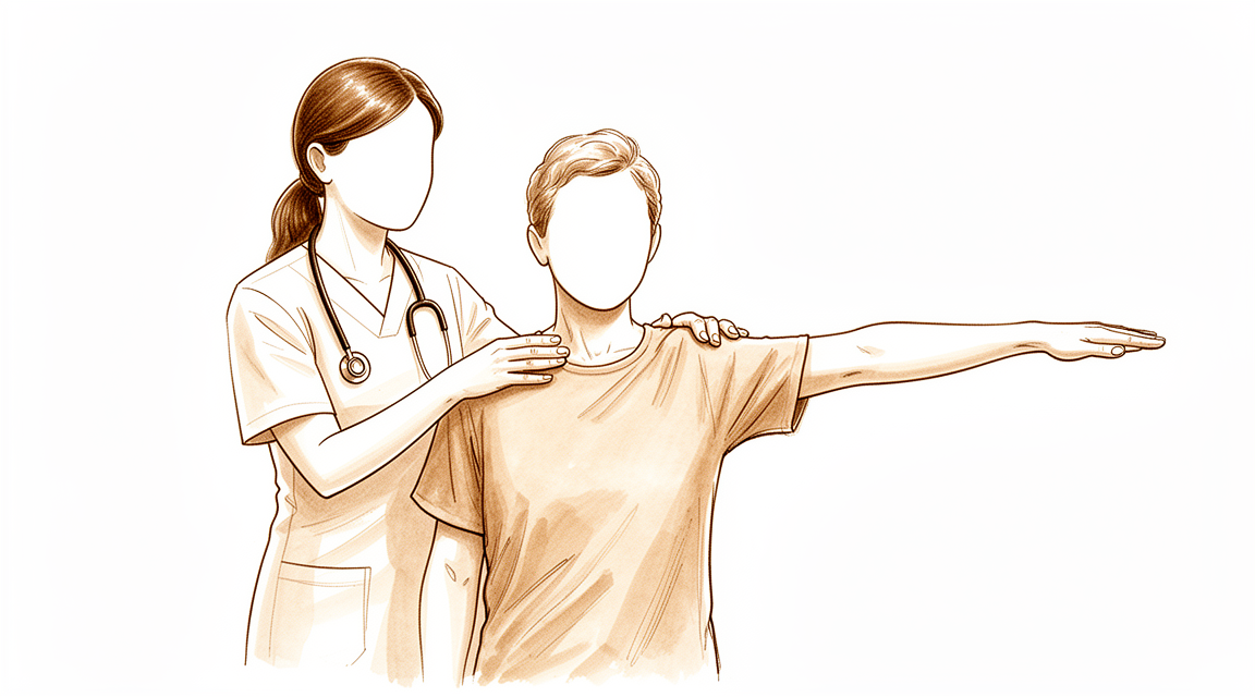
Seu ombro é uma articulação complexa do tipo bola e soquete. Ele depende de um grupo de músculos e tendões chamados manguito rotador para permanecer estável e mover-se suavemente. A cabeça longa do bíceps também desempenha um papel na manutenção da estabilidade dessa articulação. Em muitos casos, os depósitos de cálcio afetam essas estruturas. Você pode sentir dor na parte frontal do ombro. Isso ocorre porque o depósito interage com os tecidos moles circundantes, em vez de ser um problema único e isolado. A dor é o sinal do seu corpo de que algo está errado nesse equilíbrio delicado.
À medida que você envelhece, as alterações nos tendões tornam-se mais comuns. Nadadores e indivíduos ativos frequentemente apresentam essas alterações estruturais mais cedo. O próprio depósito de cálcio pode variar em tamanho. Se a lesão tiver mais de 1 cm, há quase três vezes mais chances de você precisar de cirurgia para removê-la. No entanto, muitas pessoas encontram alívio sem uma operação. Tratamentos conservadores, como a terapia por ondas de choque, são frequentemente muito bem-sucedidos no alívio da dor e na restauração da função, com muito poucos efeitos colaterais.
Se a cirurgia for necessária, seu cirurgião provavelmente removerá o depósito de cálcio. Isso é frequentemente feito usando uma pequena câmera e instrumentos (artroscopia). Em muitos casos, o cirurgião também reparará o manguito rotador ao mesmo tempo. Essa combinação leva a melhorias significativas na dor e na função do ombro. Leva tempo para que esses benefícios se manifestem totalmente. Você deve esperar pelo menos seis meses após a cirurgia para que a função do seu ombro atinja seu melhor nível. Durante esse período, seu cirurgião o guiará pela recuperação para garantir o melhor resultado possível.
O que podemos fazer a respeito
A maioria das pessoas encontra alívio sem cirurgia. Sua jornada geralmente começa com autocuidado simples e movimento guiado. É provável que você tente medicação anti-inflamatória oral para acalmar a dor. Seu cirurgião também pode recomendar fisioterapia para restaurar a amplitude de movimento e a força do seu ombro. Essas modalidades não cirúrgicas são a escolha primária para a tendinite calcificada aguda.
Você deve dar tempo ao tratamento conservador para funcionar. A condição tem boa probabilidade de resolução completa a longo prazo. De fato, 72% dos pacientes obtêm resultados excelentes ou bons com o cuidado não cirúrgico. Essa taxa de sucesso é válida independentemente de onde o cálcio está localizado, de seu tamanho ou da gravidade dos seus sintomas iniciais. Se a sua dor for prolongada, pode levar ao afastamento do trabalho e à redução da qualidade de vida. O esforço consistente com a terapia é fundamental para retornar à sua rotina diária.
Se o autocuidado e a terapia não forem suficientes, seu cirurgião pode sugerir intervenções médicas. Injeções de corticosteroides são uma opção comum para reduzir a inflamação e a dor. Outro tratamento eficaz não invasivo é a terapia por ondas de choque extracorpóreas (ESWT). Esta terapia utiliza ondas sonoras para fragmentar os depósitos de cálcio. É segura e produz uma alta taxa de sucesso no alívio da dor e na restauração funcional. Os pacientes frequentemente observam redução dos sintomas dolorosos e melhora da função do ombro após este tratamento. As complicações da ESWT são desprezíveis.
A cirurgia é considerada apenas quando o cuidado conservador atinge seu limite. Seu cirurgião pode recomendar tratamento operatório se suas lesões calcificadas tiverem mais de 1 cm, pois essas têm uma probabilidade 2,8 vezes maior de necessitar de cirurgia. A remoção artroscópica da calcificação leva a melhores resultados clínicos para os casos crônicos. A maioria dos pacientes que passam por este manejo cirúrgico também requer uma reparação concomitante do manguito rotador.
Se você prosseguir com a cirurgia, espere uma recuperação gradual. As pontuações funcionais melhoram lentamente após o procedimento. Elas tipicamente ultrapassam 75 por cento seis meses após a cirurgia. Pelo menos 6 meses de acompanhamento são necessários para que essas melhorias se tornem estatisticamente significativas. A maioria dos pacientes relata melhorias significativas na dor e na função do ombro após a operação. Seu cirurgião discutirá se a cirurgia artroscópica, endoscópica ou aberta é a melhor para o seu caso específico.
O que esperar
A dor no ombro causada pela tendinite calcificada pode durar muito tempo. Esses sintomas podem ser prolongados. Isso frequentemente significa afastamento do trabalho e uma menor qualidade de vida. A boa notícia é que essa condição tem uma boa probabilidade de resolução completa a longo prazo. Muitas pessoas percebem que seus sintomas se resolvem espontaneamente.
Se a sua dor não desaparecer, o seu cirurgião pode sugerir tratamento. Opções não cirúrgicas, como terapia por ondas de choque ou aspiração com agulha, são eficazes. A terapia por ondas de choque oferece uma alta taxa de sucesso no alívio da dor e na restauração funcional, com complicações associadas desprezíveis. A aspiração com agulha é apenas ligeiramente invasiva e dolorosa. Esses tratamentos funcionam bem para muitos pacientes.
Se for necessária cirurgia, o seu cirurgião removerá o depósito de cálcio. É provável que seja necessária uma reparação do manguito rotador ao mesmo tempo. Isso é uma parte comum do procedimento. A sua dor e função no ombro melhorarão significativamente após a cirurgia. No entanto, a recuperação não é instantânea. As pontuações funcionais dos pacientes submetidos ao tratamento artroscópico melhoraram lentamente. Você verá mais de 75% de melhora aos seis meses após a cirurgia.
Leva tempo para sentir o benefício completo da remoção artroscópica. Pelo menos 6 meses de acompanhamento são necessários para que essas melhorias se tornem estatisticamente significativas. Não apresse a sua cicatrização. O seu cirurgião irá guiá-lo durante esse processo. A maioria dos pacientes está satisfeita com os resultados finais.
Quando procurar ajuda
Consulte o seu médico de família se tiver dor no ombro persistente que não melhora com o repouso. A tendinite calcificante é uma condição aguda ou cronicamente dolorosa, causada por inflamação em torno de depósitos de cálcio nos tendões do manguito rotador. Deve procurar ajuda se os sintomas interferirem no seu sono ou trabalho, ou se experimentar fraqueza, instabilidade ou bloqueio. O tratamento conservador é a primeira opção, especialmente nos casos agudos, e mostra melhora clinicamente significativa, com 72% dos pacientes obtendo resultados excelentes ou bons. No entanto, se a sua dor permanecer intensa apesar do tratamento conservador, solicite uma avaliação por um especialista. O seu cirurgião pode determinar se são necessários passos adicionais para restaurar a função e aliviar a dor.
Evidence & references
Overview
- Shoulder MRI may be warranted for preoperative planning in select patients with chronic calcific tendinopathy and prolonged refractory pain, although the probability of identifying additional cuff pathology requiring surgical intervention is very low [1].
- Patients with calcific lesions >1 cm have a 2.8 increased likelihood to undergo operative treatment in the setting of calcific tendinitis of the shoulder [2].
- The majority of patients who undergo surgical management for removal of the calcific deposit require a concomitant rotator cuff repair and experience significant improvements in shoulder pain and function [3].
- Treatment of calcific tendinitis of the shoulder with extracorporeal shock waves produces a high rate of success in pain relief and functional restoration with negligible associated complications [6].
- Patients undergoing arthroscopic treatment of a calcific deposit in the shoulder have satisfactory clinical and radiological outcomes at final follow-up, with functional scores improving slowly and reaching more than 75 percent at six months after surgery [8].
- Arthroscopic removal of calcification leads to improved clinical outcomes in patients with chronic calcific tendinitis, but at least 6 months of follow-up is needed for these improvements to become statistically significant [11].
- The primary choice of treatment for calcific tendinitis is conservative, especially in patients with acute calcific tendinitis [12].
- Arthroscopic treatment of calcifying tendinitis provides good to excellent clinical results [16].
- Routine diagnostic glenohumeral exploration does not appear beneficial in arthroscopic treatment of calcific tendinitis due to the low prevalence of intraarticular pathologies which most frequently do not require surgical treatment [19].
- Endoscopic and open surgery are equally effective in the treatment of chronic calcifying tendinopathy, showing similar clinical and sonographic results [23].
Anatomy & Pathophysiology
- Rotator cuff disease, shoulder instability, and associated lesions are common pathologic conditions of the shoulder involving soft tissues [42].
- The long head of the biceps contributes to stability of the glenohumeral joint in all directions, although in vivo studies have not yet established this stabilizing effect or the physiologic load required [32].
- Understanding the function and pathology surrounding the teres minor is paramount in comprehensive management of patients with shoulder pathology [34].
- In the context of rotator cuff disease, anterior shoulder pain with macroscopic changes in the biceps tendon is related to the complex interaction of the tendon and surrounding soft tissues rather than a single entity [41].
- Long head of biceps tendinopathy is associated with age and cuff tendinopathy on MRI obtained for evaluation of shoulder pain [51].
- Clinicians can guide patients to understand shoulder pain as age-appropriate and safe, potentially reducing unnecessary visits, tests, and treatments [51].
- A high prevalence of structural changes in the rotator cuff and biceps tendons in masters swimmers reflects the effect of shoulder symptoms, aging, and swim training [54].
- In chronic rotator cuff tears, the shape of the deltoid muscle seems only to be influenced by natural aging and is independent of reduced shoulder motion [53].
- There seems to be no disadvantage to exhausting conservative treatment and delaying implantation of reverse total shoulder arthroplasty in patients with chronic rotator cuff tears [53].
- Surgical repair of an isolated supraspinatus tear may be sufficient to keep the torn rotator cuff intact and achieve satisfactory patient-reported outcomes, but glenohumeral joint mechanics and shoulder strength are not fully restored with current repair techniques [39].
- At a 2-year follow-up, shoulder function evaluated in terms of CMS was not significantly improved with conservative versus surgical management for rotator cuff tears [37].
- Over time, supraspinatus tendon thickness in individuals with rotator cuff-related shoulder pain tends to normalize compared to the contralateral side, regardless of the exercise intervention [28].
- The theoretical concept of a high acromion index resulting in an increased upward force against the subacromial space influencing pain and function in calcifying tendinitis of the shoulder was not supported [49].
- Ultrasound is a useful tool for discovering subjects in pre-symptomatic stages who may undergo shoulder symptomatic pathologies [40].
Classification
- Calcific tendinitis is a common shoulder disorder [9].
- Calcific tendinitis should be differentiated from dystrophic calcification because their pathogenesis and natural history are totally different [9].
- Calcific tendinitis is an acute or chronically painful condition caused by inflammation around calcium deposits in the rotator cuff tendons [15].
- Symptoms of calcific tendinitis can be protracted, resulting in time off work and impaired quality of life [5].
- Symptomatic calcific tendinitis of the shoulder has a good likelihood to completely resolve in the long-term [4].
- The demographic, radiographic, and clinical features of calcific tendinitis of the shoulder in the Korean population are not different from those of Western populations [7].
- Conservative treatment for calcific tendinitis of the shoulder showed clinically significant improvement, with 72% of excellent or good results regardless of the location, radiologic type and size, and initial symptoms of calcific deposits [10].
- The primary choice of treatment for calcific tendinitis is conservative, especially in patients with acute calcific tendinitis [12].
- Patients with calcific lesions >1 cm had a 2.8 increased likelihood to undergo operative treatment in the setting of calcific tendinitis of the shoulder [2].
- The majority of patients who undergo surgical management for removal of the calcific deposit will require a concomitant rotator cuff repair and have significant improvements in shoulder pain and function [3].
- Shoulder MRI may be warranted for preoperative planning in the select population of patients with chronic calcific tendinitis and prolonged refractory pain, although the probability of identifying additional cuff pathology requiring surgical intervention is very low [1].
- Calcific tendinitis of the rotator cuff with tuberosity osteolysis is a distinctive form of calcific tendinitis that should be considered in clinical and surgical practice [17].
- Restriction of passive glenohumeral abduction combined with normal passive external rotation is a diagnostic feature of calcific tendinitis that can be used to clinically differentiate it from adhesive capsulitis [18].
- The incidence and type of intraarticular lesions in calcifying tendinitis are comparable to age-matched shoulders with partial- rather than full-thickness rotator cuff tears [35].
Clinical Presentation
- Calcific tendinitis of the shoulder is an acute or chronically painful condition caused by inflammation around calcium deposits in the rotator cuff tendons [15].
- Symptoms of calcific tendinitis can be protracted, resulting in time off work and impaired quality of life [5].
- Symptomatic calcific tendinitis of the shoulder has a good likelihood to completely resolve in the long-term [4].
- Calcific tendinitis is a common shoulder disorder that should be differentiated from dystrophic calcification because the pathogenesis and natural history of both are totally different [9].
- The demographic, radiographic, and clinical features of calcific tendinitis of the shoulder in the Korean population are not different from those of Western populations [7].
- Restriction of passive glenohumeral abduction combined with normal passive external rotation is a diagnostic feature of calcific tendinitis that can be used to clinically differentiate it from adhesive capsulitis [18].
- Arthroscopic treatment of calcifying tendinitis provides good to excellent clinical results [16].
- The majority of patients who undergo surgical management for removal of the calcific deposit will require a concomitant rotator cuff repair and have significant improvements in shoulder pain and function [3].
- Patients with calcific lesions >1 cm had a 2.8 increased likelihood to undergo operative treatment in the setting of calcific tendinitis of the shoulder [2].
- Conservative treatment for calcific tendinitis of the shoulder showed clinically significant improvement, with 72% of excellent or good results regardless of the location, radiologic type and size, and initial symptoms of calcific deposits [10].
- The primary choice of treatment for calcific tendinitis is conservative, especially in patients with acute calcific tendinitis [12].
- Treatment of calcific tendinitis of the shoulder with extracorporeal shock waves has produced a high rate of success in pain relief and functional restoration with negligible associated complications [6].
- Both ultrasound-guided needling and extracorporeal shock wave therapy improved clinical outcomes and eliminated calcium deposits in the treatment of calcific tendinitis [22].
- Shoulder MRI may be warranted for preoperative planning in the select population of patients with chronic calcific tendinopathy and prolonged refractory pain, although the probability of identifying additional cuff pathology requiring surgical intervention is very low [1].
- Routine diagnostic glenohumeral exploration does not appear beneficial in arthroscopic treatment of calcific tendinitis due to the low prevalence of intraarticular pathologies which most frequently do not require surgical treatment [19].
- Imaging and functional data indicate that calcific tendinitis of the rotator cuff with tuberosity osteolysis is a distinctive form of calcific tendinitis that should be considered in clinical and surgical practice [17].
- Recognition of atypical presentations of calcific tendinitis with bone erosion may prevent unnecessary biopsy and overtreatment [14].
- Atypical presentations of calcific tendinitis, such as involvement of the teres minor affecting overhead movement or isolated posterior shoulder pain, should be considered in clinical practice [20].
- A patient can suffer from both a calcifying lesion within the medial collateral ligament and calcifying tendinitis of the rotator cuff in both shoulders simultaneously [13].
Investigations
- Shoulder MRI may be warranted for preoperative planning in select patients with chronic calcific tendinopathy and prolonged refractory pain, although the probability of identifying additional cuff pathology requiring surgical intervention is very low [1].
- Patients with calcific lesions >1 cm have a 2.8 increased likelihood to undergo operative treatment [2].
- The majority of patients undergoing surgical management for removal of the calcific deposit require a concomitant rotator cuff repair [3].
- Symptomatic calcific tendinitis of the shoulder has a good likelihood to completely resolve in the long-term [4].
- Calcific tendinitis symptoms can be protracted, resulting in time off work and impaired quality of life [5].
- Demographic, radiographic, and clinical features of calcific tendinitis of the shoulder in the Korean population are not different from those of Western populations [7].
- Patients undergoing arthroscopic treatment of a calcific deposit in the shoulder had satisfactory clinical and radiological outcomes at final follow-up, with functional scores improving slowly and reaching more than 75 percent at six months after surgery [8].
- Conservative treatment for calcific tendinitis of the shoulder showed clinically significant improvement, with 72% of excellent or good results regardless of the location, radiologic type and size, and initial symptoms of calcific deposits [10].
- Calcific tendinitis can present with simultaneous calcifying lesions in other locations, such as the medial collateral ligament of the knee [13].
- Atypical presentations of calcific tendinitis with bone erosion may occur, and recognition of these presentations can prevent unnecessary biopsy and overtreatment [14].
- Calcific tendinitis of the rotator cuff with tuberosity osteolysis is a distinctive form that should be considered in clinical and surgical practice [17].
- Restriction of passive glenohumeral abduction combined with normal passive external rotation is a diagnostic feature that can be used to clinically differentiate adhesive capsulitis from calcific tendinitis [18].
- Atypical presentations of calcific tendinitis, such as involvement of the teres minor affecting overhead movement, should be considered in the context of isolated posterior shoulder pain [20].
- Shoulder surgeons should be cautious about rotator cuff tears as a comorbidity in calcific tendinitis and aware of the accuracy limitations of sonographic or MRI evaluation [27].
- The use of MRI before a trial of conservative management in patients with atraumatic shoulder pain, minimal to no strength deficits on physical examination, and suspected cuff tendinopathy other than full-thickness tears provides negative value in management at both individual and population levels [30].
- MRI, but not radiography, can be used to help discriminate between traumatic and nontraumatic rotator cuff lesions [38].
- Repeating lavage and corticosteroid injections until calcific deposits resolve completely is not recommended due to lack of evidence regarding efficacy and risks [43].
- Preoperative ultrasound-guided marking of calcific deposits statistically significantly improves the clinical results of arthroscopic surgery at 6 weeks and 2 years [44].
Treatment
Non-Operative Management
- Conservative treatment is the primary choice for calcific tendinitis, especially in patients with acute calcific tendinitis [12].
- Nonsurgical management remains the mainstay of treatment for calcific tendinitis of the rotator cuff [31].
- Most patients improve with nonsurgical modalities such as oral anti-inflammatory medication, physical therapy, and corticosteroid injections [31].
- Symptomatic calcific tendinitis of the shoulder has a good likelihood to completely resolve in the long-term [4].
- Conservative treatment for calcific tendinitis of the shoulder showed clinically significant improvement, with 72% of excellent or good results regardless of the location, radiologic type and size, and initial symptoms of calcific deposits [10].
- Extracorporeal shock wave therapy (ESWT) is a safe and effective noninvasive treatment for patients with calcific tendinitis of the shoulder [21].
- ESWT produces a high rate of success in pain relief and functional restoration with negligible complications [21].
- ESWT effectively reduced painful symptomatology and increased shoulder function in patients with chronic calcific tendinitis of the shoulder [24].
- Treatment of calcific tendinitis of the shoulder with shock waves has produced a high rate of success in pain relief and functional restoration with negligible associated complications [6].
- Both ultrasound-guided needling and extracorporeal shock wave therapy improved clinical outcomes and eliminated calcium deposits in the treatment of calcific tendinitis [22].
- The use of MRI before a trial of conservative management in patients with atraumatic shoulder pain, minimal to no strength deficits on physical examination, and suspected cuff tendinopathy other than full-thickness tears provides negative value in the management of these patients [30].
Operative Management
- Patients with calcific lesions >1 cm had a 2.8 increased likelihood to undergo operative treatment in the setting of calcific tendinitis of the shoulder [2].
- Arthroscopic removal of calcification leads to improved clinical outcomes in patients with chronic calcific tendinitis [11].
- At least 6 months of follow-up is needed for improvements in clinical outcomes after arthroscopic removal of calcification to become statistically significant [11].
- Patients undergoing arthroscopic treatment of a calcific deposit in the shoulder had satisfactory clinical and radiological outcomes at the final follow-up [8].
- Functional scores in patients undergoing arthroscopic treatment of a calcific deposit improved slowly, reaching more than 75 percent at six months after surgery [8].
- Arthroscopic treatment of calcifying tendinitis provides good to excellent clinical results [16].
- The majority of patients who undergo surgical management for removal of the calcific deposit will require a concomitant rotator cuff repair [3].
- Patients who undergo surgical management for removal of the calcific deposit have significant improvements in shoulder pain and function [3].
- Endoscopic and open surgery are equally effective in the treatment of chronic calcifying tendinopathy, showing similar clinical and sonographic results [23].
- Routine diagnostic glenohumeral exploration does not appear beneficial in arthroscopic treatment of calcific tendinitis due to the low prevalence of intraarticular pathologies which most frequently do not require surgical treatment [19].
- Shoulder MRI may be warranted for preoperative planning in the select population of patients with chronic calcific tendinopathy and prolonged refractory pain [1].
- The probability of identifying additional cuff pathology requiring surgical intervention via shoulder MRI in patients with chronic calcific tendinopathy and prolonged refractory pain is very low [1].
Complications
- Calcific tendinitis symptoms can be protracted, resulting in time off work and impaired quality of life [5].
- Atypical presentations of calcific tendinitis may include bone erosion, which can lead to unnecessary biopsy and overtreatment if not recognized [14].
- Shoulder surgeons should be cautious about rotator cuff tears as a comorbidity in calcific tendinitis due to accuracy limitations in sonographic or MRI evaluation [27].
- The probability of identifying additional cuff pathology requiring surgical intervention via preoperative shoulder MRI in patients with chronic calcific tendinopathy is very low [1].
- Arthroscopic decompression is not recommended in the treatment of rotator cuff tendinopathy [25].
Recovery
- Symptomatic calcific tendinitis of the shoulder has a good likelihood to completely resolve in the long-term [4].
- Symptoms of calcific tendinitis can be protracted, resulting in time off work and impaired quality of life [5].
- Patients undergoing arthroscopic treatment of a calcific deposit in the shoulder had satisfactory clinical and radiological outcomes at the final follow-up [8].
- Functional scores for patients undergoing arthroscopic treatment of a calcific deposit improved slowly, reaching more than 75 percent at six months after surgery [8].
- Arthroscopic removal of calcification leads to improved clinical outcomes in patients with chronic calcific tendinitis, but at least 6 months of follow-up is needed for these improvements to become statistically significant [11].
- The majority of patients who undergo surgical management for removal of the calcific deposit will require a concomitant rotator cuff repair [3].
- Patients who undergo surgical management for removal of the calcific deposit have significant improvements in shoulder pain and function [3].
- Percutaneous needle aspiration and lavage is effective in the short term and in the long term in calcific tendinitis of the shoulder [29].
- Percutaneous needle aspiration and lavage results are similar to or better than those published for other techniques [29].
- Percutaneous needle aspiration and lavage is only slightly invasive and painful [29].
- Treatment of calcific tendinitis of the shoulder with shock waves has produced a high rate of success in pain relief and functional restoration with negligible associated complications [6].
- Shock wave therapy is a safe and effective noninvasive treatment for patients with calcific tendinitis of the shoulder, producing a high rate of success in pain relief and functional restoration with negligible complications [21].
- A symptom duration of ≤10 months or calcification size of ≤10.82 mm represented the clinical scenarios most likely to show resorption after extracorporeal shockwave therapy (ESWT) [56].
- The short-term functional outcome of patients with calcific tendonitis after arthroscopic bursectomy and debridement of the calcific deposit is not influenced if performed in combination with or without a subacromial decompression [26].
Key Evidence
- [Commentary] Shoulder MRI may be warranted for preoperative planning in the select population of patients with chronic calcific tendinopathy and prolonged refractory pain, although the probability of identifying additional cuff pathology requiring surgical intervention is very low. [1] (10.1016/j.arthro.2020.01.014)
- [L3] Patients with calcific lesions >1 cm had a 2.8 increased likelihood to undergo operative treatment in the setting of calcific tendinitis of the shoulder. [2] (10.1016/j.jseint.2021.01.013)
- [L3] The majority of patients who undergo surgical management for removal of the calcific deposit will require a concomitant rotator cuff repair and have significant improvements in shoulder pain and function. [3] (10.1177/2325967121s00208)
- [L1] Symptomatic calcific tendinitis of the shoulder has a good likelihood to completely resolve in the long-term. [4] (10.1097/phm.0000000000000939)
- [L3] Calcific tendinitis is a poorly understood condition in which symptoms can be protracted, resulting in time off work and impaired quality of life. [5] (10.1016/j.jse.2006.06.007)
- [L2] Treatment of calcific tendinitis of the shoulder with shock waves has produced a high rate of success in pain relief and functional restoration with negligible associated complications. [6] (10.1016/j.jse.2007.03.023)
- [L4] This study reported demographic, radiographic, and clinical features of calcific tendinitis of the shoulder in the Korean population, which were not different from those of Western populations. [7] (10.5397/cise.2020.00010)
- [Paper] Patients undergoing arthroscopic treatment of a calcific deposit in the shoulder had satisfactory clinical and radiological outcomes at the final follow-up, with functional scores improving slowly and reaching more than 75 percent at six months after surgery. [8] (10.1016/j.otsr.2020.03.005)
- [L5] Calcific tendinitis is a common shoulder disorder that should be differentiated from dystrophic calcification as the pathogenesis and natural history of both is totally different. [9] (10.5312/wjo.v7.i1.55)
- [L2] Conservative treatment for calcific tendinitis of the shoulder showed clinically significant improvement, with 72% of excellent or good results regardless of the location, radiologic type and size, and initial symptoms of calcific deposits. [10] (10.1016/j.jse.2009.07.008)
- [L4] Arthroscopic removal of calcification leads to improved clinical outcomes in patients with chronic calcific tendinitis, but at least 6 months of follow-up is needed for these improvements to become statistically significant. [11] (10.5397/cise.2018.21.2.75)
- [L5] The primary choice of treatment for calcific tendinitis is conservative, especially in patients with acute calcific tendinitis. [12] (10.5397/cise.2020.00318)
- [Case_report] This is the first case report of a patient suffering from both a calcifying lesion within the medial collateral ligament and calcifying tendinitis of the rotator cuff in both shoulders. [13] (10.1186/s12891-016-1147-z)
- [L5] Recognition of atypical presentations of calcific tendinitis with bone erosion may prevent unnecessary biopsy and overtreatment. [14] (10.1016/j.jse.2009.02.009)
- [L5] Calcific tendinitis of the shoulder is an acute or chronically painful condition caused by inflammation around calcium deposits in the rotator cuff tendons. [15] (10.1016/s0030-5898(03)00089-0)
- [L3] Arthroscopic treatment of calcifying tendinitis provides good to excellent clinical results. [16] (10.1177/03635465211037690)
- [L2] Imaging and functional data indicate that calcific tendinitis of the rotator cuff with tuberosity osteolysis is a distinctive form of calcific tendinitis that should be considered in clinical and surgical practice. [17] (10.1016/j.jse.2008.09.016)
- [L3] This finding can be used to clinically differentiate adhesive capsulitis from calcific tendinitis. [18] (10.1177/2325967117752907)
- [L3] Routine diagnostic glenohumeral exploration does not appear beneficial in arthroscopic treatment of calcific tendinitis due to the low prevalence of intraarticular pathologies which most frequently do not require surgical treatment. [19] (10.1186/s12891-017-1839-z)
- [Case_report] This case highlights the importance of considering atypical presentations of calcific tendinitis, particularly in the context of isolated posterior shoulder pain. [20] (10.1016/j.jisako.2025.101055)
- [L3] Shock wave therapy is a safe and effective noninvasive treatment for patients with calcific tendinitis of the shoulder, producing a high rate of success in pain relief and functional restoration with negligible complications. [21] (10.1177/03635465030310031701)
- [L2] Both treatment modalities for calcific tendinitis improved clinical outcomes and eliminated calcium deposits. [22] (10.1016/j.jse.2014.06.036)
- [L1] Endoscopic and open surgery are equally effective in the treatment of chronic calcifying tendinopathy, showing similar clinical and sonographic results. [23] (10.1097/01.blo.0000063786.32430.22)
- [L2] ESWT effectively reduced painful symptomatology and increased shoulder function in patients with chronic calcific tendinitis of the shoulder. [24] (10.1136/ard.62.3.248)
- [L1] The natural history of rotator cuff tendinopathy probably plays a significant role in the results in the long-term. [25] (10.1302/0301-620x.99b6.bjj-2016-0569.r1)
- [L1] This study has demonstrated that the short-term functional outcome of patients with calcific tendonitis after arthroscopic bursectomy and debridement of the calcific deposit is not influenced if performed in combination with or without a subacromial decompression. [26] (10.1016/j.arthro.2015.05.015)
- [L3] Shoulder surgeons should be cautious about rotator cuff tears as a comorbidity in calcific tendinitis and aware of the accuracy limitations of sonographic or MRI evaluation. [27] (10.5397/cise.2021.00094)
- [L2] Findings from this study suggest that, over time, supraspinatus tendon thickness in individuals with rotator cuff-related shoulder pain tends to normalize compared to the contralateral side, regardless of the exercise intervention. [28] (10.1016/j.jse.2024.03.055)
- [L4] Percutaneous needle aspiration and lavage is effective in the short term and in the long term in calcific tendinitis of the shoulder, with results similar to or better than those published for other techniques, and it is only slightly invasive and painful. [29] (10.2214/ajr.07.2254)
- [L4] The use of MRI before a trial of conservative management in patients with atraumatic shoulder pain, minimal to no strength deficits on physical examination, and suspected cuff tendinopathy other than full-thickness tears provides negative value in the management of these patients, at both the individual and population level. [30] (10.1016/j.jse.2019.04.003)
- [L5] Nonsurgical management remains the mainstay of treatment for calcific tendinitis of the rotator cuff, with most patients improving with modalities such as oral anti-inflammatory medication, physical therapy, and corticosteroid injections. [31] (10.5435/jaaos-22-11-707)
- [L5] Biomechanical studies indicate that the long head of the biceps contributes to stability of the glenohumeral joint in all directions, though in vivo studies have yet to establish this stabilizing effect and the physiologic load required remains unknown. [32] (10.1016/j.arthro.2010.10.014)
- [L5] Understanding the function and pathology surrounding the teres minor is paramount in comprehensive management of the patient with shoulder pathology. [34] (10.5435/jaaos-d-15-00258)
- [L3] The incidence and type of intraarticular lesions in calcifying tendinitis are comparable to age-matched shoulders with partial- rather than full-thickness rotator cuff tears. [35] (10.1007/s00402-011-1263-z)
- [L1] At a 2-year follow-up, shoulder function evaluated in terms of CMS was not significantly improved. [37] (10.1186/s12891-020-03872-4)
- [L2] MRI, but not radiography, can be used to help discriminate between traumatic and nontraumatic rotator cuff lesions. [38] (10.1016/j.jse.2015.06.005)
- [L4] Surgical repair of an isolated supraspinatus tear may be sufficient to keep the torn rotator cuff intact and achieve satisfactory patient-reported outcomes, but glenohumeral joint mechanics and shoulder strength are not fully restored with current repair techniques. [39] (10.1177/0363546511412164)
- [L3] Ultrasound is an useful tool for discovering in pre-symptomatic stages the subjects that may undergo shoulder symptomatic pathologies. [40] (10.1186/1471-2474-11-278)
- [L4] In the context of rotator cuff disease, the etiology of anterior shoulder pain with macroscopic changes in the biceps tendon is related to the complex interaction of the tendon and surrounding soft tissues, rather than a single entity. [41] (10.1016/j.jse.2008.05.044)
- [L5] Repeating lavage and corticosteroid injections until the calcific deposits resolve completely is not recommended due to lack of evidence regarding efficacy and risks. [43] (10.5397/cise.2021.00269)
- [L3] Preoperative ultrasound-guided marking of calcific deposits is a procedure that statistically significantly improves the clinical results of arthroscopic surgery as seen at 6 weeks and 2 years. [44] (10.1016/j.arthro.2006.08.005)
- [L2] The theoretical concept of a high acromion index resulting in an increased upward force against the subacromial space, which influences pain and function in calcifying tendinitis of the shoulder, was not supported. [49] (10.1007/s00167-011-1563-4)
- [L3] Clinicians can guide patients to understand shoulder pain as age-appropriate and safe, potentially reducing unnecessary visits, tests, and treatments. [51] (10.1097/corr.0000000000003342)
- [L3] Our data support that in chronic rotator cuff tears, there seems to be no disadvantage to exhausting conservative treatment and to delay implantation of reverse total shoulder arthroplasty, as the shape of deltoid muscle seems only to be influenced by natural aging, but to be independent of reduced shoulder motion. [53] (10.1186/1471-2474-14-247)
- [L4] A high prevalence of structural changes in the rotator cuff and biceps tendons in masters swimmers reflects the effect of shoulder symptoms, aging, and swim training. [54] (10.3390/medicina57010029)
- [L3] A symptom duration of ≤10 months or calcification size of ≤10.82 mm represented the clinical scenarios most likely to show resorption after ESWT. [56] (10.1177/23259671241231609)
References
[1] Editorial Commentary: Is Magnetic Resonance Imaging of the Shoulder Ever Appropriate in Evaluating Patients With Calcific Tendinopathy of the Rotator Cuff?. Arthroscopy: The Journal of Arthroscopic & Related Surgery. 2020. DOI: 10.1016/j.arthro.2020.01.014 [2] Predictive factors for failure of conservative management in the treatment of calcific tendinitis of the shoulder. JSES International. 2021. DOI: 10.1016/j.jseint.2021.01.013 [3] Predictive factor for failure of conservative management in the treatment of calcific tendinitis of the shoulder. Orthopaedic Journal of Sports Medicine. 2021. DOI: 10.1177/2325967121s00208 [4] Long-Term Course of Shoulders After Ultrasound Therapy for Calcific Tendinitis. American Journal of Physical Medicine & Rehabilitation. 2018. DOI: 10.1097/phm.0000000000000939 [5] Calcific tendinitis: Natural history and association with endocrine disorders. Journal of Shoulder and Elbow Surgery. 2007. DOI: 10.1016/j.jse.2006.06.007 [6] Extracorporeal shock wave therapy for calcifying tendinitis of the shoulder. Journal of Shoulder and Elbow Surgery. 2008. DOI: 10.1016/j.jse.2007.03.023 [7] Calcific tendinitis of the shoulder in the Korean population: demographics and its relation with coexisting rotator cuff tear. Clinics in Shoulder and Elbow. 2021. DOI: 10.5397/cise.2020.00010 [8] Recovery pattern after arthroscopic treatment for calcific tendinitis of the shoulder. Orthopaedics & Traumatology: Surgery & Research. 2020. DOI: 10.1016/j.otsr.2020.03.005 [9] Calcific tendinitis of the rotator cuff. World Journal of Orthopedics. 2016. DOI: 10.5312/wjo.v7.i1.55 [10] Radiologic course of the calcific deposits in calcific tendinitis of the shoulder: Does the initial radiologic aspect affect the final results?. Journal of Shoulder and Elbow Surgery. 2010. DOI: 10.1016/j.jse.2009.07.008 [11] Functional Recovery of the Shoulder after Arthroscopic Treatment for Chronic Calcific Tendinitis. Clinics in Shoulder and Elbow. 2018. DOI: 10.5397/cise.2018.21.2.75 [12] Diagnosis and treatment of calcific tendinitis of the shoulder. Clinics in Shoulder and Elbow. 2020. DOI: 10.5397/cise.2020.00318 [13] Case report - calcification of the medial collateral ligament of the knee with simultaneous calcifying tendinitis of the rotator cuff. BMC Musculoskeletal Disorders. 2016. DOI: 10.1186/s12891-016-1147-z [14] Calcific tendinitis of the rotator cuff associated with intraosseous loculation: Two case reports. Journal of Shoulder and Elbow Surgery. 2009. DOI: 10.1016/j.jse.2009.02.009 [15] Calcific tendinitis of the shoulder. Orthopedic Clinics of North America. 2003. DOI: 10.1016/s0030-5898(03)00089-0 [16] Clinical and Structural Results of Rotator Cuff Repair Compared With Rotator Cuff Debridement in Arthroscopic Treatment of Calcifying Tendinitis of the Shoulder. The American Journal of Sports Medicine. 2021. DOI: 10.1177/03635465211037690 [17] Osteolytic lesion of greater tuberosity in calcific tendinitis of the shoulder. Journal of Shoulder and Elbow Surgery. 2009. DOI: 10.1016/j.jse.2008.09.016 [18] Restriction of Passive Glenohumeral Abduction Combined With Normal Passive External Rotation Is a Diagnostic Feature of Calcific Tendinitis. Orthopaedic Journal of Sports Medicine. 2018. DOI: 10.1177/2325967117752907 [19] Examination of concomitant glenohumeral pathologies in patients treated arthroscopically for calcific tendinitis of the shoulder and implications for routine diagnostic joint exploration. BMC Musculoskeletal Disorders. 2017. DOI: 10.1186/s12891-017-1839-z [20] Atypical calcific tendinitis involving teres minor which affects overhead movement: A case report. Journal of ISAKOS. 2026. DOI: 10.1016/j.jisako.2025.101055 [21] Shock Wave Therapy for Calcific Tendinitis of the Shoulder. The American Journal of Sports Medicine. 2003. DOI: 10.1177/03635465030310031701 [22] Which method is more effective in treatment of calcific tendinitis in the shoulder? Prospective randomized comparison between ultrasound-guided needling and extracorporeal shock wave therapy. Journal of Shoulder and Elbow Surgery. 2014. DOI: 10.1016/j.jse.2014.06.036 [23] Prospective Randomized Surgical Treatments for Calcifying Tendinopathy. Clinical Orthopaedics & Related Research. 2003. DOI: 10.1097/01.blo.0000063786.32430.22 [24] Extracorporeal shock wave therapy for chronic calcific tendinitis of the shoulder: single blind study. Annals of the Rheumatic Diseases. 2003. DOI: 10.1136/ard.62.3.248 [25] Arthroscopic decompression not recommended in the treatment of rotator cuff tendinopathy. The Bone & Joint Journal. 2017. DOI: 10.1302/0301-620x.99b6.bjj-2016-0569.r1 [26] Short‐Term Outcome After Arthroscopic Bursectomy Debridement of Rotator Cuff Calcific Tendonopathy With and Without Subacromial Decompression: A Prospective Randomized Controlled Trial. Arthroscopy. 2015. DOI: 10.1016/j.arthro.2015.05.015 [27] Is common the rotator cuff tear in the calcific tendinitis?. Clinics in Shoulder and Elbow. 2021. DOI: 10.5397/cise.2021.00094 [28] Do therapeutic exercises impact supraspinatus tendon thickness? Secondary analyses of the combined dataset from two randomized controlled trials in patients with rotator cuff-related shoulder pain. Journal of Shoulder and Elbow Surgery. 2024. DOI: 10.1016/j.jse.2024.03.055 [29] Sonographically Guided Percutaneous Needle Lavage in Calcific Tendinitis of the Shoulder: Short- and Long-Term Results. American Journal of Roentgenology. 2007. DOI: 10.2214/ajr.07.2254 [30] A value-based care analysis of magnetic resonance imaging in patients with suspected rotator cuff tendinopathy and the implicated role of conservative management. Journal of Shoulder and Elbow Surgery. 2019. DOI: 10.1016/j.jse.2019.04.003 [31] Calcific Tendinitis of the Rotator Cuff. Journal of the American Academy of Orthopaedic Surgeons. 2014. DOI: 10.5435/jaaos-22-11-707 [32] Anatomy, Function, Injuries, and Treatment of the Long Head of the Biceps Brachii Tendon. Arthroscopy. 2011. DOI: 10.1016/j.arthro.2010.10.014 [34] Understanding the Importance of the Teres Minor for Shoulder Function: Functional Anatomy and Pathology. Journal of the American Academy of Orthopaedic Surgeons. 2018. DOI: 10.5435/jaaos-d-15-00258 [35] Intraarticular lesions in calcifying tendinitis: incidence and association with the acromion index. Archives of Orthopaedic and Trauma Surgery. 2011. DOI: 10.1007/s00402-011-1263-z [37] Conservative versus surgical management for patients with rotator cuff tears: a systematic review and META-analysis. BMC Musculoskeletal Disorders. 2021. DOI: 10.1186/s12891-020-03872-4 [38] How to discriminate between acute traumatic and chronic degenerative rotator cuff lesions: an analysis of specific criteria on radiography and magnetic resonance imaging. Journal of Shoulder and Elbow Surgery. 2015. DOI: 10.1016/j.jse.2015.06.005 [39] In Vivo Shoulder Function After Surgical Repair of a Torn Rotator Cuff. The American Journal of Sports Medicine. 2011. DOI: 10.1177/0363546511412164 [40] Sonographic evaluation of the shoulder in asymptomatic elderly subjects with diabetes. BMC Musculoskeletal Disorders. 2010. DOI: 10.1186/1471-2474-11-278 [41] Biceps tendinitis in chronic rotator cuff tears: A histologic perspective. Journal of Shoulder and Elbow Surgery. 2008. DOI: 10.1016/j.jse.2008.05.044 [42] Chapter 24 Shoulder Instability, Rotator Cuff Disorders, Muscular Ruptures, Adhesive Capsulitis, Calcific Tendinitis. 2020. [43] The efficacy of repeated needling for calcific tendinitis of the rotator cuff. Clinics in Shoulder and Elbow. 2021. DOI: 10.5397/cise.2021.00269 [44] Value of Preoperative Ultrasound Marking of Calcium Deposits in Patients Who Require Surgical Treatment of Calcific Tendinitis of the Shoulder. Arthroscopy. 2007. DOI: 10.1016/j.arthro.2006.08.005 [49] Do anatomic variants of the acromion shape in the frontal plane influence pain and function in calcifying tendinitis of the shoulder?. Knee Surgery, Sports Traumatology, Arthroscopy. 2011. DOI: 10.1007/s00167-011-1563-4 [51] Long Head of Biceps Tendinopathy Is Associated With Age and Cuff Tendinopathy on MRI Obtained for Evaluation of Shoulder Pain. Clinical Orthopaedics & Related Research. 2024. DOI: 10.1097/corr.0000000000003342 [53] Deltoid muscle shape analysis with magnetic resonance imaging in patients with chronic rotator cuff tears. BMC Musculoskeletal Disorders. 2013. DOI: 10.1186/1471-2474-14-247 [54] Ultrasonographic Evaluation of the Shoulders and Its Associations with Shoulder Pain, Age, and Swim Training in Masters Swimmers. Medicina. 2020. DOI: 10.3390/medicina57010029 [56] Treatment Algorithm for the Resorption of Calcific Tendinitis Using Extracorporeal Shockwave Therapy: A Data Mining Study. Orthopaedic Journal of Sports Medicine. 2024. DOI: 10.1177/23259671241231609


