Viêm gân vôi hóa
Patients › Shoulder
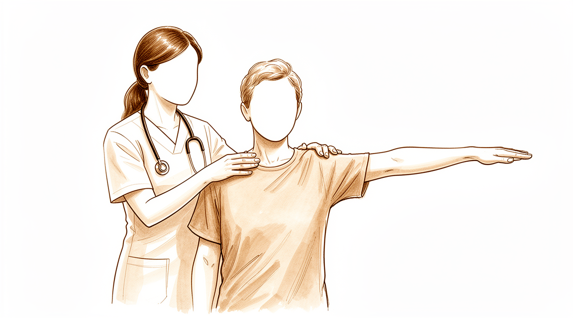
Calcific tendinitis causes shoulder pain from calcium deposits; treatment ranges from observation to washing out the calcium.
Những gì bạn đang cảm nhận
Bạn có thể trải qua tình trạng đau cấp tính hoặc mạn tính ở vai do viêm xung quanh các lắng đọng canxi trong các gân của nhóm cơ xoay cuff. Tình trạng này, được gọi là viêm gân vôi hóa, có thể ảnh hưởng đáng kể đến chất lượng cuộc sống của bạn và có thể dẫn đến việc phải nghỉ làm. Cơn đau thường dữ dội và có thể bùng phát mà không có cảnh báo trước. Bạn có thể nhận thấy rằng một số cử động cụ thể kích hoạt cảm giác khó chịu sắc nhọn, trong khi những cử động khác gây ra cảm giác âm ỉ và dai dẳng.
Các hoạt động hàng ngày đòi hỏi việc với tay hoặc nâng vật trở nên khó khăn. Những hành động đơn giản như cài áo sơ mi hoặc với tay ra sau lưng để cài móc áo ngực có thể trở nên thách thức. Bạn có thể thấy khó ngủ ở bên bị ảnh hưởng, vì áp lực có thể làm trầm trọng thêm cơn đau. Sự khó chịu vào ban đêm là phổ biến, thường làm gián đoạn giấc ngủ của bạn và khiến bạn cảm thấy mệt mỏi. Cơn đau cũng có thể trở nên nghiêm trọng hơn khi thức dậy, khiến bạn khó bắt đầu ngày mới.
Trong một số trường hợp, bạn có thể gặp các triệu chứng không điển hình. Ví dụ, cơn đau có thể chỉ giới hạn ở phía sau vai hoặc ảnh hưởng đến các cử động trên đầu nếu cơ tròn nhỏ (teres minor) bị liên quan. Hiếm khi, bạn có thể có các tổn thương vôi hóa ở các khu vực khác, chẳng hạn như dây chằng collateral giữa (medial collateral ligament), hoặc ở cả hai vai cùng lúc. Tuy nhiên, hầu hết mọi người đều trải qua tình trạng đau vai tại chỗ làm gián đoạn các hoạt động thường ngày.
Tin tốt là viêm gân vôi hóa có triệu chứng có khả năng cao sẽ hoàn toàn thuyên giảm về lâu dài. Điều trị bảo tồn thường là lựa chọn chính, đặc biệt đối với các trường hợp cấp tính, và cho thấy sự cải thiện có ý nghĩa về mặt lâm sàng. Khoảng 72% bệnh nhân đạt kết quả xuất sắc hoặc tốt với chăm sóc không phẫu thuật, bất kể kích thước hoặc vị trí của lắng đọng canxi. Nếu các triệu chứng vẫn tiếp tục, bác sĩ phẫu thuật của bạn có thể thảo luận về các lựa chọn khác, nhưng nhiều người tìm thấy sự giảm đau thông qua các biện pháp bảo tồn. Hiểu rõ các triệu chứng này giúp bạn quản lý kỳ vọng và làm việc với nhóm chăm sóc sức khỏe của mình để tìm ra hướng đi tốt nhất.
Những gì thực sự đang xảy ra
Viêm gân vôi hóa là tình trạng canxi tích tụ bên trong các gân của vai bạn. Các gân này là những dải mô cứng, giống như sợi dây, nối cơ với xương. Hãy tưởng tượng chúng như những dây cáp giúp bạn nâng và cử động cánh tay. Khi các mảng canxi hình thành, chúng tạo thành một khối cứng bên trong mô mềm này. Sự tích tụ này có thể gây kích ứng vùng xung quanh, dẫn đến đau đáng kể và hạn chế khả năng sử dụng vai của bạn.
Vai của bạn là một khớp cầu và hốc phức tạp. Nó dựa vào một nhóm cơ và gân được gọi là nhóm cơ xoay cuff (rotator cuff) để duy trì sự ổn định và cử động trơn tru. Đầu dài của cơ nhị đầu (biceps) cũng đóng vai trò trong việc giữ cho khớp này ổn định. Trong nhiều trường hợp, các mảng canxi ảnh hưởng đến các cấu trúc này. Bạn có thể cảm thấy đau ở phía trước vai. Điều này xảy ra vì các mảng canxi tương tác với các mô mềm xung quanh thay vì là một vấn đề đơn lẻ, biệt lập. Cơn đau là tín hiệu của cơ thể cho thấy có điều gì đó không ổn trong sự cân bằng tinh tế này.
Khi bạn già đi, những thay đổi ở gân trở nên phổ biến hơn. Những người bơi lội và những người năng động thường thấy những thay đổi cấu trúc này sớm hơn. Bản thân mảng canxi có thể thay đổi về kích thước. Nếu tổn thương lớn hơn 1 cm, bạn có khả năng cần phẫu thuật để loại bỏ nó cao gấp gần ba lần. Tuy nhiên, nhiều người tìm thấy sự giảm đau mà không cần phẫu thuật. Các phương pháp điều trị bảo tồn, chẳng hạn như liệu pháp sóng xung kích, thường rất thành công trong việc giảm đau và khôi phục chức năng với rất ít tác dụng phụ.
Nếu cần phẫu thuật, bác sĩ phẫu thuật của bạn có thể sẽ loại bỏ mảng canxi. Điều này thường được thực hiện bằng cách sử dụng một máy quay nhỏ và các dụng cụ (nội soi khớp). Trong nhiều trường hợp, bác sĩ phẫu thuật cũng sẽ sửa chữa rotator cuff cùng lúc. Sự kết hợp này dẫn đến những cải thiện đáng kể về giảm đau và chức năng vai. Cần có thời gian để những lợi ích này thể hiện đầy đủ. Bạn nên mong đợi phải chờ ít nhất sáu tháng sau phẫu thuật để chức năng vai của bạn đạt mức tốt nhất. Trong thời gian này, bác sĩ phẫu thuật của bạn sẽ hướng dẫn bạn quá trình phục hồi để đảm bảo kết quả tốt nhất có thể.
Những gì chúng tôi có thể làm về vấn đề này
Hầu hết mọi người tìm thấy sự giảm nhẹ triệu chứng mà không cần phẫu thuật. Hành trình của bạn thường bắt đầu với việc tự chăm sóc đơn giản và các bài tập vận động được hướng dẫn. Bạn có thể sẽ dùng thuốc chống viêm đường uống để làm giảm đau. Bác sĩ phẫu thuật của bạn cũng có thể khuyến nghị vật lý trị liệu để khôi phục tầm vận động và sức mạnh của vai. Các phương pháp điều trị không phẫu thuật này là lựa chọn chính cho viêm gân vôi hóa cấp tính.
Bạn nên dành thời gian cho điều trị bảo tồn để phát huy tác dụng. Tình trạng này có khả năng cao sẽ thuyên giảm hoàn toàn về lâu dài. Thực tế, 72% bệnh nhân đạt kết quả xuất sắc hoặc tốt với chăm sóc không phẫu thuật. Tỷ lệ thành công này đúng bất kể vị trí của canxi, kích thước của nó hoặc mức độ nghiêm trọng của các triệu chứng ban đầu của bạn. Nếu cơn đau của bạn kéo dài, nó có thể dẫn đến nghỉ làm việc và suy giảm chất lượng cuộc sống. Sự nỗ lực nhất quán trong liệu pháp là chìa khóa để quay trở lại sinh hoạt hàng ngày của bạn.
Nếu việc tự chăm sóc và liệu pháp không đủ hiệu quả, bác sĩ phẫu thuật của bạn có thể đề xuất các can thiệp y tế. Tiêm corticosteroid là một lựa chọn phổ biến để giảm viêm và đau. Một phương pháp điều trị không xâm lấn hiệu quả khác là liệu pháp sóng xung kích ngoài cơ thể (ESWT). Liệu pháp này sử dụng sóng âm để phá vỡ các lắng đọng canxi. Nó an toàn và tạo ra tỷ lệ thành công cao trong việc giảm đau và khôi phục chức năng. Bệnh nhân thường thấy các triệu chứng đau giảm và chức năng vai cải thiện sau điều trị này. Các biến chứng từ ESWT là không đáng kể.
Phẫu thuật chỉ được xem xét khi điều trị bảo tồn đạt đến giới hạn. Bác sĩ phẫu thuật của bạn có thể khuyến nghị điều trị phẫu thuật nếu các tổn thương vôi hóa của bạn lớn hơn 1 cm, vì những tổn thương này có khả năng cần phẫu thuật cao hơn 2,8 lần. Việc loại bỏ vôi hóa bằng nội soi khớp dẫn đến kết quả lâm sàng được cải thiện cho các trường hợp mạn tính. Phần lớn bệnh nhân trải qua quản lý phẫu thuật này cũng cần sửa chữa gân quay đồng thời.
Nếu bạn tiến hành phẫu thuật, hãy mong đợi một quá trình hồi phục dần dần. Các điểm số chức năng cải thiện chậm sau thủ thuật. Chúng thường đạt hơn 75 phần trăm vào sáu tháng sau phẫu thuật. Cần ít nhất 6 tháng theo dõi để những cải thiện này trở nên có ý nghĩa thống kê. Hầu hết bệnh nhân báo cáo những cải thiện đáng kể về đau vai và chức năng sau phẫu thuật. Bác sĩ phẫu thuật của bạn sẽ thảo luận xem phẫu thuật nội soi khớp, nội soi hoặc mổ mở là tốt nhất cho trường hợp cụ thể của bạn.
Những điều cần biết
Đau vai do viêm gân vôi hóa có thể kéo dài trong thời gian dài. Các triệu chứng này có thể diễn biến kéo dài. Điều này thường dẫn đến việc nghỉ làm việc và chất lượng cuộc sống giảm sút. Tin tốt là tình trạng này có khả năng cao sẽ khỏi hoàn toàn về lâu dài. Nhiều người nhận thấy các triệu chứng của họ tự thuyên giảm.
Nếu cơn đau của bạn không biến mất, bác sĩ phẫu thuật có thể đề xuất điều trị. Các lựa chọn không phẫu thuật như liệu pháp sóng xung kích hoặc chọc hút kim là hiệu quả. Liệu pháp sóng xung kích mang lại tỷ lệ thành công cao trong việc giảm đau và phục hồi chức năng với các biến chứng liên quan không đáng kể. Chọc hút kim chỉ xâm lấn và gây đau nhẹ. Những phương pháp điều trị này hoạt động tốt đối với nhiều bệnh nhân.
Nếu cần phẫu thuật, bác sĩ phẫu thuật sẽ loại bỏ khối lắng đọng canxi. Bạn có thể cần phải sửa chữa gân quay cùng lúc. Đây là một phần phổ biến của thủ thuật. Đau vai và chức năng của bạn sẽ cải thiện đáng kể sau phẫu thuật. Tuy nhiên, quá trình hồi phục không diễn ra ngay lập tức. Điểm số chức năng của bệnh nhân trải qua điều trị nội soi khớp cải thiện chậm. Bạn sẽ thấy mức cải thiện hơn 75 phần trăm sau sáu tháng phẫu thuật.
Phải mất thời gian để cảm nhận được lợi ích đầy đủ của việc loại bỏ bằng nội soi khớp. Cần ít nhất 6 tháng theo dõi để những cải thiện này trở nên có ý nghĩa thống kê. Đừng vội vàng trong quá trình lành bệnh. Bác sĩ phẫu thuật sẽ hướng dẫn bạn qua quá trình này. Hầu hết bệnh nhân hài lòng với kết quả cuối cùng.
Khi nào cần gặp bác sĩ
Hãy gặp bác sĩ đa khoa nếu bạn có đau vai dai dẳng không cải thiện khi nghỉ ngơi. Viêm gân vôi hóa là tình trạng đau cấp tính hoặc mạn tính do viêm quanh các lắng đọng canxi trong các gân của nhóm cơ xoay vai. Bạn nên tìm kiếm sự chăm sóc y tế nếu các triệu chứng ảnh hưởng đến giấc ngủ hoặc công việc, hoặc nếu bạn gặp phải tình trạng yếu, mất ổn định hoặc kẹt khớp. Điều trị bảo tồn là lựa chọn chính, đặc biệt đối với các trường hợp cấp tính, và cho thấy sự cải thiện có ý nghĩa lâm sàng với 72% bệnh nhân đạt kết quả tốt hoặc xuất sắc. Tuy nhiên, nếu cơn đau của bạn vẫn nghiêm trọng mặc dù đã được điều trị bảo tồn, hãy yêu cầu đánh giá bởi bác sĩ chuyên khoa. Bác sĩ phẫu thuật của bạn có thể xác định xem có cần thực hiện các bước tiếp theo để khôi phục chức năng và giảm đau hay không.
Evidence & references
Overview
- Shoulder MRI may be warranted for preoperative planning in select patients with chronic calcific tendinopathy and prolonged refractory pain, although the probability of identifying additional cuff pathology requiring surgical intervention is very low [1].
- Patients with calcific lesions >1 cm have a 2.8 increased likelihood to undergo operative treatment in the setting of calcific tendinitis of the shoulder [2].
- The majority of patients who undergo surgical management for removal of the calcific deposit require a concomitant rotator cuff repair and experience significant improvements in shoulder pain and function [3].
- Treatment of calcific tendinitis of the shoulder with extracorporeal shock waves produces a high rate of success in pain relief and functional restoration with negligible associated complications [6].
- Patients undergoing arthroscopic treatment of a calcific deposit in the shoulder have satisfactory clinical and radiological outcomes at final follow-up, with functional scores improving slowly and reaching more than 75 percent at six months after surgery [8].
- Arthroscopic removal of calcification leads to improved clinical outcomes in patients with chronic calcific tendinitis, but at least 6 months of follow-up is needed for these improvements to become statistically significant [11].
- The primary choice of treatment for calcific tendinitis is conservative, especially in patients with acute calcific tendinitis [12].
- Arthroscopic treatment of calcifying tendinitis provides good to excellent clinical results [16].
- Routine diagnostic glenohumeral exploration does not appear beneficial in arthroscopic treatment of calcific tendinitis due to the low prevalence of intraarticular pathologies which most frequently do not require surgical treatment [19].
- Endoscopic and open surgery are equally effective in the treatment of chronic calcifying tendinopathy, showing similar clinical and sonographic results [23].
Anatomy & Pathophysiology
- Rotator cuff disease, shoulder instability, and associated lesions are common pathologic conditions of the shoulder involving soft tissues [42].
- The long head of the biceps contributes to stability of the glenohumeral joint in all directions, although in vivo studies have not yet established this stabilizing effect or the physiologic load required [32].
- Understanding the function and pathology surrounding the teres minor is paramount in comprehensive management of patients with shoulder pathology [34].
- In the context of rotator cuff disease, anterior shoulder pain with macroscopic changes in the biceps tendon is related to the complex interaction of the tendon and surrounding soft tissues rather than a single entity [41].
- Long head of biceps tendinopathy is associated with age and cuff tendinopathy on MRI obtained for evaluation of shoulder pain [51].
- Clinicians can guide patients to understand shoulder pain as age-appropriate and safe, potentially reducing unnecessary visits, tests, and treatments [51].
- A high prevalence of structural changes in the rotator cuff and biceps tendons in masters swimmers reflects the effect of shoulder symptoms, aging, and swim training [54].
- In chronic rotator cuff tears, the shape of the deltoid muscle seems only to be influenced by natural aging and is independent of reduced shoulder motion [53].
- There seems to be no disadvantage to exhausting conservative treatment and delaying implantation of reverse total shoulder arthroplasty in patients with chronic rotator cuff tears [53].
- Surgical repair of an isolated supraspinatus tear may be sufficient to keep the torn rotator cuff intact and achieve satisfactory patient-reported outcomes, but glenohumeral joint mechanics and shoulder strength are not fully restored with current repair techniques [39].
- At a 2-year follow-up, shoulder function evaluated in terms of CMS was not significantly improved with conservative versus surgical management for rotator cuff tears [37].
- Over time, supraspinatus tendon thickness in individuals with rotator cuff-related shoulder pain tends to normalize compared to the contralateral side, regardless of the exercise intervention [28].
- The theoretical concept of a high acromion index resulting in an increased upward force against the subacromial space influencing pain and function in calcifying tendinitis of the shoulder was not supported [49].
- Ultrasound is a useful tool for discovering subjects in pre-symptomatic stages who may undergo shoulder symptomatic pathologies [40].
Classification
- Calcific tendinitis is a common shoulder disorder [9].
- Calcific tendinitis should be differentiated from dystrophic calcification because their pathogenesis and natural history are totally different [9].
- Calcific tendinitis is an acute or chronically painful condition caused by inflammation around calcium deposits in the rotator cuff tendons [15].
- Symptoms of calcific tendinitis can be protracted, resulting in time off work and impaired quality of life [5].
- Symptomatic calcific tendinitis of the shoulder has a good likelihood to completely resolve in the long-term [4].
- The demographic, radiographic, and clinical features of calcific tendinitis of the shoulder in the Korean population are not different from those of Western populations [7].
- Conservative treatment for calcific tendinitis of the shoulder showed clinically significant improvement, with 72% of excellent or good results regardless of the location, radiologic type and size, and initial symptoms of calcific deposits [10].
- The primary choice of treatment for calcific tendinitis is conservative, especially in patients with acute calcific tendinitis [12].
- Patients with calcific lesions >1 cm had a 2.8 increased likelihood to undergo operative treatment in the setting of calcific tendinitis of the shoulder [2].
- The majority of patients who undergo surgical management for removal of the calcific deposit will require a concomitant rotator cuff repair and have significant improvements in shoulder pain and function [3].
- Shoulder MRI may be warranted for preoperative planning in the select population of patients with chronic calcific tendinitis and prolonged refractory pain, although the probability of identifying additional cuff pathology requiring surgical intervention is very low [1].
- Calcific tendinitis of the rotator cuff with tuberosity osteolysis is a distinctive form of calcific tendinitis that should be considered in clinical and surgical practice [17].
- Restriction of passive glenohumeral abduction combined with normal passive external rotation is a diagnostic feature of calcific tendinitis that can be used to clinically differentiate it from adhesive capsulitis [18].
- The incidence and type of intraarticular lesions in calcifying tendinitis are comparable to age-matched shoulders with partial- rather than full-thickness rotator cuff tears [35].
Clinical Presentation
- Calcific tendinitis of the shoulder is an acute or chronically painful condition caused by inflammation around calcium deposits in the rotator cuff tendons [15].
- Symptoms of calcific tendinitis can be protracted, resulting in time off work and impaired quality of life [5].
- Symptomatic calcific tendinitis of the shoulder has a good likelihood to completely resolve in the long-term [4].
- Calcific tendinitis is a common shoulder disorder that should be differentiated from dystrophic calcification because the pathogenesis and natural history of both are totally different [9].
- The demographic, radiographic, and clinical features of calcific tendinitis of the shoulder in the Korean population are not different from those of Western populations [7].
- Restriction of passive glenohumeral abduction combined with normal passive external rotation is a diagnostic feature of calcific tendinitis that can be used to clinically differentiate it from adhesive capsulitis [18].
- Arthroscopic treatment of calcifying tendinitis provides good to excellent clinical results [16].
- The majority of patients who undergo surgical management for removal of the calcific deposit will require a concomitant rotator cuff repair and have significant improvements in shoulder pain and function [3].
- Patients with calcific lesions >1 cm had a 2.8 increased likelihood to undergo operative treatment in the setting of calcific tendinitis of the shoulder [2].
- Conservative treatment for calcific tendinitis of the shoulder showed clinically significant improvement, with 72% of excellent or good results regardless of the location, radiologic type and size, and initial symptoms of calcific deposits [10].
- The primary choice of treatment for calcific tendinitis is conservative, especially in patients with acute calcific tendinitis [12].
- Treatment of calcific tendinitis of the shoulder with extracorporeal shock waves has produced a high rate of success in pain relief and functional restoration with negligible associated complications [6].
- Both ultrasound-guided needling and extracorporeal shock wave therapy improved clinical outcomes and eliminated calcium deposits in the treatment of calcific tendinitis [22].
- Shoulder MRI may be warranted for preoperative planning in the select population of patients with chronic calcific tendinopathy and prolonged refractory pain, although the probability of identifying additional cuff pathology requiring surgical intervention is very low [1].
- Routine diagnostic glenohumeral exploration does not appear beneficial in arthroscopic treatment of calcific tendinitis due to the low prevalence of intraarticular pathologies which most frequently do not require surgical treatment [19].
- Imaging and functional data indicate that calcific tendinitis of the rotator cuff with tuberosity osteolysis is a distinctive form of calcific tendinitis that should be considered in clinical and surgical practice [17].
- Recognition of atypical presentations of calcific tendinitis with bone erosion may prevent unnecessary biopsy and overtreatment [14].
- Atypical presentations of calcific tendinitis, such as involvement of the teres minor affecting overhead movement or isolated posterior shoulder pain, should be considered in clinical practice [20].
- A patient can suffer from both a calcifying lesion within the medial collateral ligament and calcifying tendinitis of the rotator cuff in both shoulders simultaneously [13].
Investigations
- Shoulder MRI may be warranted for preoperative planning in select patients with chronic calcific tendinopathy and prolonged refractory pain, although the probability of identifying additional cuff pathology requiring surgical intervention is very low [1].
- Patients with calcific lesions >1 cm have a 2.8 increased likelihood to undergo operative treatment [2].
- The majority of patients undergoing surgical management for removal of the calcific deposit require a concomitant rotator cuff repair [3].
- Symptomatic calcific tendinitis of the shoulder has a good likelihood to completely resolve in the long-term [4].
- Calcific tendinitis symptoms can be protracted, resulting in time off work and impaired quality of life [5].
- Demographic, radiographic, and clinical features of calcific tendinitis of the shoulder in the Korean population are not different from those of Western populations [7].
- Patients undergoing arthroscopic treatment of a calcific deposit in the shoulder had satisfactory clinical and radiological outcomes at final follow-up, with functional scores improving slowly and reaching more than 75 percent at six months after surgery [8].
- Conservative treatment for calcific tendinitis of the shoulder showed clinically significant improvement, with 72% of excellent or good results regardless of the location, radiologic type and size, and initial symptoms of calcific deposits [10].
- Calcific tendinitis can present with simultaneous calcifying lesions in other locations, such as the medial collateral ligament of the knee [13].
- Atypical presentations of calcific tendinitis with bone erosion may occur, and recognition of these presentations can prevent unnecessary biopsy and overtreatment [14].
- Calcific tendinitis of the rotator cuff with tuberosity osteolysis is a distinctive form that should be considered in clinical and surgical practice [17].
- Restriction of passive glenohumeral abduction combined with normal passive external rotation is a diagnostic feature that can be used to clinically differentiate adhesive capsulitis from calcific tendinitis [18].
- Atypical presentations of calcific tendinitis, such as involvement of the teres minor affecting overhead movement, should be considered in the context of isolated posterior shoulder pain [20].
- Shoulder surgeons should be cautious about rotator cuff tears as a comorbidity in calcific tendinitis and aware of the accuracy limitations of sonographic or MRI evaluation [27].
- The use of MRI before a trial of conservative management in patients with atraumatic shoulder pain, minimal to no strength deficits on physical examination, and suspected cuff tendinopathy other than full-thickness tears provides negative value in management at both individual and population levels [30].
- MRI, but not radiography, can be used to help discriminate between traumatic and nontraumatic rotator cuff lesions [38].
- Repeating lavage and corticosteroid injections until calcific deposits resolve completely is not recommended due to lack of evidence regarding efficacy and risks [43].
- Preoperative ultrasound-guided marking of calcific deposits statistically significantly improves the clinical results of arthroscopic surgery at 6 weeks and 2 years [44].
Treatment
Non-Operative Management
- Conservative treatment is the primary choice for calcific tendinitis, especially in patients with acute calcific tendinitis [12].
- Nonsurgical management remains the mainstay of treatment for calcific tendinitis of the rotator cuff [31].
- Most patients improve with nonsurgical modalities such as oral anti-inflammatory medication, physical therapy, and corticosteroid injections [31].
- Symptomatic calcific tendinitis of the shoulder has a good likelihood to completely resolve in the long-term [4].
- Conservative treatment for calcific tendinitis of the shoulder showed clinically significant improvement, with 72% of excellent or good results regardless of the location, radiologic type and size, and initial symptoms of calcific deposits [10].
- Extracorporeal shock wave therapy (ESWT) is a safe and effective noninvasive treatment for patients with calcific tendinitis of the shoulder [21].
- ESWT produces a high rate of success in pain relief and functional restoration with negligible complications [21].
- ESWT effectively reduced painful symptomatology and increased shoulder function in patients with chronic calcific tendinitis of the shoulder [24].
- Treatment of calcific tendinitis of the shoulder with shock waves has produced a high rate of success in pain relief and functional restoration with negligible associated complications [6].
- Both ultrasound-guided needling and extracorporeal shock wave therapy improved clinical outcomes and eliminated calcium deposits in the treatment of calcific tendinitis [22].
- The use of MRI before a trial of conservative management in patients with atraumatic shoulder pain, minimal to no strength deficits on physical examination, and suspected cuff tendinopathy other than full-thickness tears provides negative value in the management of these patients [30].
Operative Management
- Patients with calcific lesions >1 cm had a 2.8 increased likelihood to undergo operative treatment in the setting of calcific tendinitis of the shoulder [2].
- Arthroscopic removal of calcification leads to improved clinical outcomes in patients with chronic calcific tendinitis [11].
- At least 6 months of follow-up is needed for improvements in clinical outcomes after arthroscopic removal of calcification to become statistically significant [11].
- Patients undergoing arthroscopic treatment of a calcific deposit in the shoulder had satisfactory clinical and radiological outcomes at the final follow-up [8].
- Functional scores in patients undergoing arthroscopic treatment of a calcific deposit improved slowly, reaching more than 75 percent at six months after surgery [8].
- Arthroscopic treatment of calcifying tendinitis provides good to excellent clinical results [16].
- The majority of patients who undergo surgical management for removal of the calcific deposit will require a concomitant rotator cuff repair [3].
- Patients who undergo surgical management for removal of the calcific deposit have significant improvements in shoulder pain and function [3].
- Endoscopic and open surgery are equally effective in the treatment of chronic calcifying tendinopathy, showing similar clinical and sonographic results [23].
- Routine diagnostic glenohumeral exploration does not appear beneficial in arthroscopic treatment of calcific tendinitis due to the low prevalence of intraarticular pathologies which most frequently do not require surgical treatment [19].
- Shoulder MRI may be warranted for preoperative planning in the select population of patients with chronic calcific tendinopathy and prolonged refractory pain [1].
- The probability of identifying additional cuff pathology requiring surgical intervention via shoulder MRI in patients with chronic calcific tendinopathy and prolonged refractory pain is very low [1].
Complications
- Calcific tendinitis symptoms can be protracted, resulting in time off work and impaired quality of life [5].
- Atypical presentations of calcific tendinitis may include bone erosion, which can lead to unnecessary biopsy and overtreatment if not recognized [14].
- Shoulder surgeons should be cautious about rotator cuff tears as a comorbidity in calcific tendinitis due to accuracy limitations in sonographic or MRI evaluation [27].
- The probability of identifying additional cuff pathology requiring surgical intervention via preoperative shoulder MRI in patients with chronic calcific tendinopathy is very low [1].
- Arthroscopic decompression is not recommended in the treatment of rotator cuff tendinopathy [25].
Recovery
- Symptomatic calcific tendinitis of the shoulder has a good likelihood to completely resolve in the long-term [4].
- Symptoms of calcific tendinitis can be protracted, resulting in time off work and impaired quality of life [5].
- Patients undergoing arthroscopic treatment of a calcific deposit in the shoulder had satisfactory clinical and radiological outcomes at the final follow-up [8].
- Functional scores for patients undergoing arthroscopic treatment of a calcific deposit improved slowly, reaching more than 75 percent at six months after surgery [8].
- Arthroscopic removal of calcification leads to improved clinical outcomes in patients with chronic calcific tendinitis, but at least 6 months of follow-up is needed for these improvements to become statistically significant [11].
- The majority of patients who undergo surgical management for removal of the calcific deposit will require a concomitant rotator cuff repair [3].
- Patients who undergo surgical management for removal of the calcific deposit have significant improvements in shoulder pain and function [3].
- Percutaneous needle aspiration and lavage is effective in the short term and in the long term in calcific tendinitis of the shoulder [29].
- Percutaneous needle aspiration and lavage results are similar to or better than those published for other techniques [29].
- Percutaneous needle aspiration and lavage is only slightly invasive and painful [29].
- Treatment of calcific tendinitis of the shoulder with shock waves has produced a high rate of success in pain relief and functional restoration with negligible associated complications [6].
- Shock wave therapy is a safe and effective noninvasive treatment for patients with calcific tendinitis of the shoulder, producing a high rate of success in pain relief and functional restoration with negligible complications [21].
- A symptom duration of ≤10 months or calcification size of ≤10.82 mm represented the clinical scenarios most likely to show resorption after extracorporeal shockwave therapy (ESWT) [56].
- The short-term functional outcome of patients with calcific tendonitis after arthroscopic bursectomy and debridement of the calcific deposit is not influenced if performed in combination with or without a subacromial decompression [26].
Key Evidence
- [Commentary] Shoulder MRI may be warranted for preoperative planning in the select population of patients with chronic calcific tendinopathy and prolonged refractory pain, although the probability of identifying additional cuff pathology requiring surgical intervention is very low. [1] (10.1016/j.arthro.2020.01.014)
- [L3] Patients with calcific lesions >1 cm had a 2.8 increased likelihood to undergo operative treatment in the setting of calcific tendinitis of the shoulder. [2] (10.1016/j.jseint.2021.01.013)
- [L3] The majority of patients who undergo surgical management for removal of the calcific deposit will require a concomitant rotator cuff repair and have significant improvements in shoulder pain and function. [3] (10.1177/2325967121s00208)
- [L1] Symptomatic calcific tendinitis of the shoulder has a good likelihood to completely resolve in the long-term. [4] (10.1097/phm.0000000000000939)
- [L3] Calcific tendinitis is a poorly understood condition in which symptoms can be protracted, resulting in time off work and impaired quality of life. [5] (10.1016/j.jse.2006.06.007)
- [L2] Treatment of calcific tendinitis of the shoulder with shock waves has produced a high rate of success in pain relief and functional restoration with negligible associated complications. [6] (10.1016/j.jse.2007.03.023)
- [L4] This study reported demographic, radiographic, and clinical features of calcific tendinitis of the shoulder in the Korean population, which were not different from those of Western populations. [7] (10.5397/cise.2020.00010)
- [Paper] Patients undergoing arthroscopic treatment of a calcific deposit in the shoulder had satisfactory clinical and radiological outcomes at the final follow-up, with functional scores improving slowly and reaching more than 75 percent at six months after surgery. [8] (10.1016/j.otsr.2020.03.005)
- [L5] Calcific tendinitis is a common shoulder disorder that should be differentiated from dystrophic calcification as the pathogenesis and natural history of both is totally different. [9] (10.5312/wjo.v7.i1.55)
- [L2] Conservative treatment for calcific tendinitis of the shoulder showed clinically significant improvement, with 72% of excellent or good results regardless of the location, radiologic type and size, and initial symptoms of calcific deposits. [10] (10.1016/j.jse.2009.07.008)
- [L4] Arthroscopic removal of calcification leads to improved clinical outcomes in patients with chronic calcific tendinitis, but at least 6 months of follow-up is needed for these improvements to become statistically significant. [11] (10.5397/cise.2018.21.2.75)
- [L5] The primary choice of treatment for calcific tendinitis is conservative, especially in patients with acute calcific tendinitis. [12] (10.5397/cise.2020.00318)
- [Case_report] This is the first case report of a patient suffering from both a calcifying lesion within the medial collateral ligament and calcifying tendinitis of the rotator cuff in both shoulders. [13] (10.1186/s12891-016-1147-z)
- [L5] Recognition of atypical presentations of calcific tendinitis with bone erosion may prevent unnecessary biopsy and overtreatment. [14] (10.1016/j.jse.2009.02.009)
- [L5] Calcific tendinitis of the shoulder is an acute or chronically painful condition caused by inflammation around calcium deposits in the rotator cuff tendons. [15] (10.1016/s0030-5898(03)00089-0)
- [L3] Arthroscopic treatment of calcifying tendinitis provides good to excellent clinical results. [16] (10.1177/03635465211037690)
- [L2] Imaging and functional data indicate that calcific tendinitis of the rotator cuff with tuberosity osteolysis is a distinctive form of calcific tendinitis that should be considered in clinical and surgical practice. [17] (10.1016/j.jse.2008.09.016)
- [L3] This finding can be used to clinically differentiate adhesive capsulitis from calcific tendinitis. [18] (10.1177/2325967117752907)
- [L3] Routine diagnostic glenohumeral exploration does not appear beneficial in arthroscopic treatment of calcific tendinitis due to the low prevalence of intraarticular pathologies which most frequently do not require surgical treatment. [19] (10.1186/s12891-017-1839-z)
- [Case_report] This case highlights the importance of considering atypical presentations of calcific tendinitis, particularly in the context of isolated posterior shoulder pain. [20] (10.1016/j.jisako.2025.101055)
- [L3] Shock wave therapy is a safe and effective noninvasive treatment for patients with calcific tendinitis of the shoulder, producing a high rate of success in pain relief and functional restoration with negligible complications. [21] (10.1177/03635465030310031701)
- [L2] Both treatment modalities for calcific tendinitis improved clinical outcomes and eliminated calcium deposits. [22] (10.1016/j.jse.2014.06.036)
- [L1] Endoscopic and open surgery are equally effective in the treatment of chronic calcifying tendinopathy, showing similar clinical and sonographic results. [23] (10.1097/01.blo.0000063786.32430.22)
- [L2] ESWT effectively reduced painful symptomatology and increased shoulder function in patients with chronic calcific tendinitis of the shoulder. [24] (10.1136/ard.62.3.248)
- [L1] The natural history of rotator cuff tendinopathy probably plays a significant role in the results in the long-term. [25] (10.1302/0301-620x.99b6.bjj-2016-0569.r1)
- [L1] This study has demonstrated that the short-term functional outcome of patients with calcific tendonitis after arthroscopic bursectomy and debridement of the calcific deposit is not influenced if performed in combination with or without a subacromial decompression. [26] (10.1016/j.arthro.2015.05.015)
- [L3] Shoulder surgeons should be cautious about rotator cuff tears as a comorbidity in calcific tendinitis and aware of the accuracy limitations of sonographic or MRI evaluation. [27] (10.5397/cise.2021.00094)
- [L2] Findings from this study suggest that, over time, supraspinatus tendon thickness in individuals with rotator cuff-related shoulder pain tends to normalize compared to the contralateral side, regardless of the exercise intervention. [28] (10.1016/j.jse.2024.03.055)
- [L4] Percutaneous needle aspiration and lavage is effective in the short term and in the long term in calcific tendinitis of the shoulder, with results similar to or better than those published for other techniques, and it is only slightly invasive and painful. [29] (10.2214/ajr.07.2254)
- [L4] The use of MRI before a trial of conservative management in patients with atraumatic shoulder pain, minimal to no strength deficits on physical examination, and suspected cuff tendinopathy other than full-thickness tears provides negative value in the management of these patients, at both the individual and population level. [30] (10.1016/j.jse.2019.04.003)
- [L5] Nonsurgical management remains the mainstay of treatment for calcific tendinitis of the rotator cuff, with most patients improving with modalities such as oral anti-inflammatory medication, physical therapy, and corticosteroid injections. [31] (10.5435/jaaos-22-11-707)
- [L5] Biomechanical studies indicate that the long head of the biceps contributes to stability of the glenohumeral joint in all directions, though in vivo studies have yet to establish this stabilizing effect and the physiologic load required remains unknown. [32] (10.1016/j.arthro.2010.10.014)
- [L5] Understanding the function and pathology surrounding the teres minor is paramount in comprehensive management of the patient with shoulder pathology. [34] (10.5435/jaaos-d-15-00258)
- [L3] The incidence and type of intraarticular lesions in calcifying tendinitis are comparable to age-matched shoulders with partial- rather than full-thickness rotator cuff tears. [35] (10.1007/s00402-011-1263-z)
- [L1] At a 2-year follow-up, shoulder function evaluated in terms of CMS was not significantly improved. [37] (10.1186/s12891-020-03872-4)
- [L2] MRI, but not radiography, can be used to help discriminate between traumatic and nontraumatic rotator cuff lesions. [38] (10.1016/j.jse.2015.06.005)
- [L4] Surgical repair of an isolated supraspinatus tear may be sufficient to keep the torn rotator cuff intact and achieve satisfactory patient-reported outcomes, but glenohumeral joint mechanics and shoulder strength are not fully restored with current repair techniques. [39] (10.1177/0363546511412164)
- [L3] Ultrasound is an useful tool for discovering in pre-symptomatic stages the subjects that may undergo shoulder symptomatic pathologies. [40] (10.1186/1471-2474-11-278)
- [L4] In the context of rotator cuff disease, the etiology of anterior shoulder pain with macroscopic changes in the biceps tendon is related to the complex interaction of the tendon and surrounding soft tissues, rather than a single entity. [41] (10.1016/j.jse.2008.05.044)
- [L5] Repeating lavage and corticosteroid injections until the calcific deposits resolve completely is not recommended due to lack of evidence regarding efficacy and risks. [43] (10.5397/cise.2021.00269)
- [L3] Preoperative ultrasound-guided marking of calcific deposits is a procedure that statistically significantly improves the clinical results of arthroscopic surgery as seen at 6 weeks and 2 years. [44] (10.1016/j.arthro.2006.08.005)
- [L2] The theoretical concept of a high acromion index resulting in an increased upward force against the subacromial space, which influences pain and function in calcifying tendinitis of the shoulder, was not supported. [49] (10.1007/s00167-011-1563-4)
- [L3] Clinicians can guide patients to understand shoulder pain as age-appropriate and safe, potentially reducing unnecessary visits, tests, and treatments. [51] (10.1097/corr.0000000000003342)
- [L3] Our data support that in chronic rotator cuff tears, there seems to be no disadvantage to exhausting conservative treatment and to delay implantation of reverse total shoulder arthroplasty, as the shape of deltoid muscle seems only to be influenced by natural aging, but to be independent of reduced shoulder motion. [53] (10.1186/1471-2474-14-247)
- [L4] A high prevalence of structural changes in the rotator cuff and biceps tendons in masters swimmers reflects the effect of shoulder symptoms, aging, and swim training. [54] (10.3390/medicina57010029)
- [L3] A symptom duration of ≤10 months or calcification size of ≤10.82 mm represented the clinical scenarios most likely to show resorption after ESWT. [56] (10.1177/23259671241231609)
References
[1] Editorial Commentary: Is Magnetic Resonance Imaging of the Shoulder Ever Appropriate in Evaluating Patients With Calcific Tendinopathy of the Rotator Cuff?. Arthroscopy: The Journal of Arthroscopic & Related Surgery. 2020. DOI: 10.1016/j.arthro.2020.01.014 [2] Predictive factors for failure of conservative management in the treatment of calcific tendinitis of the shoulder. JSES International. 2021. DOI: 10.1016/j.jseint.2021.01.013 [3] Predictive factor for failure of conservative management in the treatment of calcific tendinitis of the shoulder. Orthopaedic Journal of Sports Medicine. 2021. DOI: 10.1177/2325967121s00208 [4] Long-Term Course of Shoulders After Ultrasound Therapy for Calcific Tendinitis. American Journal of Physical Medicine & Rehabilitation. 2018. DOI: 10.1097/phm.0000000000000939 [5] Calcific tendinitis: Natural history and association with endocrine disorders. Journal of Shoulder and Elbow Surgery. 2007. DOI: 10.1016/j.jse.2006.06.007 [6] Extracorporeal shock wave therapy for calcifying tendinitis of the shoulder. Journal of Shoulder and Elbow Surgery. 2008. DOI: 10.1016/j.jse.2007.03.023 [7] Calcific tendinitis of the shoulder in the Korean population: demographics and its relation with coexisting rotator cuff tear. Clinics in Shoulder and Elbow. 2021. DOI: 10.5397/cise.2020.00010 [8] Recovery pattern after arthroscopic treatment for calcific tendinitis of the shoulder. Orthopaedics & Traumatology: Surgery & Research. 2020. DOI: 10.1016/j.otsr.2020.03.005 [9] Calcific tendinitis of the rotator cuff. World Journal of Orthopedics. 2016. DOI: 10.5312/wjo.v7.i1.55 [10] Radiologic course of the calcific deposits in calcific tendinitis of the shoulder: Does the initial radiologic aspect affect the final results?. Journal of Shoulder and Elbow Surgery. 2010. DOI: 10.1016/j.jse.2009.07.008 [11] Functional Recovery of the Shoulder after Arthroscopic Treatment for Chronic Calcific Tendinitis. Clinics in Shoulder and Elbow. 2018. DOI: 10.5397/cise.2018.21.2.75 [12] Diagnosis and treatment of calcific tendinitis of the shoulder. Clinics in Shoulder and Elbow. 2020. DOI: 10.5397/cise.2020.00318 [13] Case report - calcification of the medial collateral ligament of the knee with simultaneous calcifying tendinitis of the rotator cuff. BMC Musculoskeletal Disorders. 2016. DOI: 10.1186/s12891-016-1147-z [14] Calcific tendinitis of the rotator cuff associated with intraosseous loculation: Two case reports. Journal of Shoulder and Elbow Surgery. 2009. DOI: 10.1016/j.jse.2009.02.009 [15] Calcific tendinitis of the shoulder. Orthopedic Clinics of North America. 2003. DOI: 10.1016/s0030-5898(03)00089-0 [16] Clinical and Structural Results of Rotator Cuff Repair Compared With Rotator Cuff Debridement in Arthroscopic Treatment of Calcifying Tendinitis of the Shoulder. The American Journal of Sports Medicine. 2021. DOI: 10.1177/03635465211037690 [17] Osteolytic lesion of greater tuberosity in calcific tendinitis of the shoulder. Journal of Shoulder and Elbow Surgery. 2009. DOI: 10.1016/j.jse.2008.09.016 [18] Restriction of Passive Glenohumeral Abduction Combined With Normal Passive External Rotation Is a Diagnostic Feature of Calcific Tendinitis. Orthopaedic Journal of Sports Medicine. 2018. DOI: 10.1177/2325967117752907 [19] Examination of concomitant glenohumeral pathologies in patients treated arthroscopically for calcific tendinitis of the shoulder and implications for routine diagnostic joint exploration. BMC Musculoskeletal Disorders. 2017. DOI: 10.1186/s12891-017-1839-z [20] Atypical calcific tendinitis involving teres minor which affects overhead movement: A case report. Journal of ISAKOS. 2026. DOI: 10.1016/j.jisako.2025.101055 [21] Shock Wave Therapy for Calcific Tendinitis of the Shoulder. The American Journal of Sports Medicine. 2003. DOI: 10.1177/03635465030310031701 [22] Which method is more effective in treatment of calcific tendinitis in the shoulder? Prospective randomized comparison between ultrasound-guided needling and extracorporeal shock wave therapy. Journal of Shoulder and Elbow Surgery. 2014. DOI: 10.1016/j.jse.2014.06.036 [23] Prospective Randomized Surgical Treatments for Calcifying Tendinopathy. Clinical Orthopaedics & Related Research. 2003. DOI: 10.1097/01.blo.0000063786.32430.22 [24] Extracorporeal shock wave therapy for chronic calcific tendinitis of the shoulder: single blind study. Annals of the Rheumatic Diseases. 2003. DOI: 10.1136/ard.62.3.248 [25] Arthroscopic decompression not recommended in the treatment of rotator cuff tendinopathy. The Bone & Joint Journal. 2017. DOI: 10.1302/0301-620x.99b6.bjj-2016-0569.r1 [26] Short‐Term Outcome After Arthroscopic Bursectomy Debridement of Rotator Cuff Calcific Tendonopathy With and Without Subacromial Decompression: A Prospective Randomized Controlled Trial. Arthroscopy. 2015. DOI: 10.1016/j.arthro.2015.05.015 [27] Is common the rotator cuff tear in the calcific tendinitis?. Clinics in Shoulder and Elbow. 2021. DOI: 10.5397/cise.2021.00094 [28] Do therapeutic exercises impact supraspinatus tendon thickness? Secondary analyses of the combined dataset from two randomized controlled trials in patients with rotator cuff-related shoulder pain. Journal of Shoulder and Elbow Surgery. 2024. DOI: 10.1016/j.jse.2024.03.055 [29] Sonographically Guided Percutaneous Needle Lavage in Calcific Tendinitis of the Shoulder: Short- and Long-Term Results. American Journal of Roentgenology. 2007. DOI: 10.2214/ajr.07.2254 [30] A value-based care analysis of magnetic resonance imaging in patients with suspected rotator cuff tendinopathy and the implicated role of conservative management. Journal of Shoulder and Elbow Surgery. 2019. DOI: 10.1016/j.jse.2019.04.003 [31] Calcific Tendinitis of the Rotator Cuff. Journal of the American Academy of Orthopaedic Surgeons. 2014. DOI: 10.5435/jaaos-22-11-707 [32] Anatomy, Function, Injuries, and Treatment of the Long Head of the Biceps Brachii Tendon. Arthroscopy. 2011. DOI: 10.1016/j.arthro.2010.10.014 [34] Understanding the Importance of the Teres Minor for Shoulder Function: Functional Anatomy and Pathology. Journal of the American Academy of Orthopaedic Surgeons. 2018. DOI: 10.5435/jaaos-d-15-00258 [35] Intraarticular lesions in calcifying tendinitis: incidence and association with the acromion index. Archives of Orthopaedic and Trauma Surgery. 2011. DOI: 10.1007/s00402-011-1263-z [37] Conservative versus surgical management for patients with rotator cuff tears: a systematic review and META-analysis. BMC Musculoskeletal Disorders. 2021. DOI: 10.1186/s12891-020-03872-4 [38] How to discriminate between acute traumatic and chronic degenerative rotator cuff lesions: an analysis of specific criteria on radiography and magnetic resonance imaging. Journal of Shoulder and Elbow Surgery. 2015. DOI: 10.1016/j.jse.2015.06.005 [39] In Vivo Shoulder Function After Surgical Repair of a Torn Rotator Cuff. The American Journal of Sports Medicine. 2011. DOI: 10.1177/0363546511412164 [40] Sonographic evaluation of the shoulder in asymptomatic elderly subjects with diabetes. BMC Musculoskeletal Disorders. 2010. DOI: 10.1186/1471-2474-11-278 [41] Biceps tendinitis in chronic rotator cuff tears: A histologic perspective. Journal of Shoulder and Elbow Surgery. 2008. DOI: 10.1016/j.jse.2008.05.044 [42] Chapter 24 Shoulder Instability, Rotator Cuff Disorders, Muscular Ruptures, Adhesive Capsulitis, Calcific Tendinitis. 2020. [43] The efficacy of repeated needling for calcific tendinitis of the rotator cuff. Clinics in Shoulder and Elbow. 2021. DOI: 10.5397/cise.2021.00269 [44] Value of Preoperative Ultrasound Marking of Calcium Deposits in Patients Who Require Surgical Treatment of Calcific Tendinitis of the Shoulder. Arthroscopy. 2007. DOI: 10.1016/j.arthro.2006.08.005 [49] Do anatomic variants of the acromion shape in the frontal plane influence pain and function in calcifying tendinitis of the shoulder?. Knee Surgery, Sports Traumatology, Arthroscopy. 2011. DOI: 10.1007/s00167-011-1563-4 [51] Long Head of Biceps Tendinopathy Is Associated With Age and Cuff Tendinopathy on MRI Obtained for Evaluation of Shoulder Pain. Clinical Orthopaedics & Related Research. 2024. DOI: 10.1097/corr.0000000000003342 [53] Deltoid muscle shape analysis with magnetic resonance imaging in patients with chronic rotator cuff tears. BMC Musculoskeletal Disorders. 2013. DOI: 10.1186/1471-2474-14-247 [54] Ultrasonographic Evaluation of the Shoulders and Its Associations with Shoulder Pain, Age, and Swim Training in Masters Swimmers. Medicina. 2020. DOI: 10.3390/medicina57010029 [56] Treatment Algorithm for the Resorption of Calcific Tendinitis Using Extracorporeal Shockwave Therapy: A Data Mining Study. Orthopaedic Journal of Sports Medicine. 2024. DOI: 10.1177/23259671241231609


