钙化性肌腱炎
Patients › Shoulder
Calcific tendinitis causes shoulder pain from calcium deposits; treatment ranges from observation to washing out the calcium.
您的感受
您可能因肩袖肌腱内钙化沉积物周围的炎症而感到肩部急性或慢性疼痛。这种被称为钙化性肌腱炎的病症会显著影响您的生活质量,并可能导致您需要请假休息。疼痛通常非常剧烈,且可能毫无预兆地突然发作。您可能会发现某些动作会引发尖锐的不适,而其他动作则带来持续性的钝痛。
需要伸手或举起重物的日常任务会变得困难。简单的动作,如将衬衫塞进裤腰或伸手到背后扣内衣,都可能变得具有挑战性。您可能发现难以侧卧在患侧睡觉,因为压迫会加剧疼痛。夜间不适很常见,往往会干扰您的休息,让您感到疲惫。疼痛在醒来时可能更为严重,使得您难以开始新的一天。
在某些情况下,您可能会出现非典型症状。例如,疼痛可能仅限于肩部后方,或者如果小圆肌受累,可能会影响过头运动。极少数情况下,您可能在其他部位出现钙化病变,例如内侧副韧带,或者双肩同时出现。然而,大多数人经历的是干扰日常活动的局部肩部疼痛。
好消息是,症状性钙化性肌腱炎在长期内完全缓解的可能性很大。保守治疗通常是首选方案,尤其是对于急性病例,并且显示出具有临床意义的改善。无论沉积物的大小或位置如何,约 72% 的患者通过非手术治疗可获得极佳或良好的效果。如果症状持续存在,您的外科医生可能会讨论其他选择,但许多人通过保守措施找到了缓解。了解这些症状有助于您管理期望,并与您的医疗团队合作,找到最佳的后续治疗方案。
实际发生了什么
钙化性肌腱炎是一种肩部肌腱内钙盐沉积的疾病。这些肌腱是连接肌肉与骨骼的坚韧、绳索状结构。可以将其视为帮助抬起和移动手臂的缆绳。当钙沉积形成时,会在软组织内形成硬结。这种积聚会刺激周围区域,导致剧烈疼痛并限制肩部的活动功能。
您的肩部是一个复杂的球窝关节。它依靠一组称为肩袖的肌肉和肌腱来保持稳定并实现平滑运动。肱二头肌长头也在维持该关节稳定方面发挥作用。在许多情况下,钙沉积会影响这些结构。您可能会感到肩部前方疼痛。这是因为沉积物与周围软组织相互作用,而非单一的孤立问题。疼痛是身体发出的信号,表明该精细平衡出现了异常。
随着年龄增长,肌腱的变化更为常见。游泳者和活跃人群往往更早出现这些结构变化。钙沉积物的大小可能各不相同。如果病变大于 1 厘米,您接受手术切除的概率几乎增加三倍。然而,许多人无需手术即可获得缓解。保守治疗(如冲击波治疗)通常非常成功地缓解疼痛并恢复功能,且副作用极少。
如果需要手术,您的外科医生可能会切除钙沉积物。这通常使用小型摄像头和器械(关节镜)进行。在许多情况下,外科医生会同时修复肩袖。这种联合治疗可显著改善疼痛和肩部功能。这些益处完全显现需要时间。您应预期在手术后至少等待六个月,肩部功能才能达到最佳水平。在此期间,您的外科医生将指导您进行康复,以确保获得最佳结果。
我们能采取的措施
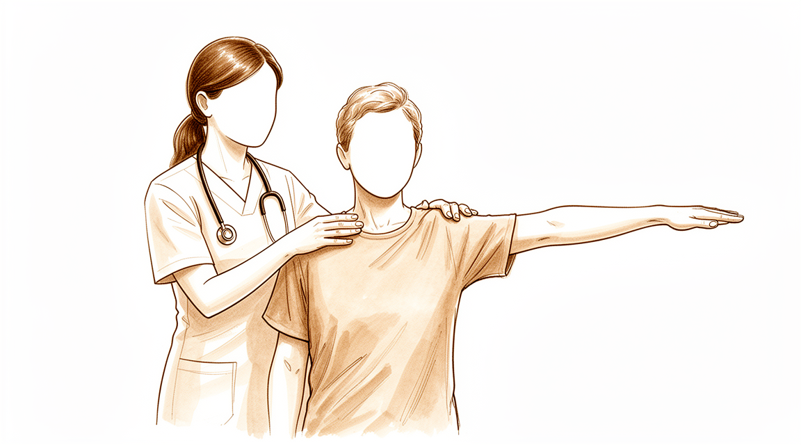
大多数人无需手术即可缓解症状。您的治疗过程通常从简单的自我护理和引导性运动开始。您可能会尝试口服抗炎药物以减轻疼痛。您的外科医生还可能推荐物理治疗,以恢复肩关节的活动范围和力量。这些非手术疗法是急性钙化性肌腱炎的首选方案。
您应给予保守治疗发挥作用的时间。该病症在长期来看有较高的完全治愈可能性。事实上,72% 的患者通过非手术治疗可获得极佳或良好的疗效。无论钙化灶的位置、大小或初始症状的严重程度如何,这一成功率均适用。如果疼痛迁延不愈,可能导致无法工作并降低生活质量。坚持进行康复治疗是重返日常生活关键。
如果自我护理和物理治疗不足以控制病情,您的外科医生可能会建议医疗干预。皮质类固醇注射是减轻炎症和疼痛的常见选择。另一种有效的非侵入性治疗是体外冲击波疗法(ESWT)。该疗法利用声波粉碎钙化沉积物。它安全性高,在缓解疼痛和恢复功能方面成功率很高。患者在接受该治疗后,通常可见疼痛症状减轻及肩关节功能改善。ESWT 的并发症微乎其微。
仅当保守治疗达到极限时,才会考虑手术。如果您的钙化病灶大于 1 厘米,外科医生可能会建议手术治疗,因为这类病灶需要手术的可能性增加 2.8 倍。对于慢性病例,关节镜下清除钙化灶可带来更好的临床结果。接受该手术治疗的大多数患者还需同时进行肩袖修复。
如果您确实接受手术,请预期恢复过程将是渐进的。术后功能评分改善缓慢。通常在术后六个月时,评分可超过 75%。至少需要 6 个月的随访,这些改善才具有统计学意义。大多数患者在术后报告肩关节疼痛和功能有显著改善。您的外科医生将根据您的具体情况,讨论关节镜、内镜或开放手术哪种方式最为适宜。
预期情况
您的钙化性肌腱炎引起的肩部疼痛可能会持续很长时间。这些症状可能会迁延不愈。这通常意味着需要请假休息以及生活质量下降。好消息是,从长远来看,这种情况完全缓解的可能性很大。许多人发现他们的症状会自行缓解。
如果您的疼痛没有消失,您的外科医生可能会建议进行治疗。非手术选项如冲击波治疗或针吸术是有效的。冲击波治疗在缓解疼痛和恢复功能方面具有很高的成功率,且相关并发症微乎其微。针吸术的侵入性和疼痛感仅轻微。这些治疗对许多患者效果良好。
如果需要手术,您的外科医生将移除钙化沉积物。您可能需要同时进行肩袖修复术。这是该手术的常见部分。手术后,您的肩部疼痛和功能将显著改善。然而,恢复并非一蹴而就。接受关节镜治疗的患者功能评分改善缓慢。您在术后六个月时会看到超过 75% 的改善。
完全感受到关节镜移除术的全部益处需要时间。至少需要 6 个月的随访,这些改善才会具有统计学意义。不要急于求成。您的外科医生将指导您度过这一过程。大多数患者对最终结果感到满意。
何时就医
若肩部持续性疼痛经休息后未见改善,请咨询全科医生。钙化性肌腱炎是一种急性或慢性疼痛性疾病,由肩袖肌腱周围钙化沉积物引起的炎症所致。若症状干扰睡眠或工作,或出现无力、不稳或交锁现象,应寻求医疗帮助。保守治疗是首选方案,尤其适用于急性病例,临床数据显示72%的患者可获得显著改善,结果评定为优秀或良好。然而,若经保守治疗后疼痛仍严重,请要求专科医生评估。您的外科医生可判断是否需要采取进一步措施以恢复功能并缓解疼痛。
Evidence & references
Overview
- Shoulder MRI may be warranted for preoperative planning in select patients with chronic calcific tendinopathy and prolonged refractory pain, although the probability of identifying additional cuff pathology requiring surgical intervention is very low [1].
- Patients with calcific lesions >1 cm have a 2.8 increased likelihood to undergo operative treatment in the setting of calcific tendinitis of the shoulder [2].
- The majority of patients who undergo surgical management for removal of the calcific deposit require a concomitant rotator cuff repair and experience significant improvements in shoulder pain and function [3].
- Treatment of calcific tendinitis of the shoulder with extracorporeal shock waves produces a high rate of success in pain relief and functional restoration with negligible associated complications [6].
- Patients undergoing arthroscopic treatment of a calcific deposit in the shoulder have satisfactory clinical and radiological outcomes at final follow-up, with functional scores improving slowly and reaching more than 75 percent at six months after surgery [8].
- Arthroscopic removal of calcification leads to improved clinical outcomes in patients with chronic calcific tendinitis, but at least 6 months of follow-up is needed for these improvements to become statistically significant [11].
- The primary choice of treatment for calcific tendinitis is conservative, especially in patients with acute calcific tendinitis [12].
- Arthroscopic treatment of calcifying tendinitis provides good to excellent clinical results [16].
- Routine diagnostic glenohumeral exploration does not appear beneficial in arthroscopic treatment of calcific tendinitis due to the low prevalence of intraarticular pathologies which most frequently do not require surgical treatment [19].
- Endoscopic and open surgery are equally effective in the treatment of chronic calcifying tendinopathy, showing similar clinical and sonographic results [23].
Anatomy & Pathophysiology
- Rotator cuff disease, shoulder instability, and associated lesions are common pathologic conditions of the shoulder involving soft tissues [42].
- The long head of the biceps contributes to stability of the glenohumeral joint in all directions, although in vivo studies have not yet established this stabilizing effect or the physiologic load required [32].
- Understanding the function and pathology surrounding the teres minor is paramount in comprehensive management of patients with shoulder pathology [34].
- In the context of rotator cuff disease, anterior shoulder pain with macroscopic changes in the biceps tendon is related to the complex interaction of the tendon and surrounding soft tissues rather than a single entity [41].
- Long head of biceps tendinopathy is associated with age and cuff tendinopathy on MRI obtained for evaluation of shoulder pain [51].
- Clinicians can guide patients to understand shoulder pain as age-appropriate and safe, potentially reducing unnecessary visits, tests, and treatments [51].
- A high prevalence of structural changes in the rotator cuff and biceps tendons in masters swimmers reflects the effect of shoulder symptoms, aging, and swim training [54].
- In chronic rotator cuff tears, the shape of the deltoid muscle seems only to be influenced by natural aging and is independent of reduced shoulder motion [53].
- There seems to be no disadvantage to exhausting conservative treatment and delaying implantation of reverse total shoulder arthroplasty in patients with chronic rotator cuff tears [53].
- Surgical repair of an isolated supraspinatus tear may be sufficient to keep the torn rotator cuff intact and achieve satisfactory patient-reported outcomes, but glenohumeral joint mechanics and shoulder strength are not fully restored with current repair techniques [39].
- At a 2-year follow-up, shoulder function evaluated in terms of CMS was not significantly improved with conservative versus surgical management for rotator cuff tears [37].
- Over time, supraspinatus tendon thickness in individuals with rotator cuff-related shoulder pain tends to normalize compared to the contralateral side, regardless of the exercise intervention [28].
- The theoretical concept of a high acromion index resulting in an increased upward force against the subacromial space influencing pain and function in calcifying tendinitis of the shoulder was not supported [49].
- Ultrasound is a useful tool for discovering subjects in pre-symptomatic stages who may undergo shoulder symptomatic pathologies [40].
Classification
- Calcific tendinitis is a common shoulder disorder [9].
- Calcific tendinitis should be differentiated from dystrophic calcification because their pathogenesis and natural history are totally different [9].
- Calcific tendinitis is an acute or chronically painful condition caused by inflammation around calcium deposits in the rotator cuff tendons [15].
- Symptoms of calcific tendinitis can be protracted, resulting in time off work and impaired quality of life [5].
- Symptomatic calcific tendinitis of the shoulder has a good likelihood to completely resolve in the long-term [4].
- The demographic, radiographic, and clinical features of calcific tendinitis of the shoulder in the Korean population are not different from those of Western populations [7].
- Conservative treatment for calcific tendinitis of the shoulder showed clinically significant improvement, with 72% of excellent or good results regardless of the location, radiologic type and size, and initial symptoms of calcific deposits [10].
- The primary choice of treatment for calcific tendinitis is conservative, especially in patients with acute calcific tendinitis [12].
- Patients with calcific lesions >1 cm had a 2.8 increased likelihood to undergo operative treatment in the setting of calcific tendinitis of the shoulder [2].
- The majority of patients who undergo surgical management for removal of the calcific deposit will require a concomitant rotator cuff repair and have significant improvements in shoulder pain and function [3].
- Shoulder MRI may be warranted for preoperative planning in the select population of patients with chronic calcific tendinitis and prolonged refractory pain, although the probability of identifying additional cuff pathology requiring surgical intervention is very low [1].
- Calcific tendinitis of the rotator cuff with tuberosity osteolysis is a distinctive form of calcific tendinitis that should be considered in clinical and surgical practice [17].
- Restriction of passive glenohumeral abduction combined with normal passive external rotation is a diagnostic feature of calcific tendinitis that can be used to clinically differentiate it from adhesive capsulitis [18].
- The incidence and type of intraarticular lesions in calcifying tendinitis are comparable to age-matched shoulders with partial- rather than full-thickness rotator cuff tears [35].
Clinical Presentation
- Calcific tendinitis of the shoulder is an acute or chronically painful condition caused by inflammation around calcium deposits in the rotator cuff tendons [15].
- Symptoms of calcific tendinitis can be protracted, resulting in time off work and impaired quality of life [5].
- Symptomatic calcific tendinitis of the shoulder has a good likelihood to completely resolve in the long-term [4].
- Calcific tendinitis is a common shoulder disorder that should be differentiated from dystrophic calcification because the pathogenesis and natural history of both are totally different [9].
- The demographic, radiographic, and clinical features of calcific tendinitis of the shoulder in the Korean population are not different from those of Western populations [7].
- Restriction of passive glenohumeral abduction combined with normal passive external rotation is a diagnostic feature of calcific tendinitis that can be used to clinically differentiate it from adhesive capsulitis [18].
- Arthroscopic treatment of calcifying tendinitis provides good to excellent clinical results [16].
- The majority of patients who undergo surgical management for removal of the calcific deposit will require a concomitant rotator cuff repair and have significant improvements in shoulder pain and function [3].
- Patients with calcific lesions >1 cm had a 2.8 increased likelihood to undergo operative treatment in the setting of calcific tendinitis of the shoulder [2].
- Conservative treatment for calcific tendinitis of the shoulder showed clinically significant improvement, with 72% of excellent or good results regardless of the location, radiologic type and size, and initial symptoms of calcific deposits [10].
- The primary choice of treatment for calcific tendinitis is conservative, especially in patients with acute calcific tendinitis [12].
- Treatment of calcific tendinitis of the shoulder with extracorporeal shock waves has produced a high rate of success in pain relief and functional restoration with negligible associated complications [6].
- Both ultrasound-guided needling and extracorporeal shock wave therapy improved clinical outcomes and eliminated calcium deposits in the treatment of calcific tendinitis [22].
- Shoulder MRI may be warranted for preoperative planning in the select population of patients with chronic calcific tendinopathy and prolonged refractory pain, although the probability of identifying additional cuff pathology requiring surgical intervention is very low [1].
- Routine diagnostic glenohumeral exploration does not appear beneficial in arthroscopic treatment of calcific tendinitis due to the low prevalence of intraarticular pathologies which most frequently do not require surgical treatment [19].
- Imaging and functional data indicate that calcific tendinitis of the rotator cuff with tuberosity osteolysis is a distinctive form of calcific tendinitis that should be considered in clinical and surgical practice [17].
- Recognition of atypical presentations of calcific tendinitis with bone erosion may prevent unnecessary biopsy and overtreatment [14].
- Atypical presentations of calcific tendinitis, such as involvement of the teres minor affecting overhead movement or isolated posterior shoulder pain, should be considered in clinical practice [20].
- A patient can suffer from both a calcifying lesion within the medial collateral ligament and calcifying tendinitis of the rotator cuff in both shoulders simultaneously [13].
Investigations
- Shoulder MRI may be warranted for preoperative planning in select patients with chronic calcific tendinopathy and prolonged refractory pain, although the probability of identifying additional cuff pathology requiring surgical intervention is very low [1].
- Patients with calcific lesions >1 cm have a 2.8 increased likelihood to undergo operative treatment [2].
- The majority of patients undergoing surgical management for removal of the calcific deposit require a concomitant rotator cuff repair [3].
- Symptomatic calcific tendinitis of the shoulder has a good likelihood to completely resolve in the long-term [4].
- Calcific tendinitis symptoms can be protracted, resulting in time off work and impaired quality of life [5].
- Demographic, radiographic, and clinical features of calcific tendinitis of the shoulder in the Korean population are not different from those of Western populations [7].
- Patients undergoing arthroscopic treatment of a calcific deposit in the shoulder had satisfactory clinical and radiological outcomes at final follow-up, with functional scores improving slowly and reaching more than 75 percent at six months after surgery [8].
- Conservative treatment for calcific tendinitis of the shoulder showed clinically significant improvement, with 72% of excellent or good results regardless of the location, radiologic type and size, and initial symptoms of calcific deposits [10].
- Calcific tendinitis can present with simultaneous calcifying lesions in other locations, such as the medial collateral ligament of the knee [13].
- Atypical presentations of calcific tendinitis with bone erosion may occur, and recognition of these presentations can prevent unnecessary biopsy and overtreatment [14].
- Calcific tendinitis of the rotator cuff with tuberosity osteolysis is a distinctive form that should be considered in clinical and surgical practice [17].
- Restriction of passive glenohumeral abduction combined with normal passive external rotation is a diagnostic feature that can be used to clinically differentiate adhesive capsulitis from calcific tendinitis [18].
- Atypical presentations of calcific tendinitis, such as involvement of the teres minor affecting overhead movement, should be considered in the context of isolated posterior shoulder pain [20].
- Shoulder surgeons should be cautious about rotator cuff tears as a comorbidity in calcific tendinitis and aware of the accuracy limitations of sonographic or MRI evaluation [27].
- The use of MRI before a trial of conservative management in patients with atraumatic shoulder pain, minimal to no strength deficits on physical examination, and suspected cuff tendinopathy other than full-thickness tears provides negative value in management at both individual and population levels [30].
- MRI, but not radiography, can be used to help discriminate between traumatic and nontraumatic rotator cuff lesions [38].
- Repeating lavage and corticosteroid injections until calcific deposits resolve completely is not recommended due to lack of evidence regarding efficacy and risks [43].
- Preoperative ultrasound-guided marking of calcific deposits statistically significantly improves the clinical results of arthroscopic surgery at 6 weeks and 2 years [44].
Treatment
Non-Operative Management
- Conservative treatment is the primary choice for calcific tendinitis, especially in patients with acute calcific tendinitis [12].
- Nonsurgical management remains the mainstay of treatment for calcific tendinitis of the rotator cuff [31].
- Most patients improve with nonsurgical modalities such as oral anti-inflammatory medication, physical therapy, and corticosteroid injections [31].
- Symptomatic calcific tendinitis of the shoulder has a good likelihood to completely resolve in the long-term [4].
- Conservative treatment for calcific tendinitis of the shoulder showed clinically significant improvement, with 72% of excellent or good results regardless of the location, radiologic type and size, and initial symptoms of calcific deposits [10].
- Extracorporeal shock wave therapy (ESWT) is a safe and effective noninvasive treatment for patients with calcific tendinitis of the shoulder [21].
- ESWT produces a high rate of success in pain relief and functional restoration with negligible complications [21].
- ESWT effectively reduced painful symptomatology and increased shoulder function in patients with chronic calcific tendinitis of the shoulder [24].
- Treatment of calcific tendinitis of the shoulder with shock waves has produced a high rate of success in pain relief and functional restoration with negligible associated complications [6].
- Both ultrasound-guided needling and extracorporeal shock wave therapy improved clinical outcomes and eliminated calcium deposits in the treatment of calcific tendinitis [22].
- The use of MRI before a trial of conservative management in patients with atraumatic shoulder pain, minimal to no strength deficits on physical examination, and suspected cuff tendinopathy other than full-thickness tears provides negative value in the management of these patients [30].
Operative Management
- Patients with calcific lesions >1 cm had a 2.8 increased likelihood to undergo operative treatment in the setting of calcific tendinitis of the shoulder [2].
- Arthroscopic removal of calcification leads to improved clinical outcomes in patients with chronic calcific tendinitis [11].
- At least 6 months of follow-up is needed for improvements in clinical outcomes after arthroscopic removal of calcification to become statistically significant [11].
- Patients undergoing arthroscopic treatment of a calcific deposit in the shoulder had satisfactory clinical and radiological outcomes at the final follow-up [8].
- Functional scores in patients undergoing arthroscopic treatment of a calcific deposit improved slowly, reaching more than 75 percent at six months after surgery [8].
- Arthroscopic treatment of calcifying tendinitis provides good to excellent clinical results [16].
- The majority of patients who undergo surgical management for removal of the calcific deposit will require a concomitant rotator cuff repair [3].
- Patients who undergo surgical management for removal of the calcific deposit have significant improvements in shoulder pain and function [3].
- Endoscopic and open surgery are equally effective in the treatment of chronic calcifying tendinopathy, showing similar clinical and sonographic results [23].
- Routine diagnostic glenohumeral exploration does not appear beneficial in arthroscopic treatment of calcific tendinitis due to the low prevalence of intraarticular pathologies which most frequently do not require surgical treatment [19].
- Shoulder MRI may be warranted for preoperative planning in the select population of patients with chronic calcific tendinopathy and prolonged refractory pain [1].
- The probability of identifying additional cuff pathology requiring surgical intervention via shoulder MRI in patients with chronic calcific tendinopathy and prolonged refractory pain is very low [1].
Complications
- Calcific tendinitis symptoms can be protracted, resulting in time off work and impaired quality of life [5].
- Atypical presentations of calcific tendinitis may include bone erosion, which can lead to unnecessary biopsy and overtreatment if not recognized [14].
- Shoulder surgeons should be cautious about rotator cuff tears as a comorbidity in calcific tendinitis due to accuracy limitations in sonographic or MRI evaluation [27].
- The probability of identifying additional cuff pathology requiring surgical intervention via preoperative shoulder MRI in patients with chronic calcific tendinopathy is very low [1].
- Arthroscopic decompression is not recommended in the treatment of rotator cuff tendinopathy [25].
Recovery
- Symptomatic calcific tendinitis of the shoulder has a good likelihood to completely resolve in the long-term [4].
- Symptoms of calcific tendinitis can be protracted, resulting in time off work and impaired quality of life [5].
- Patients undergoing arthroscopic treatment of a calcific deposit in the shoulder had satisfactory clinical and radiological outcomes at the final follow-up [8].
- Functional scores for patients undergoing arthroscopic treatment of a calcific deposit improved slowly, reaching more than 75 percent at six months after surgery [8].
- Arthroscopic removal of calcification leads to improved clinical outcomes in patients with chronic calcific tendinitis, but at least 6 months of follow-up is needed for these improvements to become statistically significant [11].
- The majority of patients who undergo surgical management for removal of the calcific deposit will require a concomitant rotator cuff repair [3].
- Patients who undergo surgical management for removal of the calcific deposit have significant improvements in shoulder pain and function [3].
- Percutaneous needle aspiration and lavage is effective in the short term and in the long term in calcific tendinitis of the shoulder [29].
- Percutaneous needle aspiration and lavage results are similar to or better than those published for other techniques [29].
- Percutaneous needle aspiration and lavage is only slightly invasive and painful [29].
- Treatment of calcific tendinitis of the shoulder with shock waves has produced a high rate of success in pain relief and functional restoration with negligible associated complications [6].
- Shock wave therapy is a safe and effective noninvasive treatment for patients with calcific tendinitis of the shoulder, producing a high rate of success in pain relief and functional restoration with negligible complications [21].
- A symptom duration of ≤10 months or calcification size of ≤10.82 mm represented the clinical scenarios most likely to show resorption after extracorporeal shockwave therapy (ESWT) [56].
- The short-term functional outcome of patients with calcific tendonitis after arthroscopic bursectomy and debridement of the calcific deposit is not influenced if performed in combination with or without a subacromial decompression [26].
Key Evidence
- [Commentary] Shoulder MRI may be warranted for preoperative planning in the select population of patients with chronic calcific tendinopathy and prolonged refractory pain, although the probability of identifying additional cuff pathology requiring surgical intervention is very low. [1] (10.1016/j.arthro.2020.01.014)
- [L3] Patients with calcific lesions >1 cm had a 2.8 increased likelihood to undergo operative treatment in the setting of calcific tendinitis of the shoulder. [2] (10.1016/j.jseint.2021.01.013)
- [L3] The majority of patients who undergo surgical management for removal of the calcific deposit will require a concomitant rotator cuff repair and have significant improvements in shoulder pain and function. [3] (10.1177/2325967121s00208)
- [L1] Symptomatic calcific tendinitis of the shoulder has a good likelihood to completely resolve in the long-term. [4] (10.1097/phm.0000000000000939)
- [L3] Calcific tendinitis is a poorly understood condition in which symptoms can be protracted, resulting in time off work and impaired quality of life. [5] (10.1016/j.jse.2006.06.007)
- [L2] Treatment of calcific tendinitis of the shoulder with shock waves has produced a high rate of success in pain relief and functional restoration with negligible associated complications. [6] (10.1016/j.jse.2007.03.023)
- [L4] This study reported demographic, radiographic, and clinical features of calcific tendinitis of the shoulder in the Korean population, which were not different from those of Western populations. [7] (10.5397/cise.2020.00010)
- [Paper] Patients undergoing arthroscopic treatment of a calcific deposit in the shoulder had satisfactory clinical and radiological outcomes at the final follow-up, with functional scores improving slowly and reaching more than 75 percent at six months after surgery. [8] (10.1016/j.otsr.2020.03.005)
- [L5] Calcific tendinitis is a common shoulder disorder that should be differentiated from dystrophic calcification as the pathogenesis and natural history of both is totally different. [9] (10.5312/wjo.v7.i1.55)
- [L2] Conservative treatment for calcific tendinitis of the shoulder showed clinically significant improvement, with 72% of excellent or good results regardless of the location, radiologic type and size, and initial symptoms of calcific deposits. [10] (10.1016/j.jse.2009.07.008)
- [L4] Arthroscopic removal of calcification leads to improved clinical outcomes in patients with chronic calcific tendinitis, but at least 6 months of follow-up is needed for these improvements to become statistically significant. [11] (10.5397/cise.2018.21.2.75)
- [L5] The primary choice of treatment for calcific tendinitis is conservative, especially in patients with acute calcific tendinitis. [12] (10.5397/cise.2020.00318)
- [Case_report] This is the first case report of a patient suffering from both a calcifying lesion within the medial collateral ligament and calcifying tendinitis of the rotator cuff in both shoulders. [13] (10.1186/s12891-016-1147-z)
- [L5] Recognition of atypical presentations of calcific tendinitis with bone erosion may prevent unnecessary biopsy and overtreatment. [14] (10.1016/j.jse.2009.02.009)
- [L5] Calcific tendinitis of the shoulder is an acute or chronically painful condition caused by inflammation around calcium deposits in the rotator cuff tendons. [15] (10.1016/s0030-5898(03)00089-0)
- [L3] Arthroscopic treatment of calcifying tendinitis provides good to excellent clinical results. [16] (10.1177/03635465211037690)
- [L2] Imaging and functional data indicate that calcific tendinitis of the rotator cuff with tuberosity osteolysis is a distinctive form of calcific tendinitis that should be considered in clinical and surgical practice. [17] (10.1016/j.jse.2008.09.016)
- [L3] This finding can be used to clinically differentiate adhesive capsulitis from calcific tendinitis. [18] (10.1177/2325967117752907)
- [L3] Routine diagnostic glenohumeral exploration does not appear beneficial in arthroscopic treatment of calcific tendinitis due to the low prevalence of intraarticular pathologies which most frequently do not require surgical treatment. [19] (10.1186/s12891-017-1839-z)
- [Case_report] This case highlights the importance of considering atypical presentations of calcific tendinitis, particularly in the context of isolated posterior shoulder pain. [20] (10.1016/j.jisako.2025.101055)
- [L3] Shock wave therapy is a safe and effective noninvasive treatment for patients with calcific tendinitis of the shoulder, producing a high rate of success in pain relief and functional restoration with negligible complications. [21] (10.1177/03635465030310031701)
- [L2] Both treatment modalities for calcific tendinitis improved clinical outcomes and eliminated calcium deposits. [22] (10.1016/j.jse.2014.06.036)
- [L1] Endoscopic and open surgery are equally effective in the treatment of chronic calcifying tendinopathy, showing similar clinical and sonographic results. [23] (10.1097/01.blo.0000063786.32430.22)
- [L2] ESWT effectively reduced painful symptomatology and increased shoulder function in patients with chronic calcific tendinitis of the shoulder. [24] (10.1136/ard.62.3.248)
- [L1] The natural history of rotator cuff tendinopathy probably plays a significant role in the results in the long-term. [25] (10.1302/0301-620x.99b6.bjj-2016-0569.r1)
- [L1] This study has demonstrated that the short-term functional outcome of patients with calcific tendonitis after arthroscopic bursectomy and debridement of the calcific deposit is not influenced if performed in combination with or without a subacromial decompression. [26] (10.1016/j.arthro.2015.05.015)
- [L3] Shoulder surgeons should be cautious about rotator cuff tears as a comorbidity in calcific tendinitis and aware of the accuracy limitations of sonographic or MRI evaluation. [27] (10.5397/cise.2021.00094)
- [L2] Findings from this study suggest that, over time, supraspinatus tendon thickness in individuals with rotator cuff-related shoulder pain tends to normalize compared to the contralateral side, regardless of the exercise intervention. [28] (10.1016/j.jse.2024.03.055)
- [L4] Percutaneous needle aspiration and lavage is effective in the short term and in the long term in calcific tendinitis of the shoulder, with results similar to or better than those published for other techniques, and it is only slightly invasive and painful. [29] (10.2214/ajr.07.2254)
- [L4] The use of MRI before a trial of conservative management in patients with atraumatic shoulder pain, minimal to no strength deficits on physical examination, and suspected cuff tendinopathy other than full-thickness tears provides negative value in the management of these patients, at both the individual and population level. [30] (10.1016/j.jse.2019.04.003)
- [L5] Nonsurgical management remains the mainstay of treatment for calcific tendinitis of the rotator cuff, with most patients improving with modalities such as oral anti-inflammatory medication, physical therapy, and corticosteroid injections. [31] (10.5435/jaaos-22-11-707)
- [L5] Biomechanical studies indicate that the long head of the biceps contributes to stability of the glenohumeral joint in all directions, though in vivo studies have yet to establish this stabilizing effect and the physiologic load required remains unknown. [32] (10.1016/j.arthro.2010.10.014)
- [L5] Understanding the function and pathology surrounding the teres minor is paramount in comprehensive management of the patient with shoulder pathology. [34] (10.5435/jaaos-d-15-00258)
- [L3] The incidence and type of intraarticular lesions in calcifying tendinitis are comparable to age-matched shoulders with partial- rather than full-thickness rotator cuff tears. [35] (10.1007/s00402-011-1263-z)
- [L1] At a 2-year follow-up, shoulder function evaluated in terms of CMS was not significantly improved. [37] (10.1186/s12891-020-03872-4)
- [L2] MRI, but not radiography, can be used to help discriminate between traumatic and nontraumatic rotator cuff lesions. [38] (10.1016/j.jse.2015.06.005)
- [L4] Surgical repair of an isolated supraspinatus tear may be sufficient to keep the torn rotator cuff intact and achieve satisfactory patient-reported outcomes, but glenohumeral joint mechanics and shoulder strength are not fully restored with current repair techniques. [39] (10.1177/0363546511412164)
- [L3] Ultrasound is an useful tool for discovering in pre-symptomatic stages the subjects that may undergo shoulder symptomatic pathologies. [40] (10.1186/1471-2474-11-278)
- [L4] In the context of rotator cuff disease, the etiology of anterior shoulder pain with macroscopic changes in the biceps tendon is related to the complex interaction of the tendon and surrounding soft tissues, rather than a single entity. [41] (10.1016/j.jse.2008.05.044)
- [L5] Repeating lavage and corticosteroid injections until the calcific deposits resolve completely is not recommended due to lack of evidence regarding efficacy and risks. [43] (10.5397/cise.2021.00269)
- [L3] Preoperative ultrasound-guided marking of calcific deposits is a procedure that statistically significantly improves the clinical results of arthroscopic surgery as seen at 6 weeks and 2 years. [44] (10.1016/j.arthro.2006.08.005)
- [L2] The theoretical concept of a high acromion index resulting in an increased upward force against the subacromial space, which influences pain and function in calcifying tendinitis of the shoulder, was not supported. [49] (10.1007/s00167-011-1563-4)
- [L3] Clinicians can guide patients to understand shoulder pain as age-appropriate and safe, potentially reducing unnecessary visits, tests, and treatments. [51] (10.1097/corr.0000000000003342)
- [L3] Our data support that in chronic rotator cuff tears, there seems to be no disadvantage to exhausting conservative treatment and to delay implantation of reverse total shoulder arthroplasty, as the shape of deltoid muscle seems only to be influenced by natural aging, but to be independent of reduced shoulder motion. [53] (10.1186/1471-2474-14-247)
- [L4] A high prevalence of structural changes in the rotator cuff and biceps tendons in masters swimmers reflects the effect of shoulder symptoms, aging, and swim training. [54] (10.3390/medicina57010029)
- [L3] A symptom duration of ≤10 months or calcification size of ≤10.82 mm represented the clinical scenarios most likely to show resorption after ESWT. [56] (10.1177/23259671241231609)
References
[1] Editorial Commentary: Is Magnetic Resonance Imaging of the Shoulder Ever Appropriate in Evaluating Patients With Calcific Tendinopathy of the Rotator Cuff?. Arthroscopy: The Journal of Arthroscopic & Related Surgery. 2020. DOI: 10.1016/j.arthro.2020.01.014 [2] Predictive factors for failure of conservative management in the treatment of calcific tendinitis of the shoulder. JSES International. 2021. DOI: 10.1016/j.jseint.2021.01.013 [3] Predictive factor for failure of conservative management in the treatment of calcific tendinitis of the shoulder. Orthopaedic Journal of Sports Medicine. 2021. DOI: 10.1177/2325967121s00208 [4] Long-Term Course of Shoulders After Ultrasound Therapy for Calcific Tendinitis. American Journal of Physical Medicine & Rehabilitation. 2018. DOI: 10.1097/phm.0000000000000939 [5] Calcific tendinitis: Natural history and association with endocrine disorders. Journal of Shoulder and Elbow Surgery. 2007. DOI: 10.1016/j.jse.2006.06.007 [6] Extracorporeal shock wave therapy for calcifying tendinitis of the shoulder. Journal of Shoulder and Elbow Surgery. 2008. DOI: 10.1016/j.jse.2007.03.023 [7] Calcific tendinitis of the shoulder in the Korean population: demographics and its relation with coexisting rotator cuff tear. Clinics in Shoulder and Elbow. 2021. DOI: 10.5397/cise.2020.00010 [8] Recovery pattern after arthroscopic treatment for calcific tendinitis of the shoulder. Orthopaedics & Traumatology: Surgery & Research. 2020. DOI: 10.1016/j.otsr.2020.03.005 [9] Calcific tendinitis of the rotator cuff. World Journal of Orthopedics. 2016. DOI: 10.5312/wjo.v7.i1.55 [10] Radiologic course of the calcific deposits in calcific tendinitis of the shoulder: Does the initial radiologic aspect affect the final results?. Journal of Shoulder and Elbow Surgery. 2010. DOI: 10.1016/j.jse.2009.07.008 [11] Functional Recovery of the Shoulder after Arthroscopic Treatment for Chronic Calcific Tendinitis. Clinics in Shoulder and Elbow. 2018. DOI: 10.5397/cise.2018.21.2.75 [12] Diagnosis and treatment of calcific tendinitis of the shoulder. Clinics in Shoulder and Elbow. 2020. DOI: 10.5397/cise.2020.00318 [13] Case report - calcification of the medial collateral ligament of the knee with simultaneous calcifying tendinitis of the rotator cuff. BMC Musculoskeletal Disorders. 2016. DOI: 10.1186/s12891-016-1147-z [14] Calcific tendinitis of the rotator cuff associated with intraosseous loculation: Two case reports. Journal of Shoulder and Elbow Surgery. 2009. DOI: 10.1016/j.jse.2009.02.009 [15] Calcific tendinitis of the shoulder. Orthopedic Clinics of North America. 2003. DOI: 10.1016/s0030-5898(03)00089-0 [16] Clinical and Structural Results of Rotator Cuff Repair Compared With Rotator Cuff Debridement in Arthroscopic Treatment of Calcifying Tendinitis of the Shoulder. The American Journal of Sports Medicine. 2021. DOI: 10.1177/03635465211037690 [17] Osteolytic lesion of greater tuberosity in calcific tendinitis of the shoulder. Journal of Shoulder and Elbow Surgery. 2009. DOI: 10.1016/j.jse.2008.09.016 [18] Restriction of Passive Glenohumeral Abduction Combined With Normal Passive External Rotation Is a Diagnostic Feature of Calcific Tendinitis. Orthopaedic Journal of Sports Medicine. 2018. DOI: 10.1177/2325967117752907 [19] Examination of concomitant glenohumeral pathologies in patients treated arthroscopically for calcific tendinitis of the shoulder and implications for routine diagnostic joint exploration. BMC Musculoskeletal Disorders. 2017. DOI: 10.1186/s12891-017-1839-z [20] Atypical calcific tendinitis involving teres minor which affects overhead movement: A case report. Journal of ISAKOS. 2026. DOI: 10.1016/j.jisako.2025.101055 [21] Shock Wave Therapy for Calcific Tendinitis of the Shoulder. The American Journal of Sports Medicine. 2003. DOI: 10.1177/03635465030310031701 [22] Which method is more effective in treatment of calcific tendinitis in the shoulder? Prospective randomized comparison between ultrasound-guided needling and extracorporeal shock wave therapy. Journal of Shoulder and Elbow Surgery. 2014. DOI: 10.1016/j.jse.2014.06.036 [23] Prospective Randomized Surgical Treatments for Calcifying Tendinopathy. Clinical Orthopaedics & Related Research. 2003. DOI: 10.1097/01.blo.0000063786.32430.22 [24] Extracorporeal shock wave therapy for chronic calcific tendinitis of the shoulder: single blind study. Annals of the Rheumatic Diseases. 2003. DOI: 10.1136/ard.62.3.248 [25] Arthroscopic decompression not recommended in the treatment of rotator cuff tendinopathy. The Bone & Joint Journal. 2017. DOI: 10.1302/0301-620x.99b6.bjj-2016-0569.r1 [26] Short‐Term Outcome After Arthroscopic Bursectomy Debridement of Rotator Cuff Calcific Tendonopathy With and Without Subacromial Decompression: A Prospective Randomized Controlled Trial. Arthroscopy. 2015. DOI: 10.1016/j.arthro.2015.05.015 [27] Is common the rotator cuff tear in the calcific tendinitis?. Clinics in Shoulder and Elbow. 2021. DOI: 10.5397/cise.2021.00094 [28] Do therapeutic exercises impact supraspinatus tendon thickness? Secondary analyses of the combined dataset from two randomized controlled trials in patients with rotator cuff-related shoulder pain. Journal of Shoulder and Elbow Surgery. 2024. DOI: 10.1016/j.jse.2024.03.055 [29] Sonographically Guided Percutaneous Needle Lavage in Calcific Tendinitis of the Shoulder: Short- and Long-Term Results. American Journal of Roentgenology. 2007. DOI: 10.2214/ajr.07.2254 [30] A value-based care analysis of magnetic resonance imaging in patients with suspected rotator cuff tendinopathy and the implicated role of conservative management. Journal of Shoulder and Elbow Surgery. 2019. DOI: 10.1016/j.jse.2019.04.003 [31] Calcific Tendinitis of the Rotator Cuff. Journal of the American Academy of Orthopaedic Surgeons. 2014. DOI: 10.5435/jaaos-22-11-707 [32] Anatomy, Function, Injuries, and Treatment of the Long Head of the Biceps Brachii Tendon. Arthroscopy. 2011. DOI: 10.1016/j.arthro.2010.10.014 [34] Understanding the Importance of the Teres Minor for Shoulder Function: Functional Anatomy and Pathology. Journal of the American Academy of Orthopaedic Surgeons. 2018. DOI: 10.5435/jaaos-d-15-00258 [35] Intraarticular lesions in calcifying tendinitis: incidence and association with the acromion index. Archives of Orthopaedic and Trauma Surgery. 2011. DOI: 10.1007/s00402-011-1263-z [37] Conservative versus surgical management for patients with rotator cuff tears: a systematic review and META-analysis. BMC Musculoskeletal Disorders. 2021. DOI: 10.1186/s12891-020-03872-4 [38] How to discriminate between acute traumatic and chronic degenerative rotator cuff lesions: an analysis of specific criteria on radiography and magnetic resonance imaging. Journal of Shoulder and Elbow Surgery. 2015. DOI: 10.1016/j.jse.2015.06.005 [39] In Vivo Shoulder Function After Surgical Repair of a Torn Rotator Cuff. The American Journal of Sports Medicine. 2011. DOI: 10.1177/0363546511412164 [40] Sonographic evaluation of the shoulder in asymptomatic elderly subjects with diabetes. BMC Musculoskeletal Disorders. 2010. DOI: 10.1186/1471-2474-11-278 [41] Biceps tendinitis in chronic rotator cuff tears: A histologic perspective. Journal of Shoulder and Elbow Surgery. 2008. DOI: 10.1016/j.jse.2008.05.044 [42] Chapter 24 Shoulder Instability, Rotator Cuff Disorders, Muscular Ruptures, Adhesive Capsulitis, Calcific Tendinitis. 2020. [43] The efficacy of repeated needling for calcific tendinitis of the rotator cuff. Clinics in Shoulder and Elbow. 2021. DOI: 10.5397/cise.2021.00269 [44] Value of Preoperative Ultrasound Marking of Calcium Deposits in Patients Who Require Surgical Treatment of Calcific Tendinitis of the Shoulder. Arthroscopy. 2007. DOI: 10.1016/j.arthro.2006.08.005 [49] Do anatomic variants of the acromion shape in the frontal plane influence pain and function in calcifying tendinitis of the shoulder?. Knee Surgery, Sports Traumatology, Arthroscopy. 2011. DOI: 10.1007/s00167-011-1563-4 [51] Long Head of Biceps Tendinopathy Is Associated With Age and Cuff Tendinopathy on MRI Obtained for Evaluation of Shoulder Pain. Clinical Orthopaedics & Related Research. 2024. DOI: 10.1097/corr.0000000000003342 [53] Deltoid muscle shape analysis with magnetic resonance imaging in patients with chronic rotator cuff tears. BMC Musculoskeletal Disorders. 2013. DOI: 10.1186/1471-2474-14-247 [54] Ultrasonographic Evaluation of the Shoulders and Its Associations with Shoulder Pain, Age, and Swim Training in Masters Swimmers. Medicina. 2020. DOI: 10.3390/medicina57010029 [56] Treatment Algorithm for the Resorption of Calcific Tendinitis Using Extracorporeal Shockwave Therapy: A Data Mining Study. Orthopaedic Journal of Sports Medicine. 2024. DOI: 10.1177/23259671241231609


