Lesão do TFCC
Patients › Wrist
TFCC injuries — pain on the ulnar side of the wrist, often with clicking, and treatment options.
O que você está sentindo
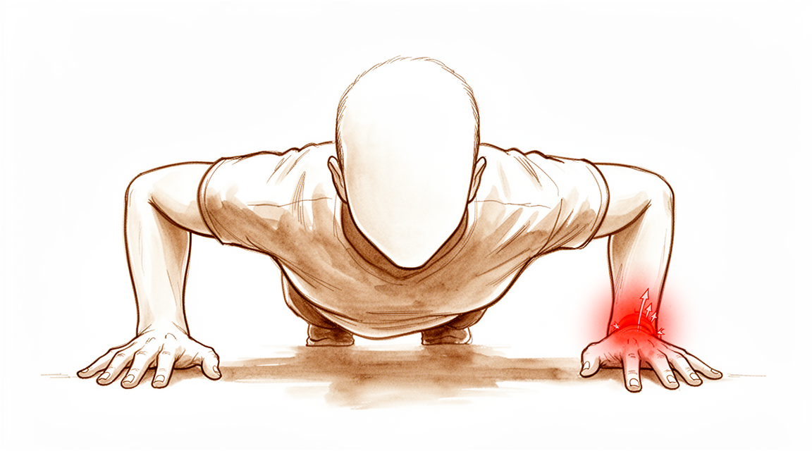
Provavelmente, você sente dor na parte externa do pulso, perto do dedo mindinho. Essa área é chamada de lado ulnar. A dor pode vir e ir, ou pode ser constante. Muitas vezes, parece uma dor profunda ou uma pontada aguda quando você move o pulso de certas maneiras.
A dor geralmente piora quando você gira o antebraço. Tarefas diárias simples podem se tornar difíceis. Você pode ter dificuldade para girar uma maçaneta, abrir um pote ou usar uma chave de fenda. Levantar objetos, especialmente com a palma da mão voltada para baixo, pode desencadear desconforto. Alcançar atrás das costas para fechar um sutiã ou guardar a camisa dentro da calça também pode doer. Algumas pessoas percebem que dormir do lado afetado piora a dor pela manhã.
Em muitos casos, o pulso parece estável. Você pode não sentir nenhuma folga ou estalo. No entanto, se você tiver um rompimento completo, pode notar instabilidade. Isso significa que seu pulso parece que pode ceder ou se deslocar inesperadamente. Isso é mais comum em tipos específicos de rompimentos que envolvem a conexão com o osso do pulso do lado do mindinho.
Se você teve uma fratura recente no osso do antebraço, também pode sentir dor na base do dedo mindinho. Isso frequentemente ocorre junto com um rompimento na almofada de cartilagem. Mesmo que seu pulso pareça normal e esteja estável, você ainda pode ter um rompimento nas fibras profundas dessa almofada. Esses rompimentos profundos causam dor, mas nem sempre causam instabilidade.
Às vezes, a dor persiste mesmo após o tratamento inicial. Se você teve cirurgia para um tipo específico de rompimento, mas ainda sente dor ou instabilidade, pode haver outra parte do rompimento que não foi tratada. Isso não é incomum. Seu cirurgião analisará cuidadosamente seu histórico e sintomas para decidir se é necessário um tratamento adicional.
A maioria dos rompimentos agudos cicatriza bem com o tempo e o repouso. No entanto, se a dor não desaparecer, pode ser porque o rompimento é degenerativo, em vez de decorrente de uma lesão súbita. Rompimentos degenerativos frequentemente exigem um manejo diferente. Seu cirurgião ajudará você a entender qual tipo de rompimento você tem e o que esperar.
O que está realmente acontecendo
O seu pulso é uma articulação complexa onde dois ossos do antebraço se encontram. Uma pequena estrutura resistente chamada complexo fibrocartilaginoso triangular situa-se entre eles. Pense nisso como um amortecedor e uma junta. Ela mantém os ossos alinhados enquanto você gira a mão ou agarra objetos.
Este complexo depende de vários ligamentos, que são como cordas fortes que mantêm a articulação unida. Os músculos ao redor do pulso também atuam como estabilizadores dinâmicos, contraindo-se para manter tudo estável durante o movimento. Quando você rotaciona o antebraço, os pontos de contato dentro da articulação mudam ligeiramente para lidar com a carga.
Uma lesão no CFT significa que este amortecedor ou uma de suas cordas de suporte está danificada. Isso geralmente ocorre após uma queda ou um torcimento súbito. A lesão pode causar dor, estalos ou uma sensação de que o pulso é instável. Pode parecer que os ossos estão escorregando de lugar quando você tenta levantar ou girar objetos.
Às vezes, a lesão está ligada ao alinhamento dos ossos. Se o osso rádio foi fraturado e cicatrizou ligeiramente fora de posição, isso altera a forma como a força se propaga pelo seu pulso. Este alinhamento alterado coloca estresse extra no CFT, dificultando a cicatrização da articulação por conta própria.
Em muitos casos, especialmente com lesões recentes, o corpo pode cicatrizar a lesão sem cirurgia. Os tecidos se reconectam ao longo do tempo. No entanto, se a lesão for completa ou se os ossos estiverem desalinhados, a articulação pode permanecer dolorosa ou fraca. Seu cirurgião analisa o tipo específico de lesão e a forma como seu pulso se move para decidir se a reparação é necessária.
O objetivo do tratamento é restaurar esse deslizamento suave e estável entre seus ossos. Seja através de repouso, terapia ou cirurgia, o objetivo é reduzir a dor e ajudar você a recuperar a força. A maioria das pessoas observa melhora significativa no movimento e na força de preensão após o cuidado adequado.
O que podemos fazer a respeito
A maioria das rupturas agudas do complexo fibrocartilaginoso triangular (CFT) cicatriza espontaneamente, sem necessidade de cirurgia. Seu cirurgião revisará cuidadosamente seu histórico e examinará seu punho para confirmar que a lesão está causando seus sintomas. Você precisará quantificar a gravidade de sua dor e rigidez para ajudar a decidir se a cirurgia é necessária.
A fisioterapia foca na restauração do movimento e da força. Embora os protocolos específicos de reabilitação variem, o objetivo é controlar os sintomas e melhorar a função. Seu cirurgião não dependerá exclusivamente do teste de estresse radio-ulnar para decidir se você precisa de uma reparação imediata. Este teste mede a laxidão articular, mas nem sempre prevê o quão bem você se sentirá ou funcionará após o tratamento.
Se o tratamento conservador não proporcionar alívio suficiente, seu cirurgião pode discutir o manejo médico. Isso geralmente inclui medicamentos para dor e anti-inflamatórios para reduzir o inchaço e o desconforto. Em alguns casos, injeções, como cortisona, ácido hialurônico ou plasma rico em plaquetas (PRP), podem ser consideradas para acalmar a inflamação e apoiar a cicatrização. Esses tratamentos visam proporcionar alívio temporário e permitir que você participe mais plenamente da fisioterapia. A duração do alívio varia de indivíduo para indivíduo, mas essas opções podem servir como ponte enquanto seu corpo cicatriza.
A cirurgia é considerada quando as medidas não operatórias falham em controlar a dor ou restaurar a estabilidade. A desbridamento artroscópico, que envolve a limpeza do tecido danificado, é segura e eficaz para rupturas centrais. Proporciona alívio da dor sustentado, melhora a qualidade de vida e restaura o movimento do punho. Para muitos pacientes, isso leva a altos índices de satisfação e benefícios funcionais a longo prazo, mesmo anos depois.
Se a ruptura envolver os pontos de inserção dos ligamentos, a reparação artroscópica pode ser recomendada. Esta técnica utiliza instrumentos minimamente invasivos para reatar o tecido rompido. Oferece melhorias significativas no movimento do punho, na força de preensão e nos níveis de dor. Em casos de rupturas crônicas, uma abordagem transóssea de um túnel pode ser utilizada para restaurar a estabilidade e fornecer escores funcionais notáveis.
Seu cirurgião também considerará a denervação cirúrgica para lesões persistentes que não respondem ao tratamento não cirúrgico ou ao desbridamento. Este procedimento reduz os sinais de dor da área afetada. Imagens avançadas, como artro-RM (ARM) ou artroscopia do punho, ajudam a confirmar o diagnóstico, pois as ressonâncias magnéticas padrão às vezes podem não detectar rupturas sutis. Seu cirurgião mantém um alto índice de suspeita, especialmente se você tiver dor no lado ulnar do punho apesar de resultados de imagem normais.
O que esperar
O seu prognóstico depende em grande parte do tipo de lesão que possui. A maioria das lesões agudas da classe 1 de Atzei cicatriza espontaneamente, sem necessidade de cirurgia. Se a sua lesão estiver no centro do disco, a maioria destas também cicatriza até ao momento do seguimento a longo prazo. De modo geral, pode esperar uma diminuição da dor e uma melhoria da mobilidade se o seu cirurgião tratar a lesão de forma eficaz.
Para lesões complexas, o seu cirurgião pode recomendar uma reparação ligamentar específica por artroscopia. Esta técnica minimamente invasiva permite uma visualização detalhada para reparar a lesão. É provável que observe melhorias significativas na mobilidade do punho, na força de preensão e nos níveis de dor. Estes benefícios são mantidos num seguimento mínimo de 2 anos. Em alguns casos, o seu cirurgião pode realizar uma ressecção assistida para remover o tecido danificado. Esta abordagem demonstra resultados satisfatórios persistentes mesmo num seguimento de 19 anos.
Se tiver uma lesão completa, o resultado a longo prazo pode ser ligeiramente inferior em comparação com outros tipos de lesão. Lesões concomitantes do tipo 2 também aumentam o risco de falha da cirurgia inicial. Deve estar ciente de que os resultados de incapacidade são piores se a sua lesão do CDTF ocorreu em conjunto com uma fratura da extremidade distal do rádio. O reconhecimento precoce da instabilidade do punho pode ajudar o seu cirurgião a fornecer um tratamento atempado. As taxas de sucesso nos casos agudos rondam os 80% quando tratados prontamente.
Não há evidências de que uma lesão do CDTF altere o seu resultado global a longo prazo. No entanto, se tiver dor persistente ou instabilidade após uma reparação bem-sucedida, pode existir uma lesão do componente proximal não detetada. O seu cirurgião irá procurar por esta lesão para garantir um tratamento adequado. A maioria dos pacientes, incluindo crianças e adolescentes, apresenta excelentes resultados funcionais após o tratamento cirúrgico. Deve sentir-se mais estável e ter menos dor à medida que se recupera.
Quando procurar um especialista
Procure uma avaliação especializada se tiver dor persistente que não melhora com o repouso. Procure atendimento se notar fraqueza ou instabilidade no punho. Consulte um médico se o seu punho travar ou ceder. Procure ajuda se os sintomas interferirem no seu sono ou no trabalho. A piora súbita da dor também é um motivo para procurar atendimento. A maioria das rupturas agudas cicatriza bem quando tratadas precocemente. No entanto, a dor sustentada após a reparação inicial pode indicar a existência de uma ruptura não identificada. Um exame cuidadoso ajuda a determinar se a cirurgia é necessária. Não assuma que uma ressonância magnética normal exclui uma lesão. É necessária uma alta suspeita clínica para dor no lado ulnar do punho.
Evidence & references
Overview
- Surgical treatment of TFCC tears and concomitant pathology in the pediatric and adolescent population results in decreased pain, improved motion and stability, and excellent functional outcomes in the majority of patients [1].
- There is no evidence that a TFCC injury influences long-term outcome, though trends suggested complete tears might have inferior outcomes [2].
- Most TFCC disc tears identified at the initial surgery had healed by long-term arthroscopic follow-up [4].
- Arthroscopic-assisted repair techniques provide detailed visualization and facilitate the repair of TFCC injuries and associated pathologies with minimally invasive techniques [5].
- Most acute Atzei class 1 tears spontaneously heal without surgical repair [6].
- In cases of sustained pain and distal radioulnar joint instability even after successful Atzei class 1 repair, an unrecognized proximal component TFCC tear may exist and appropriate treatment for the proximal component should be combined [6].
- Arthroscopic ligament-specific repair of the TFCC offers significant improvements in wrist motion, grip strength, pain, and patient-reported outcomes at a minimum 2-year follow-up [8].
- Sensory innervation data provides an initial step in planning an operative partial TFCC denervation for recalcitrant TFCC IA injuries that fail nonsurgical treatment and possibly also arthroscopic debridement [10].
- Arthroscopic assisted resection for lesions of the TFCC demonstrates persisting satisfactory subjective and functional outcomes at 19 years of follow-up [11].
- There are no outcome studies specifically examining results of TFCC debridement or repair in baseball players [13].
- Further prospective research is recommended to explore whether patient-related or rehabilitation factors influence outcomes following TFCC repair [14].
- There is a current lack of high-quality evidence required to draw firm conclusions on the merits of arthroscopic versus open repair of 1B TFCC tears [25].
- TFCC rehabilitation protocols were poorly reported and varied widely between the included studies [28].
Anatomy & Pathophysiology
- The distal oblique bundle of the interosseous membrane contributes to stabilization of the distal radioulnar joint [29].
- The effectiveness of stabilization provided by the interosseous membrane's distal oblique bundle is altered by the wrist's position and the direction of radial translation [29].
- Fractures of the distal radius interfere with the biomechanical integrity of the wrist [31].
- Distal radius fractures limit range of motion [31].
- Distal radius fractures affect hand muscle strength [31].
- Rotational malalignment of the wrist significantly affects carpal measurements [32].
- Rotational malalignment of the wrist significantly affects distal radial measurements [32].
- Rotational malalignment of the wrist significantly affects distal radioulnar joint measurements [32].
- Distal radioulnar joint arthroplasty considerably alters forearm kinematics [33].
- All wrists exhibit similar loading across the distal ulna regardless of ulnar variance [36].
- Pronation relatively increases loading across the distal ulna [36].
- The flexor carpi ulnaris muscle serves as a dynamic stabilizer of the distal radioulnar joint [37].
- The extensor carpi ulnaris muscle serves as a dynamic stabilizer of the distal radioulnar joint [37].
- The contact area of the distal radioulnar joint increases during wrist flexion [38].
- The contact area of the distal radioulnar joint decreases during wrist extension [38].
- The contact area of the distal radioulnar joint decreases during ulnar deviation [38].
- During forearm rotation, the contact site of the scaphoid on the distal radial articular surface changes minimally [41].
- During forearm rotation, the contact site of the lunate on the distal radial articular surface changes minimally [41].
- Intercarpal kinematic modifications after intercarpal arthrodeses make constant radiocarpal and midcarpal congruence during radioulnar deviation impossible [42].
- A volar ligament-sparing radiocarpal arthrotomy does not cause biomechanical radiocarpal instability [43].
- Injury to the dorsal wrist extrinsic carpal ligaments exacerbates volar radiocarpal instability after intra-articular distal radius fracture [44].
- Disruptions in forearm structures may lead to forearm instability with consequences at the remaining structures [45].
- Radial lengthening beyond the native length is not detrimental to radial loading [46].
- Radial lengthening further reduces distal ulnar loading [46].
- Achieving at least native ulnar variance appears appropriate to restore normal biomechanical loading [46].
- Four-phase grip MRI can demonstrate impairment of the articular disc and longitudinal instability of the distal radioulnar joint simultaneously [47].
- Each ligament stabilizing the distal radioulnar joint contributes to joint stability depending on the direction (palmar or dorsal) [48].
- Each ligament stabilizing the distal radioulnar joint contributes to joint stability depending on different positions of the wrist and forearm [48].
Classification
- The Palmer classification does not completely classify all peripheral TFCC tears, particularly dorsal tears [35].
- The Melone classification system does not predict the presence of TFCC lesions [50].
- Frykman Type VI and VIII fractures show a significantly higher incidence of TFCC tears [50].
- 1B TFCC injury is most common in patients with distal radius fractures (DRF) and concomitant TFCC injury [3].
- The presence of an ulnar styloid fracture associated with distal radius fracture predicts the presence of frequently occurring traumatic TFCC injury and TFCC 1B injury [9].
- A small percentage of arthroscopically validated TFCC injuries involve two tears in one wrist, with the most common pattern being a slit tear coexisting with an ulnar styloid tear [20].
- In more detailed classification of TFCC injuries, such as pc-TFCC tears classified by Atzei's classification, the diagnostic accuracy of MRI remains lower compared to wrist arthroscopy [19].
- Most acute Atzei class 1 tears spontaneously heal without surgical repair [6].
- In cases of sustained pain and distal radioulnar joint instability even after successful Atzei class 1 repair, an unrecognized proximal component TFCC tear may exist [6].
- The authors propose a new 'CUP' classification system for TFCC injuries based on anatomical location (central, ulnar, peripheral) and severity [54].
- The proposed 'CUP' classification aims to address limitations of existing systems like Palmer and Atzei by focusing exclusively on TFCC lesions and providing specific treatment recommendations for each subtype [54].
Clinical Presentation
- TFCC tears in pediatric and adolescent populations present with pain, motion limitations, and stability issues that are improved by surgical treatment [1].
- Complete TFCC tears may have inferior long-term outcomes compared to other tear types, though TFCC injury generally does not influence long-term outcome after distal radial fractures [2].
- TFCC 1B injury is the most common type of TFCC injury in patients with distal radius fractures (DRF) [3].
- Most TFCC disc tears identified at initial surgery heal by long-term arthroscopic follow-up [4].
- Arthroscopic-assisted repair techniques provide detailed visualization for managing TFCC injuries and associated pathologies [5].
- Most acute Atzei class 1 tears (isolated distal component) spontaneously heal without surgical repair [6].
- Sustained pain and distal radioulnar joint (DRUJ) instability after successful Atzei class 1 repair may indicate an unrecognized proximal component TFCC tear [6].
- Careful history and physical examination are required to determine if a TFCC tear is symptomatic and to quantify symptom severity for surgical decision-making [7].
- Arthroscopic ligament-specific repair of TFCC foveal avulsions improves wrist motion, grip strength, pain, and patient-reported outcomes at minimum 2-year follow-up [8].
- The presence of an ulnar styloid fracture associated with DRF predicts the presence of traumatic TFCC injury, specifically TFCC 1B injury [9].
- Operative partial TFCC denervation is a potential planning step for recalcitrant TFCC IA injuries that fail nonsurgical treatment and possibly arthroscopic debridement [10].
- Acute TFCC tears generally provide better results when addressed in the acute phase, while degenerative lesions are typically treated with debridement [12].
- A stable DRUJ upon clinical examination and normal MRI findings do not rule out foveal TFCC injury [17].
- High clinical suspicion is needed when managing patients with ulnar-sided wrist pain to identify foveal TFCC pathology [17].
- Imaging approaches for ulnar-sided wrist pain include discussion of anatomy, pathophysiology, and radiographic appearance of TFCC tears, DRUJ disorders, and ECU tendon disorders [18].
- A small percentage of arthroscopically validated TFCC injuries involve two tears in one wrist ("double lesion") [20].
- The most common "double lesion" pattern is a slit tear coexisting with an ulnar styloid tear [20].
- Central traumatic TFCC lesions can be treated by arthroscopic debridement, resulting in sustained pain relief, improved quality of life (DASH score), improved wrist motion, and high patient satisfaction [21].
- Wrist arthroscopy remains the gold standard for diagnosing TFCC pathologies [22].
- MR arthrography (MRA) is the preferred imaging modality for internal derangements of the wrist due to superior contrast resolution, joint distention, and contrast flow facilitating diagnosis of TFCC and intrinsic ligament lesions [23].
- A negative MRI result is unable to rule out clinically relevant injury to the TFCC, scapholunate (SL) ligament, or lunotriquetral (LT) ligament [24].
- Arthroscopic debridement or repair of wrist TFCC injury provides predictable pain relief and return to play in competitive athletes [51].
- Tears of the TFCC superficial fibers with deep fibers intact present with ulnar-sided wrist pain but without DRUJ instability [52].
Investigations
- TFCC 1B injury is the most common type of TFCC injury in patients with distal radius fractures [3].
- The presence of an ulnar styloid fracture associated with a distal radius fracture predicts the presence of traumatic TFCC injury, specifically TFCC 1B injury [9].
- A high index of clinical suspicion is required for foveal TFCC injury when managing patients with ulnar-sided wrist pain, as normal MRI findings and a stable distal radioulnar joint (DRUJ) on clinical examination do not rule out this pathology [17].
- Advanced imaging techniques like MRI and radiocarpal arthroscopy are well-suited for diagnosing central and distal TFCC tears but may report partial foveal injuries as normal despite functional incompetence [55].
- Partial and complete foveal tears without instability may be missed without a high degree of suspicion [55].
- Wrist arthroscopy remains the gold standard for diagnosing TFCC pathologies [22].
- MR arthrography (MRA) is the preferred modality for imaging internal derangements of the wrist due to superior contrast resolution, joint distention, and contrast flow facilitating the diagnosis of TFCC and intrinsic ligament lesions [23].
- MR arthrography has the potential to become a real alternative to arthroscopy for diagnosing TFCC pathologies [22].
- The diagnostic accuracy of MRI for detailed TFCC classifications, such as pc-TFCC tears by Atzei's classification, is lower compared to wrist arthroscopy [19].
- The sensitivity, specificity, and accuracy of 3.0T wrist MRI for TFCC injury detection are consistently higher than those of 1.5T wrist MRI [56].
- A negative MRI result is unable to rule out the possibility of a clinically relevant TFCC injury [24].
- Negative results of MRI or clinical provocative tests are unable to safely rule out clinically relevant TFCC tears, necessitating further diagnostic evaluation with wrist arthroscopy [57].
- Surgeons should maintain a high degree of suspicion for TFCC-related pathology in young patients with positive provocative clinical examination despite negative MRI findings [59].
- The radioulnar stress test alone cannot be recommended to decide whether to perform an acute repair of the TFCC, as radioulnar laxity and clinical outcome do not correlate after distal radius fracture [27].
- MRI-based modified radioulnar ratio technique showed significant instability parameters and diagnostic accuracy (AuC 0.787) in children and adolescents with arthroscopically-verified TFCC tears [60].
- Pisoscaphoid and radioulnar distances on lateral radiographs did not show significant differences compared to controls in children and adolescents with arthroscopically-verified TFCC tears [60].
- Load-bearing radioulnar (RaUl) measurement is a simple method to diagnose an unstable distal radioulnar joint in patients with TFCC injury [63].
Treatment
Non-Operative Management
- Most acute Atzei class 1 tears (isolated distal component) spontaneously heal without surgical repair [6].
- Careful history and physical examination are required to determine whether a TFCC tear is symptomatic [7].
- Quantifying the severity of symptoms related to TFCC pathology is important to determine whether surgical treatment is necessary [7].
- Using the radioulnar stress test alone to decide whether or not to perform an acute repair of the TFCC cannot be recommended [27].
Arthroscopic Debridement and Resection
- Arthroscopic debridement of central traumatic TFCC lesions is safe and shows sustained pain relief, significantly improved quality of life (DASH score), and wrist motion, resulting in high patient satisfaction [21].
- Arthroscopic assisted resection for lesions of the TFCC demonstrates persisting satisfactory subjective and functional outcomes at 19 years of follow-up [11].
- Arthroscopic debridement of central degenerative TFCC lesions is safe, reliable, and efficacious even for ulnar positive variance [49].
- There are no outcome studies specifically examining results of TFCC debridement or repair in baseball players [13].
Surgical Repair and Reconstruction
- Surgical treatment of TFCC tears and concomitant pathology in the pediatric and adolescent population results in decreased pain, improved motion and stability, and excellent functional outcomes in the majority of patients [1].
- Arthroscopic ligament-specific repair of the TFCC offers significant improvements in wrist motion, grip strength, pain, and patient-reported outcomes at a minimum 2-year follow-up [8].
- An arthroscopic one-tunnel transosseous approach is effective for chronic foveal tears of the TFCC with intact radioulnar ligament remnants, providing pain relief, improved joint stability, and remarkable functional ratings [53].
- Most TFCC disc tears identified at initial surgery had healed by long-term arthroscopic follow-up [4].
- Arthroscopic-assisted repair techniques provide detailed visualization and facilitate the repair of TFCC injuries and associated pathologies with minimally invasive techniques [5].
- In cases of sustained pain and distal radioulnar joint instability even after successful Atzei class 1 repair, an unrecognized proximal component TFCC tear may exist, requiring appropriate treatment for the proximal component to be combined [6].
- A systematic review demonstrates a current lack of high-quality evidence required to draw firm conclusions on the merits of arthroscopic versus open repair of 1B TFCC tears [25].
- Although statistical analysis revealed significant effects of predictor variables on treatment outcomes for arthroscopic TFCC repair, additional larger comprehensive studies are required to confirm these results so as to control for unaccounted patient variables [62].
Surgical Denervation
- Operative partial TFCC denervation is planned for recalcitrant TFCC IA injuries that fail nonsurgical treatment and possibly also arthroscopic debridement [10].
Rehabilitation
- TFCC rehabilitation protocols were poorly reported and varied widely between included studies [28].
Complications
- Complete TFCC tears may have inferior long-term outcomes compared to other tear types [2].
- Coexisting type 2 TFCC tears significantly increase the risk of index surgery failure in patients undergoing arthroscopic repair of peripheral ulnar-side TFCC tears [15].
- An unrecognized proximal component TFCC tear may exist in cases of sustained pain and distal radioulnar joint instability even after successful repair of an isolated Atzei class 1 tear [6].
Recovery
- Surgical treatment of TFCC tears and concomitant pathology in the pediatric and adolescent population results in decreased pain, improved motion and stability, and excellent functional outcomes in the majority of patients [1].
- There is no evidence that a TFCC injury influences long-term outcome, though trends suggested complete tears might have inferior outcomes [2].
- Most TFCC disc tears identified at the initial surgery had healed by long-term arthroscopic follow-up [4].
- Most acute Atzei class 1 tears spontaneously heal without surgical repair [6].
- In cases of sustained pain and distal radioulnar joint instability even after successful Atzei class 1 repair, an unrecognized proximal component TFCC tear may exist and appropriate treatment for the proximal component should be combined [6].
- Arthroscopic ligament-specific repair of the TFCC offers significant improvements in wrist motion, grip strength, pain, and patient-reported outcomes at a minimum 2-year follow-up [8].
- Arthroscopic assisted resection for lesions of the TFCC demonstrates persisting satisfactory subjective and functional outcomes at 19 years of follow-up [11].
- TFCC capsular reattachment could be performed with an arthroscopically assisted technique, providing good long-term results [26].
- Disability outcomes were worse in patients with distal radial fracture where TFCC was injured [58].
- Early recognition of longitudinal radioulnar dissociation can aid in timely treatment and improved outcomes, with success rates around 80% for acute cases [64].
Key Evidence
- [L4] Surgical treatment of TFCC tears and concomitant pathology in the pediatric and adolescent population results in decreased pain, improved motion and stability, and excellent functional outcomes in the majority of patients. [1] (10.1016/j.jhsa.2019.06.019)
- [L2] The study found no evidence that a TFCC injury influences long-term outcome, though trends suggested complete tears might have inferior outcomes. [2] (10.1016/j.jhsa.2012.05.032)
- [L3] 1B TFCC injury is most common in patients with DRF and concomitant TFCC injury. [3] (10.1186/s13018-023-04438-5)
- [L4] Most TFCC disc tears identified at the initial surgery had healed by long-term arthroscopic follow-up. [4] (10.1016/j.jhsa.2012.09.011)
- [L5] Arthroscopic-assisted repair techniques have revolutionized surgical management, providing detailed visualization and facilitating the repair of TFCC injuries and associated pathologies with minimally invasive techniques. [5] (10.1016/j.jhsg.2025.100857)
- [Commentary] Most acute Atzei class 1 tears spontaneously heal without surgical repair; however, in cases of sustained pain and distal radioulnar joint instability even after successful Atzei class 1 repair, an unrecognized proximal component TFCC tear may exist and appropriate treatment for the proximal component should be combined. [6] (10.1016/j.arthro.2022.01.019)
- [L4] Careful history and physical examination are required to determine whether a TFCC tear is symptomatic, and it is important to quantify the severity of symptoms related to TFCC pathology to determine whether surgical treatment is necessary. [7] (10.5435/jaaos-d-20-00998)
- [L4] Arthroscopic ligament-specific repair of the TFCC offers significant improvements in wrist motion, grip strength, pain, and patient-reported outcomes at a minimum 2-year follow-up. [8] (10.1177/1753193420957901)
- [L4] The presence of ulnar styloid fracture associated with distal radius fracture predicted the presence of frequently occurring traumatic triangular fibrocartilage complex injury and TFCC 1B injury. [9] (10.1016/j.arthro.2020.05.025)
- [L5] These results provide an initial step in planning an operative partial TFCC denervation for recalcitrant TFCC IA injuries that fail nonsurgical treatment and possibly also arthroscopic debridement. [10] (10.1016/j.jhsa.2014.03.007)
- [L4] This study demonstrates persisting satisfactory subjective and functional outcomes for patients following arthroscopic assisted resection for lesions of the TFCC at 19 years of follow-up. [11] (10.1177/1558944717708029)
- [L5] The article reviews the anatomy, classification, and management of TFCC injuries, noting that acute tears generally provide better results when addressed in the acute phase, while degenerative lesions are typically treated with debridement. [12] (10.1016/j.hcl.2011.05.013)
- [L5] We know of no outcome studies specifically examining results of TFCC debridement or repair in baseball players. [13] (10.1016/j.hcl.2012.05.019)
- [L4] Further prospective research is recommended to explore whether patient-related or rehabilitation factors influence outcomes following TFCC repair. [14] (10.1016/j.jht.2023.08.009)
- [L4] However, coexisting type 2 TFCC tears significantly increased the risk of index surgery failure in these patients. [15] (10.1016/j.arthro.2020.05.012)
- [L4] Having a stable distal radioulnar joint upon clinical examination and normal MRI findings does not rule out foveal TFCC injury, and a high index of clinical suspicion is needed when managing patients with ulnar sided wrist pain. [17] (10.1177/17531934231206426)
- [L5] The article provides a concise approach to the diagnosis and imaging of ulnar-sided wrist pain, discussing anatomy, pathophysiology, and radiographic appearance of common entities including TFCC tears, DRUJ disorders, and ECU tendon disorders. [18] (10.1016/j.csm.2006.02.008)
- [L4] In more detailed classification of TFCC injuries, such as pc-TFCC tears classified by Atzei's classification, the diagnostic accuracy of MRI remains lower compared to wrist arthroscopy. [19] (10.1186/s12891-023-07140-z)
- [L4] A small percentage of arthroscopically validated TFCC injuries involve two tears in one wrist, with the most common pattern being a slit tear coexisting with an ulnar styloid tear. [20] (10.1177/1753193413479479)
- [L4] Central traumatic TFCC lesions can safely be treated by arthroscopic debridement, showing sustained pain relief, significantly improved quality of life (DASH score) and wrist motion, resulting in high patient satisfaction. [21] (10.1007/s00402-018-2910-4)
- [L5] Wrist arthroscopy remains the 'gold standard' for diagnosing TFCC pathologies despite technical progress in imaging modalities, although MR arthrography may have the potential to become a real alternative in the future. [22] (10.1007/s00402-015-2153-6)
- [L4] Superior contrast resolution, joint distention, and the flow of contrast facilitate the diagnosis of lesions of the TFCC and intrinsic ligaments on contrast-sensitive sequences, making MRA the preferred modality for imaging internal derangements of the wrist. [23] (10.1007/s11552-008-9149-4)
- [L2] A negative result from MRI is unable to rule out the possibility of a clinically relevant injury to the TFCC, SL ligament, or LT ligament of the wrist. [24] (10.1016/j.arthro.2015.04.090)
- [L4] This SR demonstrates a current lack of high-quality evidence required to draw firm conclusions on the merits of arthroscopic versus open repair of 1B TFCC tears. [25] (10.1177/1558944718815244)
- [L5] TFCC capsular reattachment could be performed with an arthroscopically assisted technique, providing good long-term results. [26] (10.1016/j.hcl.2017.06.005)
- [L2] Therefore, using the radioulnar stress test alone to decide whether or not to perform an acute repair of the TFCC cannot be recommended. [27] (10.1177/1753193411403690)
- [L4] TFCC rehabilitation protocols were poorly reported and varied widely between the included studies. [28] (10.1016/j.jht.2021.10.004)
- [L5] However, the wrist's position and the direction of radial translation seem to alter the stabilization's effectiveness. [29] (10.1016/j.otsr.2020.03.041)
- [L3] These results supported the initial hypothesis that a fracture of the distal radius interferes with the biomechanical integrity of the wrist, limiting range of motion and affecting hand muscle strength. [31] (10.1177/1758998315574352)
- [L4] Rotational malalignment of the wrist has significant effects on carpal, distal radial and distal radioulnar joint measurements. [32] (10.1177/1753193408090393)
- [L4] The Aptis distal radioulnar joint arthroplasty considerably alters forearm kinematics, which can have clinical implications. [33] (10.1177/17531934241274142)
- [L4] The Palmer classification does not completely classify all peripheral TFCC tears, particularly dorsal tears. [35] (10.1016/j.arthro.2007.01.026)
- [L5] The results show that all wrists have similar loading across the distal ulna regardless of ulnar variance, while pronation relatively increases loading across the distal ulna. [36] (10.1016/j.jhsa.2014.10.001)
- [L4] The flexor carpi ulnaris and extensor carpi ulnaris muscles serve as dynamic stabilizers of the distal radioulnar joint. [37] (10.1177/17531934231168299)
- [L4] The contact area of the DRUJ increases during wrist flexion and decreases during wrist extension and ulnar deviation. [38] (10.1016/j.jhsa.2015.07.027)
- [L5] During forearm rotation, the contact site of the scaphoid and the lunate on the distal radial articular surface changed minimally. [41] (10.1016/j.jhsa.2013.01.021)
- [L5] The study confirms that constant radiocarpal and midcarpal congruence during radioulnar deviation in normal wrists is no longer possible with intercarpal kinematic modifications after these arthrodeses. [42] (10.1177/17531934231176004)
- [L5] This volar ligament-sparing radiocarpal arthrotomy did not cause biomechanical radiocarpal instability. [43] (10.1016/j.jhsa.2022.08.028)
- [L5] Injury to the dorsal wrist extrinsic carpal ligaments exacerbates volar radiocarpal instability. [44] (10.1177/1558944719851210)
- [L5] Disruptions in any of these structures may lead to forearm instability with consequences at the remaining structures. [45] (10.1016/j.jhsa.2016.10.017)
- [L5] Radial lengthening beyond the native length was not detrimental to radial loading and further reduced distal ulnar loading; achieving at least native ulnar variance seems to be appropriate to restore normal biomechanical loading based on this in vitro study. [46] (10.1016/j.jhsa.2019.03.017)
- [L4] Reconstructed animation from four-phase grip MRI demonstrated impairment of the articular disc and longitudinal instability of the distal radioulnar joint simultaneously and should be of value in investigating dynamic pathophysiology causing ulnar wrist pain. [47] (10.1177/1753193413476979)
- [L4] Arthroscopic debridement of central degenerative TFCC lesions is safe, reliable, and efficacious even for ulnar positive variance. [49] (10.1007/s00402-021-03918-9)
- [L3] The Melone classification system does not predict the presence of TFCC lesions, while Frykman Type VI and VIII fractures show a significantly higher incidence of TFCC tears. [50] (10.1177/1753193408090106)
- [L4] Arthroscopic debridement or repair of wrist TFC injury provides predictable pain relief and return to play in competitive athletes. [51] (10.1177/0363546508325921)
- [L4] Tears of the TFCC superficial fibers with the deep fibers intact present with ulnar-sided wrist pain but without distal radioulnar joint instability. [52] (10.1016/j.jhsa.2011.12.023)
- [L4] This simple arthroscopic one-tunnel transosseous approach is effective for chronic foveal tears of the TFCC with intact radioulnar ligament remnants, providing pain relief, improved joint stability, and remarkable functional ratings. [53] (10.1177/17531934211056854)
- [L5] The authors propose a new 'CUP' classification system for TFCC injuries based on anatomical location (central, ulnar, peripheral) and severity, aiming to address limitations of existing systems like Palmer and Atzei by focusing exclusively on TFCC lesions and providing specific treatment recommendations for each subtype. [54] (10.1177/17531934221121931)
- [L5] Partial and complete foveal tears without instability may be missed without a high degree of suspicion, as advanced imaging techniques like MRI and radiocarpal arthroscopy are well-suited for diagnosing central and distal TFCC tears but may report partial injuries as normal despite functional incompetence. [55] (10.1302/0301-620x.105b1.bjj-2022-0908.r1)
- [L3] The sensitivity, specificity, and accuracy of 3.0T wrist MRI for the TFCC is consistently higher compared with those of 1.5T wrist MRI, suggesting improved capability for detection of TFCC injuries. [56] (10.1016/j.jhsa.2008.02.028)
- [Letter] Negative results of MRI or clinical provocative tests are still unable to safely rule out the possibility of clinically relevant tears to the TFCC and other wrist ligaments, which makes further diagnostic evaluation with wrist arthroscopy necessary. [57] (10.1016/j.arthro.2015.08.001)
- [L2] Disability outcomes were worse in patients with distal radial fracture where TFCC was injured. [58] (10.1016/j.jht.2017.09.002)
- [L2] Surgeons should have a high degree of suspicion for TFCC-related pathology in the setting of positive provocative clinical examination despite negative MRI findings in young patients. [59] (10.1016/j.jhsa.2024.04.015)
- [L3] MRI-based modified radioulnar ratio technique showed significant instability parameters and diagnostic accuracy (AuC 0.787) in children and adolescents with arthroscopically-verified TFCC tears, whereas pisoscaphoid and radioulnar distances on lateral radiographs did not show significant differences compared to controls. [60] (10.1007/s00402-020-03470-y)
- [L5] Although statistical analysis revealed significant effects of predictor variables on treatment outcomes for arthroscopic TFCC repair, additional larger comprehensive studies are required to confirm these results so as to control for unaccounted patient variables. [62] (10.1016/j.arthro.2019.04.022)
- [L2] Load-bearing RaUl measurement is a simple method to diagnose an unstable distal radioulnar joint in patients with TFCC injury. [63] (10.1016/j.jhsa.2022.01.008)
- [L5] Early recognition of longitudinal radioulnar dissociation can aid in timely treatment and improved outcomes, with success rates around 80% for acute cases. [64] (10.1016/j.hcl.2007.01.005)
References
[1] Early Results of Surgical Treatment of Triangular Fibrocartilage Complex Tears in Children and Adolescents. The Journal of Hand Surgery. 2020. DOI: 10.1016/j.jhsa.2019.06.019 [2] The Natural Course of Traumatic Triangular Fibrocartilage Complex Tears in Distal Radial Fractures: A 13–15 Year Follow-up of Arthroscopically Diagnosed but Untreated Injuries. The Journal of Hand Surgery. 2012. DOI: 10.1016/j.jhsa.2012.05.032 [3] Association between imaging parameter changes and triangular fibrocartilage complex injury after distal radius fractures. Journal of Orthopaedic Surgery and Research. 2023. DOI: 10.1186/s13018-023-04438-5 [4] Clinical, Radiographic, and Arthroscopic Outcomes After Ulnar Shortening Osteotomy: A Long-Term Follow-Up Study. The Journal of Hand Surgery. 2012. DOI: 10.1016/j.jhsa.2012.09.011 [5] WITHDRAWN: Arthroscopic-Assisted Repair of the Triangular Fibrocartilage Complex. Journal of Hand Surgery Global Online. 2025. DOI: 10.1016/j.jhsg.2025.100857 [6] Editorial Commentary: Arthroscopic Capsular Repair of Wrist Triangular Fibrocartilage Complex Tears: Beware That Apparent Isolated Atzei Class 1 (Isolated Distal Component) Tears May Include a Proximal Component. Arthroscopy: The Journal of Arthroscopic & Related Surgery. 2022. DOI: 10.1016/j.arthro.2022.01.019 [7] Open and Arthroscopic Triangular Fibrocartilage Complex (TFCC) Repair. Journal of the American Academy of Orthopaedic Surgeons. 2021. DOI: 10.5435/jaaos-d-20-00998 [8] Arthroscopic ligament-specific repair for triangular fibrocartilage complex foveal avulsions: a minimum 2-year follow-up study. Journal of Hand Surgery (European Volume). 2020. DOI: 10.1177/1753193420957901 [9] The Presence and the Location of an Ulnar Styloid Fracture Associated With Distal Radius Fracture Predict the Presence of Triangular Fibrocartilage Complex 1B Injury. Arthroscopy: The Journal of Arthroscopic & Related Surgery. 2020. DOI: 10.1016/j.arthro.2020.05.025 [10] Sensory Innervation of the Triangular Fibrocartilage Complex: A Cadaveric Study. The Journal of Hand Surgery. 2014. DOI: 10.1016/j.jhsa.2014.03.007 [11] Arthroscopic Assisted Resection of Triangular Fibrocartilage Complex Lesions: A 19-Year Follow-up. HAND. 2017. DOI: 10.1177/1558944717708029 [12] Radial Side (1D) Tears. Hand Clinics. 2011. DOI: 10.1016/j.hcl.2011.05.013 [13] Central TFCC Tears in Baseball Players. Hand Clinics. 2012. DOI: 10.1016/j.hcl.2012.05.019 [14] Current rehabilitation recommendations following primary triangular fibrocartilage complex foveal repair surgery: A survey of Australian hand therapists. Journal of Hand Therapy. 2023. DOI: 10.1016/j.jht.2023.08.009 [15] What Is the Effect of the Ulnar‐Plus Variance on the Outcomes of Arthroscopic Repair of the Peripheral Ulnar‐Side Triangular Fibrocartilage Complex Tear?. Arthroscopy. 2020. DOI: 10.1016/j.arthro.2020.05.012 [17] Foveal triangular fibrocartilage complex pathology: a potentially under-recognized injury. Journal of Hand Surgery (European Volume). 2023. DOI: 10.1177/17531934231206426 [18] Imaging of Ulnar-Sided Wrist Pain. Clinics in Sports Medicine. 2006. DOI: 10.1016/j.csm.2006.02.008 [19] Diagnostic value of MRI in traumatic triangular fibrocartilage complex injuries: a retrospective study. BMC Musculoskeletal Disorders. 2024. DOI: 10.1186/s12891-023-07140-z [20] Incidence and diagnosis of ‘the double lesion’ of the triangular fibrocartilage complex. Journal of Hand Surgery (European Volume). 2013. DOI: 10.1177/1753193413479479 [21] Results after arthroscopic treatment of central traumatic lesions of the triangular fibrocartilage complex. Archives of Orthopaedic and Trauma Surgery. 2018. DOI: 10.1007/s00402-018-2910-4 [22] Update TFCC: histology and pathology, classification, examination and diagnostics. Archives of Orthopaedic and Trauma Surgery. 2015. DOI: 10.1007/s00402-015-2153-6 [23] MR Arthrography of the Wrist: Controversies and Concepts. HAND. 2008. DOI: 10.1007/s11552-008-9149-4 [24] Efficacy of Magnetic Resonance Imaging and Clinical Tests in Diagnostics of Wrist Ligament Injuries: A Systematic Review. Arthroscopy. 2015. DOI: 10.1016/j.arthro.2015.04.090 [25] Open Versus Arthroscopic Repair of 1B Ulnar-Sided Triangular Fibrocartilage Complex Tears: A Systematic Review. HAND. 2019. DOI: 10.1177/1558944718815244 [26] Arthroscopic Management of Triangular Fibrocartilage Complex Peripheral Injury. Hand Clinics. 2017. DOI: 10.1016/j.hcl.2017.06.005 [27] Radioulnar laxity and clinical outcome do not correlate after a distal radius fracture. Journal of Hand Surgery (European Volume). 2011. DOI: 10.1177/1753193411403690 [28] Wrist and forearm range of motion commencement time following primary triangular fibrocartilage complex foveal repair surgery: A scoping review. Journal of Hand Therapy. 2023. DOI: 10.1016/j.jht.2021.10.004 [29] Stabilization of the distal radioulnar joint by reconstructing the interosseous membrane's distal oblique bundle: Cadaver study. Orthopaedics & Traumatology: Surgery & Research. 2020. DOI: 10.1016/j.otsr.2020.03.041 [31] Pathomechanics of the wrist following fractures of the distal radius. Hand Therapy. 2015. DOI: 10.1177/1758998315574352 [32] The Effect of Rotational Malalignment on X-rays of the Wrist. Journal of Hand Surgery (European Volume). 2009. DOI: 10.1177/1753193408090393 [33] Performance of the Aptis distal radioulnar joint implant: kinematic and geometric analysis. Journal of Hand Surgery (European Volume). 2024. DOI: 10.1177/17531934241274142 [35] Arthroscopic Repair of Triangular Fibrocartilage Complex Tears. Arthroscopy. 2007. DOI: 10.1016/j.arthro.2007.01.026 [36] Force Variations in the Distal Radius and Ulna: Effect of Ulnar Variance and Forearm Motion. The Journal of Hand Surgery. 2015. DOI: 10.1016/j.jhsa.2014.10.001 [37] Stability of the distal radioulnar joint with and without activation of forearm muscles. Journal of Hand Surgery (European Volume). 2023. DOI: 10.1177/17531934231168299 [38] In Vivo Contact Characteristics of Distal Radioulnar Joint With Malunited Distal Radius During Wrist Motion. The Journal of Hand Surgery. 2015. DOI: 10.1016/j.jhsa.2015.07.027 [41] Changes in Contact Site of the Radiocarpal Joint and Lengths of the Carpal Ligaments in Forearm Rotation: An In Vivo Study. The Journal of Hand Surgery. 2013. DOI: 10.1016/j.jhsa.2013.01.021 [42] The effect of intercarpal arthrodeses on wrist kinematics during radial and ulnar deviation: a cadaveric study using four-dimensional CT. Journal of Hand Surgery (European Volume). 2023. DOI: 10.1177/17531934231176004 [43] Ligament-Sparing Volar Radiocarpal Arthrotomy During Distal Radius Fracture Repair: Biomechanical Implications on Wrist Stability in a Cadaveric Model. The Journal of Hand Surgery. 2024. DOI: 10.1016/j.jhsa.2022.08.028 [44] Dorsal Wrist Extrinsic Carpal Ligament Injury Exacerbates Volar Radiocarpal Instability After Intra-Articular Distal Radius Fracture. HAND. 2019. DOI: 10.1177/1558944719851210 [45] Forearm Instability: Anatomy, Biomechanics, and Treatment Options. The Journal of Hand Surgery. 2017. DOI: 10.1016/j.jhsa.2016.10.017 [46] Effect of Radial Lengthening on Distal Forearm Loading Following Simulated In Vitro Radial Shortening During Simulated Dynamic Wrist Motion. The Journal of Hand Surgery. 2019. DOI: 10.1016/j.jhsa.2019.03.017 [47] Reconstructed animation from four-phase grip MRI of the wrist with ulnar-sided pain. Journal of Hand Surgery (European Volume). 2013. DOI: 10.1177/1753193413476979 [48] 10.1055-s-0037-1601367. n.d.. [49] Long-term outcome after arthroscopic debridement of Palmer type 2C central degenerative lesions of the triangular fibrocartilage complex. Archives of Orthopaedic and Trauma Surgery. 2021. DOI: 10.1007/s00402-021-03918-9 [50] The Value of Plain X-Rays in Predicting TFCC Injury after Distal Radial Fractures. Journal of Hand Surgery (European Volume). 2008. DOI: 10.1177/1753193408090106 [51] Arthroscopic Treatment of Triangular Fibrocartilage Wrist Injuries in the Athlete. The American Journal of Sports Medicine. 2008. DOI: 10.1177/0363546508325921 [52] Arthroscopic Treatment of Peripheral Triangular Fibrocartilage Complex Tears With the Deep Fibers Intact. The Journal of Hand Surgery. 2012. DOI: 10.1016/j.jhsa.2011.12.023 [53] Arthroscopic one-tunnel transosseous reconstruction of chronic triangular fibrocartilage complex foveal tears: outcomes in 12 patients. Journal of Hand Surgery (European Volume). 2021. DOI: 10.1177/17531934211056854 [54] Triangular fibrocartilage complex injuries – limitations of the current classification systems and the proposed new ‘CUP’ classification. Journal of Hand Surgery (European Volume). 2022. DOI: 10.1177/17531934221121931 [55] Structurally intact and functionally incompetent foveal triangular fibrocartilage complex injuries. The Bone & Joint Journal. 2023. DOI: 10.1302/0301-620x.105b1.bjj-2022-0908.r1 [56] Diagnostic Comparison of 1.5 Tesla and 3.0 Tesla Preoperative MRI of the Wrist in Patients With Ulnar-Sided Wrist Pain. The Journal of Hand Surgery. 2008. DOI: 10.1016/j.jhsa.2008.02.028 [57] Regarding “Efficacy of Magnetic Resonance Imaging and Clinical Tests in Diagnostics of Wrist Ligament Injuries: A Systematic Review”. Arthroscopy. 2015. DOI: 10.1016/j.arthro.2015.08.001 [58] Outcomes of surgically treated distal radial fractures with associated triangular fibrocartilage complex injury. Journal of Hand Therapy. 2019. DOI: 10.1016/j.jht.2017.09.002 [59] Etiology and Diagnostic Challenges of Ulnar Wrist Pain in Pediatric and Adolescent Patients. The Journal of Hand Surgery. 2024. DOI: 10.1016/j.jhsa.2024.04.015 [60] Evaluation of radiological instability signs in the distal radioulnar joint in children and adolescents with arthroscopically-verified TFCC tears. Archives of Orthopaedic and Trauma Surgery. 2020. DOI: 10.1007/s00402-020-03470-y [62] Editorial Commentary: Outcomes of Arthroscopic Triangular Fibrocartilage Complex Repair: Can We Predict Who Will Benefit or Not?. Arthroscopy. 2019. DOI: 10.1016/j.arthro.2019.04.022 [63] Load-Bearing Radioulnar Distances to Evaluate an Unstable Distal Radioulnar Joint in Patients With Triangular Fibrocartilage Complex Tears. The Journal of Hand Surgery. 2022. DOI: 10.1016/j.jhsa.2022.01.008 [64] Longitudinal Radioulnar Dissociation: Identification and Treatment of Acute and Chronic Injuries. Hand Clinics. 2007. DOI: 10.1016/j.hcl.2007.01.005


