TFCC சேதம்
Patients › Wrist
TFCC injuries — pain on the ulnar side of the wrist, often with clicking, and treatment options.
நீங்கள் என்ன உணர்கிறீர்கள்
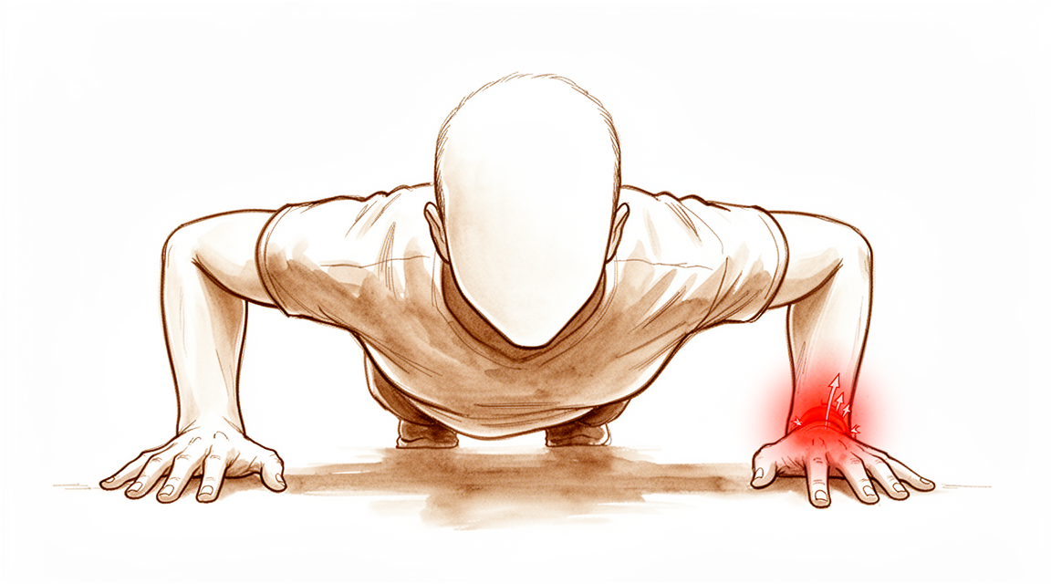
உங்கள் மார்பகத்தின் வெளிப்புறத்தில், உங்கள் குட்டி விரலுக்கு அருகில் வலியை நீங்கள் உணரலாம். இந்த பகுதி அல்னார் பக்கமாக அழைக்கப்படுகிறது. வலி வந்து போகலாம், அல்லது அது நிலையானதாக இருக்கலாம். உங்கள் மார்பகத்தை சில வழிகளில் நகர்த்தும்போது அது பெரும்பாலும் ஒரு ஆழமான வலி அல்லது கூர்மையான புண் போல உணர்கிறது.
உங்கள் முதுகெலும்பை திருகும்போது வலி பொதுவாக மோசமடைகிறது. எளிய அன்றாட பணிகள் கடினமாகிவிடும். நீங்கள் ஒரு கதவு கைப்பிடியைத் திருப்புவது, ஒரு ஜாடி திறப்பது அல்லது திருகுவிளக்கைப் பயன்படுத்துவது ஆகியவற்றில் சிரமப்படலாம். பொருட்களை தூக்குவது, குறிப்பாக உங்கள் உள்ளங்கைகளை கீழே திருப்புவது, அச om கரியத்தைத் தூண்டக்கூடும். உங்கள் முதுகுக்குப் பின்னால் அடைந்து பிராவைப் பிணைப்பது அல்லது சட்டைக்குள் நுழைப்பது கூட வலிக்கக்கூடும். பாதிக்கப்பட்ட பக்கத்தில் தூங்குவது காலையில் வலியை மோசமாக்குகிறது என்று சிலருக்குத் தெரியும்.
பல சந்தர்ப்பங்களில், மணிக்கட்டு நிலையானதாக உணர்கிறது. நீங்கள் எந்த தளர்வையும் அல்லது கிளிக் செய்வதையும் உணரக்கூடாது. இருப்பினும், உங்களுக்கு ஒரு முழுமையான கண்ணீர் இருந்தால், நீங்கள் உறுதியற்ற தன்மையைக் கவனிக்கலாம். இதன் பொருள் உங்கள் மணிக்கட்டு எதிர்பாராத விதமாக வழிநடத்தலாம் அல்லது நகர்த்தலாம். உங்கள் பிங்கி-சைட் மணிக்கட்டு எலும்புடன் இணைப்பை உள்ளடக்கிய குறிப்பிட்ட வகையான கண்ணீர் இது மிகவும் பொதுவானது.
உங்கள் முதுகெலும்பில் சமீபத்தில் எலும்பு முறிவு ஏற்பட்டிருந்தால், உங்கள் சிறிய விரலின் அடிப்பகுதியிலும் உங்களுக்கு வலி ஏற்படலாம். இது பெரும்பாலும் மண்டை ஓடு தலையணையில் ஒரு கண்ணீருடன் நிகழ்கிறது. உங்கள் மணிக்கட்டு சாதாரணமாகவும் நிலையானதாகவும் தோன்றினாலும், அந்த தலையணையின் ஆழமான இழைகளில் உங்களுக்கு இன்னும் ஒரு கண்ணீர் இருக்கலாம். இந்த ஆழமான கண்ணீர் வலியை ஏற்படுத்துகிறது, ஆனால் எப்போதும் நிலையற்ற தன்மையை ஏற்படுத்தாது.
சில நேரங்களில், ஆரம்ப சிகிச்சைக்குப் பிறகும் வலி நீடிக்கும். ஒரு குறிப்பிட்ட வகை கண்ணீருக்கான அறுவை சிகிச்சையை நீங்கள் செய்திருந்தாலும், இன்னும் வலி அல்லது உறுதியற்ற தன்மையை உணர்ந்தால், கண்ணீரின் மற்றொரு பகுதி கவனிக்கப்படாமல் இருக்கலாம். இது அசாதாரணமானது அல்ல. உங்கள் அறுவை சிகிச்சையாளர் உங்கள் வரலாறு மற்றும் அறிகுறிகளை கவனமாகப் பார்த்து, மேலும் சிகிச்சை தேவை என்பதை தீர்மானிப்பார்.
பெரும்பாலான கூர்மையான கண்ணீர் நேரம் மற்றும் ஓய்வில் நன்றாக குணமடைகிறது. இருப்பினும், வலி போகவில்லை என்றால், கண்ணீர் திடீரென காயம் ஏற்படுவதை விட சீரழிவு காரணமாக இருக்கலாம். சீரழிவு கண்ணீர் பெரும்பாலும் வேறுபட்ட மேலாண்மை தேவைப்படுகிறது. உங்கள் அறுவை சிகிச்சையாளர் உங்களுக்கு எந்த வகையான கண்ணீர் உள்ளது என்பதையும் என்ன எதிர்பார்க்கலாம் என்பதையும் புரிந்து கொள்ள உதவும்.
உண்மையில் என்ன நடக்கிறது
உங்கள் மணிக்கட்டு ஒரு சிக்கலான கீறல் ஆகும், அங்கு இரண்டு முன்கை எலும்புகள் சந்திக்கின்றன. ஒரு சிறிய, கடினமான அமைப்பு முக்கோண ஃபைப்ரோ கார்டிலேஜ் வளாகம் என்று அழைக்கப்படுகிறது, அவைகளுக்கு இடையில் அமர்ந்திருக்கிறது. அதை ஒரு அதிர்ச்சி தணிப்பான் மற்றும் ஒரு அடைப்பு என்று நினைத்துப் பாருங்கள். நீங்கள் உங்கள் கையை திருப்பும்போது அல்லது பொருட்களைப் பிடிக்கும்போது எலும்புகளை சீரமைக்க வைக்கிறது.
இந்த சிக்கலானது பல இழைகளை நம்பியுள்ளது, அவை மூட்டு ஒன்றாக இணைந்திருக்கும் வலுவான கயிறுகளைப் போன்றவை. மணிக்கட்டு சுற்றியுள்ள தசைகளும் டைனமிக் ஸ்திரப்படுத்திகளாக செயல்படுகின்றன, இயக்கத்தின் போது எல்லாவற்றையும் சீராக வைத்திருக்க இறுக்கமாகின்றன. உங்கள் முன் காலை சுழற்றும்போது, மூட்டுக்குள் உள்ள தொடர்பு புள்ளிகள் சுமைகளை கையாள சற்று நகர்கின்றன.
ஒரு TFCC காயம் என்பது இந்த அதிர்ச்சி அப்சர்வர் அல்லது அதன் ஆதரவு கயிறுகளில் ஒன்று சேதமடைந்துள்ளது என்பதாகும். இது பெரும்பாலும் ஒரு வீழ்ச்சி அல்லது திடீர் திருப்பத்திற்குப் பிறகு நிகழ்கிறது. சேதம் வலி, கிளிக் செய்தல் அல்லது மணிக்கட்டு நிலையற்றது என்ற உணர்வை ஏற்படுத்தும். நீங்கள் பொருட்களை தூக்க அல்லது திருப்ப முயற்சிக்கும்போது எலும்புகள் இடத்திலிருந்து நழுவுவதைப் போல உணரலாம்.
சில நேரங்களில், எலும்புகள் எவ்வாறு சீரமைக்கப்படுகின்றன என்பதில் காயம் இணைக்கப்பட்டுள்ளது. கதிர் எலும்பு உடைந்து, சற்று வெளியேறினால், அது உங்கள் மணிக்கட்டில் சக்தியை எவ்வாறு பயணிக்கிறது என்பதை மாற்றுகிறது. இந்த மாற்றப்பட்ட சீரமைப்பு TFCC மீது கூடுதல் அழுத்தத்தை ஏற்படுத்துகிறது, இது மூட்டு தனியாக குணமடைய கடினமாக்குகிறது.
பல சந்தர்ப்பங்களில், குறிப்பாக புதிய கண்ணீருடன், உடல் அறுவை சிகிச்சை இல்லாமல் சேதத்தை குணப்படுத்த முடியும். திசுக்கள் காலப்போக்கில் மீண்டும் ஒன்றிணைகின்றன. இருப்பினும், கண்ணீர் முழுமையாக இருந்தால் அல்லது எலும்புகள் தவறாக இருந்தால், மூட்டு வலி அல்லது பலவீனமாக இருக்கலாம். உங்கள் அறுவை சிகிச்சையாளர் குறிப்பிட்ட வகை கண்ணீரைப் பார்க்கிறார், உங்கள் மணிக்கட்டு எவ்வாறு நகர்கிறது என்பதை சரிசெய்ய வேண்டுமா என்பதை தீர்மானிக்க.
சிகிச்சையின் நோக்கம் உங்கள் எலும்புகளுக்கு இடையில் அந்த மென்மையான, நிலையான சறுக்கலை மீட்டெடுப்பதாகும். ஓய்வு, சிகிச்சை அல்லது அறுவை சிகிச்சை மூலம், நோக்கம் வலியைக் குறைத்து, உங்கள் பலத்தை மீட்டெடுக்க உதவுவதாகும்.
நாம் என்ன செய்ய முடியும்
முக்கோண ஃபைப்ரோகார்டிலேஜ் வளாகத்தின் (TFCC) பெரும்பாலான கடுமையான கண்ணீர் அறுவை சிகிச்சை இல்லாமல் தானாகவே குணமடைகிறது. உங்கள் அறுவை சிகிச்சை நிபுணர் உங்கள் வரலாற்றை கவனமாக மதிப்பாய்வு செய்து, காயம் உங்கள் அறிகுறிகளை ஏற்படுத்துகிறது என்பதை உறுதிப்படுத்த உங்கள் மணிக்கட்டை பரிசோதிப்பார். அறுவை சிகிச்சை தேவையா என்பதை தீர்மானிக்க உதவும் வகையில் உங்கள் வலி மற்றும் கடினத்தன்மை எவ்வளவு கடுமையானது என்பதை நீங்கள் அளவிட வேண்டும்.
பிசியோதெரபி இயக்கம் மற்றும் வலிமையை மீட்டெடுப்பதில் கவனம் செலுத்துகிறது. குறிப்பிட்ட மறுவாழ்வு நெறிமுறைகள் மாறுபடும் போது, நோய்க்குறிகளை நிர்வகிப்பது மற்றும் செயல்பாட்டை மேம்படுத்துவதே குறிக்கோள். உங்களுக்கு உடனடி பழுது தேவை என்பதை தீர்மானிக்க உங்கள் அறுவை சிகிச்சை நிபுணர் ரேடியோ அல்னார் அழுத்த சோதனையை மட்டுமே நம்ப மாட்டார். இந்த சோதனை மூட்டு தளர்வான தன்மையை அளவிடுகிறது, ஆனால் சிகிச்சையின் பின்னர் நீங்கள் எவ்வளவு நன்றாக உணருவீர்கள் அல்லது செயல்படுவீர்கள் என்பதை எப்போதும் கணிக்க முடியாது.
கன்சர்வேடிவ் பராமரிப்பு போதுமான நிவாரணத்தை வழங்கவில்லை என்றால், உங்கள் அறுவை சிகிச்சையாளர் மருத்துவ மேலாண்மை பற்றி விவாதிக்கலாம். இது பெரும்பாலும் வீக்கம் மற்றும் அச om கரியத்தை குறைக்க வலி மருந்துகள் மற்றும் அழற்சி எதிர்ப்பு மருந்துகளை உள்ளடக்கியது. சில சந்தர்ப்பங்களில், கோர்டிசோன், ஹைலூரோனிக் அமிலம் அல்லது பிளேட்லெட் நிறைந்த பிளாஸ்மா (பிஆர்பி) போன்ற ஊசிகள் அழற்சியை அமைதிப்படுத்தவும் குணமடையவும் பரிசீலிக்கப்படலாம். இந்த சிகிச்சைகள் தற்காலிக நிவாரணத்தை வழங்குவதை நோக்கமாகக் கொண்டுள்ளன, மேலும் உடல் சிகிச்சையில் முழுமையாக பங்கேற்க உங்களை அனுமதிக்கின்றன. நிவாரணத்தின் காலம் தனிநபருக்கு ஏற்ப மாறுபடும், ஆனால் உங்கள் உடல் குணமடையும் போது இந்த விருப்பங்கள் இடைவெளியைக் குறைக்கலாம்.
அறுவை சிகிச்சை அல்லாத நடவடிக்கைகள் வலியைக் கட்டுப்படுத்தவோ அல்லது ஸ்திரத்தன்மையை மீட்டெடுக்கவோ தவறும் போது அறுவை சிகிச்சை பரிசீலிக்கப்படுகிறது. சேதமடைந்த திசுவை சுத்தம் செய்வதை உள்ளடக்கிய ஆர்த்த்ரோஸ்கோபிக் டிப்ரிட்மென்ட், மத்திய கண்ணீருக்கு பாதுகாப்பானது மற்றும் பயனுள்ளதாக இருக்கும். இது நீடித்த வலி நிவாரணத்தை அளிக்கிறது, வாழ்க்கைத் தரத்தை மேம்படுத்துகிறது, மற்றும் மணிக்கட்டு இயக்கத்தை மீட்டெடுக்கிறது. பல நோயாளிகளுக்கு, இது அதிக திருப்தி மற்றும் நீண்ட கால செயல்பாட்டு நன்மைகளுக்கு வழிவகுக்கிறது, பல ஆண்டுகளுக்குப் பிறகும்.
இந்த நுட்பம் குறைந்தபட்ச ஆக்கிரமிப்பு கருவிகளைப் பயன்படுத்துகிறது. இது கிழிந்த திசுக்களை மீண்டும் இணைக்க உதவுகிறது. இது மணிக்கட்டு இயக்கம், பிடியின் வலிமை மற்றும் வலி அளவுகளில் குறிப்பிடத்தக்க முன்னேற்றங்களை வழங்குகிறது. நாள்பட்ட கண்ணீர் நிகழ்வுகளில், ஒரு சுரங்கப்பாதை டிரான்ஸ்சோஸ் அணுகுமுறை ஸ்திரத்தன்மையை மீட்டெடுக்கவும் குறிப்பிடத்தக்க செயல்பாட்டு மதிப்பீடுகளை வழங்கவும் பயன்படுத்தப்படலாம்.
உங்கள் அறுவை சிகிச்சையானது அறுவை சிகிச்சையற்ற சிகிச்சைக்கு அல்லது சிகிச்சைக்கு பதிலளிக்காத நீடித்த காயங்களுக்கு அறுவை சிகிச்சை டெனெர்வேஷனைக் கருத்தில் கொள்ளும். இந்த நடைமுறை பாதிக்கப்பட்ட பகுதியிலிருந்து வலி சமிக்ஞைகளை குறைக்கிறது. எம்ஆர் ஆர்த்ரோகிராஃபி (எம்ஆர்ஏ) அல்லது மணிக்கட்டு ஆர்த்ரோஸ்கோபி போன்ற மேம்பட்ட இமேஜிங் நோயறிதலை உறுதிப்படுத்த உதவுகிறது, ஏனெனில் நிலையான எம்ஆர்ஐ ஸ்கேன்கள் சில நேரங்களில் நுட்பமான கண்ணீரைத் தவறவிடக்கூடும். உங்கள் அறுவை சிகிச்சையாளர் சந்தேகத்தின் உயர் குறியீட்டைப் பராமரிக்கிறார், குறிப்பாக சாதாரண இமேஜிங் முடிவுகள் இருந்தபோதிலும் உங்களுக்கு அல்னார் பக்க மணிக்கட்டு வலி இருந்தால்.
எதிர்பார்ப்பது என்ன
பெரும்பாலான கடுமையான அட்ஸீ வகுப்பு 1 கண்ணீர் அறுவை சிகிச்சை தேவையில்லாமல் தானாகவே குணமடைகிறது. உங்கள் கண்ணீர் வட்டு மையத்தில் இருந்தால், இவை பெரும்பாலானவை நீண்டகால பின்தொடர்தல் நேரத்தில் குணமடைகின்றன. உங்கள் அறுவை சிகிச்சையாளர் காயத்தை திறம்பட சிகிச்சையளித்தால், பொதுவாக குறைக்கப்பட்ட வலி மற்றும் மேம்பட்ட இயக்கத்தை நீங்கள் எதிர்பார்க்கலாம்.
சிக்கலான கண்ணீருக்காக, உங்கள் அறுவை சிகிச்சை நிபுணர் ஆர்த்த்ரோஸ்கோபிக் இழை-குறிப்பிட்ட பழுதுபார்ப்பை பரிந்துரைக்கலாம். இந்த குறைந்தபட்ச ஆக்கிரமிப்பு நுட்பம் காயத்தை சரிசெய்ய விரிவான காட்சிப்படுத்தலை அனுமதிக்கிறது. மணிக்கட்டு இயக்கம், பிடியின் வலிமை மற்றும் வலி நிலைகளில் குறிப்பிடத்தக்க முன்னேற்றங்களை நீங்கள் காணலாம். இந்த நன்மைகள் குறைந்தபட்சம் 2 ஆண்டு பின்தொடர்தலில் பராமரிக்கப்படுகின்றன. சில சந்தர்ப்பங்களில், சேதமடைந்த திசுக்களை அகற்ற உங்கள் அறுவை சிகிச்சை நிபுணர் உதவியுடன் வெட்டுதல் செய்யலாம். இந்த அணுகுமுறை 19 ஆண்டு பின்தொடர்தலில் கூட திருப்திகரமான முடிவுகளைத் தொடர்ந்து காட்டுகிறது.
நீங்கள் ஒரு முழுமையான கண்ணீர் இருந்தால், நீண்ட கால விளைவு மற்ற கண்ணீர் வகைகளுடன் ஒப்பிடும்போது சற்று குறைவாக இருக்கலாம். இணைந்த வகை 2 கண்ணீர் ஆரம்ப அறுவை சிகிச்சையின் தோல்வியின் அபாயத்தையும் அதிகரிக்கிறது. உங்கள் டி. எஃப். சி. சி காயம் ஒரு தூர கதிரியக்க எலும்பு முறிவுடன் ஏற்பட்டால் இயலாமை விளைவுகள் மோசமாக இருக்கும் என்பதை நீங்கள் அறிந்திருக்க வேண்டும். மணிக்கட்டு நிலையற்ற தன்மையை ஆரம்பத்தில் அங்கீகரிப்பது உங்கள் அறுவை சிகிச்சையாளருக்கு சரியான நேரத்தில் சிகிச்சையளிக்க உதவும். கடுமையான வழக்குகளுக்கு வெற்றிகரமான விகிதங்கள் 80% ஆகும்.
TFCC காயம் உங்கள் நீண்ட கால முடிவை ஒட்டுமொத்தமாக மாற்றுகிறது என்பதற்கு எந்த ஆதாரமும் இல்லை. இருப்பினும், ஒரு வெற்றிகரமான பழுதுபார்ப்புக்குப் பிறகு நீங்கள் வலி அல்லது நிலையற்ற தன்மையைத் தொடர்ந்தால், அங்கீகரிக்கப்படாத அருகிலுள்ள கூறு கண்ணீர் இருக்கலாம். உங்கள் அறுவை சிகிச்சை நிபுணர் சரியான சிகிச்சையை உறுதிப்படுத்த இதைத் தேடுவார். குழந்தைகள் மற்றும் இளம் பருவத்தினர் உட்பட பெரும்பாலான நோயாளிகள் அறுவை சிகிச்சைக்குப் பிறகு சிறந்த செயல்பாட்டு முடிவுகளை அனுபவிக்கிறார்கள். நீங்கள் குணமடையும்போது நீங்கள் மிகவும் நிலையானதாகவும் குறைவான வலியாகவும் உணர வேண்டும்.
யாரையாவது எப்போது பார்க்க வேண்டும்
உங்கள் மணிக்கட்டில் பலவீனம் அல்லது நிலையற்ற தன்மை இருப்பதை நீங்கள் கவனித்தால் ஒரு நிபுணர் மதிப்பாய்வைக் கேளுங்கள். உங்கள் மணிக்கட்டில் பூட்டப்பட்டால் அல்லது வழி கொடுத்தால் ஒரு மருத்துவரைப் பார்க்கவும். உங்கள் தூக்கம் அல்லது வேலையில் அறிகுறிகள் தலையிட்டால் உதவியைப் பெறுங்கள். வலியின் திடீர் மோசமடைவதும் செல்ல ஒரு காரணமாகும். ஆரம்பகால சிகிச்சையின் போது பெரும்பாலான கடுமையான கண்ணீர் நன்றாக குணமடைகிறது. இருப்பினும், ஆரம்ப பழுதுபார்ப்புக்குப் பிறகு நீடித்த வலி அங்கீகரிக்கப்படாத கண்ணீர் இருப்பதைக் குறிக்கலாம். அறுவை சிகிச்சை தேவை என்பதை தீர்மானிக்க கவனமான பரிசோதனை உதவுகிறது. சாதாரண எம்ஆர்ஐ காயத்தை நிராகரிக்காது என்று கருத வேண்டாம். அல்னார் பக்க மணிக்கட்டு வலிக்கு அதிக மருத்துவ சந்தேகம் தேவைப்படுகிறது.
Evidence & references
Overview
- Surgical treatment of TFCC tears and concomitant pathology in the pediatric and adolescent population results in decreased pain, improved motion and stability, and excellent functional outcomes in the majority of patients [1].
- There is no evidence that a TFCC injury influences long-term outcome, though trends suggested complete tears might have inferior outcomes [2].
- Most TFCC disc tears identified at the initial surgery had healed by long-term arthroscopic follow-up [4].
- Arthroscopic-assisted repair techniques provide detailed visualization and facilitate the repair of TFCC injuries and associated pathologies with minimally invasive techniques [5].
- Most acute Atzei class 1 tears spontaneously heal without surgical repair [6].
- In cases of sustained pain and distal radioulnar joint instability even after successful Atzei class 1 repair, an unrecognized proximal component TFCC tear may exist and appropriate treatment for the proximal component should be combined [6].
- Arthroscopic ligament-specific repair of the TFCC offers significant improvements in wrist motion, grip strength, pain, and patient-reported outcomes at a minimum 2-year follow-up [8].
- Sensory innervation data provides an initial step in planning an operative partial TFCC denervation for recalcitrant TFCC IA injuries that fail nonsurgical treatment and possibly also arthroscopic debridement [10].
- Arthroscopic assisted resection for lesions of the TFCC demonstrates persisting satisfactory subjective and functional outcomes at 19 years of follow-up [11].
- There are no outcome studies specifically examining results of TFCC debridement or repair in baseball players [13].
- Further prospective research is recommended to explore whether patient-related or rehabilitation factors influence outcomes following TFCC repair [14].
- There is a current lack of high-quality evidence required to draw firm conclusions on the merits of arthroscopic versus open repair of 1B TFCC tears [25].
- TFCC rehabilitation protocols were poorly reported and varied widely between the included studies [28].
Anatomy & Pathophysiology
- The distal oblique bundle of the interosseous membrane contributes to stabilization of the distal radioulnar joint [29].
- The effectiveness of stabilization provided by the interosseous membrane's distal oblique bundle is altered by the wrist's position and the direction of radial translation [29].
- Fractures of the distal radius interfere with the biomechanical integrity of the wrist [31].
- Distal radius fractures limit range of motion [31].
- Distal radius fractures affect hand muscle strength [31].
- Rotational malalignment of the wrist significantly affects carpal measurements [32].
- Rotational malalignment of the wrist significantly affects distal radial measurements [32].
- Rotational malalignment of the wrist significantly affects distal radioulnar joint measurements [32].
- Distal radioulnar joint arthroplasty considerably alters forearm kinematics [33].
- All wrists exhibit similar loading across the distal ulna regardless of ulnar variance [36].
- Pronation relatively increases loading across the distal ulna [36].
- The flexor carpi ulnaris muscle serves as a dynamic stabilizer of the distal radioulnar joint [37].
- The extensor carpi ulnaris muscle serves as a dynamic stabilizer of the distal radioulnar joint [37].
- The contact area of the distal radioulnar joint increases during wrist flexion [38].
- The contact area of the distal radioulnar joint decreases during wrist extension [38].
- The contact area of the distal radioulnar joint decreases during ulnar deviation [38].
- During forearm rotation, the contact site of the scaphoid on the distal radial articular surface changes minimally [41].
- During forearm rotation, the contact site of the lunate on the distal radial articular surface changes minimally [41].
- Intercarpal kinematic modifications after intercarpal arthrodeses make constant radiocarpal and midcarpal congruence during radioulnar deviation impossible [42].
- A volar ligament-sparing radiocarpal arthrotomy does not cause biomechanical radiocarpal instability [43].
- Injury to the dorsal wrist extrinsic carpal ligaments exacerbates volar radiocarpal instability after intra-articular distal radius fracture [44].
- Disruptions in forearm structures may lead to forearm instability with consequences at the remaining structures [45].
- Radial lengthening beyond the native length is not detrimental to radial loading [46].
- Radial lengthening further reduces distal ulnar loading [46].
- Achieving at least native ulnar variance appears appropriate to restore normal biomechanical loading [46].
- Four-phase grip MRI can demonstrate impairment of the articular disc and longitudinal instability of the distal radioulnar joint simultaneously [47].
- Each ligament stabilizing the distal radioulnar joint contributes to joint stability depending on the direction (palmar or dorsal) [48].
- Each ligament stabilizing the distal radioulnar joint contributes to joint stability depending on different positions of the wrist and forearm [48].
Classification
- The Palmer classification does not completely classify all peripheral TFCC tears, particularly dorsal tears [35].
- The Melone classification system does not predict the presence of TFCC lesions [50].
- Frykman Type VI and VIII fractures show a significantly higher incidence of TFCC tears [50].
- 1B TFCC injury is most common in patients with distal radius fractures (DRF) and concomitant TFCC injury [3].
- The presence of an ulnar styloid fracture associated with distal radius fracture predicts the presence of frequently occurring traumatic TFCC injury and TFCC 1B injury [9].
- A small percentage of arthroscopically validated TFCC injuries involve two tears in one wrist, with the most common pattern being a slit tear coexisting with an ulnar styloid tear [20].
- In more detailed classification of TFCC injuries, such as pc-TFCC tears classified by Atzei's classification, the diagnostic accuracy of MRI remains lower compared to wrist arthroscopy [19].
- Most acute Atzei class 1 tears spontaneously heal without surgical repair [6].
- In cases of sustained pain and distal radioulnar joint instability even after successful Atzei class 1 repair, an unrecognized proximal component TFCC tear may exist [6].
- The authors propose a new 'CUP' classification system for TFCC injuries based on anatomical location (central, ulnar, peripheral) and severity [54].
- The proposed 'CUP' classification aims to address limitations of existing systems like Palmer and Atzei by focusing exclusively on TFCC lesions and providing specific treatment recommendations for each subtype [54].
Clinical Presentation
- TFCC tears in pediatric and adolescent populations present with pain, motion limitations, and stability issues that are improved by surgical treatment [1].
- Complete TFCC tears may have inferior long-term outcomes compared to other tear types, though TFCC injury generally does not influence long-term outcome after distal radial fractures [2].
- TFCC 1B injury is the most common type of TFCC injury in patients with distal radius fractures (DRF) [3].
- Most TFCC disc tears identified at initial surgery heal by long-term arthroscopic follow-up [4].
- Arthroscopic-assisted repair techniques provide detailed visualization for managing TFCC injuries and associated pathologies [5].
- Most acute Atzei class 1 tears (isolated distal component) spontaneously heal without surgical repair [6].
- Sustained pain and distal radioulnar joint (DRUJ) instability after successful Atzei class 1 repair may indicate an unrecognized proximal component TFCC tear [6].
- Careful history and physical examination are required to determine if a TFCC tear is symptomatic and to quantify symptom severity for surgical decision-making [7].
- Arthroscopic ligament-specific repair of TFCC foveal avulsions improves wrist motion, grip strength, pain, and patient-reported outcomes at minimum 2-year follow-up [8].
- The presence of an ulnar styloid fracture associated with DRF predicts the presence of traumatic TFCC injury, specifically TFCC 1B injury [9].
- Operative partial TFCC denervation is a potential planning step for recalcitrant TFCC IA injuries that fail nonsurgical treatment and possibly arthroscopic debridement [10].
- Acute TFCC tears generally provide better results when addressed in the acute phase, while degenerative lesions are typically treated with debridement [12].
- A stable DRUJ upon clinical examination and normal MRI findings do not rule out foveal TFCC injury [17].
- High clinical suspicion is needed when managing patients with ulnar-sided wrist pain to identify foveal TFCC pathology [17].
- Imaging approaches for ulnar-sided wrist pain include discussion of anatomy, pathophysiology, and radiographic appearance of TFCC tears, DRUJ disorders, and ECU tendon disorders [18].
- A small percentage of arthroscopically validated TFCC injuries involve two tears in one wrist ("double lesion") [20].
- The most common "double lesion" pattern is a slit tear coexisting with an ulnar styloid tear [20].
- Central traumatic TFCC lesions can be treated by arthroscopic debridement, resulting in sustained pain relief, improved quality of life (DASH score), improved wrist motion, and high patient satisfaction [21].
- Wrist arthroscopy remains the gold standard for diagnosing TFCC pathologies [22].
- MR arthrography (MRA) is the preferred imaging modality for internal derangements of the wrist due to superior contrast resolution, joint distention, and contrast flow facilitating diagnosis of TFCC and intrinsic ligament lesions [23].
- A negative MRI result is unable to rule out clinically relevant injury to the TFCC, scapholunate (SL) ligament, or lunotriquetral (LT) ligament [24].
- Arthroscopic debridement or repair of wrist TFCC injury provides predictable pain relief and return to play in competitive athletes [51].
- Tears of the TFCC superficial fibers with deep fibers intact present with ulnar-sided wrist pain but without DRUJ instability [52].
Investigations
- TFCC 1B injury is the most common type of TFCC injury in patients with distal radius fractures [3].
- The presence of an ulnar styloid fracture associated with a distal radius fracture predicts the presence of traumatic TFCC injury, specifically TFCC 1B injury [9].
- A high index of clinical suspicion is required for foveal TFCC injury when managing patients with ulnar-sided wrist pain, as normal MRI findings and a stable distal radioulnar joint (DRUJ) on clinical examination do not rule out this pathology [17].
- Advanced imaging techniques like MRI and radiocarpal arthroscopy are well-suited for diagnosing central and distal TFCC tears but may report partial foveal injuries as normal despite functional incompetence [55].
- Partial and complete foveal tears without instability may be missed without a high degree of suspicion [55].
- Wrist arthroscopy remains the gold standard for diagnosing TFCC pathologies [22].
- MR arthrography (MRA) is the preferred modality for imaging internal derangements of the wrist due to superior contrast resolution, joint distention, and contrast flow facilitating the diagnosis of TFCC and intrinsic ligament lesions [23].
- MR arthrography has the potential to become a real alternative to arthroscopy for diagnosing TFCC pathologies [22].
- The diagnostic accuracy of MRI for detailed TFCC classifications, such as pc-TFCC tears by Atzei's classification, is lower compared to wrist arthroscopy [19].
- The sensitivity, specificity, and accuracy of 3.0T wrist MRI for TFCC injury detection are consistently higher than those of 1.5T wrist MRI [56].
- A negative MRI result is unable to rule out the possibility of a clinically relevant TFCC injury [24].
- Negative results of MRI or clinical provocative tests are unable to safely rule out clinically relevant TFCC tears, necessitating further diagnostic evaluation with wrist arthroscopy [57].
- Surgeons should maintain a high degree of suspicion for TFCC-related pathology in young patients with positive provocative clinical examination despite negative MRI findings [59].
- The radioulnar stress test alone cannot be recommended to decide whether to perform an acute repair of the TFCC, as radioulnar laxity and clinical outcome do not correlate after distal radius fracture [27].
- MRI-based modified radioulnar ratio technique showed significant instability parameters and diagnostic accuracy (AuC 0.787) in children and adolescents with arthroscopically-verified TFCC tears [60].
- Pisoscaphoid and radioulnar distances on lateral radiographs did not show significant differences compared to controls in children and adolescents with arthroscopically-verified TFCC tears [60].
- Load-bearing radioulnar (RaUl) measurement is a simple method to diagnose an unstable distal radioulnar joint in patients with TFCC injury [63].
Treatment
Non-Operative Management
- Most acute Atzei class 1 tears (isolated distal component) spontaneously heal without surgical repair [6].
- Careful history and physical examination are required to determine whether a TFCC tear is symptomatic [7].
- Quantifying the severity of symptoms related to TFCC pathology is important to determine whether surgical treatment is necessary [7].
- Using the radioulnar stress test alone to decide whether or not to perform an acute repair of the TFCC cannot be recommended [27].
Arthroscopic Debridement and Resection
- Arthroscopic debridement of central traumatic TFCC lesions is safe and shows sustained pain relief, significantly improved quality of life (DASH score), and wrist motion, resulting in high patient satisfaction [21].
- Arthroscopic assisted resection for lesions of the TFCC demonstrates persisting satisfactory subjective and functional outcomes at 19 years of follow-up [11].
- Arthroscopic debridement of central degenerative TFCC lesions is safe, reliable, and efficacious even for ulnar positive variance [49].
- There are no outcome studies specifically examining results of TFCC debridement or repair in baseball players [13].
Surgical Repair and Reconstruction
- Surgical treatment of TFCC tears and concomitant pathology in the pediatric and adolescent population results in decreased pain, improved motion and stability, and excellent functional outcomes in the majority of patients [1].
- Arthroscopic ligament-specific repair of the TFCC offers significant improvements in wrist motion, grip strength, pain, and patient-reported outcomes at a minimum 2-year follow-up [8].
- An arthroscopic one-tunnel transosseous approach is effective for chronic foveal tears of the TFCC with intact radioulnar ligament remnants, providing pain relief, improved joint stability, and remarkable functional ratings [53].
- Most TFCC disc tears identified at initial surgery had healed by long-term arthroscopic follow-up [4].
- Arthroscopic-assisted repair techniques provide detailed visualization and facilitate the repair of TFCC injuries and associated pathologies with minimally invasive techniques [5].
- In cases of sustained pain and distal radioulnar joint instability even after successful Atzei class 1 repair, an unrecognized proximal component TFCC tear may exist, requiring appropriate treatment for the proximal component to be combined [6].
- A systematic review demonstrates a current lack of high-quality evidence required to draw firm conclusions on the merits of arthroscopic versus open repair of 1B TFCC tears [25].
- Although statistical analysis revealed significant effects of predictor variables on treatment outcomes for arthroscopic TFCC repair, additional larger comprehensive studies are required to confirm these results so as to control for unaccounted patient variables [62].
Surgical Denervation
- Operative partial TFCC denervation is planned for recalcitrant TFCC IA injuries that fail nonsurgical treatment and possibly also arthroscopic debridement [10].
Rehabilitation
- TFCC rehabilitation protocols were poorly reported and varied widely between included studies [28].
Complications
- Complete TFCC tears may have inferior long-term outcomes compared to other tear types [2].
- Coexisting type 2 TFCC tears significantly increase the risk of index surgery failure in patients undergoing arthroscopic repair of peripheral ulnar-side TFCC tears [15].
- An unrecognized proximal component TFCC tear may exist in cases of sustained pain and distal radioulnar joint instability even after successful repair of an isolated Atzei class 1 tear [6].
Recovery
- Surgical treatment of TFCC tears and concomitant pathology in the pediatric and adolescent population results in decreased pain, improved motion and stability, and excellent functional outcomes in the majority of patients [1].
- There is no evidence that a TFCC injury influences long-term outcome, though trends suggested complete tears might have inferior outcomes [2].
- Most TFCC disc tears identified at the initial surgery had healed by long-term arthroscopic follow-up [4].
- Most acute Atzei class 1 tears spontaneously heal without surgical repair [6].
- In cases of sustained pain and distal radioulnar joint instability even after successful Atzei class 1 repair, an unrecognized proximal component TFCC tear may exist and appropriate treatment for the proximal component should be combined [6].
- Arthroscopic ligament-specific repair of the TFCC offers significant improvements in wrist motion, grip strength, pain, and patient-reported outcomes at a minimum 2-year follow-up [8].
- Arthroscopic assisted resection for lesions of the TFCC demonstrates persisting satisfactory subjective and functional outcomes at 19 years of follow-up [11].
- TFCC capsular reattachment could be performed with an arthroscopically assisted technique, providing good long-term results [26].
- Disability outcomes were worse in patients with distal radial fracture where TFCC was injured [58].
- Early recognition of longitudinal radioulnar dissociation can aid in timely treatment and improved outcomes, with success rates around 80% for acute cases [64].
Key Evidence
- [L4] Surgical treatment of TFCC tears and concomitant pathology in the pediatric and adolescent population results in decreased pain, improved motion and stability, and excellent functional outcomes in the majority of patients. [1] (10.1016/j.jhsa.2019.06.019)
- [L2] The study found no evidence that a TFCC injury influences long-term outcome, though trends suggested complete tears might have inferior outcomes. [2] (10.1016/j.jhsa.2012.05.032)
- [L3] 1B TFCC injury is most common in patients with DRF and concomitant TFCC injury. [3] (10.1186/s13018-023-04438-5)
- [L4] Most TFCC disc tears identified at the initial surgery had healed by long-term arthroscopic follow-up. [4] (10.1016/j.jhsa.2012.09.011)
- [L5] Arthroscopic-assisted repair techniques have revolutionized surgical management, providing detailed visualization and facilitating the repair of TFCC injuries and associated pathologies with minimally invasive techniques. [5] (10.1016/j.jhsg.2025.100857)
- [Commentary] Most acute Atzei class 1 tears spontaneously heal without surgical repair; however, in cases of sustained pain and distal radioulnar joint instability even after successful Atzei class 1 repair, an unrecognized proximal component TFCC tear may exist and appropriate treatment for the proximal component should be combined. [6] (10.1016/j.arthro.2022.01.019)
- [L4] Careful history and physical examination are required to determine whether a TFCC tear is symptomatic, and it is important to quantify the severity of symptoms related to TFCC pathology to determine whether surgical treatment is necessary. [7] (10.5435/jaaos-d-20-00998)
- [L4] Arthroscopic ligament-specific repair of the TFCC offers significant improvements in wrist motion, grip strength, pain, and patient-reported outcomes at a minimum 2-year follow-up. [8] (10.1177/1753193420957901)
- [L4] The presence of ulnar styloid fracture associated with distal radius fracture predicted the presence of frequently occurring traumatic triangular fibrocartilage complex injury and TFCC 1B injury. [9] (10.1016/j.arthro.2020.05.025)
- [L5] These results provide an initial step in planning an operative partial TFCC denervation for recalcitrant TFCC IA injuries that fail nonsurgical treatment and possibly also arthroscopic debridement. [10] (10.1016/j.jhsa.2014.03.007)
- [L4] This study demonstrates persisting satisfactory subjective and functional outcomes for patients following arthroscopic assisted resection for lesions of the TFCC at 19 years of follow-up. [11] (10.1177/1558944717708029)
- [L5] The article reviews the anatomy, classification, and management of TFCC injuries, noting that acute tears generally provide better results when addressed in the acute phase, while degenerative lesions are typically treated with debridement. [12] (10.1016/j.hcl.2011.05.013)
- [L5] We know of no outcome studies specifically examining results of TFCC debridement or repair in baseball players. [13] (10.1016/j.hcl.2012.05.019)
- [L4] Further prospective research is recommended to explore whether patient-related or rehabilitation factors influence outcomes following TFCC repair. [14] (10.1016/j.jht.2023.08.009)
- [L4] However, coexisting type 2 TFCC tears significantly increased the risk of index surgery failure in these patients. [15] (10.1016/j.arthro.2020.05.012)
- [L4] Having a stable distal radioulnar joint upon clinical examination and normal MRI findings does not rule out foveal TFCC injury, and a high index of clinical suspicion is needed when managing patients with ulnar sided wrist pain. [17] (10.1177/17531934231206426)
- [L5] The article provides a concise approach to the diagnosis and imaging of ulnar-sided wrist pain, discussing anatomy, pathophysiology, and radiographic appearance of common entities including TFCC tears, DRUJ disorders, and ECU tendon disorders. [18] (10.1016/j.csm.2006.02.008)
- [L4] In more detailed classification of TFCC injuries, such as pc-TFCC tears classified by Atzei's classification, the diagnostic accuracy of MRI remains lower compared to wrist arthroscopy. [19] (10.1186/s12891-023-07140-z)
- [L4] A small percentage of arthroscopically validated TFCC injuries involve two tears in one wrist, with the most common pattern being a slit tear coexisting with an ulnar styloid tear. [20] (10.1177/1753193413479479)
- [L4] Central traumatic TFCC lesions can safely be treated by arthroscopic debridement, showing sustained pain relief, significantly improved quality of life (DASH score) and wrist motion, resulting in high patient satisfaction. [21] (10.1007/s00402-018-2910-4)
- [L5] Wrist arthroscopy remains the 'gold standard' for diagnosing TFCC pathologies despite technical progress in imaging modalities, although MR arthrography may have the potential to become a real alternative in the future. [22] (10.1007/s00402-015-2153-6)
- [L4] Superior contrast resolution, joint distention, and the flow of contrast facilitate the diagnosis of lesions of the TFCC and intrinsic ligaments on contrast-sensitive sequences, making MRA the preferred modality for imaging internal derangements of the wrist. [23] (10.1007/s11552-008-9149-4)
- [L2] A negative result from MRI is unable to rule out the possibility of a clinically relevant injury to the TFCC, SL ligament, or LT ligament of the wrist. [24] (10.1016/j.arthro.2015.04.090)
- [L4] This SR demonstrates a current lack of high-quality evidence required to draw firm conclusions on the merits of arthroscopic versus open repair of 1B TFCC tears. [25] (10.1177/1558944718815244)
- [L5] TFCC capsular reattachment could be performed with an arthroscopically assisted technique, providing good long-term results. [26] (10.1016/j.hcl.2017.06.005)
- [L2] Therefore, using the radioulnar stress test alone to decide whether or not to perform an acute repair of the TFCC cannot be recommended. [27] (10.1177/1753193411403690)
- [L4] TFCC rehabilitation protocols were poorly reported and varied widely between the included studies. [28] (10.1016/j.jht.2021.10.004)
- [L5] However, the wrist's position and the direction of radial translation seem to alter the stabilization's effectiveness. [29] (10.1016/j.otsr.2020.03.041)
- [L3] These results supported the initial hypothesis that a fracture of the distal radius interferes with the biomechanical integrity of the wrist, limiting range of motion and affecting hand muscle strength. [31] (10.1177/1758998315574352)
- [L4] Rotational malalignment of the wrist has significant effects on carpal, distal radial and distal radioulnar joint measurements. [32] (10.1177/1753193408090393)
- [L4] The Aptis distal radioulnar joint arthroplasty considerably alters forearm kinematics, which can have clinical implications. [33] (10.1177/17531934241274142)
- [L4] The Palmer classification does not completely classify all peripheral TFCC tears, particularly dorsal tears. [35] (10.1016/j.arthro.2007.01.026)
- [L5] The results show that all wrists have similar loading across the distal ulna regardless of ulnar variance, while pronation relatively increases loading across the distal ulna. [36] (10.1016/j.jhsa.2014.10.001)
- [L4] The flexor carpi ulnaris and extensor carpi ulnaris muscles serve as dynamic stabilizers of the distal radioulnar joint. [37] (10.1177/17531934231168299)
- [L4] The contact area of the DRUJ increases during wrist flexion and decreases during wrist extension and ulnar deviation. [38] (10.1016/j.jhsa.2015.07.027)
- [L5] During forearm rotation, the contact site of the scaphoid and the lunate on the distal radial articular surface changed minimally. [41] (10.1016/j.jhsa.2013.01.021)
- [L5] The study confirms that constant radiocarpal and midcarpal congruence during radioulnar deviation in normal wrists is no longer possible with intercarpal kinematic modifications after these arthrodeses. [42] (10.1177/17531934231176004)
- [L5] This volar ligament-sparing radiocarpal arthrotomy did not cause biomechanical radiocarpal instability. [43] (10.1016/j.jhsa.2022.08.028)
- [L5] Injury to the dorsal wrist extrinsic carpal ligaments exacerbates volar radiocarpal instability. [44] (10.1177/1558944719851210)
- [L5] Disruptions in any of these structures may lead to forearm instability with consequences at the remaining structures. [45] (10.1016/j.jhsa.2016.10.017)
- [L5] Radial lengthening beyond the native length was not detrimental to radial loading and further reduced distal ulnar loading; achieving at least native ulnar variance seems to be appropriate to restore normal biomechanical loading based on this in vitro study. [46] (10.1016/j.jhsa.2019.03.017)
- [L4] Reconstructed animation from four-phase grip MRI demonstrated impairment of the articular disc and longitudinal instability of the distal radioulnar joint simultaneously and should be of value in investigating dynamic pathophysiology causing ulnar wrist pain. [47] (10.1177/1753193413476979)
- [L4] Arthroscopic debridement of central degenerative TFCC lesions is safe, reliable, and efficacious even for ulnar positive variance. [49] (10.1007/s00402-021-03918-9)
- [L3] The Melone classification system does not predict the presence of TFCC lesions, while Frykman Type VI and VIII fractures show a significantly higher incidence of TFCC tears. [50] (10.1177/1753193408090106)
- [L4] Arthroscopic debridement or repair of wrist TFC injury provides predictable pain relief and return to play in competitive athletes. [51] (10.1177/0363546508325921)
- [L4] Tears of the TFCC superficial fibers with the deep fibers intact present with ulnar-sided wrist pain but without distal radioulnar joint instability. [52] (10.1016/j.jhsa.2011.12.023)
- [L4] This simple arthroscopic one-tunnel transosseous approach is effective for chronic foveal tears of the TFCC with intact radioulnar ligament remnants, providing pain relief, improved joint stability, and remarkable functional ratings. [53] (10.1177/17531934211056854)
- [L5] The authors propose a new 'CUP' classification system for TFCC injuries based on anatomical location (central, ulnar, peripheral) and severity, aiming to address limitations of existing systems like Palmer and Atzei by focusing exclusively on TFCC lesions and providing specific treatment recommendations for each subtype. [54] (10.1177/17531934221121931)
- [L5] Partial and complete foveal tears without instability may be missed without a high degree of suspicion, as advanced imaging techniques like MRI and radiocarpal arthroscopy are well-suited for diagnosing central and distal TFCC tears but may report partial injuries as normal despite functional incompetence. [55] (10.1302/0301-620x.105b1.bjj-2022-0908.r1)
- [L3] The sensitivity, specificity, and accuracy of 3.0T wrist MRI for the TFCC is consistently higher compared with those of 1.5T wrist MRI, suggesting improved capability for detection of TFCC injuries. [56] (10.1016/j.jhsa.2008.02.028)
- [Letter] Negative results of MRI or clinical provocative tests are still unable to safely rule out the possibility of clinically relevant tears to the TFCC and other wrist ligaments, which makes further diagnostic evaluation with wrist arthroscopy necessary. [57] (10.1016/j.arthro.2015.08.001)
- [L2] Disability outcomes were worse in patients with distal radial fracture where TFCC was injured. [58] (10.1016/j.jht.2017.09.002)
- [L2] Surgeons should have a high degree of suspicion for TFCC-related pathology in the setting of positive provocative clinical examination despite negative MRI findings in young patients. [59] (10.1016/j.jhsa.2024.04.015)
- [L3] MRI-based modified radioulnar ratio technique showed significant instability parameters and diagnostic accuracy (AuC 0.787) in children and adolescents with arthroscopically-verified TFCC tears, whereas pisoscaphoid and radioulnar distances on lateral radiographs did not show significant differences compared to controls. [60] (10.1007/s00402-020-03470-y)
- [L5] Although statistical analysis revealed significant effects of predictor variables on treatment outcomes for arthroscopic TFCC repair, additional larger comprehensive studies are required to confirm these results so as to control for unaccounted patient variables. [62] (10.1016/j.arthro.2019.04.022)
- [L2] Load-bearing RaUl measurement is a simple method to diagnose an unstable distal radioulnar joint in patients with TFCC injury. [63] (10.1016/j.jhsa.2022.01.008)
- [L5] Early recognition of longitudinal radioulnar dissociation can aid in timely treatment and improved outcomes, with success rates around 80% for acute cases. [64] (10.1016/j.hcl.2007.01.005)
References
[1] Early Results of Surgical Treatment of Triangular Fibrocartilage Complex Tears in Children and Adolescents. The Journal of Hand Surgery. 2020. DOI: 10.1016/j.jhsa.2019.06.019 [2] The Natural Course of Traumatic Triangular Fibrocartilage Complex Tears in Distal Radial Fractures: A 13–15 Year Follow-up of Arthroscopically Diagnosed but Untreated Injuries. The Journal of Hand Surgery. 2012. DOI: 10.1016/j.jhsa.2012.05.032 [3] Association between imaging parameter changes and triangular fibrocartilage complex injury after distal radius fractures. Journal of Orthopaedic Surgery and Research. 2023. DOI: 10.1186/s13018-023-04438-5 [4] Clinical, Radiographic, and Arthroscopic Outcomes After Ulnar Shortening Osteotomy: A Long-Term Follow-Up Study. The Journal of Hand Surgery. 2012. DOI: 10.1016/j.jhsa.2012.09.011 [5] WITHDRAWN: Arthroscopic-Assisted Repair of the Triangular Fibrocartilage Complex. Journal of Hand Surgery Global Online. 2025. DOI: 10.1016/j.jhsg.2025.100857 [6] Editorial Commentary: Arthroscopic Capsular Repair of Wrist Triangular Fibrocartilage Complex Tears: Beware That Apparent Isolated Atzei Class 1 (Isolated Distal Component) Tears May Include a Proximal Component. Arthroscopy: The Journal of Arthroscopic & Related Surgery. 2022. DOI: 10.1016/j.arthro.2022.01.019 [7] Open and Arthroscopic Triangular Fibrocartilage Complex (TFCC) Repair. Journal of the American Academy of Orthopaedic Surgeons. 2021. DOI: 10.5435/jaaos-d-20-00998 [8] Arthroscopic ligament-specific repair for triangular fibrocartilage complex foveal avulsions: a minimum 2-year follow-up study. Journal of Hand Surgery (European Volume). 2020. DOI: 10.1177/1753193420957901 [9] The Presence and the Location of an Ulnar Styloid Fracture Associated With Distal Radius Fracture Predict the Presence of Triangular Fibrocartilage Complex 1B Injury. Arthroscopy: The Journal of Arthroscopic & Related Surgery. 2020. DOI: 10.1016/j.arthro.2020.05.025 [10] Sensory Innervation of the Triangular Fibrocartilage Complex: A Cadaveric Study. The Journal of Hand Surgery. 2014. DOI: 10.1016/j.jhsa.2014.03.007 [11] Arthroscopic Assisted Resection of Triangular Fibrocartilage Complex Lesions: A 19-Year Follow-up. HAND. 2017. DOI: 10.1177/1558944717708029 [12] Radial Side (1D) Tears. Hand Clinics. 2011. DOI: 10.1016/j.hcl.2011.05.013 [13] Central TFCC Tears in Baseball Players. Hand Clinics. 2012. DOI: 10.1016/j.hcl.2012.05.019 [14] Current rehabilitation recommendations following primary triangular fibrocartilage complex foveal repair surgery: A survey of Australian hand therapists. Journal of Hand Therapy. 2023. DOI: 10.1016/j.jht.2023.08.009 [15] What Is the Effect of the Ulnar‐Plus Variance on the Outcomes of Arthroscopic Repair of the Peripheral Ulnar‐Side Triangular Fibrocartilage Complex Tear?. Arthroscopy. 2020. DOI: 10.1016/j.arthro.2020.05.012 [17] Foveal triangular fibrocartilage complex pathology: a potentially under-recognized injury. Journal of Hand Surgery (European Volume). 2023. DOI: 10.1177/17531934231206426 [18] Imaging of Ulnar-Sided Wrist Pain. Clinics in Sports Medicine. 2006. DOI: 10.1016/j.csm.2006.02.008 [19] Diagnostic value of MRI in traumatic triangular fibrocartilage complex injuries: a retrospective study. BMC Musculoskeletal Disorders. 2024. DOI: 10.1186/s12891-023-07140-z [20] Incidence and diagnosis of ‘the double lesion’ of the triangular fibrocartilage complex. Journal of Hand Surgery (European Volume). 2013. DOI: 10.1177/1753193413479479 [21] Results after arthroscopic treatment of central traumatic lesions of the triangular fibrocartilage complex. Archives of Orthopaedic and Trauma Surgery. 2018. DOI: 10.1007/s00402-018-2910-4 [22] Update TFCC: histology and pathology, classification, examination and diagnostics. Archives of Orthopaedic and Trauma Surgery. 2015. DOI: 10.1007/s00402-015-2153-6 [23] MR Arthrography of the Wrist: Controversies and Concepts. HAND. 2008. DOI: 10.1007/s11552-008-9149-4 [24] Efficacy of Magnetic Resonance Imaging and Clinical Tests in Diagnostics of Wrist Ligament Injuries: A Systematic Review. Arthroscopy. 2015. DOI: 10.1016/j.arthro.2015.04.090 [25] Open Versus Arthroscopic Repair of 1B Ulnar-Sided Triangular Fibrocartilage Complex Tears: A Systematic Review. HAND. 2019. DOI: 10.1177/1558944718815244 [26] Arthroscopic Management of Triangular Fibrocartilage Complex Peripheral Injury. Hand Clinics. 2017. DOI: 10.1016/j.hcl.2017.06.005 [27] Radioulnar laxity and clinical outcome do not correlate after a distal radius fracture. Journal of Hand Surgery (European Volume). 2011. DOI: 10.1177/1753193411403690 [28] Wrist and forearm range of motion commencement time following primary triangular fibrocartilage complex foveal repair surgery: A scoping review. Journal of Hand Therapy. 2023. DOI: 10.1016/j.jht.2021.10.004 [29] Stabilization of the distal radioulnar joint by reconstructing the interosseous membrane's distal oblique bundle: Cadaver study. Orthopaedics & Traumatology: Surgery & Research. 2020. DOI: 10.1016/j.otsr.2020.03.041 [31] Pathomechanics of the wrist following fractures of the distal radius. Hand Therapy. 2015. DOI: 10.1177/1758998315574352 [32] The Effect of Rotational Malalignment on X-rays of the Wrist. Journal of Hand Surgery (European Volume). 2009. DOI: 10.1177/1753193408090393 [33] Performance of the Aptis distal radioulnar joint implant: kinematic and geometric analysis. Journal of Hand Surgery (European Volume). 2024. DOI: 10.1177/17531934241274142 [35] Arthroscopic Repair of Triangular Fibrocartilage Complex Tears. Arthroscopy. 2007. DOI: 10.1016/j.arthro.2007.01.026 [36] Force Variations in the Distal Radius and Ulna: Effect of Ulnar Variance and Forearm Motion. The Journal of Hand Surgery. 2015. DOI: 10.1016/j.jhsa.2014.10.001 [37] Stability of the distal radioulnar joint with and without activation of forearm muscles. Journal of Hand Surgery (European Volume). 2023. DOI: 10.1177/17531934231168299 [38] In Vivo Contact Characteristics of Distal Radioulnar Joint With Malunited Distal Radius During Wrist Motion. The Journal of Hand Surgery. 2015. DOI: 10.1016/j.jhsa.2015.07.027 [41] Changes in Contact Site of the Radiocarpal Joint and Lengths of the Carpal Ligaments in Forearm Rotation: An In Vivo Study. The Journal of Hand Surgery. 2013. DOI: 10.1016/j.jhsa.2013.01.021 [42] The effect of intercarpal arthrodeses on wrist kinematics during radial and ulnar deviation: a cadaveric study using four-dimensional CT. Journal of Hand Surgery (European Volume). 2023. DOI: 10.1177/17531934231176004 [43] Ligament-Sparing Volar Radiocarpal Arthrotomy During Distal Radius Fracture Repair: Biomechanical Implications on Wrist Stability in a Cadaveric Model. The Journal of Hand Surgery. 2024. DOI: 10.1016/j.jhsa.2022.08.028 [44] Dorsal Wrist Extrinsic Carpal Ligament Injury Exacerbates Volar Radiocarpal Instability After Intra-Articular Distal Radius Fracture. HAND. 2019. DOI: 10.1177/1558944719851210 [45] Forearm Instability: Anatomy, Biomechanics, and Treatment Options. The Journal of Hand Surgery. 2017. DOI: 10.1016/j.jhsa.2016.10.017 [46] Effect of Radial Lengthening on Distal Forearm Loading Following Simulated In Vitro Radial Shortening During Simulated Dynamic Wrist Motion. The Journal of Hand Surgery. 2019. DOI: 10.1016/j.jhsa.2019.03.017 [47] Reconstructed animation from four-phase grip MRI of the wrist with ulnar-sided pain. Journal of Hand Surgery (European Volume). 2013. DOI: 10.1177/1753193413476979 [48] 10.1055-s-0037-1601367. n.d.. [49] Long-term outcome after arthroscopic debridement of Palmer type 2C central degenerative lesions of the triangular fibrocartilage complex. Archives of Orthopaedic and Trauma Surgery. 2021. DOI: 10.1007/s00402-021-03918-9 [50] The Value of Plain X-Rays in Predicting TFCC Injury after Distal Radial Fractures. Journal of Hand Surgery (European Volume). 2008. DOI: 10.1177/1753193408090106 [51] Arthroscopic Treatment of Triangular Fibrocartilage Wrist Injuries in the Athlete. The American Journal of Sports Medicine. 2008. DOI: 10.1177/0363546508325921 [52] Arthroscopic Treatment of Peripheral Triangular Fibrocartilage Complex Tears With the Deep Fibers Intact. The Journal of Hand Surgery. 2012. DOI: 10.1016/j.jhsa.2011.12.023 [53] Arthroscopic one-tunnel transosseous reconstruction of chronic triangular fibrocartilage complex foveal tears: outcomes in 12 patients. Journal of Hand Surgery (European Volume). 2021. DOI: 10.1177/17531934211056854 [54] Triangular fibrocartilage complex injuries – limitations of the current classification systems and the proposed new ‘CUP’ classification. Journal of Hand Surgery (European Volume). 2022. DOI: 10.1177/17531934221121931 [55] Structurally intact and functionally incompetent foveal triangular fibrocartilage complex injuries. The Bone & Joint Journal. 2023. DOI: 10.1302/0301-620x.105b1.bjj-2022-0908.r1 [56] Diagnostic Comparison of 1.5 Tesla and 3.0 Tesla Preoperative MRI of the Wrist in Patients With Ulnar-Sided Wrist Pain. The Journal of Hand Surgery. 2008. DOI: 10.1016/j.jhsa.2008.02.028 [57] Regarding “Efficacy of Magnetic Resonance Imaging and Clinical Tests in Diagnostics of Wrist Ligament Injuries: A Systematic Review”. Arthroscopy. 2015. DOI: 10.1016/j.arthro.2015.08.001 [58] Outcomes of surgically treated distal radial fractures with associated triangular fibrocartilage complex injury. Journal of Hand Therapy. 2019. DOI: 10.1016/j.jht.2017.09.002 [59] Etiology and Diagnostic Challenges of Ulnar Wrist Pain in Pediatric and Adolescent Patients. The Journal of Hand Surgery. 2024. DOI: 10.1016/j.jhsa.2024.04.015 [60] Evaluation of radiological instability signs in the distal radioulnar joint in children and adolescents with arthroscopically-verified TFCC tears. Archives of Orthopaedic and Trauma Surgery. 2020. DOI: 10.1007/s00402-020-03470-y [62] Editorial Commentary: Outcomes of Arthroscopic Triangular Fibrocartilage Complex Repair: Can We Predict Who Will Benefit or Not?. Arthroscopy. 2019. DOI: 10.1016/j.arthro.2019.04.022 [63] Load-Bearing Radioulnar Distances to Evaluate an Unstable Distal Radioulnar Joint in Patients With Triangular Fibrocartilage Complex Tears. The Journal of Hand Surgery. 2022. DOI: 10.1016/j.jhsa.2022.01.008 [64] Longitudinal Radioulnar Dissociation: Identification and Treatment of Acute and Chronic Injuries. Hand Clinics. 2007. DOI: 10.1016/j.hcl.2007.01.005


