Chấn thương TFCC
Patients › Wrist
TFCC injuries — pain on the ulnar side of the wrist, often with clicking, and treatment options.
Những gì bạn đang cảm nhận
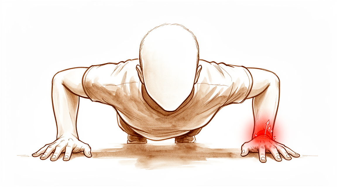
Bạn có thể cảm thấy đau ở mặt ngoài cổ tay, gần ngón út. Khu vực này được gọi là mặt trụ. Cơn đau có thể xuất hiện rồi biến mất, hoặc có thể duy trì liên tục. Cảm giác đau thường giống như một cơn đau âm ỉ sâu hoặc một cơn đau nhói sắc khi bạn cử động cổ tay theo một số cách nhất định.
Cơn đau thường trở nên nghiêm trọng hơn khi bạn xoay cẳng tay. Các hoạt động hàng ngày đơn giản có thể trở nên khó khăn. Bạn có thể gặp khó khăn khi xoay núm cửa, mở lọ hoặc sử dụng tua vít. Nhấc các vật nặng, đặc biệt là khi lòng bàn tay hướng xuống, có thể kích hoạt sự khó chịu. Với với ra sau lưng để cài áo ngực hoặc nhét áo vào quần cũng có thể gây đau. Một số người nhận thấy rằng nằm ngủ ở bên bị ảnh hưởng làm cho cơn đau trở nên nghiêm trọng hơn vào buổi sáng.
Trong nhiều trường hợp, cổ tay cảm thấy ổn định. Bạn có thể không cảm thấy bất kỳ sự lỏng lẻo hoặc tiếng kêu lục cục nào. Tuy nhiên, nếu bạn bị rách hoàn toàn, bạn có thể nhận thấy sự mất ổn định. Điều này có nghĩa là cổ tay của bạn cảm thấy như thể nó có thể bị yếu đi hoặc dịch chuyển một cách bất ngờ. Điều này phổ biến hơn với các loại rách cụ thể liên quan đến sự kết nối với xương cổ tay phía ngón út.
Nếu bạn đã từng bị gãy xương cẳng tay gần đây, bạn cũng có thể cảm thấy đau ở gốc ngón út. Điều này thường xảy ra cùng với sự rách của lớp đệm sụn. Ngay cả khi cổ tay của bạn trông bình thường và cảm thấy ổn định, bạn vẫn có thể bị rách các sợi sâu của lớp đệm đó. Những vết rách sâu này gây đau nhưng không phải lúc nào cũng gây mất ổn định.
Đôi khi, cơn đau vẫn tiếp tục ngay cả sau khi điều trị ban đầu. Nếu bạn đã phẫu thuật cho một loại rách cụ thể nhưng vẫn cảm thấy đau hoặc mất ổn định, có thể có một phần khác của vết rách chưa được xử lý. Điều này không phải là hiếm. Bác sĩ phẫu thuật của bạn sẽ xem xét cẩn thận tiền sử và triệu chứng của bạn để quyết định xem có cần điều trị thêm hay không.
Hầu hết các vết rách cấp tính sẽ lành tốt theo thời gian và nghỉ ngơi. Tuy nhiên, nếu cơn đau không biến mất, có thể là do vết rách thoái hóa thay vì do chấn thương đột ngột. Các vết rách thoái hóa thường đòi hỏi cách quản lý khác. Bác sĩ phẫu thuật của bạn sẽ giúp bạn hiểu loại vết rách bạn có và những gì cần mong đợi.
Những gì thực sự đang xảy ra
Cổ tay của bạn là một khớp bản lề phức tạp nơi hai xương cẳng tay gặp nhau. Một cấu trúc nhỏ, chắc chắn gọi là phức hợp sụn chêm tam giác (TFCC) nằm giữa chúng. Hãy tưởng tượng nó như một bộ giảm xóc và một vòng đệm. Nó giữ cho các xương thẳng hàng khi bạn xoay bàn tay hoặc nắm giữ các vật thể.
Phức hợp này dựa vào nhiều dây chằng, đóng vai trò như những sợi dây thừng chắc chắn giữ khớp lại với nhau. Các cơ xung quanh cổ tay cũng hoạt động như những chất ổn định động, co lại để giữ mọi thứ ổn định trong khi vận động. Khi bạn xoay cẳng tay, các điểm tiếp xúc bên trong khớp dịch chuyển nhẹ để chịu tải.
Chấn thương TFCC có nghĩa là bộ giảm xóc này hoặc một trong những sợi dây hỗ trợ nó bị tổn thương. Điều này thường xảy ra sau khi ngã hoặc xoay đột ngột. Tổn thương có thể gây đau, tiếng kêu lách cách hoặc cảm giác cổ tay không ổn định. Bạn có thể cảm thấy như các xương bị trượt ra khỏi vị trí khi bạn cố gắng nâng hoặc xoay các vật.
Đôi khi, chấn thương có liên quan đến cách các xương thẳng hàng. Nếu xương trụ (radius) bị gãy và liền hơi lệch vị trí, nó sẽ thay đổi cách lực truyền qua cổ tay của bạn. Sự thẳng hàng thay thế này gây thêm căng thẳng lên TFCC, khiến khớp khó tự lành hơn.
Trong nhiều trường hợp, đặc biệt là với các vết rách mới, cơ thể có thể tự lành tổn thương mà không cần phẫu thuật. Các mô sẽ nối lại với nhau theo thời gian. Tuy nhiên, nếu vết rách hoàn toàn hoặc nếu các xương bị lệch vị trí, khớp có thể vẫn đau hoặc yếu. Bác sĩ phẫu thuật của bạn xem xét loại vết rách cụ thể và cách cổ tay của bạn vận động để quyết định có cần sửa chữa hay không.
Mục tiêu của điều trị là khôi phục sự trượt mượt mà, ổn định giữa các xương của bạn. Dù thông qua nghỉ ngơi, trị liệu hay phẫu thuật, mục đích là giảm đau và giúp bạn phục hồi sức mạnh. Hầu hết mọi người đều thấy cải thiện đáng kể về tầm vận động và sức mạnh nắm giữ sau khi được chăm sóc đúng cách.
Những gì chúng tôi có thể làm về vấn đề này
Hầu hết các vết rách cấp tính của phức hợp sụn chêm tam giác (TFCC) sẽ tự lành mà không cần phẫu thuật. Bác sĩ phẫu thuật của bạn sẽ xem xét kỹ lưỡng tiền sử bệnh và thăm khám cổ tay để xác nhận rằng chấn thương là nguyên nhân gây ra các triệu chứng của bạn. Bạn cần định lượng mức độ nghiêm trọng của cơn đau và tình trạng cứng khớp để giúp quyết định có cần phẫu thuật hay không.
Vật lý trị liệu tập trung vào việc khôi phục phạm vi chuyển động và sức mạnh. Mặc dù các phác đồ phục hồi chức năng cụ thể có thể khác nhau, nhưng mục tiêu là kiểm soát các triệu chứng và cải thiện chức năng. Bác sĩ phẫu thuật của bạn sẽ không chỉ dựa vào bài kiểm tra căng thẳng quay-trụ (radioulnar stress test) để quyết định xem bạn có cần sửa chữa ngay lập tức hay không. Bài kiểm tra này đo lường độ chùng khớp, nhưng không phải lúc nào cũng dự đoán chính xác mức độ cảm giác hoặc chức năng của bạn sau khi điều trị.
Nếu chăm sóc bảo tồn không mang lại đủ sự giảm đau, bác sĩ phẫu thuật của bạn có thể thảo luận về các biện pháp quản lý bằng thuốc. Điều này thường bao gồm thuốc giảm đau và thuốc chống viêm để giảm sưng và khó chịu. Trong một số trường hợp, các phương pháp tiêm như cortisone, axit hyaluronic hoặc huyết tương giàu tiểu cầu (PRP) có thể được xem xét để làm dịu tình trạng viêm và hỗ trợ quá trình lành thương. Các phương pháp điều trị này nhằm mục đích cung cấp sự giảm đau tạm thời và cho phép bạn tham gia tích cực hơn vào vật lý trị liệu. Thời gian giảm đau thay đổi tùy theo từng cá nhân, nhưng các lựa chọn này có thể giúp bạn vượt qua giai đoạn chờ đợi trong khi cơ thể bạn tự lành.
Phẫu thuật được xem xét khi các biện pháp không phẫu thuật không kiểm soát được cơn đau hoặc khôi phục được sự ổn định. Cắt lọc nội soi (arthroscopic debridement), liên quan đến việc làm sạch mô bị tổn thương, là an toàn và hiệu quả đối với các vết rách ở vùng trung tâm. Nó mang lại sự giảm đau kéo dài, cải thiện chất lượng cuộc sống và khôi phục phạm vi chuyển động của cổ tay. Đối với nhiều bệnh nhân, điều này dẫn đến mức độ hài lòng cao và những lợi ích chức năng lâu dài, thậm chí nhiều năm sau đó.
Nếu vết rách liên quan đến các điểm bám dây chằng, sửa chữa nội soi (arthroscopic repair) có thể được khuyến nghị. Kỹ thuật này sử dụng các dụng cụ xâm lấn tối thiểu để gắn lại mô bị rách. Nó mang lại những cải thiện đáng kể về phạm vi chuyển động của cổ tay, sức mạnh nắm tay và mức độ đau. Trong các trường hợp vết rách mạn tính, phương pháp xuyên xương một ống (one-tunnel transosseous approach) có thể được sử dụng để khôi phục sự ổn định và mang lại các chỉ số chức năng đáng chú ý.
Bác sĩ phẫu thuật của bạn cũng sẽ xem xét phẫu thuật cắt thần kinh (surgical denervation) đối với các chấn thương dai dẳng không đáp ứng với điều trị không phẫu thuật hoặc cắt lọc. Thủ thuật này làm giảm các tín hiệu đau từ vùng bị ảnh hưởng. Hình ảnh học tiên tiến như chụp cộng hưởng từ khớp cổ tay (MR arthrography - MRA) hoặc nội soi khớp cổ tay giúp xác nhận chẩn đoán, vì các chụp MRI tiêu chuẩn đôi khi có thể bỏ sót các vết rách tinh vi. Bác sĩ phẫu thuật của bạn luôn duy trì mức độ nghi ngờ cao, đặc biệt nếu bạn có đau vùng trụ của cổ tay mặc dù kết quả hình ảnh học bình thường.
Những gì bạn có thể mong đợi
Tiên lượng của bạn phụ thuộc phần lớn vào loại rách mà bạn mắc phải. Hầu hết các rách cấp tính loại Atzei 1 tự lành mà không cần phẫu thuật. Nếu vết rách của bạn nằm ở trung tâm của đĩa, phần lớn các trường hợp này cũng sẽ lành vào thời điểm theo dõi dài hạn. Bạn thường có thể mong đợi giảm đau và cải thiện tầm vận động nếu bác sĩ phẫu thuật điều trị chấn thương một cách hiệu quả.
Đối với các vết rách phức tạp, bác sĩ phẫu thuật của bạn có thể khuyến nghị sửa chữa dây chằng chuyên biệt bằng nội soi khớp. Kỹ thuật xâm lấn tối thiểu này cho phép quan sát chi tiết để sửa chữa chấn thương. Bạn có khả năng sẽ thấy những cải thiện đáng kể về tầm vận động cổ tay, sức mạnh nắm tay và mức độ đau. Những lợi ích này được duy trì trong ít nhất 2 năm theo dõi. Trong một số trường hợp, bác sĩ phẫu thuật của bạn có thể thực hiện cắt bỏ hỗ trợ để loại bỏ mô bị tổn thương. Phương pháp này cho thấy kết quả hài lòng kéo dài ngay cả sau 19 năm theo dõi.
Nếu bạn bị rách hoàn toàn, kết quả dài hạn có thể kém hơn một chút so với các loại rách khác. Các vết rách loại 2 đi kèm cũng làm tăng nguy cơ phẫu thuật ban đầu thất bại. Bạn nên nhận thức rằng kết quả tàn tật sẽ tồi tệ hơn nếu chấn thương TFCC của bạn xảy ra cùng với gãy xương trụ xa. Việc nhận biết sớm tình trạng mất ổn định cổ tay có thể giúp bác sĩ phẫu thuật cung cấp điều trị kịp thời. Tỷ lệ thành công đối với các trường hợp cấp tính là khoảng 80% khi được điều trị kịp thời.
Không có bằng chứng cho thấy chấn thương TFCC làm thay đổi kết quả dài hạn tổng thể của bạn. Tuy nhiên, nếu bạn vẫn còn đau hoặc mất ổn định sau khi sửa chữa thành công, có thể tồn tại một vết rách thành phần gần chưa được phát hiện. Bác sĩ phẫu thuật của bạn sẽ tìm kiếm tình trạng này để đảm bảo điều trị thích hợp. Hầu hết bệnh nhân, bao gồm cả trẻ em và thanh thiếu niên, đều đạt được kết quả chức năng xuất sắc sau khi điều trị phẫu thuật. Bạn sẽ cảm thấy ổn định hơn và giảm đau khi bạn hồi phục.
Khi nào cần gặp bác sĩ
Yêu cầu đánh giá bởi chuyên gia nếu bạn có cơn đau dai dẳng không cải thiện khi nghỉ ngơi. Tìm kiếm sự chăm sóc y tế nếu bạn nhận thấy tình trạng yếu hoặc mất ổn định ở cổ tay. Gặp bác sĩ nếu cổ tay của bạn bị khóa hoặc đột ngột mất lực. Nhận sự trợ giúp nếu các triệu chứng ảnh hưởng đến giấc ngủ hoặc công việc của bạn. Tình trạng đau tăng lên đột ngột cũng là lý do để đi khám. Hầu hết các vết rách cấp tính sẽ lành tốt khi được điều trị sớm. Tuy nhiên, tình trạng đau kéo dài sau khi sửa chữa ban đầu có thể cho thấy sự tồn tại của một vết rách chưa được phát hiện. Việc khám xét cẩn thận giúp xác định xem có cần phẫu thuật hay không. Đừng giả định rằng kết quả MRI bình thường loại trừ chấn thương. Cần duy trì mức độ nghi ngờ lâm sàng cao đối với cơn đau ở mặt trụ của cổ tay.
Evidence & references
Overview
- Surgical treatment of TFCC tears and concomitant pathology in the pediatric and adolescent population results in decreased pain, improved motion and stability, and excellent functional outcomes in the majority of patients [1].
- There is no evidence that a TFCC injury influences long-term outcome, though trends suggested complete tears might have inferior outcomes [2].
- Most TFCC disc tears identified at the initial surgery had healed by long-term arthroscopic follow-up [4].
- Arthroscopic-assisted repair techniques provide detailed visualization and facilitate the repair of TFCC injuries and associated pathologies with minimally invasive techniques [5].
- Most acute Atzei class 1 tears spontaneously heal without surgical repair [6].
- In cases of sustained pain and distal radioulnar joint instability even after successful Atzei class 1 repair, an unrecognized proximal component TFCC tear may exist and appropriate treatment for the proximal component should be combined [6].
- Arthroscopic ligament-specific repair of the TFCC offers significant improvements in wrist motion, grip strength, pain, and patient-reported outcomes at a minimum 2-year follow-up [8].
- Sensory innervation data provides an initial step in planning an operative partial TFCC denervation for recalcitrant TFCC IA injuries that fail nonsurgical treatment and possibly also arthroscopic debridement [10].
- Arthroscopic assisted resection for lesions of the TFCC demonstrates persisting satisfactory subjective and functional outcomes at 19 years of follow-up [11].
- There are no outcome studies specifically examining results of TFCC debridement or repair in baseball players [13].
- Further prospective research is recommended to explore whether patient-related or rehabilitation factors influence outcomes following TFCC repair [14].
- There is a current lack of high-quality evidence required to draw firm conclusions on the merits of arthroscopic versus open repair of 1B TFCC tears [25].
- TFCC rehabilitation protocols were poorly reported and varied widely between the included studies [28].
Anatomy & Pathophysiology
- The distal oblique bundle of the interosseous membrane contributes to stabilization of the distal radioulnar joint [29].
- The effectiveness of stabilization provided by the interosseous membrane's distal oblique bundle is altered by the wrist's position and the direction of radial translation [29].
- Fractures of the distal radius interfere with the biomechanical integrity of the wrist [31].
- Distal radius fractures limit range of motion [31].
- Distal radius fractures affect hand muscle strength [31].
- Rotational malalignment of the wrist significantly affects carpal measurements [32].
- Rotational malalignment of the wrist significantly affects distal radial measurements [32].
- Rotational malalignment of the wrist significantly affects distal radioulnar joint measurements [32].
- Distal radioulnar joint arthroplasty considerably alters forearm kinematics [33].
- All wrists exhibit similar loading across the distal ulna regardless of ulnar variance [36].
- Pronation relatively increases loading across the distal ulna [36].
- The flexor carpi ulnaris muscle serves as a dynamic stabilizer of the distal radioulnar joint [37].
- The extensor carpi ulnaris muscle serves as a dynamic stabilizer of the distal radioulnar joint [37].
- The contact area of the distal radioulnar joint increases during wrist flexion [38].
- The contact area of the distal radioulnar joint decreases during wrist extension [38].
- The contact area of the distal radioulnar joint decreases during ulnar deviation [38].
- During forearm rotation, the contact site of the scaphoid on the distal radial articular surface changes minimally [41].
- During forearm rotation, the contact site of the lunate on the distal radial articular surface changes minimally [41].
- Intercarpal kinematic modifications after intercarpal arthrodeses make constant radiocarpal and midcarpal congruence during radioulnar deviation impossible [42].
- A volar ligament-sparing radiocarpal arthrotomy does not cause biomechanical radiocarpal instability [43].
- Injury to the dorsal wrist extrinsic carpal ligaments exacerbates volar radiocarpal instability after intra-articular distal radius fracture [44].
- Disruptions in forearm structures may lead to forearm instability with consequences at the remaining structures [45].
- Radial lengthening beyond the native length is not detrimental to radial loading [46].
- Radial lengthening further reduces distal ulnar loading [46].
- Achieving at least native ulnar variance appears appropriate to restore normal biomechanical loading [46].
- Four-phase grip MRI can demonstrate impairment of the articular disc and longitudinal instability of the distal radioulnar joint simultaneously [47].
- Each ligament stabilizing the distal radioulnar joint contributes to joint stability depending on the direction (palmar or dorsal) [48].
- Each ligament stabilizing the distal radioulnar joint contributes to joint stability depending on different positions of the wrist and forearm [48].
Classification
- The Palmer classification does not completely classify all peripheral TFCC tears, particularly dorsal tears [35].
- The Melone classification system does not predict the presence of TFCC lesions [50].
- Frykman Type VI and VIII fractures show a significantly higher incidence of TFCC tears [50].
- 1B TFCC injury is most common in patients with distal radius fractures (DRF) and concomitant TFCC injury [3].
- The presence of an ulnar styloid fracture associated with distal radius fracture predicts the presence of frequently occurring traumatic TFCC injury and TFCC 1B injury [9].
- A small percentage of arthroscopically validated TFCC injuries involve two tears in one wrist, with the most common pattern being a slit tear coexisting with an ulnar styloid tear [20].
- In more detailed classification of TFCC injuries, such as pc-TFCC tears classified by Atzei's classification, the diagnostic accuracy of MRI remains lower compared to wrist arthroscopy [19].
- Most acute Atzei class 1 tears spontaneously heal without surgical repair [6].
- In cases of sustained pain and distal radioulnar joint instability even after successful Atzei class 1 repair, an unrecognized proximal component TFCC tear may exist [6].
- The authors propose a new 'CUP' classification system for TFCC injuries based on anatomical location (central, ulnar, peripheral) and severity [54].
- The proposed 'CUP' classification aims to address limitations of existing systems like Palmer and Atzei by focusing exclusively on TFCC lesions and providing specific treatment recommendations for each subtype [54].
Clinical Presentation
- TFCC tears in pediatric and adolescent populations present with pain, motion limitations, and stability issues that are improved by surgical treatment [1].
- Complete TFCC tears may have inferior long-term outcomes compared to other tear types, though TFCC injury generally does not influence long-term outcome after distal radial fractures [2].
- TFCC 1B injury is the most common type of TFCC injury in patients with distal radius fractures (DRF) [3].
- Most TFCC disc tears identified at initial surgery heal by long-term arthroscopic follow-up [4].
- Arthroscopic-assisted repair techniques provide detailed visualization for managing TFCC injuries and associated pathologies [5].
- Most acute Atzei class 1 tears (isolated distal component) spontaneously heal without surgical repair [6].
- Sustained pain and distal radioulnar joint (DRUJ) instability after successful Atzei class 1 repair may indicate an unrecognized proximal component TFCC tear [6].
- Careful history and physical examination are required to determine if a TFCC tear is symptomatic and to quantify symptom severity for surgical decision-making [7].
- Arthroscopic ligament-specific repair of TFCC foveal avulsions improves wrist motion, grip strength, pain, and patient-reported outcomes at minimum 2-year follow-up [8].
- The presence of an ulnar styloid fracture associated with DRF predicts the presence of traumatic TFCC injury, specifically TFCC 1B injury [9].
- Operative partial TFCC denervation is a potential planning step for recalcitrant TFCC IA injuries that fail nonsurgical treatment and possibly arthroscopic debridement [10].
- Acute TFCC tears generally provide better results when addressed in the acute phase, while degenerative lesions are typically treated with debridement [12].
- A stable DRUJ upon clinical examination and normal MRI findings do not rule out foveal TFCC injury [17].
- High clinical suspicion is needed when managing patients with ulnar-sided wrist pain to identify foveal TFCC pathology [17].
- Imaging approaches for ulnar-sided wrist pain include discussion of anatomy, pathophysiology, and radiographic appearance of TFCC tears, DRUJ disorders, and ECU tendon disorders [18].
- A small percentage of arthroscopically validated TFCC injuries involve two tears in one wrist ("double lesion") [20].
- The most common "double lesion" pattern is a slit tear coexisting with an ulnar styloid tear [20].
- Central traumatic TFCC lesions can be treated by arthroscopic debridement, resulting in sustained pain relief, improved quality of life (DASH score), improved wrist motion, and high patient satisfaction [21].
- Wrist arthroscopy remains the gold standard for diagnosing TFCC pathologies [22].
- MR arthrography (MRA) is the preferred imaging modality for internal derangements of the wrist due to superior contrast resolution, joint distention, and contrast flow facilitating diagnosis of TFCC and intrinsic ligament lesions [23].
- A negative MRI result is unable to rule out clinically relevant injury to the TFCC, scapholunate (SL) ligament, or lunotriquetral (LT) ligament [24].
- Arthroscopic debridement or repair of wrist TFCC injury provides predictable pain relief and return to play in competitive athletes [51].
- Tears of the TFCC superficial fibers with deep fibers intact present with ulnar-sided wrist pain but without DRUJ instability [52].
Investigations
- TFCC 1B injury is the most common type of TFCC injury in patients with distal radius fractures [3].
- The presence of an ulnar styloid fracture associated with a distal radius fracture predicts the presence of traumatic TFCC injury, specifically TFCC 1B injury [9].
- A high index of clinical suspicion is required for foveal TFCC injury when managing patients with ulnar-sided wrist pain, as normal MRI findings and a stable distal radioulnar joint (DRUJ) on clinical examination do not rule out this pathology [17].
- Advanced imaging techniques like MRI and radiocarpal arthroscopy are well-suited for diagnosing central and distal TFCC tears but may report partial foveal injuries as normal despite functional incompetence [55].
- Partial and complete foveal tears without instability may be missed without a high degree of suspicion [55].
- Wrist arthroscopy remains the gold standard for diagnosing TFCC pathologies [22].
- MR arthrography (MRA) is the preferred modality for imaging internal derangements of the wrist due to superior contrast resolution, joint distention, and contrast flow facilitating the diagnosis of TFCC and intrinsic ligament lesions [23].
- MR arthrography has the potential to become a real alternative to arthroscopy for diagnosing TFCC pathologies [22].
- The diagnostic accuracy of MRI for detailed TFCC classifications, such as pc-TFCC tears by Atzei's classification, is lower compared to wrist arthroscopy [19].
- The sensitivity, specificity, and accuracy of 3.0T wrist MRI for TFCC injury detection are consistently higher than those of 1.5T wrist MRI [56].
- A negative MRI result is unable to rule out the possibility of a clinically relevant TFCC injury [24].
- Negative results of MRI or clinical provocative tests are unable to safely rule out clinically relevant TFCC tears, necessitating further diagnostic evaluation with wrist arthroscopy [57].
- Surgeons should maintain a high degree of suspicion for TFCC-related pathology in young patients with positive provocative clinical examination despite negative MRI findings [59].
- The radioulnar stress test alone cannot be recommended to decide whether to perform an acute repair of the TFCC, as radioulnar laxity and clinical outcome do not correlate after distal radius fracture [27].
- MRI-based modified radioulnar ratio technique showed significant instability parameters and diagnostic accuracy (AuC 0.787) in children and adolescents with arthroscopically-verified TFCC tears [60].
- Pisoscaphoid and radioulnar distances on lateral radiographs did not show significant differences compared to controls in children and adolescents with arthroscopically-verified TFCC tears [60].
- Load-bearing radioulnar (RaUl) measurement is a simple method to diagnose an unstable distal radioulnar joint in patients with TFCC injury [63].
Treatment
Non-Operative Management
- Most acute Atzei class 1 tears (isolated distal component) spontaneously heal without surgical repair [6].
- Careful history and physical examination are required to determine whether a TFCC tear is symptomatic [7].
- Quantifying the severity of symptoms related to TFCC pathology is important to determine whether surgical treatment is necessary [7].
- Using the radioulnar stress test alone to decide whether or not to perform an acute repair of the TFCC cannot be recommended [27].
Arthroscopic Debridement and Resection
- Arthroscopic debridement of central traumatic TFCC lesions is safe and shows sustained pain relief, significantly improved quality of life (DASH score), and wrist motion, resulting in high patient satisfaction [21].
- Arthroscopic assisted resection for lesions of the TFCC demonstrates persisting satisfactory subjective and functional outcomes at 19 years of follow-up [11].
- Arthroscopic debridement of central degenerative TFCC lesions is safe, reliable, and efficacious even for ulnar positive variance [49].
- There are no outcome studies specifically examining results of TFCC debridement or repair in baseball players [13].
Surgical Repair and Reconstruction
- Surgical treatment of TFCC tears and concomitant pathology in the pediatric and adolescent population results in decreased pain, improved motion and stability, and excellent functional outcomes in the majority of patients [1].
- Arthroscopic ligament-specific repair of the TFCC offers significant improvements in wrist motion, grip strength, pain, and patient-reported outcomes at a minimum 2-year follow-up [8].
- An arthroscopic one-tunnel transosseous approach is effective for chronic foveal tears of the TFCC with intact radioulnar ligament remnants, providing pain relief, improved joint stability, and remarkable functional ratings [53].
- Most TFCC disc tears identified at initial surgery had healed by long-term arthroscopic follow-up [4].
- Arthroscopic-assisted repair techniques provide detailed visualization and facilitate the repair of TFCC injuries and associated pathologies with minimally invasive techniques [5].
- In cases of sustained pain and distal radioulnar joint instability even after successful Atzei class 1 repair, an unrecognized proximal component TFCC tear may exist, requiring appropriate treatment for the proximal component to be combined [6].
- A systematic review demonstrates a current lack of high-quality evidence required to draw firm conclusions on the merits of arthroscopic versus open repair of 1B TFCC tears [25].
- Although statistical analysis revealed significant effects of predictor variables on treatment outcomes for arthroscopic TFCC repair, additional larger comprehensive studies are required to confirm these results so as to control for unaccounted patient variables [62].
Surgical Denervation
- Operative partial TFCC denervation is planned for recalcitrant TFCC IA injuries that fail nonsurgical treatment and possibly also arthroscopic debridement [10].
Rehabilitation
- TFCC rehabilitation protocols were poorly reported and varied widely between included studies [28].
Complications
- Complete TFCC tears may have inferior long-term outcomes compared to other tear types [2].
- Coexisting type 2 TFCC tears significantly increase the risk of index surgery failure in patients undergoing arthroscopic repair of peripheral ulnar-side TFCC tears [15].
- An unrecognized proximal component TFCC tear may exist in cases of sustained pain and distal radioulnar joint instability even after successful repair of an isolated Atzei class 1 tear [6].
Recovery
- Surgical treatment of TFCC tears and concomitant pathology in the pediatric and adolescent population results in decreased pain, improved motion and stability, and excellent functional outcomes in the majority of patients [1].
- There is no evidence that a TFCC injury influences long-term outcome, though trends suggested complete tears might have inferior outcomes [2].
- Most TFCC disc tears identified at the initial surgery had healed by long-term arthroscopic follow-up [4].
- Most acute Atzei class 1 tears spontaneously heal without surgical repair [6].
- In cases of sustained pain and distal radioulnar joint instability even after successful Atzei class 1 repair, an unrecognized proximal component TFCC tear may exist and appropriate treatment for the proximal component should be combined [6].
- Arthroscopic ligament-specific repair of the TFCC offers significant improvements in wrist motion, grip strength, pain, and patient-reported outcomes at a minimum 2-year follow-up [8].
- Arthroscopic assisted resection for lesions of the TFCC demonstrates persisting satisfactory subjective and functional outcomes at 19 years of follow-up [11].
- TFCC capsular reattachment could be performed with an arthroscopically assisted technique, providing good long-term results [26].
- Disability outcomes were worse in patients with distal radial fracture where TFCC was injured [58].
- Early recognition of longitudinal radioulnar dissociation can aid in timely treatment and improved outcomes, with success rates around 80% for acute cases [64].
Key Evidence
- [L4] Surgical treatment of TFCC tears and concomitant pathology in the pediatric and adolescent population results in decreased pain, improved motion and stability, and excellent functional outcomes in the majority of patients. [1] (10.1016/j.jhsa.2019.06.019)
- [L2] The study found no evidence that a TFCC injury influences long-term outcome, though trends suggested complete tears might have inferior outcomes. [2] (10.1016/j.jhsa.2012.05.032)
- [L3] 1B TFCC injury is most common in patients with DRF and concomitant TFCC injury. [3] (10.1186/s13018-023-04438-5)
- [L4] Most TFCC disc tears identified at the initial surgery had healed by long-term arthroscopic follow-up. [4] (10.1016/j.jhsa.2012.09.011)
- [L5] Arthroscopic-assisted repair techniques have revolutionized surgical management, providing detailed visualization and facilitating the repair of TFCC injuries and associated pathologies with minimally invasive techniques. [5] (10.1016/j.jhsg.2025.100857)
- [Commentary] Most acute Atzei class 1 tears spontaneously heal without surgical repair; however, in cases of sustained pain and distal radioulnar joint instability even after successful Atzei class 1 repair, an unrecognized proximal component TFCC tear may exist and appropriate treatment for the proximal component should be combined. [6] (10.1016/j.arthro.2022.01.019)
- [L4] Careful history and physical examination are required to determine whether a TFCC tear is symptomatic, and it is important to quantify the severity of symptoms related to TFCC pathology to determine whether surgical treatment is necessary. [7] (10.5435/jaaos-d-20-00998)
- [L4] Arthroscopic ligament-specific repair of the TFCC offers significant improvements in wrist motion, grip strength, pain, and patient-reported outcomes at a minimum 2-year follow-up. [8] (10.1177/1753193420957901)
- [L4] The presence of ulnar styloid fracture associated with distal radius fracture predicted the presence of frequently occurring traumatic triangular fibrocartilage complex injury and TFCC 1B injury. [9] (10.1016/j.arthro.2020.05.025)
- [L5] These results provide an initial step in planning an operative partial TFCC denervation for recalcitrant TFCC IA injuries that fail nonsurgical treatment and possibly also arthroscopic debridement. [10] (10.1016/j.jhsa.2014.03.007)
- [L4] This study demonstrates persisting satisfactory subjective and functional outcomes for patients following arthroscopic assisted resection for lesions of the TFCC at 19 years of follow-up. [11] (10.1177/1558944717708029)
- [L5] The article reviews the anatomy, classification, and management of TFCC injuries, noting that acute tears generally provide better results when addressed in the acute phase, while degenerative lesions are typically treated with debridement. [12] (10.1016/j.hcl.2011.05.013)
- [L5] We know of no outcome studies specifically examining results of TFCC debridement or repair in baseball players. [13] (10.1016/j.hcl.2012.05.019)
- [L4] Further prospective research is recommended to explore whether patient-related or rehabilitation factors influence outcomes following TFCC repair. [14] (10.1016/j.jht.2023.08.009)
- [L4] However, coexisting type 2 TFCC tears significantly increased the risk of index surgery failure in these patients. [15] (10.1016/j.arthro.2020.05.012)
- [L4] Having a stable distal radioulnar joint upon clinical examination and normal MRI findings does not rule out foveal TFCC injury, and a high index of clinical suspicion is needed when managing patients with ulnar sided wrist pain. [17] (10.1177/17531934231206426)
- [L5] The article provides a concise approach to the diagnosis and imaging of ulnar-sided wrist pain, discussing anatomy, pathophysiology, and radiographic appearance of common entities including TFCC tears, DRUJ disorders, and ECU tendon disorders. [18] (10.1016/j.csm.2006.02.008)
- [L4] In more detailed classification of TFCC injuries, such as pc-TFCC tears classified by Atzei's classification, the diagnostic accuracy of MRI remains lower compared to wrist arthroscopy. [19] (10.1186/s12891-023-07140-z)
- [L4] A small percentage of arthroscopically validated TFCC injuries involve two tears in one wrist, with the most common pattern being a slit tear coexisting with an ulnar styloid tear. [20] (10.1177/1753193413479479)
- [L4] Central traumatic TFCC lesions can safely be treated by arthroscopic debridement, showing sustained pain relief, significantly improved quality of life (DASH score) and wrist motion, resulting in high patient satisfaction. [21] (10.1007/s00402-018-2910-4)
- [L5] Wrist arthroscopy remains the 'gold standard' for diagnosing TFCC pathologies despite technical progress in imaging modalities, although MR arthrography may have the potential to become a real alternative in the future. [22] (10.1007/s00402-015-2153-6)
- [L4] Superior contrast resolution, joint distention, and the flow of contrast facilitate the diagnosis of lesions of the TFCC and intrinsic ligaments on contrast-sensitive sequences, making MRA the preferred modality for imaging internal derangements of the wrist. [23] (10.1007/s11552-008-9149-4)
- [L2] A negative result from MRI is unable to rule out the possibility of a clinically relevant injury to the TFCC, SL ligament, or LT ligament of the wrist. [24] (10.1016/j.arthro.2015.04.090)
- [L4] This SR demonstrates a current lack of high-quality evidence required to draw firm conclusions on the merits of arthroscopic versus open repair of 1B TFCC tears. [25] (10.1177/1558944718815244)
- [L5] TFCC capsular reattachment could be performed with an arthroscopically assisted technique, providing good long-term results. [26] (10.1016/j.hcl.2017.06.005)
- [L2] Therefore, using the radioulnar stress test alone to decide whether or not to perform an acute repair of the TFCC cannot be recommended. [27] (10.1177/1753193411403690)
- [L4] TFCC rehabilitation protocols were poorly reported and varied widely between the included studies. [28] (10.1016/j.jht.2021.10.004)
- [L5] However, the wrist's position and the direction of radial translation seem to alter the stabilization's effectiveness. [29] (10.1016/j.otsr.2020.03.041)
- [L3] These results supported the initial hypothesis that a fracture of the distal radius interferes with the biomechanical integrity of the wrist, limiting range of motion and affecting hand muscle strength. [31] (10.1177/1758998315574352)
- [L4] Rotational malalignment of the wrist has significant effects on carpal, distal radial and distal radioulnar joint measurements. [32] (10.1177/1753193408090393)
- [L4] The Aptis distal radioulnar joint arthroplasty considerably alters forearm kinematics, which can have clinical implications. [33] (10.1177/17531934241274142)
- [L4] The Palmer classification does not completely classify all peripheral TFCC tears, particularly dorsal tears. [35] (10.1016/j.arthro.2007.01.026)
- [L5] The results show that all wrists have similar loading across the distal ulna regardless of ulnar variance, while pronation relatively increases loading across the distal ulna. [36] (10.1016/j.jhsa.2014.10.001)
- [L4] The flexor carpi ulnaris and extensor carpi ulnaris muscles serve as dynamic stabilizers of the distal radioulnar joint. [37] (10.1177/17531934231168299)
- [L4] The contact area of the DRUJ increases during wrist flexion and decreases during wrist extension and ulnar deviation. [38] (10.1016/j.jhsa.2015.07.027)
- [L5] During forearm rotation, the contact site of the scaphoid and the lunate on the distal radial articular surface changed minimally. [41] (10.1016/j.jhsa.2013.01.021)
- [L5] The study confirms that constant radiocarpal and midcarpal congruence during radioulnar deviation in normal wrists is no longer possible with intercarpal kinematic modifications after these arthrodeses. [42] (10.1177/17531934231176004)
- [L5] This volar ligament-sparing radiocarpal arthrotomy did not cause biomechanical radiocarpal instability. [43] (10.1016/j.jhsa.2022.08.028)
- [L5] Injury to the dorsal wrist extrinsic carpal ligaments exacerbates volar radiocarpal instability. [44] (10.1177/1558944719851210)
- [L5] Disruptions in any of these structures may lead to forearm instability with consequences at the remaining structures. [45] (10.1016/j.jhsa.2016.10.017)
- [L5] Radial lengthening beyond the native length was not detrimental to radial loading and further reduced distal ulnar loading; achieving at least native ulnar variance seems to be appropriate to restore normal biomechanical loading based on this in vitro study. [46] (10.1016/j.jhsa.2019.03.017)
- [L4] Reconstructed animation from four-phase grip MRI demonstrated impairment of the articular disc and longitudinal instability of the distal radioulnar joint simultaneously and should be of value in investigating dynamic pathophysiology causing ulnar wrist pain. [47] (10.1177/1753193413476979)
- [L4] Arthroscopic debridement of central degenerative TFCC lesions is safe, reliable, and efficacious even for ulnar positive variance. [49] (10.1007/s00402-021-03918-9)
- [L3] The Melone classification system does not predict the presence of TFCC lesions, while Frykman Type VI and VIII fractures show a significantly higher incidence of TFCC tears. [50] (10.1177/1753193408090106)
- [L4] Arthroscopic debridement or repair of wrist TFC injury provides predictable pain relief and return to play in competitive athletes. [51] (10.1177/0363546508325921)
- [L4] Tears of the TFCC superficial fibers with the deep fibers intact present with ulnar-sided wrist pain but without distal radioulnar joint instability. [52] (10.1016/j.jhsa.2011.12.023)
- [L4] This simple arthroscopic one-tunnel transosseous approach is effective for chronic foveal tears of the TFCC with intact radioulnar ligament remnants, providing pain relief, improved joint stability, and remarkable functional ratings. [53] (10.1177/17531934211056854)
- [L5] The authors propose a new 'CUP' classification system for TFCC injuries based on anatomical location (central, ulnar, peripheral) and severity, aiming to address limitations of existing systems like Palmer and Atzei by focusing exclusively on TFCC lesions and providing specific treatment recommendations for each subtype. [54] (10.1177/17531934221121931)
- [L5] Partial and complete foveal tears without instability may be missed without a high degree of suspicion, as advanced imaging techniques like MRI and radiocarpal arthroscopy are well-suited for diagnosing central and distal TFCC tears but may report partial injuries as normal despite functional incompetence. [55] (10.1302/0301-620x.105b1.bjj-2022-0908.r1)
- [L3] The sensitivity, specificity, and accuracy of 3.0T wrist MRI for the TFCC is consistently higher compared with those of 1.5T wrist MRI, suggesting improved capability for detection of TFCC injuries. [56] (10.1016/j.jhsa.2008.02.028)
- [Letter] Negative results of MRI or clinical provocative tests are still unable to safely rule out the possibility of clinically relevant tears to the TFCC and other wrist ligaments, which makes further diagnostic evaluation with wrist arthroscopy necessary. [57] (10.1016/j.arthro.2015.08.001)
- [L2] Disability outcomes were worse in patients with distal radial fracture where TFCC was injured. [58] (10.1016/j.jht.2017.09.002)
- [L2] Surgeons should have a high degree of suspicion for TFCC-related pathology in the setting of positive provocative clinical examination despite negative MRI findings in young patients. [59] (10.1016/j.jhsa.2024.04.015)
- [L3] MRI-based modified radioulnar ratio technique showed significant instability parameters and diagnostic accuracy (AuC 0.787) in children and adolescents with arthroscopically-verified TFCC tears, whereas pisoscaphoid and radioulnar distances on lateral radiographs did not show significant differences compared to controls. [60] (10.1007/s00402-020-03470-y)
- [L5] Although statistical analysis revealed significant effects of predictor variables on treatment outcomes for arthroscopic TFCC repair, additional larger comprehensive studies are required to confirm these results so as to control for unaccounted patient variables. [62] (10.1016/j.arthro.2019.04.022)
- [L2] Load-bearing RaUl measurement is a simple method to diagnose an unstable distal radioulnar joint in patients with TFCC injury. [63] (10.1016/j.jhsa.2022.01.008)
- [L5] Early recognition of longitudinal radioulnar dissociation can aid in timely treatment and improved outcomes, with success rates around 80% for acute cases. [64] (10.1016/j.hcl.2007.01.005)
References
[1] Early Results of Surgical Treatment of Triangular Fibrocartilage Complex Tears in Children and Adolescents. The Journal of Hand Surgery. 2020. DOI: 10.1016/j.jhsa.2019.06.019 [2] The Natural Course of Traumatic Triangular Fibrocartilage Complex Tears in Distal Radial Fractures: A 13–15 Year Follow-up of Arthroscopically Diagnosed but Untreated Injuries. The Journal of Hand Surgery. 2012. DOI: 10.1016/j.jhsa.2012.05.032 [3] Association between imaging parameter changes and triangular fibrocartilage complex injury after distal radius fractures. Journal of Orthopaedic Surgery and Research. 2023. DOI: 10.1186/s13018-023-04438-5 [4] Clinical, Radiographic, and Arthroscopic Outcomes After Ulnar Shortening Osteotomy: A Long-Term Follow-Up Study. The Journal of Hand Surgery. 2012. DOI: 10.1016/j.jhsa.2012.09.011 [5] WITHDRAWN: Arthroscopic-Assisted Repair of the Triangular Fibrocartilage Complex. Journal of Hand Surgery Global Online. 2025. DOI: 10.1016/j.jhsg.2025.100857 [6] Editorial Commentary: Arthroscopic Capsular Repair of Wrist Triangular Fibrocartilage Complex Tears: Beware That Apparent Isolated Atzei Class 1 (Isolated Distal Component) Tears May Include a Proximal Component. Arthroscopy: The Journal of Arthroscopic & Related Surgery. 2022. DOI: 10.1016/j.arthro.2022.01.019 [7] Open and Arthroscopic Triangular Fibrocartilage Complex (TFCC) Repair. Journal of the American Academy of Orthopaedic Surgeons. 2021. DOI: 10.5435/jaaos-d-20-00998 [8] Arthroscopic ligament-specific repair for triangular fibrocartilage complex foveal avulsions: a minimum 2-year follow-up study. Journal of Hand Surgery (European Volume). 2020. DOI: 10.1177/1753193420957901 [9] The Presence and the Location of an Ulnar Styloid Fracture Associated With Distal Radius Fracture Predict the Presence of Triangular Fibrocartilage Complex 1B Injury. Arthroscopy: The Journal of Arthroscopic & Related Surgery. 2020. DOI: 10.1016/j.arthro.2020.05.025 [10] Sensory Innervation of the Triangular Fibrocartilage Complex: A Cadaveric Study. The Journal of Hand Surgery. 2014. DOI: 10.1016/j.jhsa.2014.03.007 [11] Arthroscopic Assisted Resection of Triangular Fibrocartilage Complex Lesions: A 19-Year Follow-up. HAND. 2017. DOI: 10.1177/1558944717708029 [12] Radial Side (1D) Tears. Hand Clinics. 2011. DOI: 10.1016/j.hcl.2011.05.013 [13] Central TFCC Tears in Baseball Players. Hand Clinics. 2012. DOI: 10.1016/j.hcl.2012.05.019 [14] Current rehabilitation recommendations following primary triangular fibrocartilage complex foveal repair surgery: A survey of Australian hand therapists. Journal of Hand Therapy. 2023. DOI: 10.1016/j.jht.2023.08.009 [15] What Is the Effect of the Ulnar‐Plus Variance on the Outcomes of Arthroscopic Repair of the Peripheral Ulnar‐Side Triangular Fibrocartilage Complex Tear?. Arthroscopy. 2020. DOI: 10.1016/j.arthro.2020.05.012 [17] Foveal triangular fibrocartilage complex pathology: a potentially under-recognized injury. Journal of Hand Surgery (European Volume). 2023. DOI: 10.1177/17531934231206426 [18] Imaging of Ulnar-Sided Wrist Pain. Clinics in Sports Medicine. 2006. DOI: 10.1016/j.csm.2006.02.008 [19] Diagnostic value of MRI in traumatic triangular fibrocartilage complex injuries: a retrospective study. BMC Musculoskeletal Disorders. 2024. DOI: 10.1186/s12891-023-07140-z [20] Incidence and diagnosis of ‘the double lesion’ of the triangular fibrocartilage complex. Journal of Hand Surgery (European Volume). 2013. DOI: 10.1177/1753193413479479 [21] Results after arthroscopic treatment of central traumatic lesions of the triangular fibrocartilage complex. Archives of Orthopaedic and Trauma Surgery. 2018. DOI: 10.1007/s00402-018-2910-4 [22] Update TFCC: histology and pathology, classification, examination and diagnostics. Archives of Orthopaedic and Trauma Surgery. 2015. DOI: 10.1007/s00402-015-2153-6 [23] MR Arthrography of the Wrist: Controversies and Concepts. HAND. 2008. DOI: 10.1007/s11552-008-9149-4 [24] Efficacy of Magnetic Resonance Imaging and Clinical Tests in Diagnostics of Wrist Ligament Injuries: A Systematic Review. Arthroscopy. 2015. DOI: 10.1016/j.arthro.2015.04.090 [25] Open Versus Arthroscopic Repair of 1B Ulnar-Sided Triangular Fibrocartilage Complex Tears: A Systematic Review. HAND. 2019. DOI: 10.1177/1558944718815244 [26] Arthroscopic Management of Triangular Fibrocartilage Complex Peripheral Injury. Hand Clinics. 2017. DOI: 10.1016/j.hcl.2017.06.005 [27] Radioulnar laxity and clinical outcome do not correlate after a distal radius fracture. Journal of Hand Surgery (European Volume). 2011. DOI: 10.1177/1753193411403690 [28] Wrist and forearm range of motion commencement time following primary triangular fibrocartilage complex foveal repair surgery: A scoping review. Journal of Hand Therapy. 2023. DOI: 10.1016/j.jht.2021.10.004 [29] Stabilization of the distal radioulnar joint by reconstructing the interosseous membrane's distal oblique bundle: Cadaver study. Orthopaedics & Traumatology: Surgery & Research. 2020. DOI: 10.1016/j.otsr.2020.03.041 [31] Pathomechanics of the wrist following fractures of the distal radius. Hand Therapy. 2015. DOI: 10.1177/1758998315574352 [32] The Effect of Rotational Malalignment on X-rays of the Wrist. Journal of Hand Surgery (European Volume). 2009. DOI: 10.1177/1753193408090393 [33] Performance of the Aptis distal radioulnar joint implant: kinematic and geometric analysis. Journal of Hand Surgery (European Volume). 2024. DOI: 10.1177/17531934241274142 [35] Arthroscopic Repair of Triangular Fibrocartilage Complex Tears. Arthroscopy. 2007. DOI: 10.1016/j.arthro.2007.01.026 [36] Force Variations in the Distal Radius and Ulna: Effect of Ulnar Variance and Forearm Motion. The Journal of Hand Surgery. 2015. DOI: 10.1016/j.jhsa.2014.10.001 [37] Stability of the distal radioulnar joint with and without activation of forearm muscles. Journal of Hand Surgery (European Volume). 2023. DOI: 10.1177/17531934231168299 [38] In Vivo Contact Characteristics of Distal Radioulnar Joint With Malunited Distal Radius During Wrist Motion. The Journal of Hand Surgery. 2015. DOI: 10.1016/j.jhsa.2015.07.027 [41] Changes in Contact Site of the Radiocarpal Joint and Lengths of the Carpal Ligaments in Forearm Rotation: An In Vivo Study. The Journal of Hand Surgery. 2013. DOI: 10.1016/j.jhsa.2013.01.021 [42] The effect of intercarpal arthrodeses on wrist kinematics during radial and ulnar deviation: a cadaveric study using four-dimensional CT. Journal of Hand Surgery (European Volume). 2023. DOI: 10.1177/17531934231176004 [43] Ligament-Sparing Volar Radiocarpal Arthrotomy During Distal Radius Fracture Repair: Biomechanical Implications on Wrist Stability in a Cadaveric Model. The Journal of Hand Surgery. 2024. DOI: 10.1016/j.jhsa.2022.08.028 [44] Dorsal Wrist Extrinsic Carpal Ligament Injury Exacerbates Volar Radiocarpal Instability After Intra-Articular Distal Radius Fracture. HAND. 2019. DOI: 10.1177/1558944719851210 [45] Forearm Instability: Anatomy, Biomechanics, and Treatment Options. The Journal of Hand Surgery. 2017. DOI: 10.1016/j.jhsa.2016.10.017 [46] Effect of Radial Lengthening on Distal Forearm Loading Following Simulated In Vitro Radial Shortening During Simulated Dynamic Wrist Motion. The Journal of Hand Surgery. 2019. DOI: 10.1016/j.jhsa.2019.03.017 [47] Reconstructed animation from four-phase grip MRI of the wrist with ulnar-sided pain. Journal of Hand Surgery (European Volume). 2013. DOI: 10.1177/1753193413476979 [48] 10.1055-s-0037-1601367. n.d.. [49] Long-term outcome after arthroscopic debridement of Palmer type 2C central degenerative lesions of the triangular fibrocartilage complex. Archives of Orthopaedic and Trauma Surgery. 2021. DOI: 10.1007/s00402-021-03918-9 [50] The Value of Plain X-Rays in Predicting TFCC Injury after Distal Radial Fractures. Journal of Hand Surgery (European Volume). 2008. DOI: 10.1177/1753193408090106 [51] Arthroscopic Treatment of Triangular Fibrocartilage Wrist Injuries in the Athlete. The American Journal of Sports Medicine. 2008. DOI: 10.1177/0363546508325921 [52] Arthroscopic Treatment of Peripheral Triangular Fibrocartilage Complex Tears With the Deep Fibers Intact. The Journal of Hand Surgery. 2012. DOI: 10.1016/j.jhsa.2011.12.023 [53] Arthroscopic one-tunnel transosseous reconstruction of chronic triangular fibrocartilage complex foveal tears: outcomes in 12 patients. Journal of Hand Surgery (European Volume). 2021. DOI: 10.1177/17531934211056854 [54] Triangular fibrocartilage complex injuries – limitations of the current classification systems and the proposed new ‘CUP’ classification. Journal of Hand Surgery (European Volume). 2022. DOI: 10.1177/17531934221121931 [55] Structurally intact and functionally incompetent foveal triangular fibrocartilage complex injuries. The Bone & Joint Journal. 2023. DOI: 10.1302/0301-620x.105b1.bjj-2022-0908.r1 [56] Diagnostic Comparison of 1.5 Tesla and 3.0 Tesla Preoperative MRI of the Wrist in Patients With Ulnar-Sided Wrist Pain. The Journal of Hand Surgery. 2008. DOI: 10.1016/j.jhsa.2008.02.028 [57] Regarding “Efficacy of Magnetic Resonance Imaging and Clinical Tests in Diagnostics of Wrist Ligament Injuries: A Systematic Review”. Arthroscopy. 2015. DOI: 10.1016/j.arthro.2015.08.001 [58] Outcomes of surgically treated distal radial fractures with associated triangular fibrocartilage complex injury. Journal of Hand Therapy. 2019. DOI: 10.1016/j.jht.2017.09.002 [59] Etiology and Diagnostic Challenges of Ulnar Wrist Pain in Pediatric and Adolescent Patients. The Journal of Hand Surgery. 2024. DOI: 10.1016/j.jhsa.2024.04.015 [60] Evaluation of radiological instability signs in the distal radioulnar joint in children and adolescents with arthroscopically-verified TFCC tears. Archives of Orthopaedic and Trauma Surgery. 2020. DOI: 10.1007/s00402-020-03470-y [62] Editorial Commentary: Outcomes of Arthroscopic Triangular Fibrocartilage Complex Repair: Can We Predict Who Will Benefit or Not?. Arthroscopy. 2019. DOI: 10.1016/j.arthro.2019.04.022 [63] Load-Bearing Radioulnar Distances to Evaluate an Unstable Distal Radioulnar Joint in Patients With Triangular Fibrocartilage Complex Tears. The Journal of Hand Surgery. 2022. DOI: 10.1016/j.jhsa.2022.01.008 [64] Longitudinal Radioulnar Dissociation: Identification and Treatment of Acute and Chronic Injuries. Hand Clinics. 2007. DOI: 10.1016/j.hcl.2007.01.005


