Ganglios do Pulso
Patients › Wrist
Wrist ganglia are common, fluid-filled lumps – often painless – and this page covers observation, aspiration, and excision.
O que você está sentindo
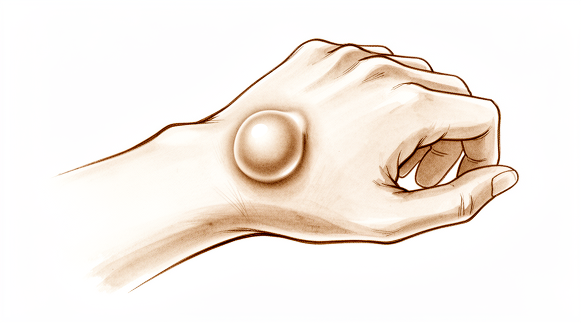
Você pode notar um caroço no pulso. Ele costuma ser macio e pode variar de tamanho. Se o caroço estiver na parte dorsal do pulso, você pode sentir dor nessa região. Mulheres com esse tipo de cisto sinovial têm maior probabilidade de apresentar dor persistente após a cirurgia. A dor pode intensificar-se quando você move o pulso.
Se o caroço estiver na face palmar do pulso, ele pode parecer uma faixa tensa. Isso pode prender seu dedo. Você pode experimentar uma sensação de estalo ou bloqueio ao flexionar o dedo. Isso se assemelha ao dedo em gatilho. Você pode ter dificuldade para segurar objetos ou digitar com conforto.
As tarefas diárias podem tornar-se difíceis. Alcançar as costas para fechar o sutiã pode causar dor. Guardar a camisa dentro da calça pode ser incômodo se esticar a pele sobre o caroço. Dormir do lado do pulso afetado pode perturbar seu descanso. Se seu trabalho ou hobbies exigirem flexão forçada do pulso para trás, você corre risco considerável de dor persistente e limitação de movimento após o tratamento.
Crianças frequentemente apresentam caroços na parte dorsal do pulso. Meninas têm maior probabilidade de ter esses caroços do que meninos. Para crianças menores de 10 anos, o caroço geralmente está na face palmar. Na maioria dos casos, esses caroços desaparecem espontaneamente em 12 a 18 meses. Não são necessárias radiografias de rotina para avaliar isso, pois elas raramente alteram o tratamento cirúrgico.
Se o caroço não desaparecer ou causar dor, seu cirurgião pode sugerir observá-lo por cerca de 2 meses. O uso de talas pode ajudar. Se persistir, a cirurgia é uma opção. A cirurgia remove o caroço e reduz significativamente os sintomas. A chance de recorrência do caroço após a cirurgia é baixa. A artroscopia, que utiliza pequenas câmeras, é uma maneira segura e eficaz de remover caroços dorsais do pulso.
Evite tratamentos que injetem agentes espessantes no caroço. Eles podem causar danos graves, incluindo lesão na artéria radial. Se você precisar de uma ressonância magnética para confirmar o diagnóstico, ela é confiável. Ela identifica corretamente o caroço em 83% dos casos quando comparada aos achados cirúrgicos.
O que está realmente acontecendo
Um cisto sinovial é um saco preenchido por líquido que se forma próximo à articulação do punho ou aos tendões. Pense nele como um pequeno balão de água que vaza da membrana sinovial da articulação. A cápsula articular é a capa resistente que mantém os ossos do punho unidos e retém o líquido lubrificante no lugar. Por vezes, essa membrana enfraquece ou se rompe, permitindo que o líquido se infiltre e forme um nódulo.
Você pode notar esse nódulo na parte dorsal (superior) ou volar (inferior) do punho. As mulheres têm significativamente mais probabilidade de receber um diagnóstico de cisto sinovial volar do punho, independentemente da idade ou do status militar. Nos cistos sinoviais pediátricos, eles afetam mais comumente o punho dorsal e apresentam maior prevalência no sexo feminino. Os cistos sinoviais da mão em crianças têm uma maior taxa de resolução do que os cistos sinoviais do punho.
O líquido no interior é espesso e de consistência gelatinosa, semelhante ao lubrificante que permite o deslizamento suave dos tendões. Quando esse saco cresce, pode comprimir nervos ou estruturas próximas. Essa compressão é frequentemente o que causa sua dor ou limita seu movimento. Pacientes com hiperlaxidão do punho têm predisposição para o desenvolvimento de cistos sinoviais. Isso significa que, se suas articulações forem naturalmente mais frouxas, você pode estar mais propenso ao desenvolvimento desses cistos.
Seu cirurgião pode utilizar ressonância magnética para avaliar a dor no punho quando a sequência de pulso adequada é utilizada. Essa imagem fornece uma visualização clara e não invasiva dos tecidos moles. No entanto, a realização rotineira de radiografias do punho não é custo-efetiva no processo de avaliação e tomada de decisão terapêutica para pacientes com cisto sinovial do punho, devido à baixa prevalência de achados clinicamente significativos. A maioria das ressonâncias magnéticas do punho em instituições pediátricas foi solicitada para dor no punho, ajudando seu médico a ver exatamente de onde o líquido está vindo.
Compreender a origem do nódulo ajuda a explicar por que alguns tratamentos funcionam melhor do que outros. Como o saco está conectado à articulação, simplesmente drená-lo frequentemente leva ao seu preenchimento novamente. É por isso que seu cirurgião discute opções como observação, imobilização com talas ou excisão cirúrgica com base nos seus sintomas específicos e estilo de vida.
O que podemos fazer a respeito
Você pode começar observando o nódulo e permitindo que ele descanse. Isso é chamado de manejo expectante. Funciona bem para muitas pessoas, especialmente crianças. Em crianças com menos de 10 anos, 69% a 79% desses cistos desaparecem espontaneamente dentro de 12 a 18 meses. Seu cirurgião pode sugerir o uso de tala para manter o pulso imóvel. Isso ajuda a reduzir a irritação. A maioria dos cistos ganglionares pediátricos da mão e do pulso resolve-se apenas com observação ou uso de tala. Você deve dar a essa abordagem pelo menos dois meses para surtir efeito. Se o cisto for doloroso ou não diminuir de tamanho, discutiremos outras opções.
Não solicitamos radiografias de rotina para essa condição. Elas raramente alteram o tratamento, pois raramente apresentam achados úteis. Se você tiver dor, seu cirurgião pode prescrever medicação anti-inflamatória. Esses medicamentos ajudam a reduzir o inchaço e aliviar o desconforto. Eles não removem o cisto, mas tornam a vida diária mais confortável. Alguns pacientes encontram alívio com o uso de uma tala durante o dia ou à noite. Isso limita o movimento e reduz o estresse sobre a articulação. Evitamos injetar substâncias como esclerosantes no cisto. Essa prática foi abandonada porque pode causar danos graves, como lesão da artéria radial no pulso.
A cirurgia é considerada se o cisto permanecer doloroso após o fracasso do tratamento conservador. Também é uma opção se o nódulo retornar após o tratamento inicial. A excisão cirúrgica reduz significativamente os sintomas e apresenta uma baixa taxa de recorrência do cisto. A maioria dos pacientes relata alta satisfação após o procedimento. Seu cirurgião escolherá entre cirurgia aberta ou cirurgia artroscópica (usando pequenas câmeras). A cirurgia aberta tem menor chance de recorrência do cisto em comparação com outros métodos. No entanto, se seu trabalho ou hobbies exigirem extensão forçada do pulso, você pode enfrentar um risco considerável de dor residual ou limitações funcionais após a excisão aberta. O tratamento artroscópico é uma alternativa segura e eficaz, embora exija expertise cirúrgica específica. Revisaremos seus riscos e benefícios específicos antes de decidir o melhor caminho para você.
O que esperar
Sua perspectiva depende principalmente da sua idade e da localização do cisto. Se você é uma criança com menos de 10 anos, é provável que o cisto esteja no lado palmar do seu pulso. Nesse caso, ele frequentemente desaparece espontaneamente. Cerca de 69% a 79% desses cistos desaparecem em 12 a 18 meses sem qualquer tratamento. Seu cirurgião pode sugerir observação cuidadosa ou o uso de uma tala.
Em adultos, o cisto geralmente está na parte dorsal do pulso. Esses raramente se resolvem sem ajuda. Cerca de 40% dos cistos ganglionares do pulso diminuem de tamanho nos primeiros seis anos após a consulta com um cirurgião de mão. No entanto, a maioria dos cistos não desaparece completamente por conta própria. Se você optar por deixá-lo sem tratamento, pode experimentar desconforto contínuo ou um nódulo visível.
Se você decidir pelo tratamento, a remoção cirúrgica reduz significativamente seus sintomas. A maioria dos pacientes relata alta satisfação com os resultados. A chance de o cisto retornar após a cirurgia é baixa, de cerca de 10%. Isso é muito melhor do que tentar drená-lo com uma agulha, o que frequentemente leva ao retorno do cisto.
Esteja ciente de que alguns fatores podem afetar sua recuperação. Se você é do sexo feminino e teve dor ao redor do cisto antes da cirurgia, é mais provável que tenha alguma dor residual após o procedimento. Além disso, se seu trabalho ou hobbies exigirem flexão forçada do seu pulso para trás, você enfrenta um risco maior de dor persistente ou limitação de movimento após a cirurgia aberta. Seu cirurgião discutirá esses riscos com você para garantir o melhor resultado para o seu estilo de vida específico.
Quando procurar um especialista
Procure uma avaliação especializada se tiver dor persistente que não melhora com o repouso. Procure atendimento se notar fraqueza ou instabilidade no pulso. Consulte o seu médico se o seu pulso travar ou ceder durante o uso. Deve também procurar ajuda se os sintomas interferirem no seu sono ou no trabalho. Solicite uma avaliação se experimentar uma piora súbita da sua condição. O seu cirurgião pode determinar se o cisto sinovial requer tratamento ou se a observação é a melhor opção. Uma avaliação precoce ajuda a prevenir complicações e garante que receba o tratamento adequado para a sua situação específica.
Evidence & references
Overview
- Female patients with preoperative pain around dorsal wrist ganglia are the most likely to have residual pain after surgery [1].
- There is no consensus within the literature regarding the best management of pediatric wrist ganglia [2].
- No single treatment modality confers a particular advantage or disadvantage over another for pediatric wrist ganglia [2].
- Sonography-assisted arthroscopic resection is safer and more reliable for treating volar wrist ganglia [4].
- Routine midcarpal joint exploration during arthroscopic excision of dorsal wrist ganglions appears to reduce recurrence at 1 year without negatively impacting patient outcomes [5].
- Open excision of dorsal wrist ganglia leads to a lower recurrence rate than arthroscopic excision [6].
- Surgical excision of primary wrist ganglia significantly reduces patient symptoms with low recurrence rates and high patient satisfaction [8].
- Outcomes, recurrence, and complication rates after 4 years of follow-up support the use of arthroscopy as a treatment for dorsal wrist ganglion [9].
- Patients whose occupation or activities require forceful wrist extension should be counseled on the considerable risk of residual pain and functional limitations that may occur after open dorsal wrist ganglion excision [12].
- Open surgical excision offers a significantly lower chance of recurrence compared with aspiration in the treatment of wrist ganglions [15].
- Arthroscopic ganglionectomy through an intrafocal cystic portal is a safe and efficacious option for the treatment of painful wrist ganglia [17].
- High patient satisfaction can be achieved for arthroscopic treatment of occult dorsal wrist ganglia [18].
- Arthroscopic treatment of a dorsal wrist ganglion is a good alternative to open surgery, though it is a difficult procedure requiring adequate experience [20].
Anatomy & Pathophysiology
- Sonography-assisted arthroscopic resection is a safer and more reliable technique for treating volar wrist ganglia [4].
- Determining the etiology of ulnar-sided wrist pain is challenging due to overlapping history and physical examination findings [14].
- Diagnosis of ulnar-sided wrist pain requires a detailed history, systematic physical examination with provocative maneuvers, and appropriate diagnostic imaging [14].
- Four-dimensional CT complements conventional imaging and arthroscopy by providing functional information on wrist biomechanics [25].
- Four-dimensional CT should be used selectively when dynamic instability is suspected and conventional imaging is inconclusive [25].
- The radioscapholunate fusion shows the most biomechanically similar behavior to the healthy wrist among compared fusion types [26].
- The scaphoid, lunate, and capitate move synergistically throughout planar wrist motion [27].
- The row theory more clearly accounts for the function of the wrist than the column theory regarding carpal instability [28].
- Carpal instability is a multifactorial phenomenon involving inadequate wrist proprioception, poor interaction between ligaments and muscles, and lack of control by the sensorimotor system [33].
- Combined wrist hyperextension with radial deviation causes the scaphoid to contact the radius over the radial styloid [35].
- Anatomical differences in Liebenberg syndrome are biomechanically normal for the individual, resulting in near-normal function and painless joints [37].
Classification
- Female patients with preoperative pain around dorsal wrist ganglia are the most likely to have residual pain after surgery [1].
- There is no consensus within the literature regarding the best management of pediatric wrist ganglia [2].
- No single treatment modality confers a particular advantage or disadvantage over another for pediatric wrist ganglia [2].
- In children aged <10 years, ganglions mainly occur on the volar wrist [3].
- 69% to 79% of pediatric ganglions in children aged <10 years display spontaneous regression within a span of 12-18 months [3].
- Sonography-assisted arthroscopic resection is considered safer and more reliable for treating volar wrist ganglia [4].
- Open excision of dorsal wrist ganglia leads to a lower recurrence rate than arthroscopic excision [6].
- Ganglions in pediatric populations most commonly affect the dorsal wrist [7].
- Pediatric ganglions demonstrate a female predilection [7].
- Military service members have higher rates of volar wrist ganglia diagnoses than their age- and sex-matched civilian counterparts [10].
- The incidence of dorsal wrist ganglia was higher in the military compared with the civilian population [11].
- Clinicians should be careful ascribing symptoms to anatomical variations on radiographs in patients with nonspecific wrist pain [13].
- Open surgical excision offers a significantly lower chance of recurrence compared with aspiration in the treatment of wrist ganglions [15].
- Joint denervation is a symptomatic treatment for osteoarthritis of the wrist and hand [21].
- Cystic soft tissue tumours of the dorsal aspect of the wrist have two distinct histological subtypes [41].
- Both histologically distinct tissue types coexist at recurrence in dorsal wrist ganglia [41].
- There are equal recurrence rates in both initial synovial and ganglion groups for dorsal wrist cystic soft tissue tumours [41].
Clinical Presentation
- Female patients with preoperative pain around dorsal wrist ganglia are the most likely to have residual pain after surgery [1].
- Pediatric wrist ganglions most commonly affect the dorsal wrist and demonstrate a female predilection [7].
- In children aged <10 years, ganglions mainly occur on the volar wrist [3].
- Ganglions in children usually resolve within 18 months if they resolve spontaneously [16].
- Military service members have higher rates of volar wrist ganglia diagnoses than their age- and sex-matched civilian counterparts [10].
- The incidence of dorsal wrist ganglia is higher in the military compared with the civilian population [11].
- Patients whose occupation or activities require forceful wrist extension face a considerable risk of residual pain and functional limitations after open dorsal wrist ganglion excision [12].
- Clinicians should be careful ascribing symptoms to anatomical variations on radiographs in patients with nonspecific wrist pain [13].
- Determining the etiology of ulnar-sided wrist pain is often challenging due to overlapping history and physical examination findings [14].
Investigations
- Female patients with preoperative pain around dorsal wrist ganglia are the most likely to have residual pain after arthroscopic excision [1].
- There is no consensus within the literature regarding the best management of pediatric wrist ganglia [2].
- No single treatment modality confers a particular advantage or disadvantage over another for pediatric wrist ganglia [2].
- In children aged <10 years, ganglions mainly occur on the volar wrist [3].
- In children aged <10 years, 69% to 79% of volar wrist ganglions display spontaneous regression within a span of 12-18 months [3].
- Sonography-assisted arthroscopic resection is considered safer and more reliable for treating volar wrist ganglia [4].
- Routine midcarpal joint exploration during arthroscopic excision of dorsal wrist ganglions appeared to reduce recurrence at 1 year [5].
- Routine midcarpal joint exploration during arthroscopic excision of dorsal wrist ganglions did not negatively impact patient outcomes [5].
- Ganglions in pediatric populations most commonly affect the dorsal wrist [7].
- Ganglions in pediatric populations demonstrate a female predilection [7].
- Military service members have higher rates of volar wrist ganglia diagnoses than their age- and sex-matched civilian counterparts [10].
- The incidence of dorsal wrist ganglia was higher in the military compared with the civilian population [11].
- Clinicians should be careful ascribing symptoms to anatomical variations on radiographs in patients with nonspecific wrist pain [13].
- Determining the etiology of ulnar-sided wrist pain is often challenging due to overlapping history and physical examination findings [14].
- A detailed history, systematic physical examination with provocative maneuvers, and appropriate diagnostic imaging are essential for diagnosing ulnar-sided wrist pain [14].
- If a pediatric wrist ganglion resolves, it usually does so within 18 months [16].
- Radiological evaluation showed normal radiocarpal angles, volar tilt, and radial length in patients treated arthroscopically for scapholunate ligament lesions associated with intra-articular distal radius fractures [22].
- When the appropriate pulse sequence is used, magnetic resonance imaging is an accurate and effective method for the non-invasive evaluation of pain in the wrist [32].
- For young subjects, MRI is valuable in diagnosing ulnar detachment of the triangular fibrocartilage complex [34].
- The ability to distinguish between proximal and distal laminae of the triangular fibrocartilage complex using MRI remains questionable for young subjects [34].
- Convolutional neural networks can detect ganglion cysts in wrist MRI [36].
- Intraosseous carpal bone cysts are a rare cause of chronic wrist pain that can progress to pathological fracture and tendon compromise [40].
- Once identified, intraosseous carpal bone cysts require careful clinical and radiographic assessment [40].
- Surgical intervention is indicated for symptomatic intraosseous carpal bone cysts [40].
Treatment
Non-Operative Management
- In children aged <10 years, volar wrist ganglions can be treated expectantly, with 69% to 79% displaying spontaneous regression within 12-18 months [3].
- There is no consensus within the literature regarding the best management of pediatric wrist ganglia, and no single treatment modality confers a particular advantage or disadvantage over another [2].
Surgical Excision: Open vs. Arthroscopic vs. Aspiration
- Open surgical excision offers a significantly lower chance of recurrence compared with aspiration in the treatment of wrist ganglions [15].
- Open excision of dorsal wrist ganglia leads to a lower recurrence rate than does arthroscopic excision [6].
- Arthroscopic treatment of a dorsal wrist ganglion is a good alternative to open surgery, though it is a difficult procedure requiring adequate experience [20].
- Arthroscopic resection of dorsal wrist ganglions with or without midcarpal exploration supports the use of arthroscopy as a treatment for dorsal wrist ganglion with favorable outcomes, recurrence, and complication rates at 4 years of follow-up [9].
- Arthroscopic ganglionectomy through an intrafocal cystic portal is a safe and efficacious option for the treatment of painful wrist ganglia [17].
- High patient satisfaction can be achieved for arthroscopic treatment of occult dorsal wrist ganglia [18].
- Sonography-assisted arthroscopic resection is a safer and more reliable method for treating volar wrist ganglia [4].
- Surgical excision of primary wrist ganglia significantly reduces patient symptoms with low recurrence rates and high patient satisfaction [8].
Recurrence and Technical Considerations
- Routine midcarpal joint exploration during arthroscopic excision of dorsal wrist ganglions appears to reduce recurrence at 1 year without negatively impacting patient outcomes [5].
Patient-Specific Factors and Outcomes
- Female patients who have preoperative pain around dorsal wrist ganglia were the most likely to have residual pain after surgery [1].
- Patients whose occupation or activities require forceful wrist extension should be counseled on the considerable risk of residual pain and functional limitations that may occur after open dorsal wrist ganglion excision [12].
Complications
- Female patients with preoperative pain around dorsal wrist ganglia are the most likely to have residual pain after surgery [1].
- Patients whose occupation or activities require forceful wrist extension should be counseled on the considerable risk of residual pain and functional limitations that may occur after open dorsal wrist ganglion excision [12].
- Routine midcarpal joint exploration during arthroscopic excision of dorsal wrist ganglions appeared to reduce recurrence at 1 year without negatively impacting patient outcomes [5].
- Open excision of dorsal wrist ganglia leads to a lower recurrence rate than does arthroscopic excision [6].
- Surgical excision of primary wrist ganglia significantly reduces patient symptoms with low recurrence rates and high patient satisfaction [8].
- The outcomes, recurrence, and complications rates after 4 years of follow-up support the use of arthroscopy as a treatment for dorsal wrist ganglion [9].
Recovery
- Female patients with preoperative pain around dorsal wrist ganglia are the most likely to have residual pain after surgery [1].
- There is no consensus within the literature regarding the best management of pediatric wrist ganglia [2].
- No single treatment modality confers a particular advantage or disadvantage over another for pediatric wrist ganglia [2].
- In children aged <10 years, ganglions mainly occur on the volar wrist [3].
- Ganglions in children aged <10 years can be treated expectantly [3].
- 69% to 79% of ganglions in children aged <10 years display spontaneous regression within a span of 12-18 months [3].
- Routine midcarpal joint exploration during arthroscopic excision of dorsal wrist ganglions appeared to reduce recurrence at 1 year without negatively impacting patient outcomes [5].
- Open excision of dorsal wrist ganglia leads to a lower recurrence rate than does arthroscopic excision [6].
- Surgical excision of primary wrist ganglia significantly reduces patient symptoms [8].
- Surgical excision of primary wrist ganglia is associated with low recurrence rates [8].
- Surgical excision of primary wrist ganglia is associated with high patient satisfaction [8].
- Outcomes, recurrence, and complications rates after 4 years of follow-up support the use of arthroscopy as a treatment for dorsal wrist ganglion [9].
- Military service members have higher rates of volar wrist ganglia diagnoses than their age- and sex-matched civilian counterparts [10].
- The incidence of dorsal wrist ganglia was higher in the military compared with the civilian population [11].
- Patients whose occupation or activities require forceful wrist extension should be counseled on the considerable risk of residual pain and functional limitations that may occur after open dorsal wrist ganglion excision [12].
- Open surgical excision offers a significantly lower chance of recurrence compared with aspiration in the treatment of wrist ganglions [15].
Key Evidence
- [L4] Female patients who have preoperative pain around dorsal wrist ganglia were the most likely to have residual pain after surgery. [1] (10.1016/j.arthro.2013.04.002)
- [L4] There is no consensus within the literature regarding the best management of pediatric wrist ganglia, and no single treatment modality confers a particular advantage or disadvantage over another. [2] (10.1177/1558944720966716)
- [L4] In children aged <10 years, ganglions mainly occur on the volar wrist and can be treated expectantly, with 69% to 79% displaying spontaneous regression within a span of 12-18 months. [3] (10.1016/j.jhsa.2021.12.015)
- [Paper] This method is safer and more reliable for treating volar wrist ganglia. [4] (10.1016/j.eats.2011.12.007)
- [L3] Routine midcarpal joint exploration during arthroscopic excision of dorsal wrist ganglions appeared to reduce recurrence at 1 year without negatively impacting patient outcomes. [5] (10.1177/17531934251405730)
- [L3] This study suggests that open excision of dorsal wrist ganglia leads to a lower recurrence rate than does arthroscopic excision. [6] (10.1177/15589447211003184)
- [L2] Ganglions in pediatric populations, which most commonly affect the dorsal wrist, demonstrate a female predilection. [7] (10.1016/j.jhsa.2021.02.026)
- [L4] Surgical excision of primary wrist ganglia significantly reduces patient symptoms with low recurrence rates and high patient satisfaction. [8] (10.1177/1753193411434376)
- [L4] The outcomes, recurrence, and complications rates after 4 years of follow-up presented in this study support the use of arthroscopy as a treatment for dorsal wrist ganglion. [9] (10.1177/1558944717743601)
- [L3] Military service members have higher rates of volar wrist ganglia diagnoses than their age- and sex-matched civilian counterparts. [10] (10.1016/j.jhsa.2016.08.008)
- [L3] The incidence of dorsal wrist ganglia was higher in the military compared with the civilian population. [11] (10.1016/j.jhsg.2020.08.001)
- [L4] Patients whose occupation or activities require forceful wrist extension should be counseled on the considerable risk of residual pain and functional limitations that may occur after open dorsal wrist ganglion excision. [12] (10.1016/j.jhsa.2015.05.030)
- [L3] Clinicians should be careful ascribing symptoms to anatomical variations on radiographs in patients with nonspecific wrist pain. [13] (10.1016/j.jhsa.2017.02.002)
- [L5] Determining the etiology of ulnar-sided wrist pain is often challenging due to overlapping history and physical examination findings; a detailed history, systematic physical examination with provocative maneuvers, and appropriate diagnostic imaging are essential for diagnosis. [14] (10.5435/jaaos-d-16-00407)
- [L1] Open surgical excision offers significantly lower chance of recurrence compared with aspiration in the treatment of wrist ganglions. [15] (10.1016/j.jhsa.2014.12.014)
- [L4] In a child with a wrist ganglion, if the cyst ultimately resolved, it usually did so within 18 months. [16] (10.1016/j.jhsa.2019.10.032)
- [L4] Arthroscopic ganglionectomy through an intrafocal cystic portal is a safe and efficacious option for the treatment of painful wrist ganglia. [17] (10.1016/j.arthro.2009.08.021)
- [L4] The results confirm that high patient satisfaction can be achieved for arthroscopic treatment of occult dorsal wrist ganglia. [18] (10.1007/s00402-016-2539-0)
- [L4] Arthroscopic treatment of a dorsal wrist ganglion is a good alternative to open surgery, though it is a difficult procedure requiring adequate experience. [20] (10.1054/jhsb.1999.0290)
- [L5] Joint denervation deserves a place of choice in the surgical arsenal for osteoarthritis of the wrist and hand, provided new anatomical observations are integrated, the procedure is meticulous, and patients are informed that it is a symptomatic treatment. [21] (10.1016/j.otsr.2021.102986)
- [L4] Radiological evaluation showed normal radiocarpal angles, volar tilt, and radial length in all patients. [22] (10.1007/s001670050172)
- [L5] Four-dimensional CT complements conventional imaging and arthroscopy by providing functional information on wrist biomechanics and should be used selectively when dynamic instability is suspected and conventional imaging is inconclusive. [25] (10.1530/eor-2026-0051)
- [L5] The article summarizes current thinking regarding the diagnosis and treatment of clinically important carpal instabilities, emphasizing that the row theory more clearly accounts for the function of the wrist than the column theory. [28] (10.2106/00004623-199503000-00019)
- [L2] When the appropriate pulse sequence is used, magnetic resonance imaging is an accurate and effective method for the non-invasive evaluation of pain in the wrist. [32] (10.2106/00004623-199711000-00009)
- [L5] Carpal instability is a multifactorial phenomenon involving inadequate wrist proprioception, poor interaction between ligaments and muscles, and lack of control of the entire process by the sensorimotor system. [33] (10.1016/j.hcl.2017.04.007)
- [L3] For young subjects, MRI is still valuable, especially in diagnosing ulnar detachment, although the ability to distinguish between proximal and distal laminae remains questionable. [34] (10.1177/17531934221141986)
- [L4] Combined wrist hyperextension with radial deviation caused the scaphoid to contact the radius over the radial styloid. [35] (10.1016/j.jhsa.2012.08.030)
- [L4] CNNs can detect ganglion cysts in wrist MRI. [36] (10.1186/s12891-025-09011-1)
- [L4] Conservative management is the guiding principle as the anatomical differences are biomechanically normal for the individual, resulting in near-normal function and painless joints. [37] (10.1177/1753193413502162)
- [L4] Intraosseous carpal bone cysts are a rare cause of chronic wrist pain that can progress to pathological fracture and tendon compromise; once identified, they require careful clinical and radiographic assessment with surgical intervention indicated for symptomatic cases. [40] (10.1007/s11552-015-9750-2)
- [L4] The study demonstrated two histologically distinct tissue types at primary surgery and the coexistence of both tissue types at recurrence, with equal recurrence rates in both initial synovial and ganglion groups. [41] (10.1177/17531934241251721)
References
[1] Arthroscopic Excision of Dorsal Wrist Ganglion: Factors Related to Recurrence and Postoperative Residual Pain. Arthroscopy. 2013. DOI: 10.1016/j.arthro.2013.04.002 [2] Wrist Ganglion Cysts in Children: An Update and Review of the Literature. HAND. 2020. DOI: 10.1177/1558944720966716 [3] Pediatric Ganglions of the Hand and Wrist: A Review of Current Literature. The Journal of Hand Surgery. 2022. DOI: 10.1016/j.jhsa.2021.12.015 [4] Sonography‐Assisted Arthroscopic Resection of Volar Wrist Ganglia: A New Technique. Arthroscopy Techniques. 2012. DOI: 10.1016/j.eats.2011.12.007 [5] Arthroscopic resection of dorsal wrist ganglions with or without midcarpal exploration. Journal of Hand Surgery (European Volume). 2025. DOI: 10.1177/17531934251405730 [6] Recurrence Rates of Dorsal Wrist Ganglion Cysts After Arthroscopic Versus Open Surgical Excision: A Retrospective Comparison. HAND. 2021. DOI: 10.1177/15589447211003184 [7] Clinical Presentation and Characteristics of Hand and Wrist Ganglion Cysts in Children. The Journal of Hand Surgery. 2021. DOI: 10.1016/j.jhsa.2021.02.026 [8] Patient outcomes following wrist ganglion excision surgery. Journal of Hand Surgery (European Volume). 2012. DOI: 10.1177/1753193411434376 [9] Arthroscopic Resection of Dorsal Wrist Ganglion: Results and Rate of Recurrence Over a Minimum Follow-up of 4 Years. HAND. 2017. DOI: 10.1177/1558944717743601 [10] Incidence and Risk Factors for Volar Wrist Ganglia in the U.S. Military and Civilian Populations. The Journal of Hand Surgery. 2016. DOI: 10.1016/j.jhsa.2016.08.008 [11] Epidemiology of Symptomatic Dorsal Wrist Ganglia in Active Duty Military and Civilian Populations. Journal of Hand Surgery Global Online. 2020. DOI: 10.1016/j.jhsg.2020.08.001 [12] Outcomes of Open Dorsal Wrist Ganglion Excision in Active-Duty Military Personnel. The Journal of Hand Surgery. 2015. DOI: 10.1016/j.jhsa.2015.05.030 [13] Carpal Coalitions on Radiographs: Prevalence and Association With Ordering Indication. The Journal of Hand Surgery. 2017. DOI: 10.1016/j.jhsa.2017.02.002 [14] Evaluation of Ulnar-sided Wrist Pain. Journal of the American Academy of Orthopaedic Surgeons. 2017. DOI: 10.5435/jaaos-d-16-00407 [15] Wrist Ganglion Treatment: Systematic Review and Meta-Analysis. The Journal of Hand Surgery. 2015. DOI: 10.1016/j.jhsa.2014.12.014 [16] Wrist Ganglia in Children: Nonsurgical Versus Surgical Treatment. The Journal of Hand Surgery. 2020. DOI: 10.1016/j.jhsa.2019.10.032 [17] Arthroscopic Ganglionectomy Through an Intrafocal Cystic Portal for Wrist Ganglia. Arthroscopy. 2010. DOI: 10.1016/j.arthro.2009.08.021 [18] Arthroscopic resection of occult dorsal wrist ganglia. Archives of Orthopaedic and Trauma Surgery. 2016. DOI: 10.1007/s00402-016-2539-0 [20] Arthroscopic Resection of Dorsal Wrist Ganglia and Treatment of Recurrences. Journal of Hand Surgery. 2000. DOI: 10.1054/jhsb.1999.0290 [21] Is there still a place for denervation in the treatment of osteoarthritis of the wrist and hand?. Orthopaedics & Traumatology: Surgery & Research. 2021. DOI: 10.1016/j.otsr.2021.102986 [22] Midterm results of arthroscopic treatment of scapholunate ligament lesions associated with intra‐articular distal radius fractures. Knee Surgery, Sports Traumatology, Arthroscopy. 1999. DOI: 10.1007/s001670050172 [25] Dynamic wrist imaging using four-dimensional CT: current concepts, clinical applications, and future perspectives. EFORT Open Reviews. 2026. DOI: 10.1530/eor-2026-0051 [26] Load_transfer_through_the_radiocarpal_joint_and_the_effects_of_partial_wrist_art_1753193412441761. 1934. [27] 10.1055-s-0036-1588025. n.d.. [28] Carpal Instability. The Journal of Bone & Joint Surgery. 1995. DOI: 10.2106/00004623-199503000-00019 [32] The Utility of High-Resolution Magnetic Resonance Imaging in the Evaluation of the Triangular Fibrocartilage Complex of the Wrist. The Journal of Bone and Joint Surgery (American Volume). 1997. DOI: 10.2106/00004623-199711000-00009 [33] Carpal Ligaments. Hand Clinics. 2017. DOI: 10.1016/j.hcl.2017.04.007 [34] Abnormal MRI signal intensity of the triangular fibrocartilage complex in asymptomatic wrists. Journal of Hand Surgery (European Volume). 2022. DOI: 10.1177/17531934221141986 [35] In Vivo Changes in Contact Regions of the Radiocarpal Joint During Wrist Hyperextension. The Journal of Hand Surgery. 2012. DOI: 10.1016/j.jhsa.2012.08.030 [36] Automated detection of wrist ganglia in MRI using convolutional neural networks. BMC Musculoskeletal Disorders. 2025. DOI: 10.1186/s12891-025-09011-1 [37] The Liebenberg syndrome: in depth analysis of the original family. Journal of Hand Surgery (European Volume). 2013. DOI: 10.1177/1753193413502162 [40] Intraosseous Ganglion Cysts of the Carpus: Current Practice. HAND. 2015. DOI: 10.1007/s11552-015-9750-2 [41] Cystic soft tissue tumours of the dorsal aspect of the wrist have two distinct histological subtypes. Journal of Hand Surgery (European Volume)*. 2024. DOI: 10.1177/17531934241251721


