மணிக்கட்டு கங்லியா
Patients › Wrist
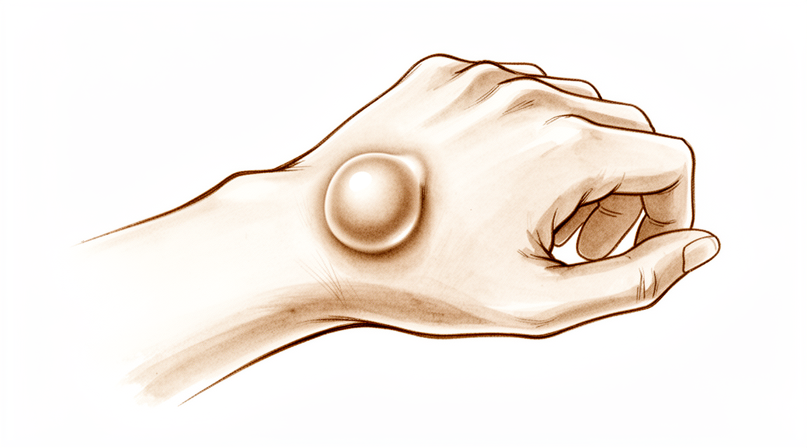
Wrist ganglia are common, fluid-filled lumps – often painless – and this page covers observation, aspiration, and excision.
நீங்கள் என்ன உணர்கிறீர்கள்
உங்கள் மணிக்கட்டில் ஒரு கட்டி இருப்பதை நீங்கள் கவனிக்கலாம். இது பெரும்பாலும் மென்மையானது மற்றும் அளவை மாற்றக்கூடும். கட்டி உங்கள் மணிக்கட்டின் பின்புறத்தில் இருந்தால், நீங்கள் அங்கு வலியை உணரலாம். இந்த வகை கட்டி கொண்ட பெண்கள் அறுவை சிகிச்சைக்குப் பிறகு வலியைக் கொண்டிருக்கலாம். உங்கள் மணிக்கட்டை நகர்த்தும்போது வலி அதிகரிக்கக்கூடும்.
உங்கள் விரலை வளைக்கும்போது கிளிக் அல்லது பூட்டுதல் உணர்வை நீங்கள் அனுபவிக்கலாம். இது தூண்டு விரல் போல் உணர்கிறது. பொருட்களைப் பிடிப்பது அல்லது வசதியாக தட்டச்சு செய்வது உங்களுக்கு கடினமாக இருக்கலாம்.
தினசரி பணிகள் கடினமாகிவிடும். உங்கள் முதுகுக்குப் பின்னால் ஒரு பிராவைப் பிணைப்பது வலிக்கக்கூடும். ஒரு சட்டைக்குள் நுழைவது சருமத்தை கட்டிக்கு மேல் நீட்டினால் சங்கடமாக இருக்கலாம். உங்கள் பாதிக்கப்பட்ட மணிக்கட்டில் ஒரு பக்கத்தில் தூங்குவது உங்கள் ஓய்வைத் தொந்தரவு செய்யலாம். உங்கள் வேலை அல்லது பொழுதுபோக்குகளுக்கு உங்கள் மணிக்கட்டில் பின்னால் வலுக்கட்டாயமாக வளைப்பது தேவைப்பட்டால், சிகிச்சையின் பின்னர் தொடர்ச்சியான வலி மற்றும் வரையறுக்கப்பட்ட இயக்கத்திற்கான கணிசமான ஆபத்து உங்களுக்கு உள்ளது.
குழந்தைகளுக்கு அடிக்கடி மணிக்கட்டின் பின்புறத்தில் கட்டிகள் உள்ளன. சிறுமிகளுக்கு இதைப் பெறுவதற்கான வாய்ப்புகள் சிறுவர்களை விட அதிகம். 10 வயதிற்குட்பட்ட குழந்தைகளுக்கு, கட்டி பொதுவாக பனைப் பக்கத்தில் இருக்கும். பெரும்பாலான சந்தர்ப்பங்களில், இந்த கட்டிகள் 12 முதல் 18 மாதங்களுக்குள் தானாகவே மறைந்துவிடும். இதைச் சரிபார்க்க உங்களுக்கு வழக்கமான எக்ஸ்-கதிர்கள் தேவையில்லை, ஏனென்றால் உங்கள் அறுவை சிகிச்சையாளர் அதை எவ்வாறு நடத்துகிறார் என்பதை அவை அரிதாகவே மாற்றுகின்றன.
கட்டி போவதில்லை அல்லது வலியை ஏற்படுத்துகிறது என்றால், உங்கள் அறுவை சிகிச்சை நிபுணர் அதை சுமார் 2 மாதங்கள் கண்காணிக்க பரிந்துரைக்கலாம். அறுவை சிகிச்சை உதவக்கூடும். அது தொடர்ந்தால், அறுவை சிகிச்சை ஒரு விருப்பமாகும். அறுவை சிகிச்சை கட்டி நீக்குகிறது மற்றும் அறிகுறிகளை கணிசமாகக் குறைக்கிறது. அறுவை சிகிச்சைக்குப் பிறகு கட்டி மீண்டும் வருவதற்கான வாய்ப்பு குறைவு. சிறிய கேமராக்களைப் பயன்படுத்தும் ஆர்த்ரோஸ்கோபி, முதுகெலும்பு கட்டி கட்டிகளை அகற்றுவதற்கான பாதுகாப்பான மற்றும் பயனுள்ள வழியாகும்.
கட்டிக்குள் தடிமனான பொருட்களை செலுத்தும் சிகிச்சைகளைத் தவிர்க்கவும். இவை ரேடியல் தமனிக்கு காயம் உட்பட கடுமையான தீங்குகளை ஏற்படுத்தும். நோயறிதலை உறுதிப்படுத்த உங்களுக்கு ஒரு எம்ஆர்ஐ தேவைப்பட்டால், அது நம்பகமானது. அறுவை சிகிச்சை கண்டுபிடிப்புகளுடன் ஒப்பிடும்போது 83% வழக்குகளில் கட்டி சரியாக அடையாளம் காணப்படுகிறது.
உண்மையில் என்ன நடக்கிறது
ஒரு காங்லியன் என்பது உங்கள் மணிக்கட்டு மூட்டு அல்லது தசைகளுக்கு அருகில் உருவாகும் திரவத்தால் நிரப்பப்பட்ட பை ஆகும். அதை ஒரு சிறிய நீர் பலூன் என்று நினைத்துப் பாருங்கள், இது மூட்டு புறணிகளிலிருந்து கசிகிறது. கூட்டு காப்ஸ்யூல் என்பது உங்கள் மணிக்கட்டு எலும்புகளை ஒன்றாக வைத்திருக்கும் மற்றும் உயவு திரவத்தை இடத்தில் வைத்திருக்கும் கடினமான ஸ்லீவ் ஆகும். சில நேரங்களில், இந்த புறணி பலவீனமடைகிறது அல்லது கிழிக்கிறது, திரவத்தை ஊடுருவி ஒரு கட்டி உருவாக்க அனுமதிக்கிறது.
உங்கள் மணிக்கட்டில் மேலே (முதுகில்) அல்லது கீழே (வாலர்) இந்த கட்டி இருப்பதை நீங்கள் கவனிக்கலாம். வயது அல்லது இராணுவ அந்தஸ்தைப் பொருட்படுத்தாமல், பெண்களுக்கு ஒரு வாலர் மணிக்கட்டு குழாய் இருப்பதாகக் கண்டறியப்படுவதற்கான வாய்ப்புகள் கணிசமாக அதிகம். உங்களுக்கு குழந்தை குழாய்கள் இருந்தால், அவை பெரும்பாலும் முதுகில் மணிக்கட்டை பாதிக்கின்றன மற்றும் பெண் விருப்பத்தைக் காட்டுகின்றன. கையில் உள்ள குழந்தை குழாய்கள் மணிக்கட்டு குழாய்களை விட அதிக விகிதத்தில் தீர்மானிக்கப்படுகின்றன.
உங்கள் மூட்டுகளில் உள்ள திரவம் தடிமனாகவும் ஜெல்லி போன்றதாகவும் இருக்கிறது, இது உங்கள் இடுப்புகளை மென்மையாக சறுக்க உதவுகிறது. இந்த சாக் வளரும்போது, அது அருகிலுள்ள நரம்புகள் அல்லது கட்டமைப்புகளை அழுத்தலாம். இந்த அழுத்தம் பெரும்பாலும் உங்கள் வலியை ஏற்படுத்துகிறது அல்லது உங்கள் இயக்கத்தை கட்டுப்படுத்துகிறது. மணிக்கட்டு ஹைப்பர்லாக்ஸிட்டி நோயாளிகள் கட்டிகளை உருவாக்குவதற்கு முன்கூட்டியே உள்ளனர். இதன் பொருள் உங்கள் மூட்டுகள் இயற்கையாகவே தளர்வானவை என்றால், இந்த கருப்பைகள் உருவாக அதிக வாய்ப்புள்ளது.
உங்கள் அறுவை சிகிச்சையாளர் முறையான துடிப்பு வரிசை பயன்படுத்தப்படும் போது மணிக்கட்டில் வலியை மதிப்பிடுவதற்கு காந்த அதிர்வு காட்சிப்படுத்தலைப் பயன்படுத்தலாம். இந்த ஸ்கேன் மென்மையான திசுக்களில் தெளிவான, ஆக்கிரமிப்பு அல்லாத தோற்றத்தை வழங்குகிறது. இருப்பினும், வழக்கமான முறையில் மணிக்கட்டு ரேடியோகிராஃபியை மேற்கொள்வது மணிக்கட்டு கேங்க்லியுடன் நோயாளிகளுக்கு மதிப்பீடு மற்றும் சிகிச்சை முடிவெடுக்கும் செயல்பாட்டில் செலவு குறைந்ததாக இல்லை, ஏனெனில் சிகிச்சை ரீதியாக குறிப்பிடத்தக்க கண்டுபிடிப்புகள் குறைவாக உள்ளன. ஒரு குழந்தை மருத்துவ நிறுவனத்தில் பெரும்பாலான மணிக்கட்டு எம்ஆர்ஐகள் மணிக்கட்டு வலிக்கு உத்தரவிடப்பட்டன, திரவம் எங்கிருந்து வருகிறது என்பதை உங்கள் மருத்துவர் சரியாகக் காண உதவுகிறது.
கட்டிகளின் மூலத்தை புரிந்துகொள்வது ஏன் சில சிகிச்சைகள் மற்றவர்களை விட சிறப்பாக செயல்படுகின்றன என்பதை விளக்க உதவுகிறது. பை மூட்டுடன் இணைக்கப்பட்டுள்ளதால், வெறுமனே அதை வடிகட்டுவது பெரும்பாலும் அதை மீண்டும் நிரப்புவதற்கு வழிவகுக்கிறது. அதனால்தான் உங்கள் அறுவை சிகிச்சையாளர் உங்கள் குறிப்பிட்ட அறிகுறிகள் மற்றும் வாழ்க்கை முறையின் அடிப்படையில் கண்காணிப்பு, அறுவை சிகிச்சை அல்லது அறுவை சிகிச்சை போன்ற விருப்பங்களைப் பற்றி விவாதிக்கிறார்.
நாம் என்ன செய்ய முடியும்
நீங்கள் கட்டி கவனித்து அதை ஓய்வெடுக்க அனுமதிப்பதன் மூலம் தொடங்கலாம். இது எதிர்பார்ப்பு மேலாண்மை என்று அழைக்கப்படுகிறது. இது பலருக்கு, குறிப்பாக குழந்தைகளுக்கு நன்றாக வேலை செய்கிறது. 10 வயதிற்குட்பட்ட குழந்தைகளில், இந்த கருப்பைகளில் 69% முதல் 79% வரை 12-18 மாதங்களுக்குள் தானாகவே போய்விடும். உங்கள் அறுவை சிகிச்சை நிபுணர் உங்கள் மணிக்கட்டை இன்னும் வைத்திருக்க அடுப்பை பரிந்துரைக்கலாம். இது எரிச்சலைக் குறைக்க உதவுகிறது. பெரும்பாலான குழந்தைகளின் கை மற்றும் மணிக்கட்டு கோளங்கள் கண்காணிப்பு அல்லது அடுப்பு மூலம் மட்டுமே தீர்க்கப்படுகின்றன. இந்த அணுகுமுறை வேலை செய்ய குறைந்தது இரண்டு மாதங்கள் கொடுக்க வேண்டும். கருப்பை வலி அல்லது சுருங்கவில்லை என்றால், நாங்கள் மற்ற விருப்பங்களைப் பற்றி விவாதிப்போம்.
இந்த நிலைக்கு நாங்கள் வழக்கமாக எக்ஸ்-கதிர்களை ஆர்டர் செய்வதில்லை. அவை அரிதாகவே பயனுள்ள கண்டுபிடிப்புகளைக் காண்பிப்பதால் நாங்கள் உங்களை எவ்வாறு நடத்துகிறோம் என்பதை அவை அரிதாகவே மாற்றுகின்றன. உங்களுக்கு வலி இருந்தால், உங்கள் அறுவை சிகிச்சை நிபுணர் அழற்சி எதிர்ப்பு மருந்துகளை பரிந்துரைக்கலாம். இந்த மருந்துகள் வீக்கத்தை அமைதிப்படுத்தவும், அச om கரியத்தை குறைக்கவும் உதவுகின்றன. அவை சிறுநீரகத்தை அகற்றாது, ஆனால் அவை அன்றாட வாழ்க்கையை மிகவும் வசதியாக ஆக்குகின்றன. சில நோயாளிகள் பகல் அல்லது இரவில் அணிந்திருக்கும் ஒரு அடுக்கு மூலம் நிவாரணம் பெறுகிறார்கள். இது இயக்கத்தை கட்டுப்படுத்துகிறது மற்றும் மூட்டுக்கு அழுத்தத்தை குறைக்கிறது. ஸ்க்லெரோசாண்ட்ஸ் போன்ற பொருட்களை சிறுநீரகத்தில் செலுத்துவதை நாங்கள் தவிர்க்கிறோம். உங்கள் மணிக்கட்டில் உள்ள ரேடியல் தமனிக்கு காயம் போன்ற கடுமையான தீங்குகளை ஏற்படுத்தக்கூடும் என்பதால் இந்த நடைமுறை நிறுத்தப்பட்டுள்ளது.
கன்சர்வேடிவ் பராமரிப்பு தோல்வியுற்றபின் கட்டி வலியுடன் இருந்தால் அறுவை சிகிச்சை பரிசீலிக்கப்படுகிறது. ஆரம்ப சிகிச்சைக்குப் பிறகு கட்டி மீண்டும் வந்தால் இது ஒரு விருப்பமாகும். அறுவை சிகிச்சை அறுவை சிகிச்சை அறிகுறிகளை கணிசமாகக் குறைக்கிறது மற்றும் கட்டி மீண்டும் வருவதற்கான குறைந்த விகிதத்தைக் கொண்டுள்ளது. பெரும்பாலான நோயாளிகள் இந்த நடைமுறைக்குப் பிறகு அதிக திருப்தியைப் பற்றி தெரிவிக்கின்றனர். உங்கள் அறுவை சிகிச்சையாளர் திறந்த அறுவை சிகிச்சை அல்லது ஆர்த்ரோஸ்கோபிக் அறுவை சிகிச்சை (சிறிய கேமராக்களைப் பயன்படுத்தி) இடையே தேர்வு செய்வார். திறந்த அறுவை சிகிச்சை மற்ற முறைகளுடன் ஒப்பிடும்போது கட்டி மீண்டும் வருவதற்கான குறைந்த வாய்ப்பைக் கொண்டுள்ளது. இருப்பினும், உங்கள் வேலை அல்லது பொழுதுபோக்குகளுக்கு வலுவான மணிக்கட்டு நீட்டி தேவைப்பட்டால், திறந்த அறுவை சிகிச்சைக்குப் பிறகு மீதமுள்ள வலி அல்லது செயல்பாட்டு வரம்புகளை நீங்கள் கணிசமான ஆபத்தை எதிர்கொள்ளலாம். ஆர்த்ரோஸ்கோபி
எதிர்பார்ப்பது என்ன
உங்கள் நோய்க்கான நோய்த் தோற்றம் உங்கள் வயதைப் பொறுத்தது. உங்கள் குழந்தை 10 வயதிற்குட்பட்டவராக இருந்தால், அது உங்கள் மணிக்கட்டில் இருக்கும். இந்த விஷயத்தில், அது பெரும்பாலும் தானாகவே மறைந்துவிடும். இந்த நோய்க்குறிகளில் சுமார் 69% முதல் 79% வரை 12 முதல் 18 மாதங்களுக்குள் எந்த சிகிச்சையும் இல்லாமல் மறைந்துவிடும். உங்கள் அறுவை சிகிச்சையாளர் அதை உன்னிப்பாகக் கண்காணிக்கவோ அல்லது ஒரு அடுப்பைப் பயன்படுத்தவோ பரிந்துரைக்கலாம்.
கை அறுவை சிகிச்சையாளரைப் பார்த்த பிறகு முதல் ஆறு ஆண்டுகளில் சுமார் 40% மார்பக கட்டிகள் சுருங்குகின்றன. இருப்பினும், பெரும்பாலான சிஸ்ட்கள் தங்களைத் தாங்களே முற்றிலுமாக விட்டுவிடாது. நீங்கள் அதை தனியாக விட்டுவிட விரும்பினால், நீங்கள் தொடர்ந்து அச om கரியத்தை அனுபவிக்கலாம் அல்லது காணக்கூடிய கட்டி ஏற்படலாம்.
நீங்கள் சிகிச்சையைத் தேர்வுசெய்தால், அறுவை சிகிச்சை மூலம் அகற்றுவது உங்கள் அறிகுறிகளை கணிசமாகக் குறைக்கிறது. பெரும்பாலான நோயாளிகள் முடிவுகளில் அதிக திருப்தியைப் பற்றி தெரிவிக்கின்றனர். அறுவை சிகிச்சைக்குப் பிறகு சிறுநீரக மீண்டும் வருவதற்கான வாய்ப்பு குறைவாக உள்ளது, சுமார் 10%. இது ஒரு ஊசியுடன் வடிகட்ட முயற்சிப்பதை விட மிகச் சிறந்தது, இது பெரும்பாலும் சிறுநீரக மீண்டும் வருவதற்கு வழிவகுக்கிறது.
சில காரணிகள் உங்கள் மீட்பை பாதிக்கும் என்பதை நினைவில் கொள்ளுங்கள். நீங்கள் ஒரு பெண்ணாக இருந்தால், அறுவை சிகிச்சைக்கு முன்னர் சிறுநீரகத்தைச் சுற்றி வலி இருந்தால், அதற்குப் பிறகு உங்களுக்கு சில மீதமுள்ள வலிகள் ஏற்பட வாய்ப்பு அதிகம். மேலும், உங்கள் வேலை அல்லது பொழுதுபோக்குகளுக்கு உங்கள் மணிக்கட்டை பின்னோக்கி வலுவாக வளைக்க வேண்டியிருந்தால், திறந்த அறுவை சிகிச்சைக்குப் பிறகு நீடித்த வலி அல்லது வரையறுக்கப்பட்ட இயக்கம் அதிக ஆபத்தை எதிர்கொள்கிறது. உங்கள் குறிப்பிட்ட வாழ்க்கை முறைக்கு சிறந்த முடிவை உறுதிப்படுத்த உங்கள் அறுவை சிகிச்சையாளர் உங்களுடன் இந்த அபாயங்களைப் பற்றி விவாதிப்பார்.
யாரையாவது எப்போது பார்க்க வேண்டும்
உங்கள் கையில் பலவீனம் அல்லது நிலையற்ற தன்மை இருப்பதை நீங்கள் கவனித்தால், ஒரு நிபுணர் மதிப்பாய்வைக் கேளுங்கள். உங்கள் கையில் பூட்டுதல் அல்லது பயன்பாட்டின் போது வழியைக் கொடுத்தால் உங்கள் மருத்துவரை அணுகவும். உங்கள் தூக்கத்திலோ அல்லது வேலையிலோ அறிகுறிகள் தலையிட்டால் நீங்கள் உதவியை நாட வேண்டும். உங்கள் நிலை திடீரென மோசமடைந்தால் ஒரு மதிப்பீட்டைக் கேளுங்கள். உங்கள் அறுவை சிகிச்சையாளர் சிகிச்சை தேவைப்படுகிறதா அல்லது கண்காணிப்பு சிறந்ததா என்பதை தீர்மானிக்க முடியும். ஆரம்ப மதிப்பீடு சிக்கல்களைத் தடுக்க உதவுகிறது மற்றும் உங்கள் குறிப்பிட்ட சூழ்நிலைக்கு சரியான கவனிப்பைப் பெறுவதை உறுதி செய்கிறது.
Evidence & references
Overview
- Female patients with preoperative pain around dorsal wrist ganglia are the most likely to have residual pain after surgery [1].
- There is no consensus within the literature regarding the best management of pediatric wrist ganglia [2].
- No single treatment modality confers a particular advantage or disadvantage over another for pediatric wrist ganglia [2].
- Sonography-assisted arthroscopic resection is safer and more reliable for treating volar wrist ganglia [4].
- Routine midcarpal joint exploration during arthroscopic excision of dorsal wrist ganglions appears to reduce recurrence at 1 year without negatively impacting patient outcomes [5].
- Open excision of dorsal wrist ganglia leads to a lower recurrence rate than arthroscopic excision [6].
- Surgical excision of primary wrist ganglia significantly reduces patient symptoms with low recurrence rates and high patient satisfaction [8].
- Outcomes, recurrence, and complication rates after 4 years of follow-up support the use of arthroscopy as a treatment for dorsal wrist ganglion [9].
- Patients whose occupation or activities require forceful wrist extension should be counseled on the considerable risk of residual pain and functional limitations that may occur after open dorsal wrist ganglion excision [12].
- Open surgical excision offers a significantly lower chance of recurrence compared with aspiration in the treatment of wrist ganglions [15].
- Arthroscopic ganglionectomy through an intrafocal cystic portal is a safe and efficacious option for the treatment of painful wrist ganglia [17].
- High patient satisfaction can be achieved for arthroscopic treatment of occult dorsal wrist ganglia [18].
- Arthroscopic treatment of a dorsal wrist ganglion is a good alternative to open surgery, though it is a difficult procedure requiring adequate experience [20].
Anatomy & Pathophysiology
- Sonography-assisted arthroscopic resection is a safer and more reliable technique for treating volar wrist ganglia [4].
- Determining the etiology of ulnar-sided wrist pain is challenging due to overlapping history and physical examination findings [14].
- Diagnosis of ulnar-sided wrist pain requires a detailed history, systematic physical examination with provocative maneuvers, and appropriate diagnostic imaging [14].
- Four-dimensional CT complements conventional imaging and arthroscopy by providing functional information on wrist biomechanics [25].
- Four-dimensional CT should be used selectively when dynamic instability is suspected and conventional imaging is inconclusive [25].
- The radioscapholunate fusion shows the most biomechanically similar behavior to the healthy wrist among compared fusion types [26].
- The scaphoid, lunate, and capitate move synergistically throughout planar wrist motion [27].
- The row theory more clearly accounts for the function of the wrist than the column theory regarding carpal instability [28].
- Carpal instability is a multifactorial phenomenon involving inadequate wrist proprioception, poor interaction between ligaments and muscles, and lack of control by the sensorimotor system [33].
- Combined wrist hyperextension with radial deviation causes the scaphoid to contact the radius over the radial styloid [35].
- Anatomical differences in Liebenberg syndrome are biomechanically normal for the individual, resulting in near-normal function and painless joints [37].
Classification
- Female patients with preoperative pain around dorsal wrist ganglia are the most likely to have residual pain after surgery [1].
- There is no consensus within the literature regarding the best management of pediatric wrist ganglia [2].
- No single treatment modality confers a particular advantage or disadvantage over another for pediatric wrist ganglia [2].
- In children aged <10 years, ganglions mainly occur on the volar wrist [3].
- 69% to 79% of pediatric ganglions in children aged <10 years display spontaneous regression within a span of 12-18 months [3].
- Sonography-assisted arthroscopic resection is considered safer and more reliable for treating volar wrist ganglia [4].
- Open excision of dorsal wrist ganglia leads to a lower recurrence rate than arthroscopic excision [6].
- Ganglions in pediatric populations most commonly affect the dorsal wrist [7].
- Pediatric ganglions demonstrate a female predilection [7].
- Military service members have higher rates of volar wrist ganglia diagnoses than their age- and sex-matched civilian counterparts [10].
- The incidence of dorsal wrist ganglia was higher in the military compared with the civilian population [11].
- Clinicians should be careful ascribing symptoms to anatomical variations on radiographs in patients with nonspecific wrist pain [13].
- Open surgical excision offers a significantly lower chance of recurrence compared with aspiration in the treatment of wrist ganglions [15].
- Joint denervation is a symptomatic treatment for osteoarthritis of the wrist and hand [21].
- Cystic soft tissue tumours of the dorsal aspect of the wrist have two distinct histological subtypes [41].
- Both histologically distinct tissue types coexist at recurrence in dorsal wrist ganglia [41].
- There are equal recurrence rates in both initial synovial and ganglion groups for dorsal wrist cystic soft tissue tumours [41].
Clinical Presentation
- Female patients with preoperative pain around dorsal wrist ganglia are the most likely to have residual pain after surgery [1].
- Pediatric wrist ganglions most commonly affect the dorsal wrist and demonstrate a female predilection [7].
- In children aged <10 years, ganglions mainly occur on the volar wrist [3].
- Ganglions in children usually resolve within 18 months if they resolve spontaneously [16].
- Military service members have higher rates of volar wrist ganglia diagnoses than their age- and sex-matched civilian counterparts [10].
- The incidence of dorsal wrist ganglia is higher in the military compared with the civilian population [11].
- Patients whose occupation or activities require forceful wrist extension face a considerable risk of residual pain and functional limitations after open dorsal wrist ganglion excision [12].
- Clinicians should be careful ascribing symptoms to anatomical variations on radiographs in patients with nonspecific wrist pain [13].
- Determining the etiology of ulnar-sided wrist pain is often challenging due to overlapping history and physical examination findings [14].
Investigations
- Female patients with preoperative pain around dorsal wrist ganglia are the most likely to have residual pain after arthroscopic excision [1].
- There is no consensus within the literature regarding the best management of pediatric wrist ganglia [2].
- No single treatment modality confers a particular advantage or disadvantage over another for pediatric wrist ganglia [2].
- In children aged <10 years, ganglions mainly occur on the volar wrist [3].
- In children aged <10 years, 69% to 79% of volar wrist ganglions display spontaneous regression within a span of 12-18 months [3].
- Sonography-assisted arthroscopic resection is considered safer and more reliable for treating volar wrist ganglia [4].
- Routine midcarpal joint exploration during arthroscopic excision of dorsal wrist ganglions appeared to reduce recurrence at 1 year [5].
- Routine midcarpal joint exploration during arthroscopic excision of dorsal wrist ganglions did not negatively impact patient outcomes [5].
- Ganglions in pediatric populations most commonly affect the dorsal wrist [7].
- Ganglions in pediatric populations demonstrate a female predilection [7].
- Military service members have higher rates of volar wrist ganglia diagnoses than their age- and sex-matched civilian counterparts [10].
- The incidence of dorsal wrist ganglia was higher in the military compared with the civilian population [11].
- Clinicians should be careful ascribing symptoms to anatomical variations on radiographs in patients with nonspecific wrist pain [13].
- Determining the etiology of ulnar-sided wrist pain is often challenging due to overlapping history and physical examination findings [14].
- A detailed history, systematic physical examination with provocative maneuvers, and appropriate diagnostic imaging are essential for diagnosing ulnar-sided wrist pain [14].
- If a pediatric wrist ganglion resolves, it usually does so within 18 months [16].
- Radiological evaluation showed normal radiocarpal angles, volar tilt, and radial length in patients treated arthroscopically for scapholunate ligament lesions associated with intra-articular distal radius fractures [22].
- When the appropriate pulse sequence is used, magnetic resonance imaging is an accurate and effective method for the non-invasive evaluation of pain in the wrist [32].
- For young subjects, MRI is valuable in diagnosing ulnar detachment of the triangular fibrocartilage complex [34].
- The ability to distinguish between proximal and distal laminae of the triangular fibrocartilage complex using MRI remains questionable for young subjects [34].
- Convolutional neural networks can detect ganglion cysts in wrist MRI [36].
- Intraosseous carpal bone cysts are a rare cause of chronic wrist pain that can progress to pathological fracture and tendon compromise [40].
- Once identified, intraosseous carpal bone cysts require careful clinical and radiographic assessment [40].
- Surgical intervention is indicated for symptomatic intraosseous carpal bone cysts [40].
Treatment
Non-Operative Management
- In children aged <10 years, volar wrist ganglions can be treated expectantly, with 69% to 79% displaying spontaneous regression within 12-18 months [3].
- There is no consensus within the literature regarding the best management of pediatric wrist ganglia, and no single treatment modality confers a particular advantage or disadvantage over another [2].
Surgical Excision: Open vs. Arthroscopic vs. Aspiration
- Open surgical excision offers a significantly lower chance of recurrence compared with aspiration in the treatment of wrist ganglions [15].
- Open excision of dorsal wrist ganglia leads to a lower recurrence rate than does arthroscopic excision [6].
- Arthroscopic treatment of a dorsal wrist ganglion is a good alternative to open surgery, though it is a difficult procedure requiring adequate experience [20].
- Arthroscopic resection of dorsal wrist ganglions with or without midcarpal exploration supports the use of arthroscopy as a treatment for dorsal wrist ganglion with favorable outcomes, recurrence, and complication rates at 4 years of follow-up [9].
- Arthroscopic ganglionectomy through an intrafocal cystic portal is a safe and efficacious option for the treatment of painful wrist ganglia [17].
- High patient satisfaction can be achieved for arthroscopic treatment of occult dorsal wrist ganglia [18].
- Sonography-assisted arthroscopic resection is a safer and more reliable method for treating volar wrist ganglia [4].
- Surgical excision of primary wrist ganglia significantly reduces patient symptoms with low recurrence rates and high patient satisfaction [8].
Recurrence and Technical Considerations
- Routine midcarpal joint exploration during arthroscopic excision of dorsal wrist ganglions appears to reduce recurrence at 1 year without negatively impacting patient outcomes [5].
Patient-Specific Factors and Outcomes
- Female patients who have preoperative pain around dorsal wrist ganglia were the most likely to have residual pain after surgery [1].
- Patients whose occupation or activities require forceful wrist extension should be counseled on the considerable risk of residual pain and functional limitations that may occur after open dorsal wrist ganglion excision [12].
Complications
- Female patients with preoperative pain around dorsal wrist ganglia are the most likely to have residual pain after surgery [1].
- Patients whose occupation or activities require forceful wrist extension should be counseled on the considerable risk of residual pain and functional limitations that may occur after open dorsal wrist ganglion excision [12].
- Routine midcarpal joint exploration during arthroscopic excision of dorsal wrist ganglions appeared to reduce recurrence at 1 year without negatively impacting patient outcomes [5].
- Open excision of dorsal wrist ganglia leads to a lower recurrence rate than does arthroscopic excision [6].
- Surgical excision of primary wrist ganglia significantly reduces patient symptoms with low recurrence rates and high patient satisfaction [8].
- The outcomes, recurrence, and complications rates after 4 years of follow-up support the use of arthroscopy as a treatment for dorsal wrist ganglion [9].
Recovery
- Female patients with preoperative pain around dorsal wrist ganglia are the most likely to have residual pain after surgery [1].
- There is no consensus within the literature regarding the best management of pediatric wrist ganglia [2].
- No single treatment modality confers a particular advantage or disadvantage over another for pediatric wrist ganglia [2].
- In children aged <10 years, ganglions mainly occur on the volar wrist [3].
- Ganglions in children aged <10 years can be treated expectantly [3].
- 69% to 79% of ganglions in children aged <10 years display spontaneous regression within a span of 12-18 months [3].
- Routine midcarpal joint exploration during arthroscopic excision of dorsal wrist ganglions appeared to reduce recurrence at 1 year without negatively impacting patient outcomes [5].
- Open excision of dorsal wrist ganglia leads to a lower recurrence rate than does arthroscopic excision [6].
- Surgical excision of primary wrist ganglia significantly reduces patient symptoms [8].
- Surgical excision of primary wrist ganglia is associated with low recurrence rates [8].
- Surgical excision of primary wrist ganglia is associated with high patient satisfaction [8].
- Outcomes, recurrence, and complications rates after 4 years of follow-up support the use of arthroscopy as a treatment for dorsal wrist ganglion [9].
- Military service members have higher rates of volar wrist ganglia diagnoses than their age- and sex-matched civilian counterparts [10].
- The incidence of dorsal wrist ganglia was higher in the military compared with the civilian population [11].
- Patients whose occupation or activities require forceful wrist extension should be counseled on the considerable risk of residual pain and functional limitations that may occur after open dorsal wrist ganglion excision [12].
- Open surgical excision offers a significantly lower chance of recurrence compared with aspiration in the treatment of wrist ganglions [15].
Key Evidence
- [L4] Female patients who have preoperative pain around dorsal wrist ganglia were the most likely to have residual pain after surgery. [1] (10.1016/j.arthro.2013.04.002)
- [L4] There is no consensus within the literature regarding the best management of pediatric wrist ganglia, and no single treatment modality confers a particular advantage or disadvantage over another. [2] (10.1177/1558944720966716)
- [L4] In children aged <10 years, ganglions mainly occur on the volar wrist and can be treated expectantly, with 69% to 79% displaying spontaneous regression within a span of 12-18 months. [3] (10.1016/j.jhsa.2021.12.015)
- [Paper] This method is safer and more reliable for treating volar wrist ganglia. [4] (10.1016/j.eats.2011.12.007)
- [L3] Routine midcarpal joint exploration during arthroscopic excision of dorsal wrist ganglions appeared to reduce recurrence at 1 year without negatively impacting patient outcomes. [5] (10.1177/17531934251405730)
- [L3] This study suggests that open excision of dorsal wrist ganglia leads to a lower recurrence rate than does arthroscopic excision. [6] (10.1177/15589447211003184)
- [L2] Ganglions in pediatric populations, which most commonly affect the dorsal wrist, demonstrate a female predilection. [7] (10.1016/j.jhsa.2021.02.026)
- [L4] Surgical excision of primary wrist ganglia significantly reduces patient symptoms with low recurrence rates and high patient satisfaction. [8] (10.1177/1753193411434376)
- [L4] The outcomes, recurrence, and complications rates after 4 years of follow-up presented in this study support the use of arthroscopy as a treatment for dorsal wrist ganglion. [9] (10.1177/1558944717743601)
- [L3] Military service members have higher rates of volar wrist ganglia diagnoses than their age- and sex-matched civilian counterparts. [10] (10.1016/j.jhsa.2016.08.008)
- [L3] The incidence of dorsal wrist ganglia was higher in the military compared with the civilian population. [11] (10.1016/j.jhsg.2020.08.001)
- [L4] Patients whose occupation or activities require forceful wrist extension should be counseled on the considerable risk of residual pain and functional limitations that may occur after open dorsal wrist ganglion excision. [12] (10.1016/j.jhsa.2015.05.030)
- [L3] Clinicians should be careful ascribing symptoms to anatomical variations on radiographs in patients with nonspecific wrist pain. [13] (10.1016/j.jhsa.2017.02.002)
- [L5] Determining the etiology of ulnar-sided wrist pain is often challenging due to overlapping history and physical examination findings; a detailed history, systematic physical examination with provocative maneuvers, and appropriate diagnostic imaging are essential for diagnosis. [14] (10.5435/jaaos-d-16-00407)
- [L1] Open surgical excision offers significantly lower chance of recurrence compared with aspiration in the treatment of wrist ganglions. [15] (10.1016/j.jhsa.2014.12.014)
- [L4] In a child with a wrist ganglion, if the cyst ultimately resolved, it usually did so within 18 months. [16] (10.1016/j.jhsa.2019.10.032)
- [L4] Arthroscopic ganglionectomy through an intrafocal cystic portal is a safe and efficacious option for the treatment of painful wrist ganglia. [17] (10.1016/j.arthro.2009.08.021)
- [L4] The results confirm that high patient satisfaction can be achieved for arthroscopic treatment of occult dorsal wrist ganglia. [18] (10.1007/s00402-016-2539-0)
- [L4] Arthroscopic treatment of a dorsal wrist ganglion is a good alternative to open surgery, though it is a difficult procedure requiring adequate experience. [20] (10.1054/jhsb.1999.0290)
- [L5] Joint denervation deserves a place of choice in the surgical arsenal for osteoarthritis of the wrist and hand, provided new anatomical observations are integrated, the procedure is meticulous, and patients are informed that it is a symptomatic treatment. [21] (10.1016/j.otsr.2021.102986)
- [L4] Radiological evaluation showed normal radiocarpal angles, volar tilt, and radial length in all patients. [22] (10.1007/s001670050172)
- [L5] Four-dimensional CT complements conventional imaging and arthroscopy by providing functional information on wrist biomechanics and should be used selectively when dynamic instability is suspected and conventional imaging is inconclusive. [25] (10.1530/eor-2026-0051)
- [L5] The article summarizes current thinking regarding the diagnosis and treatment of clinically important carpal instabilities, emphasizing that the row theory more clearly accounts for the function of the wrist than the column theory. [28] (10.2106/00004623-199503000-00019)
- [L2] When the appropriate pulse sequence is used, magnetic resonance imaging is an accurate and effective method for the non-invasive evaluation of pain in the wrist. [32] (10.2106/00004623-199711000-00009)
- [L5] Carpal instability is a multifactorial phenomenon involving inadequate wrist proprioception, poor interaction between ligaments and muscles, and lack of control of the entire process by the sensorimotor system. [33] (10.1016/j.hcl.2017.04.007)
- [L3] For young subjects, MRI is still valuable, especially in diagnosing ulnar detachment, although the ability to distinguish between proximal and distal laminae remains questionable. [34] (10.1177/17531934221141986)
- [L4] Combined wrist hyperextension with radial deviation caused the scaphoid to contact the radius over the radial styloid. [35] (10.1016/j.jhsa.2012.08.030)
- [L4] CNNs can detect ganglion cysts in wrist MRI. [36] (10.1186/s12891-025-09011-1)
- [L4] Conservative management is the guiding principle as the anatomical differences are biomechanically normal for the individual, resulting in near-normal function and painless joints. [37] (10.1177/1753193413502162)
- [L4] Intraosseous carpal bone cysts are a rare cause of chronic wrist pain that can progress to pathological fracture and tendon compromise; once identified, they require careful clinical and radiographic assessment with surgical intervention indicated for symptomatic cases. [40] (10.1007/s11552-015-9750-2)
- [L4] The study demonstrated two histologically distinct tissue types at primary surgery and the coexistence of both tissue types at recurrence, with equal recurrence rates in both initial synovial and ganglion groups. [41] (10.1177/17531934241251721)
References
[1] Arthroscopic Excision of Dorsal Wrist Ganglion: Factors Related to Recurrence and Postoperative Residual Pain. Arthroscopy. 2013. DOI: 10.1016/j.arthro.2013.04.002 [2] Wrist Ganglion Cysts in Children: An Update and Review of the Literature. HAND. 2020. DOI: 10.1177/1558944720966716 [3] Pediatric Ganglions of the Hand and Wrist: A Review of Current Literature. The Journal of Hand Surgery. 2022. DOI: 10.1016/j.jhsa.2021.12.015 [4] Sonography‐Assisted Arthroscopic Resection of Volar Wrist Ganglia: A New Technique. Arthroscopy Techniques. 2012. DOI: 10.1016/j.eats.2011.12.007 [5] Arthroscopic resection of dorsal wrist ganglions with or without midcarpal exploration. Journal of Hand Surgery (European Volume). 2025. DOI: 10.1177/17531934251405730 [6] Recurrence Rates of Dorsal Wrist Ganglion Cysts After Arthroscopic Versus Open Surgical Excision: A Retrospective Comparison. HAND. 2021. DOI: 10.1177/15589447211003184 [7] Clinical Presentation and Characteristics of Hand and Wrist Ganglion Cysts in Children. The Journal of Hand Surgery. 2021. DOI: 10.1016/j.jhsa.2021.02.026 [8] Patient outcomes following wrist ganglion excision surgery. Journal of Hand Surgery (European Volume). 2012. DOI: 10.1177/1753193411434376 [9] Arthroscopic Resection of Dorsal Wrist Ganglion: Results and Rate of Recurrence Over a Minimum Follow-up of 4 Years. HAND. 2017. DOI: 10.1177/1558944717743601 [10] Incidence and Risk Factors for Volar Wrist Ganglia in the U.S. Military and Civilian Populations. The Journal of Hand Surgery. 2016. DOI: 10.1016/j.jhsa.2016.08.008 [11] Epidemiology of Symptomatic Dorsal Wrist Ganglia in Active Duty Military and Civilian Populations. Journal of Hand Surgery Global Online. 2020. DOI: 10.1016/j.jhsg.2020.08.001 [12] Outcomes of Open Dorsal Wrist Ganglion Excision in Active-Duty Military Personnel. The Journal of Hand Surgery. 2015. DOI: 10.1016/j.jhsa.2015.05.030 [13] Carpal Coalitions on Radiographs: Prevalence and Association With Ordering Indication. The Journal of Hand Surgery. 2017. DOI: 10.1016/j.jhsa.2017.02.002 [14] Evaluation of Ulnar-sided Wrist Pain. Journal of the American Academy of Orthopaedic Surgeons. 2017. DOI: 10.5435/jaaos-d-16-00407 [15] Wrist Ganglion Treatment: Systematic Review and Meta-Analysis. The Journal of Hand Surgery. 2015. DOI: 10.1016/j.jhsa.2014.12.014 [16] Wrist Ganglia in Children: Nonsurgical Versus Surgical Treatment. The Journal of Hand Surgery. 2020. DOI: 10.1016/j.jhsa.2019.10.032 [17] Arthroscopic Ganglionectomy Through an Intrafocal Cystic Portal for Wrist Ganglia. Arthroscopy. 2010. DOI: 10.1016/j.arthro.2009.08.021 [18] Arthroscopic resection of occult dorsal wrist ganglia. Archives of Orthopaedic and Trauma Surgery. 2016. DOI: 10.1007/s00402-016-2539-0 [20] Arthroscopic Resection of Dorsal Wrist Ganglia and Treatment of Recurrences. Journal of Hand Surgery. 2000. DOI: 10.1054/jhsb.1999.0290 [21] Is there still a place for denervation in the treatment of osteoarthritis of the wrist and hand?. Orthopaedics & Traumatology: Surgery & Research. 2021. DOI: 10.1016/j.otsr.2021.102986 [22] Midterm results of arthroscopic treatment of scapholunate ligament lesions associated with intra‐articular distal radius fractures. Knee Surgery, Sports Traumatology, Arthroscopy. 1999. DOI: 10.1007/s001670050172 [25] Dynamic wrist imaging using four-dimensional CT: current concepts, clinical applications, and future perspectives. EFORT Open Reviews. 2026. DOI: 10.1530/eor-2026-0051 [26] Load_transfer_through_the_radiocarpal_joint_and_the_effects_of_partial_wrist_art_1753193412441761. 1934. [27] 10.1055-s-0036-1588025. n.d.. [28] Carpal Instability. The Journal of Bone & Joint Surgery. 1995. DOI: 10.2106/00004623-199503000-00019 [32] The Utility of High-Resolution Magnetic Resonance Imaging in the Evaluation of the Triangular Fibrocartilage Complex of the Wrist. The Journal of Bone and Joint Surgery (American Volume). 1997. DOI: 10.2106/00004623-199711000-00009 [33] Carpal Ligaments. Hand Clinics. 2017. DOI: 10.1016/j.hcl.2017.04.007 [34] Abnormal MRI signal intensity of the triangular fibrocartilage complex in asymptomatic wrists. Journal of Hand Surgery (European Volume). 2022. DOI: 10.1177/17531934221141986 [35] In Vivo Changes in Contact Regions of the Radiocarpal Joint During Wrist Hyperextension. The Journal of Hand Surgery. 2012. DOI: 10.1016/j.jhsa.2012.08.030 [36] Automated detection of wrist ganglia in MRI using convolutional neural networks. BMC Musculoskeletal Disorders. 2025. DOI: 10.1186/s12891-025-09011-1 [37] The Liebenberg syndrome: in depth analysis of the original family. Journal of Hand Surgery (European Volume). 2013. DOI: 10.1177/1753193413502162 [40] Intraosseous Ganglion Cysts of the Carpus: Current Practice. HAND. 2015. DOI: 10.1007/s11552-015-9750-2 [41] Cystic soft tissue tumours of the dorsal aspect of the wrist have two distinct histological subtypes. Journal of Hand Surgery (European Volume)*. 2024. DOI: 10.1177/17531934241251721


