U nang cổ tay
Patients › Wrist
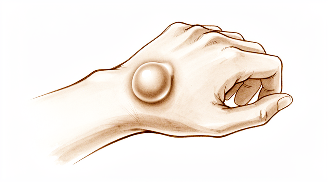
Wrist ganglia are common, fluid-filled lumps – often painless – and this page covers observation, aspiration, and excision.
Những gì bạn đang cảm thấy
Bạn có thể nhận thấy một khối u ở cổ tay. Khối u này thường mềm và có thể thay đổi kích thước. Nếu khối u nằm ở mặt sau cổ tay, bạn có thể cảm thấy đau tại vị trí đó. Phụ nữ mắc loại u nang này có khả năng cao nhất sẽ bị đau kéo dài sau phẫu thuật. Cơn đau có thể bùng phát khi bạn cử động cổ tay.
Nếu khối u nằm ở mặt lòng cổ tay, nó có thể cảm giác như một dải dây căng. Điều này có thể gây kẹt ngón tay của bạn. Bạn có thể trải nghiệm cảm giác kêu lách cách hoặc khóa khớp khi bạn gập ngón tay. Cảm giác này giống như ngón tay bị khóa (trigger finger). Bạn có thể thấy khó khăn khi cầm nắm đồ vật hoặc đánh máy một cách thoải mái.
Các hoạt động hàng ngày có thể trở nên khó khăn. Việc với ra sau lưng để cài áo ngực có thể gây đau. Việc nhét áo vào quần có thể trở nên bất tiện nếu nó kéo căng da trên khối u. Ngủ nghiêng lên cổ tay bị ảnh hưởng có thể làm gián đoạn giấc ngủ của bạn. Nếu công việc hoặc sở thích của bạn đòi hỏi việc bẻ cổ tay mạnh về phía sau, bạn có nguy cơ đáng kể bị đau dai dẳng và hạn chế vận động sau khi điều trị.
Trẻ em thường có khối u ở mặt sau cổ tay. Bé gái có khả năng mắc phải những khối u này cao hơn bé trai. Đối với trẻ em dưới 10 tuổi, khối u thường nằm ở mặt lòng cổ tay. Trong hầu hết các trường hợp, những khối u này tự biến mất trong vòng 12 đến 18 tháng. Bạn không cần chụp X-quang thường quy để kiểm tra tình trạng này, vì chúng hiếm khi thay đổi cách bác sĩ phẫu thuật điều trị cho bạn.
Nếu khối u không biến mất hoặc gây đau, bác sĩ phẫu thuật của bạn có thể đề nghị theo dõi trong khoảng 2 tháng. Nẹp cố định có thể giúp ích. Nếu tình trạng vẫn tiếp diễn, phẫu thuật là một lựa chọn. Phẫu thuật loại bỏ khối u và làm giảm đáng kể các triệu chứng. Khả năng khối u tái phát sau phẫu thuật là thấp. Nội soi khớp, sử dụng các camera nhỏ, là một phương pháp an toàn và hiệu quả để loại bỏ các khối u ở mặt sau cổ tay.
Tránh các phương pháp điều trị tiêm các chất làm đặc vào khối u. Những phương pháp này có thể gây tổn hại nghiêm trọng, bao gồm cả tổn thương động mạch quay. Nếu bạn cần chụp cộng hưởng từ (MRI) để xác nhận chẩn đoán, nó có độ tin cậy cao. Nó xác định chính xác khối u trong 83% các trường hợp khi so sánh với kết quả phẫu thuật.
Những gì thực sự đang xảy ra
U nang (ganglion) là một túi chứa dịch hình thành gần khớp cổ tay hoặc gân của bạn. Hãy tưởng tượng nó giống như một quả bóng nước nhỏ bị rò rỉ ra từ lớp màng bao khớp. Bao khớp là lớp vỏ chắc chắn giữ các xương cổ tay lại với nhau và duy trì dịch bôi trơn tại chỗ. Đôi khi, lớp màng này bị suy yếu hoặc rách, cho phép dịch đẩy ra ngoài và tạo thành một khối u.
Bạn có thể nhận thấy khối u này ở mặt trên (mu bàn tay) hoặc mặt dưới (bàn tay) của cổ tay. Phụ nữ có khả năng được chẩn đoán mắc u nang cổ tay lòng bàn tay cao hơn đáng kể, bất kể độ tuổi hoặc tình trạng quân dịch. Nếu bạn mắc u nang ở trẻ em, chúng thường ảnh hưởng đến cổ tay mu bàn tay và có xu hướng gặp ở nữ giới nhiều hơn. U nang ở bàn tay trẻ em có tỷ lệ tự khỏi cao hơn so với u nang ở cổ tay.
Dịch bên trong có đặc tính đặc và giống như thạch, tương tự như chất bôi trơn giúp gân của bạn trượt mượt mà. Khi túi này phát triển, nó có thể chèn ép lên các dây thần kinh hoặc cấu trúc lân cận. Sự chèn ép này thường là nguyên nhân gây đau hoặc hạn chế vận động của bạn. Bệnh nhân có tính lỏng khớp cổ tay có xu hướng dễ phát triển u nang. Điều này có nghĩa là nếu khớp của bạn vốn dĩ lỏng lẻo hơn, bạn có thể dễ bị hình thành các u nang này hơn.
Bác sĩ phẫu thuật của bạn có thể sử dụng chụp cộng hưởng từ (MRI) để đánh giá cơn đau ở cổ tay khi sử dụng chuỗi xung thích hợp. Chụp cắt lớp này cung cấp cái nhìn rõ ràng, không xâm lấn vào các mô mềm. Tuy nhiên, việc thực hiện chụp X-quang cổ tay một cách thường quy không mang lại hiệu quả về chi phí trong quá trình đánh giá và ra quyết định điều trị cho bệnh nhân mắc u nang cổ tay, do tỷ lệ thấp của các phát hiện có ý nghĩa lâm sàng quan trọng đối với điều trị. Hầu hết các chụp MRI cổ tay tại một cơ sở y tế chuyên khoa nhi được chỉ định cho cơn đau cổ tay, giúp bác sĩ của bạn nhìn thấy chính xác nguồn gốc của dịch.
Hiểu rõ nguồn gốc của khối u giúp giải thích tại sao một số phương pháp điều trị lại hiệu quả hơn những phương pháp khác. Vì túi này được kết nối với khớp, việc chỉ chọc hút dịch thường dẫn đến việc nó sẽ lại đầy dịch. Đây là lý do tại sao bác sĩ phẫu thuật của bạn thảo luận về các lựa chọn như theo dõi, nẹp cố định hoặc cắt bỏ phẫu thuật dựa trên các triệu chứng cụ thể và lối sống của bạn.
Những gì chúng tôi có thể làm về vấn đề này
Bạn có thể bắt đầu bằng cách theo dõi khối u và cho nó nghỉ ngơi. Đây được gọi là quản trị chờ đợi. Phương pháp này hiệu quả với nhiều người, đặc biệt là trẻ em. Ở trẻ em dưới 10 tuổi, từ 69% đến 79% các u nang này tự biến mất trong vòng 12-18 tháng. Bác sĩ phẫu thuật của bạn có thể đề nghị nẹp cố định để giữ cổ tay của bạn bất động. Điều này giúp giảm kích ứng. Hầu hết các u nang ganglion ở bàn tay và cổ tay của trẻ em sẽ tự khỏi chỉ với việc theo dõi hoặc nẹp cố định. Bạn nên dành cho phương pháp này ít nhất hai tháng để phát huy tác dụng. Nếu u nang gây đau hoặc không giảm kích thước, chúng tôi sẽ thảo luận về các lựa chọn khác.
Chúng tôi không thường xuyên chỉ định chụp X-quang cho tình trạng này. Các xét nghiệm này hiếm khi thay đổi cách điều trị của chúng tôi vì chúng hiếm khi cho thấy các phát hiện hữu ích. Nếu bạn bị đau, bác sĩ phẫu thuật có thể kê đơn thuốc chống viêm. Những loại thuốc này giúp làm giảm sưng và giảm khó chịu. Chúng không loại bỏ u nang, nhưng giúp cuộc sống hàng ngày dễ chịu hơn. Một số bệnh nhân tìm thấy sự giảm đau khi đeo nẹp vào ban ngày hoặc ban đêm. Điều này hạn chế cử động và giảm căng thẳng lên khớp. Chúng tôi tránh tiêm các chất như chất gây xơ hóa vào u nang. Thực hành này đã bị dừng lại vì nó có thể gây hại nghiêm trọng, chẳng hạn như tổn thương động mạch quay ở cổ tay của bạn.
Phẫu thuật được xem xét nếu u nang vẫn gây đau sau khi điều trị bảo thủ thất bại. Đây cũng là một lựa chọn nếu khối u tái phát sau điều trị ban đầu. Cắt bỏ phẫu thuật làm giảm đáng kể các triệu chứng và có tỷ lệ u nang tái phát thấp. Hầu hết bệnh nhân báo cáo sự hài lòng cao sau thủ thuật. Bác sĩ phẫu thuật của bạn sẽ lựa chọn giữa phẫu thuật mở hoặc phẫu thuật nội soi (sử dụng camera nhỏ). Phẫu thuật mở có khả năng u nang tái phát thấp hơn so với các phương pháp khác. Tuy nhiên, nếu công việc hoặc sở thích của bạn đòi hỏi duỗi cổ tay mạnh mẽ, bạn có thể đối mặt với nguy cơ đáng kể về đau tồn dư hoặc hạn chế chức năng sau khi cắt bỏ mở. Điều trị nội soi là một lựa chọn thay thế an toàn và hiệu quả, mặc dù nó đòi hỏi chuyên môn phẫu thuật cụ thể. Chúng tôi sẽ xem xét các rủi ro và lợi ích cụ thể của bạn trước khi quyết định con đường tốt nhất cho bạn.
Những điều cần biết
Tiên lượng của bạn phụ thuộc chủ yếu vào độ tuổi và vị trí của khối u nang. Nếu bạn là trẻ em dưới 10 tuổi, khối u nang thường nằm ở mặt lòng cổ tay. Trong trường hợp này, khối u nang thường tự biến mất. Khoảng 69% đến 79% các khối u nang này sẽ biến mất trong vòng 12 đến 18 tháng mà không cần bất kỳ điều trị nào. Bác sĩ phẫu thuật của bạn có thể đề nghị theo dõi chặt chẽ hoặc sử dụng nẹp cố định.
Đối với người lớn, khối u nang thường nằm ở mặt sau cổ tay. Những khối u nang này hiếm khi tự khỏi mà không có sự hỗ trợ. Khoảng 40% các khối u nang cổ tay sẽ teo nhỏ trong sáu năm đầu tiên sau khi bạn gặp bác sĩ phẫu thuật tay. Tuy nhiên, phần lớn các khối u nang không tự biến mất hoàn toàn. Nếu bạn chọn để yên nó, bạn có thể trải qua tình trạng khó chịu kéo dài hoặc một khối u nhìn thấy được.
Nếu bạn quyết định điều trị, việc cắt bỏ bằng phẫu thuật sẽ làm giảm đáng kể các triệu chứng của bạn. Hầu hết bệnh nhân báo cáo mức độ hài lòng cao với kết quả. Khả năng khối u nang tái phát sau phẫu thuật là thấp, khoảng 10%. Điều này tốt hơn nhiều so với việc cố gắng chọc hút bằng kim, vì phương pháp này thường dẫn đến việc khối u nang tái phát.
Hãy lưu ý rằng một số yếu tố có thể ảnh hưởng đến quá trình hồi phục của bạn. Nếu bạn là nữ và đã có cơn đau xung quanh khối u nang trước khi phẫu thuật, bạn có nhiều khả năng bị đau sót lại sau đó. Ngoài ra, nếu công việc hoặc sở thích của bạn đòi hỏi phải bẻ cổ tay mạnh về phía sau, bạn sẽ đối mặt với nguy cơ cao hơn về tình trạng đau dai dẳng hoặc hạn chế vận động sau phẫu thuật mở. Bác sĩ phẫu thuật của bạn sẽ thảo luận về những rủi ro này với bạn để đảm bảo kết quả tốt nhất cho lối sống cụ thể của bạn.
Khi nào cần gặp bác sĩ
Yêu cầu đánh giá từ chuyên gia nếu bạn có tình trạng đau dai dẳng không cải thiện khi nghỉ ngơi. Tìm kiếm sự chăm sóc y tế nếu bạn nhận thấy sự suy yếu hoặc mất ổn định ở cổ tay. Hãy gặp bác sĩ nếu cổ tay của bạn bị khóa hoặc đột ngột mất lực khi sử dụng. Bạn cũng nên tìm kiếm sự trợ giúp nếu các triệu chứng ảnh hưởng đến giấc ngủ hoặc công việc của bạn. Hãy yêu cầu đánh giá nếu bạn trải qua tình trạng xấu đi đột ngột của bệnh. Bác sĩ phẫu thuật của bạn có thể xác định xem khối u nang (ganglion) có cần điều trị hay quan sát theo dõi là tốt nhất. Đánh giá sớm giúp ngăn ngừa biến chứng và đảm bảo bạn nhận được sự chăm sóc phù hợp với tình trạng cụ thể của mình.
Evidence & references
Overview
- Female patients with preoperative pain around dorsal wrist ganglia are the most likely to have residual pain after surgery [1].
- There is no consensus within the literature regarding the best management of pediatric wrist ganglia [2].
- No single treatment modality confers a particular advantage or disadvantage over another for pediatric wrist ganglia [2].
- Sonography-assisted arthroscopic resection is safer and more reliable for treating volar wrist ganglia [4].
- Routine midcarpal joint exploration during arthroscopic excision of dorsal wrist ganglions appears to reduce recurrence at 1 year without negatively impacting patient outcomes [5].
- Open excision of dorsal wrist ganglia leads to a lower recurrence rate than arthroscopic excision [6].
- Surgical excision of primary wrist ganglia significantly reduces patient symptoms with low recurrence rates and high patient satisfaction [8].
- Outcomes, recurrence, and complication rates after 4 years of follow-up support the use of arthroscopy as a treatment for dorsal wrist ganglion [9].
- Patients whose occupation or activities require forceful wrist extension should be counseled on the considerable risk of residual pain and functional limitations that may occur after open dorsal wrist ganglion excision [12].
- Open surgical excision offers a significantly lower chance of recurrence compared with aspiration in the treatment of wrist ganglions [15].
- Arthroscopic ganglionectomy through an intrafocal cystic portal is a safe and efficacious option for the treatment of painful wrist ganglia [17].
- High patient satisfaction can be achieved for arthroscopic treatment of occult dorsal wrist ganglia [18].
- Arthroscopic treatment of a dorsal wrist ganglion is a good alternative to open surgery, though it is a difficult procedure requiring adequate experience [20].
Anatomy & Pathophysiology
- Sonography-assisted arthroscopic resection is a safer and more reliable technique for treating volar wrist ganglia [4].
- Determining the etiology of ulnar-sided wrist pain is challenging due to overlapping history and physical examination findings [14].
- Diagnosis of ulnar-sided wrist pain requires a detailed history, systematic physical examination with provocative maneuvers, and appropriate diagnostic imaging [14].
- Four-dimensional CT complements conventional imaging and arthroscopy by providing functional information on wrist biomechanics [25].
- Four-dimensional CT should be used selectively when dynamic instability is suspected and conventional imaging is inconclusive [25].
- The radioscapholunate fusion shows the most biomechanically similar behavior to the healthy wrist among compared fusion types [26].
- The scaphoid, lunate, and capitate move synergistically throughout planar wrist motion [27].
- The row theory more clearly accounts for the function of the wrist than the column theory regarding carpal instability [28].
- Carpal instability is a multifactorial phenomenon involving inadequate wrist proprioception, poor interaction between ligaments and muscles, and lack of control by the sensorimotor system [33].
- Combined wrist hyperextension with radial deviation causes the scaphoid to contact the radius over the radial styloid [35].
- Anatomical differences in Liebenberg syndrome are biomechanically normal for the individual, resulting in near-normal function and painless joints [37].
Classification
- Female patients with preoperative pain around dorsal wrist ganglia are the most likely to have residual pain after surgery [1].
- There is no consensus within the literature regarding the best management of pediatric wrist ganglia [2].
- No single treatment modality confers a particular advantage or disadvantage over another for pediatric wrist ganglia [2].
- In children aged <10 years, ganglions mainly occur on the volar wrist [3].
- 69% to 79% of pediatric ganglions in children aged <10 years display spontaneous regression within a span of 12-18 months [3].
- Sonography-assisted arthroscopic resection is considered safer and more reliable for treating volar wrist ganglia [4].
- Open excision of dorsal wrist ganglia leads to a lower recurrence rate than arthroscopic excision [6].
- Ganglions in pediatric populations most commonly affect the dorsal wrist [7].
- Pediatric ganglions demonstrate a female predilection [7].
- Military service members have higher rates of volar wrist ganglia diagnoses than their age- and sex-matched civilian counterparts [10].
- The incidence of dorsal wrist ganglia was higher in the military compared with the civilian population [11].
- Clinicians should be careful ascribing symptoms to anatomical variations on radiographs in patients with nonspecific wrist pain [13].
- Open surgical excision offers a significantly lower chance of recurrence compared with aspiration in the treatment of wrist ganglions [15].
- Joint denervation is a symptomatic treatment for osteoarthritis of the wrist and hand [21].
- Cystic soft tissue tumours of the dorsal aspect of the wrist have two distinct histological subtypes [41].
- Both histologically distinct tissue types coexist at recurrence in dorsal wrist ganglia [41].
- There are equal recurrence rates in both initial synovial and ganglion groups for dorsal wrist cystic soft tissue tumours [41].
Clinical Presentation
- Female patients with preoperative pain around dorsal wrist ganglia are the most likely to have residual pain after surgery [1].
- Pediatric wrist ganglions most commonly affect the dorsal wrist and demonstrate a female predilection [7].
- In children aged <10 years, ganglions mainly occur on the volar wrist [3].
- Ganglions in children usually resolve within 18 months if they resolve spontaneously [16].
- Military service members have higher rates of volar wrist ganglia diagnoses than their age- and sex-matched civilian counterparts [10].
- The incidence of dorsal wrist ganglia is higher in the military compared with the civilian population [11].
- Patients whose occupation or activities require forceful wrist extension face a considerable risk of residual pain and functional limitations after open dorsal wrist ganglion excision [12].
- Clinicians should be careful ascribing symptoms to anatomical variations on radiographs in patients with nonspecific wrist pain [13].
- Determining the etiology of ulnar-sided wrist pain is often challenging due to overlapping history and physical examination findings [14].
Investigations
- Female patients with preoperative pain around dorsal wrist ganglia are the most likely to have residual pain after arthroscopic excision [1].
- There is no consensus within the literature regarding the best management of pediatric wrist ganglia [2].
- No single treatment modality confers a particular advantage or disadvantage over another for pediatric wrist ganglia [2].
- In children aged <10 years, ganglions mainly occur on the volar wrist [3].
- In children aged <10 years, 69% to 79% of volar wrist ganglions display spontaneous regression within a span of 12-18 months [3].
- Sonography-assisted arthroscopic resection is considered safer and more reliable for treating volar wrist ganglia [4].
- Routine midcarpal joint exploration during arthroscopic excision of dorsal wrist ganglions appeared to reduce recurrence at 1 year [5].
- Routine midcarpal joint exploration during arthroscopic excision of dorsal wrist ganglions did not negatively impact patient outcomes [5].
- Ganglions in pediatric populations most commonly affect the dorsal wrist [7].
- Ganglions in pediatric populations demonstrate a female predilection [7].
- Military service members have higher rates of volar wrist ganglia diagnoses than their age- and sex-matched civilian counterparts [10].
- The incidence of dorsal wrist ganglia was higher in the military compared with the civilian population [11].
- Clinicians should be careful ascribing symptoms to anatomical variations on radiographs in patients with nonspecific wrist pain [13].
- Determining the etiology of ulnar-sided wrist pain is often challenging due to overlapping history and physical examination findings [14].
- A detailed history, systematic physical examination with provocative maneuvers, and appropriate diagnostic imaging are essential for diagnosing ulnar-sided wrist pain [14].
- If a pediatric wrist ganglion resolves, it usually does so within 18 months [16].
- Radiological evaluation showed normal radiocarpal angles, volar tilt, and radial length in patients treated arthroscopically for scapholunate ligament lesions associated with intra-articular distal radius fractures [22].
- When the appropriate pulse sequence is used, magnetic resonance imaging is an accurate and effective method for the non-invasive evaluation of pain in the wrist [32].
- For young subjects, MRI is valuable in diagnosing ulnar detachment of the triangular fibrocartilage complex [34].
- The ability to distinguish between proximal and distal laminae of the triangular fibrocartilage complex using MRI remains questionable for young subjects [34].
- Convolutional neural networks can detect ganglion cysts in wrist MRI [36].
- Intraosseous carpal bone cysts are a rare cause of chronic wrist pain that can progress to pathological fracture and tendon compromise [40].
- Once identified, intraosseous carpal bone cysts require careful clinical and radiographic assessment [40].
- Surgical intervention is indicated for symptomatic intraosseous carpal bone cysts [40].
Treatment
Non-Operative Management
- In children aged <10 years, volar wrist ganglions can be treated expectantly, with 69% to 79% displaying spontaneous regression within 12-18 months [3].
- There is no consensus within the literature regarding the best management of pediatric wrist ganglia, and no single treatment modality confers a particular advantage or disadvantage over another [2].
Surgical Excision: Open vs. Arthroscopic vs. Aspiration
- Open surgical excision offers a significantly lower chance of recurrence compared with aspiration in the treatment of wrist ganglions [15].
- Open excision of dorsal wrist ganglia leads to a lower recurrence rate than does arthroscopic excision [6].
- Arthroscopic treatment of a dorsal wrist ganglion is a good alternative to open surgery, though it is a difficult procedure requiring adequate experience [20].
- Arthroscopic resection of dorsal wrist ganglions with or without midcarpal exploration supports the use of arthroscopy as a treatment for dorsal wrist ganglion with favorable outcomes, recurrence, and complication rates at 4 years of follow-up [9].
- Arthroscopic ganglionectomy through an intrafocal cystic portal is a safe and efficacious option for the treatment of painful wrist ganglia [17].
- High patient satisfaction can be achieved for arthroscopic treatment of occult dorsal wrist ganglia [18].
- Sonography-assisted arthroscopic resection is a safer and more reliable method for treating volar wrist ganglia [4].
- Surgical excision of primary wrist ganglia significantly reduces patient symptoms with low recurrence rates and high patient satisfaction [8].
Recurrence and Technical Considerations
- Routine midcarpal joint exploration during arthroscopic excision of dorsal wrist ganglions appears to reduce recurrence at 1 year without negatively impacting patient outcomes [5].
Patient-Specific Factors and Outcomes
- Female patients who have preoperative pain around dorsal wrist ganglia were the most likely to have residual pain after surgery [1].
- Patients whose occupation or activities require forceful wrist extension should be counseled on the considerable risk of residual pain and functional limitations that may occur after open dorsal wrist ganglion excision [12].
Complications
- Female patients with preoperative pain around dorsal wrist ganglia are the most likely to have residual pain after surgery [1].
- Patients whose occupation or activities require forceful wrist extension should be counseled on the considerable risk of residual pain and functional limitations that may occur after open dorsal wrist ganglion excision [12].
- Routine midcarpal joint exploration during arthroscopic excision of dorsal wrist ganglions appeared to reduce recurrence at 1 year without negatively impacting patient outcomes [5].
- Open excision of dorsal wrist ganglia leads to a lower recurrence rate than does arthroscopic excision [6].
- Surgical excision of primary wrist ganglia significantly reduces patient symptoms with low recurrence rates and high patient satisfaction [8].
- The outcomes, recurrence, and complications rates after 4 years of follow-up support the use of arthroscopy as a treatment for dorsal wrist ganglion [9].
Recovery
- Female patients with preoperative pain around dorsal wrist ganglia are the most likely to have residual pain after surgery [1].
- There is no consensus within the literature regarding the best management of pediatric wrist ganglia [2].
- No single treatment modality confers a particular advantage or disadvantage over another for pediatric wrist ganglia [2].
- In children aged <10 years, ganglions mainly occur on the volar wrist [3].
- Ganglions in children aged <10 years can be treated expectantly [3].
- 69% to 79% of ganglions in children aged <10 years display spontaneous regression within a span of 12-18 months [3].
- Routine midcarpal joint exploration during arthroscopic excision of dorsal wrist ganglions appeared to reduce recurrence at 1 year without negatively impacting patient outcomes [5].
- Open excision of dorsal wrist ganglia leads to a lower recurrence rate than does arthroscopic excision [6].
- Surgical excision of primary wrist ganglia significantly reduces patient symptoms [8].
- Surgical excision of primary wrist ganglia is associated with low recurrence rates [8].
- Surgical excision of primary wrist ganglia is associated with high patient satisfaction [8].
- Outcomes, recurrence, and complications rates after 4 years of follow-up support the use of arthroscopy as a treatment for dorsal wrist ganglion [9].
- Military service members have higher rates of volar wrist ganglia diagnoses than their age- and sex-matched civilian counterparts [10].
- The incidence of dorsal wrist ganglia was higher in the military compared with the civilian population [11].
- Patients whose occupation or activities require forceful wrist extension should be counseled on the considerable risk of residual pain and functional limitations that may occur after open dorsal wrist ganglion excision [12].
- Open surgical excision offers a significantly lower chance of recurrence compared with aspiration in the treatment of wrist ganglions [15].
Key Evidence
- [L4] Female patients who have preoperative pain around dorsal wrist ganglia were the most likely to have residual pain after surgery. [1] (10.1016/j.arthro.2013.04.002)
- [L4] There is no consensus within the literature regarding the best management of pediatric wrist ganglia, and no single treatment modality confers a particular advantage or disadvantage over another. [2] (10.1177/1558944720966716)
- [L4] In children aged <10 years, ganglions mainly occur on the volar wrist and can be treated expectantly, with 69% to 79% displaying spontaneous regression within a span of 12-18 months. [3] (10.1016/j.jhsa.2021.12.015)
- [Paper] This method is safer and more reliable for treating volar wrist ganglia. [4] (10.1016/j.eats.2011.12.007)
- [L3] Routine midcarpal joint exploration during arthroscopic excision of dorsal wrist ganglions appeared to reduce recurrence at 1 year without negatively impacting patient outcomes. [5] (10.1177/17531934251405730)
- [L3] This study suggests that open excision of dorsal wrist ganglia leads to a lower recurrence rate than does arthroscopic excision. [6] (10.1177/15589447211003184)
- [L2] Ganglions in pediatric populations, which most commonly affect the dorsal wrist, demonstrate a female predilection. [7] (10.1016/j.jhsa.2021.02.026)
- [L4] Surgical excision of primary wrist ganglia significantly reduces patient symptoms with low recurrence rates and high patient satisfaction. [8] (10.1177/1753193411434376)
- [L4] The outcomes, recurrence, and complications rates after 4 years of follow-up presented in this study support the use of arthroscopy as a treatment for dorsal wrist ganglion. [9] (10.1177/1558944717743601)
- [L3] Military service members have higher rates of volar wrist ganglia diagnoses than their age- and sex-matched civilian counterparts. [10] (10.1016/j.jhsa.2016.08.008)
- [L3] The incidence of dorsal wrist ganglia was higher in the military compared with the civilian population. [11] (10.1016/j.jhsg.2020.08.001)
- [L4] Patients whose occupation or activities require forceful wrist extension should be counseled on the considerable risk of residual pain and functional limitations that may occur after open dorsal wrist ganglion excision. [12] (10.1016/j.jhsa.2015.05.030)
- [L3] Clinicians should be careful ascribing symptoms to anatomical variations on radiographs in patients with nonspecific wrist pain. [13] (10.1016/j.jhsa.2017.02.002)
- [L5] Determining the etiology of ulnar-sided wrist pain is often challenging due to overlapping history and physical examination findings; a detailed history, systematic physical examination with provocative maneuvers, and appropriate diagnostic imaging are essential for diagnosis. [14] (10.5435/jaaos-d-16-00407)
- [L1] Open surgical excision offers significantly lower chance of recurrence compared with aspiration in the treatment of wrist ganglions. [15] (10.1016/j.jhsa.2014.12.014)
- [L4] In a child with a wrist ganglion, if the cyst ultimately resolved, it usually did so within 18 months. [16] (10.1016/j.jhsa.2019.10.032)
- [L4] Arthroscopic ganglionectomy through an intrafocal cystic portal is a safe and efficacious option for the treatment of painful wrist ganglia. [17] (10.1016/j.arthro.2009.08.021)
- [L4] The results confirm that high patient satisfaction can be achieved for arthroscopic treatment of occult dorsal wrist ganglia. [18] (10.1007/s00402-016-2539-0)
- [L4] Arthroscopic treatment of a dorsal wrist ganglion is a good alternative to open surgery, though it is a difficult procedure requiring adequate experience. [20] (10.1054/jhsb.1999.0290)
- [L5] Joint denervation deserves a place of choice in the surgical arsenal for osteoarthritis of the wrist and hand, provided new anatomical observations are integrated, the procedure is meticulous, and patients are informed that it is a symptomatic treatment. [21] (10.1016/j.otsr.2021.102986)
- [L4] Radiological evaluation showed normal radiocarpal angles, volar tilt, and radial length in all patients. [22] (10.1007/s001670050172)
- [L5] Four-dimensional CT complements conventional imaging and arthroscopy by providing functional information on wrist biomechanics and should be used selectively when dynamic instability is suspected and conventional imaging is inconclusive. [25] (10.1530/eor-2026-0051)
- [L5] The article summarizes current thinking regarding the diagnosis and treatment of clinically important carpal instabilities, emphasizing that the row theory more clearly accounts for the function of the wrist than the column theory. [28] (10.2106/00004623-199503000-00019)
- [L2] When the appropriate pulse sequence is used, magnetic resonance imaging is an accurate and effective method for the non-invasive evaluation of pain in the wrist. [32] (10.2106/00004623-199711000-00009)
- [L5] Carpal instability is a multifactorial phenomenon involving inadequate wrist proprioception, poor interaction between ligaments and muscles, and lack of control of the entire process by the sensorimotor system. [33] (10.1016/j.hcl.2017.04.007)
- [L3] For young subjects, MRI is still valuable, especially in diagnosing ulnar detachment, although the ability to distinguish between proximal and distal laminae remains questionable. [34] (10.1177/17531934221141986)
- [L4] Combined wrist hyperextension with radial deviation caused the scaphoid to contact the radius over the radial styloid. [35] (10.1016/j.jhsa.2012.08.030)
- [L4] CNNs can detect ganglion cysts in wrist MRI. [36] (10.1186/s12891-025-09011-1)
- [L4] Conservative management is the guiding principle as the anatomical differences are biomechanically normal for the individual, resulting in near-normal function and painless joints. [37] (10.1177/1753193413502162)
- [L4] Intraosseous carpal bone cysts are a rare cause of chronic wrist pain that can progress to pathological fracture and tendon compromise; once identified, they require careful clinical and radiographic assessment with surgical intervention indicated for symptomatic cases. [40] (10.1007/s11552-015-9750-2)
- [L4] The study demonstrated two histologically distinct tissue types at primary surgery and the coexistence of both tissue types at recurrence, with equal recurrence rates in both initial synovial and ganglion groups. [41] (10.1177/17531934241251721)
References
[1] Arthroscopic Excision of Dorsal Wrist Ganglion: Factors Related to Recurrence and Postoperative Residual Pain. Arthroscopy. 2013. DOI: 10.1016/j.arthro.2013.04.002 [2] Wrist Ganglion Cysts in Children: An Update and Review of the Literature. HAND. 2020. DOI: 10.1177/1558944720966716 [3] Pediatric Ganglions of the Hand and Wrist: A Review of Current Literature. The Journal of Hand Surgery. 2022. DOI: 10.1016/j.jhsa.2021.12.015 [4] Sonography‐Assisted Arthroscopic Resection of Volar Wrist Ganglia: A New Technique. Arthroscopy Techniques. 2012. DOI: 10.1016/j.eats.2011.12.007 [5] Arthroscopic resection of dorsal wrist ganglions with or without midcarpal exploration. Journal of Hand Surgery (European Volume). 2025. DOI: 10.1177/17531934251405730 [6] Recurrence Rates of Dorsal Wrist Ganglion Cysts After Arthroscopic Versus Open Surgical Excision: A Retrospective Comparison. HAND. 2021. DOI: 10.1177/15589447211003184 [7] Clinical Presentation and Characteristics of Hand and Wrist Ganglion Cysts in Children. The Journal of Hand Surgery. 2021. DOI: 10.1016/j.jhsa.2021.02.026 [8] Patient outcomes following wrist ganglion excision surgery. Journal of Hand Surgery (European Volume). 2012. DOI: 10.1177/1753193411434376 [9] Arthroscopic Resection of Dorsal Wrist Ganglion: Results and Rate of Recurrence Over a Minimum Follow-up of 4 Years. HAND. 2017. DOI: 10.1177/1558944717743601 [10] Incidence and Risk Factors for Volar Wrist Ganglia in the U.S. Military and Civilian Populations. The Journal of Hand Surgery. 2016. DOI: 10.1016/j.jhsa.2016.08.008 [11] Epidemiology of Symptomatic Dorsal Wrist Ganglia in Active Duty Military and Civilian Populations. Journal of Hand Surgery Global Online. 2020. DOI: 10.1016/j.jhsg.2020.08.001 [12] Outcomes of Open Dorsal Wrist Ganglion Excision in Active-Duty Military Personnel. The Journal of Hand Surgery. 2015. DOI: 10.1016/j.jhsa.2015.05.030 [13] Carpal Coalitions on Radiographs: Prevalence and Association With Ordering Indication. The Journal of Hand Surgery. 2017. DOI: 10.1016/j.jhsa.2017.02.002 [14] Evaluation of Ulnar-sided Wrist Pain. Journal of the American Academy of Orthopaedic Surgeons. 2017. DOI: 10.5435/jaaos-d-16-00407 [15] Wrist Ganglion Treatment: Systematic Review and Meta-Analysis. The Journal of Hand Surgery. 2015. DOI: 10.1016/j.jhsa.2014.12.014 [16] Wrist Ganglia in Children: Nonsurgical Versus Surgical Treatment. The Journal of Hand Surgery. 2020. DOI: 10.1016/j.jhsa.2019.10.032 [17] Arthroscopic Ganglionectomy Through an Intrafocal Cystic Portal for Wrist Ganglia. Arthroscopy. 2010. DOI: 10.1016/j.arthro.2009.08.021 [18] Arthroscopic resection of occult dorsal wrist ganglia. Archives of Orthopaedic and Trauma Surgery. 2016. DOI: 10.1007/s00402-016-2539-0 [20] Arthroscopic Resection of Dorsal Wrist Ganglia and Treatment of Recurrences. Journal of Hand Surgery. 2000. DOI: 10.1054/jhsb.1999.0290 [21] Is there still a place for denervation in the treatment of osteoarthritis of the wrist and hand?. Orthopaedics & Traumatology: Surgery & Research. 2021. DOI: 10.1016/j.otsr.2021.102986 [22] Midterm results of arthroscopic treatment of scapholunate ligament lesions associated with intra‐articular distal radius fractures. Knee Surgery, Sports Traumatology, Arthroscopy. 1999. DOI: 10.1007/s001670050172 [25] Dynamic wrist imaging using four-dimensional CT: current concepts, clinical applications, and future perspectives. EFORT Open Reviews. 2026. DOI: 10.1530/eor-2026-0051 [26] Load_transfer_through_the_radiocarpal_joint_and_the_effects_of_partial_wrist_art_1753193412441761. 1934. [27] 10.1055-s-0036-1588025. n.d.. [28] Carpal Instability. The Journal of Bone & Joint Surgery. 1995. DOI: 10.2106/00004623-199503000-00019 [32] The Utility of High-Resolution Magnetic Resonance Imaging in the Evaluation of the Triangular Fibrocartilage Complex of the Wrist. The Journal of Bone and Joint Surgery (American Volume). 1997. DOI: 10.2106/00004623-199711000-00009 [33] Carpal Ligaments. Hand Clinics. 2017. DOI: 10.1016/j.hcl.2017.04.007 [34] Abnormal MRI signal intensity of the triangular fibrocartilage complex in asymptomatic wrists. Journal of Hand Surgery (European Volume). 2022. DOI: 10.1177/17531934221141986 [35] In Vivo Changes in Contact Regions of the Radiocarpal Joint During Wrist Hyperextension. The Journal of Hand Surgery. 2012. DOI: 10.1016/j.jhsa.2012.08.030 [36] Automated detection of wrist ganglia in MRI using convolutional neural networks. BMC Musculoskeletal Disorders. 2025. DOI: 10.1186/s12891-025-09011-1 [37] The Liebenberg syndrome: in depth analysis of the original family. Journal of Hand Surgery (European Volume). 2013. DOI: 10.1177/1753193413502162 [40] Intraosseous Ganglion Cysts of the Carpus: Current Practice. HAND. 2015. DOI: 10.1007/s11552-015-9750-2 [41] Cystic soft tissue tumours of the dorsal aspect of the wrist have two distinct histological subtypes. Journal of Hand Surgery (European Volume)*. 2024. DOI: 10.1177/17531934241251721


