ஓலெக்ரான் புர்சிடிஸ்
Patients › Elbow
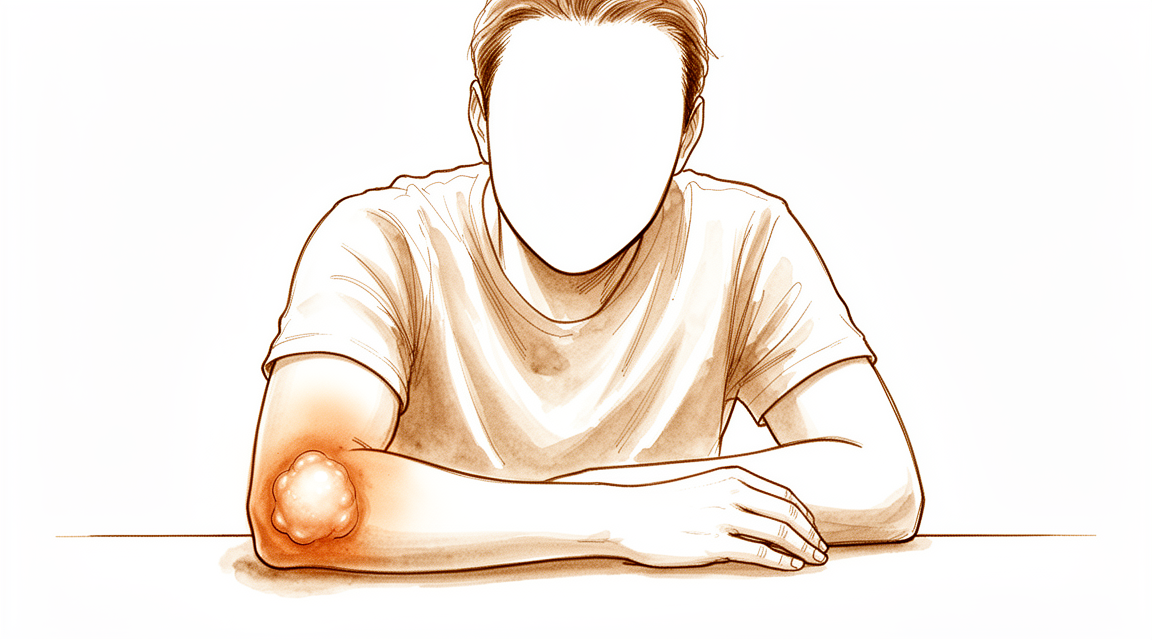
Olecranon bursitis — causes, symptoms, and when to seek urgent medical attention for infection.
நீங்கள் என்ன உணர்கிறீர்கள்
உங்கள் முழங்காலின் நுனியில் ஒரு வீக்கம் ஏற்படுவதை நீங்கள் கவனிப்பீர்கள். இந்த வீக்கம் உங்கள் எலும்பைப் பாதுகாக்கும் ஒரு சிறிய திரவ நிறைந்த பையில் திரவம் குவிப்பதால் ஏற்படுகிறது. இந்த பகுதி தொடுவதற்கு மென்மையாக உணரலாம். குறிப்பாக வீக்கம் ஒரு தொற்றுநோயால் ஏற்பட்டால் அல்லது எலும்பு திசு இரத்த விநியோகத்தை இழந்துவிட்டால், நீங்கள் வலியை உணரலாம். வீக்கம் மற்றும் வலியின் இந்த கலவையானது அன்றாட வாழ்க்கையை சங்கடமாக்கும்.
உங்கள் முழங்கை வீங்கியிருக்கும்போது எளிய இயக்கங்கள் கடினமாகின்றன. உங்கள் முழங்கைகளை ஒரு மேஜை அல்லது மேசையில் ஓய்வெடுக்க நீங்கள் போராடலாம். உங்கள் கையை முற்றிலும் வளைக்க வேண்டிய பணிகள், உங்கள் முதுகுக்குப் பின்னால் ஒரு பிராவைப் பிணைப்பது போன்றவை, வேதனையாக இருக்கலாம். ஒரு சட்டைக்குள் நுழைவது அல்லது இலகுவான பொருட்களை தூக்குவது கூட சங்கடமாகவோ அல்லது சங்கடமாகவோ உணரலாம். வீக்கம் உங்கள் கையை எவ்வளவு நேராக அல்லது வளைக்க முடியும் என்பதைக் கட்டுப்படுத்தலாம்.
உங்கள் கைகளை சிறிது நேரம் பயன்படுத்திய பிறகு வலி பெரும்பாலும் வெடிக்கிறது. இரவில் இது மோசமாக இருக்கலாம், இது ஒரு வசதியான தூக்க நிலையைக் கண்டுபிடிப்பதை கடினமாக்குகிறது. பாதிக்கப்பட்ட பக்கத்தில் படுத்து வீங்கிய புர்சா மீது நேரடி அழுத்தத்தை செலுத்துகிறது, இது மிகவும் வேதனையாக இருக்கும். உங்கள் கையை ஓய்வெடுப்பது அச om கரியத்தை குறைக்க உதவுகிறது, ஆனால் நீண்டகால செயலற்ற தன்மை எப்போதும் வீக்கத்தை தீர்க்காது. வீக்கம் நீண்ட காலமாக நீடித்தால், இது உங்கள் அறுவை சிகிச்சையாளரிடமிருந்து குறிப்பிட்ட கவனம் தேவைப்படும் மிகவும் சிக்கலான பிரச்சினையைக் குறிக்கலாம்.
உண்மையில் என்ன நடக்கிறது
உங்கள் முழங்காலில் ஒரு சிறிய, திரவத்தால் நிரப்பப்பட்ட பை உள்ளது. இது ஒலெக்ரானன் பர்சா என்று அழைக்கப்படுகிறது. இது உங்கள் முழங்காலின் எலும்பு முனையின் மேல் அமைந்துள்ளது. அதை ஒரு சிறிய நீர் பலூன் என்று நினைத்துப் பாருங்கள் இது ஒரு தலையணையாக செயல்படுகிறது. நீங்கள் உங்கள் கையை வளைக்கும்போது உங்கள் தோல் எலும்பு மீது மென்மையாக சரிய அனுமதிக்கிறது.
இந்த பை எரிச்சலடைந்தால், அது அதிக திரவத்துடன் நிரப்பப்படுகிறது. இது உங்கள் முழங்கை முனையில் வீக்கம் மற்றும் வலியை ஏற்படுத்துகிறது. இந்த நிலை ஒலெக்ரானன் பர்சிடிஸ் என்று அழைக்கப்படுகிறது. இது உங்கள் முழங்கை மீது விழுவது போன்ற திடீர் தாக்கத்தால் ஏற்படலாம். இது நீண்ட காலமாக கடினமான மேற்பரப்புகளில் சாய்ந்திருப்பதால் வரலாம். சில நேரங்களில், ஒரு தொற்று அல்லது பிற நோய் அதைத் தூண்டுகிறது.
சில சந்தர்ப்பங்களில், வீக்கம் தானாகவே போகாது. திரவம் தடிமனாக இருக்கலாம் அல்லது வடுக்கள் திசுவாக மாறலாம். இது நாள்பட்ட பர்சிடிஸ் என்று அழைக்கப்படுகிறது. நீங்கள் தோலின் கீழ் ஒரு கடினமான கட்டி உணரலாம். இது ஒரு ஓலெக்ரானன் கோட் என்று அழைக்கப்படுகிறது. இது உங்கள் முழங்கை இறுக்கமாக அல்லது இறுக்கமாக உணரக்கூடும்.
தொற்றுநோயால் வீக்கம் ஏற்பட்டால், அது செப்டிக் புர்சைடிஸ் என்று அழைக்கப்படுகிறது. உங்கள் அறுவை சிகிச்சையாளர் இதை விரைவாக சிகிச்சையளிக்க வேண்டும். சிகிச்சையளிக்கப்படாத தொற்றுநோய்கள் பரவக்கூடும். சில சந்தர்ப்பங்களில், முழங்கை சுற்றியுள்ள மென்மையான திசுக்களில் உடல் கூடுதல் எலும்புகளை உருவாக்குகிறது. இது ஹெட்டரோடோபிக் ஆஸிஃபிகேஷன் என்று அழைக்கப்படுகிறது. இது உங்கள் கையை எவ்வளவு தூரம் வளைக்க முடியும் என்பதைக் கட்டுப்படுத்தலாம். இந்த கூடுதல் எலும்பை ஆரம்பத்தில் அகற்றுவது பெரும்பாலும் நீங்கள் விரைவாக இயக்கம் பெற உதவுகிறது.
உங்கள் அறுவை சிகிச்சையாளர் வீக்கத்திற்கு காரணம் என்ன என்று பார்ப்பார். பெரும்பாலான நோயாளிகள் ஓய்வு, பனி, மற்றும் சுருக்கத்துடன் சிறப்பாக வருவார்கள். உங்களுக்கு அறுவை சிகிச்சை தேவையில்லை. வீக்கம் தொடர்ந்து வந்தால், உங்கள் அறுவை சிகிச்சையாளர் திரவத்தை வடிகட்டலாம். அவர்கள் சாக் சுருக்க மருந்து செலுத்த ஒரு ஊசி பயன்படுத்தலாம். இது ஸ்க்லெரோதெரபி என்று அழைக்கப்படுகிறது.
அரிதான சந்தர்ப்பங்களில், அறுவை சிகிச்சை தேவைப்படுகிறது. உங்கள் அறுவை சிகிச்சையாளர் முழு புர்சா சாக்கையும் அகற்றலாம். இது ஒரு புர்செக்டோமி என்று அழைக்கப்படுகிறது. நவீன நுட்பங்கள் இதை சிறிய வெட்டுக்களுடன் செய்ய அனுமதிக்கின்றன. மீட்பு பொதுவாக விரைவாக உள்ளது. பெரும்பாலான மக்கள் விரைவில் இயல்பான நடவடிக்கைகளுக்குத் திரும்புகிறார்கள். இருப்பினும், சுமார் 11.5% நோயாளிகளுக்கு அறுவை சிகிச்சைக்குப் பிறகு மற்றொரு செயல்முறை தேவைப்படுகிறது. வடு திசு கயிறுகள் கொண்ட சில நோயாளிகள் விளைவாக குறைவாக திருப்தி அடைகிறார்கள். உங்கள் குறிப்பிட்ட சூழ்நிலையின் அடிப்படையில் உங்கள் அறுவை சிகிச்சையாளர் உங்களுக்கான சிறந்த விருப்பத்தைப் பற்றி விவாதிப்பார்.
நாம் என்ன செய்ய முடியும்
சுய-பராமரிப்பு மற்றும் உடலியல் சிகிச்சையுடன் தொடங்குகிறோம். வீக்கத்தைக் குறைக்க நீங்கள் முழங்கை ஓய்வெடுக்கலாம் மற்றும் பனியைப் பயன்படுத்தலாம். மூட்டு நெகிழ்வாக இருக்க உங்கள் உடலியல் சிகிச்சையாளர் மென்மையான இயக்கங்கள் மூலம் உங்களுக்கு வழிகாட்டுவார். இந்த அணுகுமுறை ஆக்கிரமிப்பு நடைமுறைகளைத் தவிர்க்கிறது. சமீபத்திய இலக்கியம், ஆக்கிரமிப்பு அல்லாத மேலாண்மை பெரும்பாலும் நோய்க்குறைப்பு நோய்க்கு சிறந்த ஆரம்ப படியாகும் என்பதைக் காட்டுகிறது. ஊசி அல்லது அறுவை சிகிச்சையுடன் ஒப்பிடும்போது இது அபாயங்களைக் குறைக்கிறது. சிக்கலற்ற நோய்க்குறைப்பு நோய்க்குறி பெரும்பாலான வழக்குகள் அனுபவ மேலாண் மூலம் மட்டுமே தீர்க்கப்படுகின்றன. அதிக ஆக்கிரமிப்பு விருப்பங்களைக் கருத்தில் கொள்வதற்கு முன்பு இந்த பழமைவாத பராமரிப்புக்கு போதுமான நேரம் கொடுக்க வேண்டும்.
வலி நீடித்தால், உங்கள் அறுவை சிகிச்சை நிபுணர் மருத்துவ மேலாண்மை பற்றி விவாதிக்கலாம். பாக்டீரியா தொற்றுநோய்களுக்கு, நுண்ணுயிர் எதிர்ப்பிகள் முதன்மை சிகிச்சையாகும். நிலையான கவனிப்புக்கு பதிலளிக்காத தொடர்ச்சியான புர்சிடிஸின் சில சந்தர்ப்பங்களில், உங்கள் அறுவை சிகிச்சை நிபுணர் இன்ட்ராபிரஸ்ஸல் டாக்ஸிசைக்லின் ஸ்க்லெரோதெரபி பரிந்துரைக்கலாம். இது சுருக்கத்திற்கு ஒரு கரைசலை ஊசிவிடுவதை உள்ளடக்கியது. இது பிடிவாதமான வழக்குகளுக்கு அறுவை சிகிச்சைக்கு ஒரு சிறந்த மாற்றாகும். நாள்பட்ட அல்லது தொடர்ச்சியான தொற்றுநோயற்ற புர்சிடிஸுக்கு, ஹைட்ரோதெர்மல் அப்லேஷன் மற்றொரு விருப்பமாகும். இந்த பகுதிக்கு சிகிச்சையளிக்க 50C மற்றும் 52C க்கு இடையில் வெப்பத்தைப் பயன்படுத்துகிறது. இது திறந்த அறுவை சிகிச்சையை விட குறைவான சிக்கல்களைக் கொண்டுள்ளது மற்றும் அதேபோல் செயல்படுகிறது. பழைய ஆய்வுகள் நோயற்ற புர்சிடிஸுக்கு ஊசிகளை
தற்காப்பு பராமரிப்பு தோல்வியுற்றால் மட்டுமே அறுவை சிகிச்சை கருதப்படுகிறது. உங்கள் பர்சிடிஸ் மீண்டும் வந்தால் அல்லது மருந்து மற்றும் ஓய்வோடு மேம்படவில்லை என்றால், அறுவை சிகிச்சை நீக்கம் செய்யப்படலாம். இந்த நடைமுறை அழற்சி செய்யப்பட்ட பர்சாவை முழுவதுமாக நீக்குகிறது. இது மீண்டும் நிகழும் நிகழ்வுகளுக்கு இது ஒரு நம்பகமான தீர்வாகும். அறுவை சிகிச்சைக்குப் பிறகு முடிவுகள் பொதுவாக செயல்பாடு மற்றும் தோற்றம் இரண்டிற்கும் நல்லது. நாள்பட்ட அதிர்ச்சிகரமான பர்சிடிஸின் சில தேர்ந்தெடுக்கப்பட்ட நிகழ்வுகளில், பர்சல் தையல் பழுது பர்சாவை முழுவதுமாக அகற்றுவதற்கு ஒரு சாத்தியமான மாற்றாகும். இந்த முறை செயல்பாட்டு மற்றும் ஒப்பனை நன்மைகளை ஒருங்கிணைக்கிறது. சுருக்க தையலுடன் இணைந்த எண்டோஸ்கோபிக் டிப்ரிட்மென்ட் மற்றொரு குறைந்தபட்சமாக ஆக்கிரமிப்பு விருப்பமாகும். இது குறைந்தபட்ச அறுவைக்குப் பிந்தைய வலி மற்றும் குறைந்த மறுபரிசீலனை விகிதத்துடன் விரைவாக மீட்க அனுமதிக்கிறது. உங்கள் அறுவை சிகிச்சை உங்கள்
எதிர்பார்ப்பது என்ன
உங்கள் முன்னோக்கு பெரும்பாலும் வீக்கத்தின் காரணத்தையும் அதை நீங்கள் எவ்வாறு நிர்வகிக்கிறீர்கள் என்பதையும் பொறுத்தது. சிக்கலற்ற செப்டிக் புர்சைடிஸின் பெரும்பாலான சந்தர்ப்பங்களில், உங்கள் அறுவை சிகிச்சையாளர் நுண்ணுயிர் எதிர்ப்பிகள் மூலம் மட்டுமே தொற்றுநோயை சிகிச்சையளிக்க முடியும். இந்த சூழ்நிலைகளில், எந்த அறுவை சிகிச்சையும் தேவையில்லை, மேலும் நிலைமை பொதுவாக சரியான கவனிப்புடன் அமைகிறது. உங்கள் புர்சைடிஸ் ஒரு தொற்றுநோயால் ஏற்படவில்லை என்றால், சமீபத்திய சான்றுகள் அறுவை சிகிச்சை அல்லது அறுவை சிகிச்சையை விட அறுவை சிகிச்சை பெரும்பாலும் பாதுகாப்பானது மற்றும் மிகவும் பயனுள்ளதாக இருக்கும் என்பதைக் காட்டுகிறது.
சில நோயாளிகள் புர்ஸெக்டோமி என்று அழைக்கப்படும் ஒரு நடைமுறைக்கு உட்படுகிறார்கள், அங்கு வீங்கிய புர்சா அகற்றப்படுகிறது. இந்த அறுவை சிகிச்சைக்குப் பிறகு சுமார் 11.5% நோயாளிகளுக்கு இரண்டாவது நடைமுறை தேவைப்படுகிறது. ஓலெக்ரானன் கயிறுகள் எனப்படும் குறிப்பிட்ட திசு மாற்றங்கள் இருந்தால், கயிறுகள் இல்லாதவர்களுடன் ஒப்பிடும்போது அறுவை சிகிச்சையின் முடிவுகளில் நீங்கள் குறைவாக திருப்தி அடையலாம்.
சில சந்தர்ப்பங்களில், வீக்கம் நீடிக்கலாம் அல்லது மீண்டும் வரலாம். உங்கள் பர்சிடிஸ் மீண்டும் மீண்டும் வந்தால், இது ஒரு குறிப்பிட்ட வகை பாக்டீரியா தொற்று போன்ற மிகவும் அசாதாரண காரணத்தைக் குறிக்கலாம். உங்கள் அறுவை சிகிச்சை நிபுணர் இந்த குறைவான பொதுவான காரணங்களை சரிபார்க்க திசு மாதிரிகளை எடுப்பார். நாள்பட்ட அல்லது தொடர்ச்சியான வழக்குகளுக்கு, ஹைட்ரோதர்மல் அப்லேஷன் (50 ° C மற்றும் 52 ° C க்கு இடையில் வெப்பத்தைப் பயன்படுத்தி) போன்ற புதிய நுட்பங்கள் திறந்த அறுவை சிகிச்சையை விட குறைவான சிக்கல்களுடன் பாதுகாப்பான மாற்றீட்டை வழங்குகின்றன. மற்றொரு விருப்பம் எண்டோஸ்கோபிக் டிப்ரிட்மென்ட் ஆகும், இது குறைந்தபட்ச ஆக்கிரமிப்பு மற்றும் குறைந்த தொடர்ச்சியான விகிதத்துடன் விரைவான மீட்பு ஆகியவற்றை உள்ளடக்கியது.
பழைய ஆய்வுகள் ஊசி மற்றும் அறுவை சிகிச்சை என்பது செப்டிக் அல்லாத பர்சிடிஸுக்கு நிலையான தீர்மானங்கள் என்று பரிந்துரைத்திருந்தாலும், தற்போதைய தரவு இந்த அணுகுமுறைகளிலிருந்து சாத்தியமான பாதகமான விளைவுகளை முன்னிலைப்படுத்துகிறது. எனவே, உங்கள் அறுவை சிகிச்சையாளர் முதலில் எளிமையான, ஆக்கிரமிப்பு அல்லாத படிகளுக்கு முன்னுரிமை அளிப்பார். அறுவை சிகிச்சை தேவைப்பட்டால், எண்டோஸ்கோபிக் பர்செக்டோமி போன்ற நவீன முறைகள் ஆய்வு செய்யப்பட்ட குழுக்களில் அறுவை சிகிச்சை அறைக்குத் திரும்புதல் அல்லது காயத்தை குணப்படுத்தும் சிக்கல்களைக் காட்டவில்லை. தேர்ந்தெடுக்கப்பட்ட பாதையைப் பொறுத்து உங்கள் மீட்பு வித்தியாசமாக இருக்கும், ஆனால் குறிக்கோள் எப்போதும் வீக்கத்தை தீர்க்கவும், குறைந்தபட்சமான ஆக்கிரமிப்பு சிகிச்சையுடன் ஆறுதலை மீட்டெடுக்கவும் ஆகும்.
யாரையாவது எப்போது பார்க்க வேண்டும்
உங்கள் முழங்கை வீக்கம் ஓய்வில் குணமடையாவிட்டால் உங்கள் GP ஐப் பார்க்கவும். நீங்கள் ஆரோக்கியமாக இருந்தாலும், வீக்கம் நீண்ட நேரம் நீடித்தால் ஒரு நிபுணர் மதிப்பாய்வை நாடுங்கள். இது ஒரு அசாதாரண தொற்றுநோயைக் குறிக்கலாம். உங்களுக்கு மீண்டும் மீண்டும் வீக்கம் இருந்தால் உதவியை கேளுங்கள், ஏனெனில் இது சிறப்பு சோதனைகள் தேவைப்படலாம். வீக்கம் தொற்றுநோயால் அல்லது பிற சிக்கல்களால் ஏற்படுகிறதா என்பதைக் கூறுவது கடினம். தொற்றுநோயைப் போல தோற்றமளிக்கும் தோலில் திறந்த காயங்கள் ஏற்பட்டால் உங்கள் மருத்துவரை அணுகவும். முழங்கையில் திடீர் வலி அல்லது பலவீனம் ஏற்பட்டால் சரிபார்க்கப்படுங்கள். இந்த அறிகுறிகளுக்கு சரியான சிகிச்சையைக் கண்டறிய சரியான மதிப்பீடு தேவை.
Evidence & references
Overview
- Non-operative treatment of olecranon fracture in patients aged ≥75 years provided excellent functional results at 6 months, without associated complications [1].
- Nonsurgical management of olecranon bursitis is significantly more effective and safer than surgical management [2].
- Nontuberculous mycobacterial olecranon bursitis should be considered in any patient with a swollen bursa and protracted course, regardless of immune status [3].
- Intrabursal doxycycline sclerotherapy may be an effective alternative to surgical bursectomy for patients with recurrent olecranon bursitis refractory to conservative management [4].
- Patients who underwent endoscopic olecranon bursectomy experienced no recurrences or wound-healing complications necessitating return to the operating room [5].
- Empirical management of uncomplicated septic olecranon bursitis was found to be effective with no patients requiring bursectomy, whereas 8 of 11 patients in the traditional aspiration group required bursectomy [6].
- More recent literature demonstrates adverse effects of intrabursal injections and surgery compared with noninvasive management for initial treatment of nonseptic olecranon bursitis [7].
- The revision rate after bursectomy for olecranon bursitis was 11.5% [8].
- Hydrothermal ablation at temperatures between 50C and 52C is a safe treatment option for recurrent or chronic olecranon bursitis with fewer complications than open bursectomy and a comparable efficacy [11].
- Patients with olecranon cords were less satisfied after surgical excision compared to those without cords [13].
- Low-profile double-plate osteosynthesis is a safe and effective alternative treatment of olecranon fractures with excellent subjective and objective clinical outcome measures [14].
- Endoscopic debridement combined with compression suture for the treatment of aseptic olecranon bursitis has several advantages: simple operation, minimal invasiveness, minimal postoperative pain, rapid recovery, a low recurrence rate, and satisfactory overall efficacy [16].
Anatomy & Pathophysiology
- Posteromedial elbow impingement is a source of disability in overhead throwing athletes [12].
- Boxers are prone to the development of anterior and posterior elbow impingement lesions involving the coronoid and olecranon process [33].
- In boxer's elbow, the lead arm is more vulnerable to impingement lesions than the non-lead arm [33].
- Evaluation and management of elbow injuries in young athletes requires knowledge of immature developing anatomy [25].
Classification
- Non-operative treatment of olecranon fracture in patients aged ≥75 years provided excellent functional results at 6 months, without associated complications [1].
- Nonsurgical management of olecranon bursitis is significantly more effective and safer than surgical management [2].
- Nontuberculous mycobacterial olecranon bursitis should be considered in any patient with a swollen bursa and protracted course, regardless of immune status [3].
- Intrabursal doxycycline sclerotherapy may be an effective alternative to surgical bursectomy for patients with recurrent olecranon bursitis refractory to conservative management [4].
- Patients who underwent endoscopic olecranon bursectomy for recalcitrant olecranon bursitis experienced no recurrences or wound-healing complications necessitating return to the operating room [5].
- Empirical management of uncomplicated septic olecranon bursitis was found to be effective with no patients requiring bursectomy, whereas 8 of 11 patients in the traditional aspiration group required bursectomy [6].
- More recent literature demonstrates adverse effects of intrabursal injections and surgery compared with noninvasive management for initial treatment of nonseptic olecranon bursitis [7].
- The revision rate after bursectomy for olecranon bursitis was 11.5% [8].
- Excision has been curative for all lesions of the olecranon bursa, whereas multiple medications have been tried for cutaneous and systemic infections without clear-cut success [9].
- Distinguishing between septic and aseptic olecranon bursitis can be difficult because the physical and laboratory data overlap [10].
- Posteromedial elbow impingement is a source of disability in the overhead throwing athlete [12].
- The first treatment line for olecranon bursitis is conservative, including ice, rest, anti-inflammatory and analgesic drugs and, occasionally, bursal fluid aspiration [15].
- Pyoderma gangrenosum must be considered in the differential diagnosis whenever a patient presents with ulcerative cutaneous lesions that resemble an infectious process such as olecranon bursitis [17].
- The available evidence did not support the central European concept of immediate bursectomy in cases of septic bursitis [22].
Clinical Presentation
- Non-operative treatment of olecranon fracture in patients aged ≥75 years provided excellent functional results at 6 months, without associated complications [1].
- Nonsurgical management of olecranon bursitis is significantly more effective and safer than surgical management [2].
- Nontuberculous mycobacterial olecranon bursitis should be considered in any patient with a swollen bursa and protracted course, regardless of immune status [3].
- Empirical management of uncomplicated septic olecranon bursitis was found to be effective with no patients requiring bursectomy, whereas 8 of 11 patients in the traditional aspiration group required bursectomy [6].
- Older studies showed resolution with injections and surgery, but more recent literature demonstrates adverse effects of intrabursal injections and surgery compared with noninvasive management for initial treatment of nonseptic olecranon bursitis [7].
- The revision rate after bursectomy for olecranon bursitis was 11.5% [8].
- Excision has been curative for all lesions of the olecranon bursa, whereas multiple medications have been tried for cutaneous and systemic infections without clear-cut success [9].
- Distinguishing between septic and aseptic olecranon bursitis can be difficult because the physical and laboratory data overlap [10].
- The first treatment line for olecranon bursitis is conservative, including ice, rest, anti-inflammatory and analgesic drugs and, occasionally, bursal fluid aspiration [15].
- PG must be considered in the differential diagnosis whenever a patient presents with ulcerative cutaneous lesions that resemble an infectious process such as olecranon bursitis [17].
- Diagnosis and proper management of the infected bursa and dermatitis have prevented recurrence [31].
- In the rheumatoid patient, septic arthritis of the elbow joint can mimic septic olecranon bursitis, and the fact that the elbow joint may also be involved should be suspected in the rheumatoid patient who has what appears to be a septic olecranon bursitis [34].
Investigations
- Distinguishing between septic and aseptic olecranon bursitis can be difficult because physical and laboratory data overlap [10].
- Nontuberculous mycobacterial olecranon bursitis should be considered in any patient with a swollen bursa and protracted course, regardless of immune status [3].
- Pyoderma gangrenosum must be considered in the differential diagnosis whenever a patient presents with ulcerative cutaneous lesions that resemble an infectious process such as olecranon bursitis [17].
- Early use of MRI and cautious interpretation of posterior elbow palpation signs are crucial parts of the diagnosis of osteochondral injury of the elbow trochlea [21].
Treatment
Non-Operative Management
- Nonsurgical management of olecranon bursitis is significantly more effective and safer than surgical management [2].
- Noninvasive management is preferred for the initial treatment of nonseptic olecranon bursitis due to adverse effects associated with intrabursal injections and surgery [7].
- The first treatment line for olecranon bursitis is conservative, including ice, rest, anti-inflammatory and analgesic drugs, and occasionally bursal fluid aspiration [15].
- Empirical management of uncomplicated septic olecranon bursitis without aspiration was effective, with no patients requiring bursectomy [6].
- In a comparison of empirical management versus traditional aspiration for uncomplicated septic olecranon bursitis, 8 of 11 patients in the traditional aspiration group required bursectomy [6].
Surgical and Interventional Management
- Intrabursal doxycycline sclerotherapy may be an effective alternative to surgical bursectomy for patients with recurrent olecranon bursitis refractory to conservative management [4].
- Hydrothermal ablation at temperatures between 50°C and 52°C is a safe treatment option for recurrent or chronic olecranon bursitis, offering fewer complications than open bursectomy and comparable efficacy [11].
- Endoscopic debridement combined with compression suture for recalcitrant aseptic olecranon bursitis offers minimal invasiveness, minimal postoperative pain, rapid recovery, a low recurrence rate, and satisfactory overall efficacy [16].
- Patients undergoing endoscopic olecranon bursectomy for recalcitrant olecranon bursitis experienced no recurrences or wound-healing complications necessitating return to the operating room [5].
- The revision rate after bursectomy for olecranon bursitis is 11.5% [8].
- Excision has been curative for lesions of the olecranon bursa, whereas multiple medications have been tried for cutaneous and systemic infections without clear-cut success [9].
Diagnostic Considerations Relevant to Treatment
- Distinguishing between septic and aseptic olecranon bursitis can be difficult because physical and laboratory data overlap [10].
- Nontuberculous mycobacterial olecranon bursitis should be considered in any patient with a swollen bursa and protracted course, regardless of immune status [3].
Complications
- Non-operative treatment of olecranon fracture in patients aged ≥75 years provided excellent functional results at 6 months without associated complications [1].
- Nonsurgical management of olecranon bursitis is significantly more effective and safer than surgical management [2].
- Intrabursal injections and surgery have adverse effects compared with noninvasive management for initial treatment of nonseptic olecranon bursitis [7].
- The revision rate after bursectomy for olecranon bursitis was 11.5% [8].
- Patients with olecranon cords were less satisfied after surgical excision compared to those without cords [13].
- Plating of the olecranon leads to predictable union, though the most common complication was lack of full extension in 39% of patients [19].
Recovery
Non-Operative Management
- Non-surgical management of olecranon bursitis is significantly more effective and safer than surgical management [2].
- More recent literature demonstrates adverse effects of intrabursal injections and surgery compared with noninvasive management for initial treatment of nonseptic olecranon bursitis [7].
- Empirical management of uncomplicated septic olecranon bursitis without aspiration was found to be effective, with no patients requiring bursectomy [6].
- In a comparison group, 8 of 11 patients in the traditional aspiration group required bursectomy for uncomplicated septic olecranon bursitis [6].
- The available evidence did not support the central European concept of immediate bursectomy in cases of septic bursitis [22].
Interventional and Operative Management
- Intrabursal doxycycline sclerotherapy may be an effective alternative to surgical bursectomy for patients with recurrent olecranon bursitis refractory to conservative management [4].
- Hydrothermal ablation at temperatures between 50C and 52C is a safe treatment option for recurrent or chronic olecranon bursitis with fewer complications than open bursectomy and comparable efficacy [11].
- Patients who underwent endoscopic olecranon bursectomy for recalcitrant olecranon bursitis experienced no recurrences or wound-healing complications necessitating return to the operating room [5].
- The revision rate after bursectomy for olecranon bursitis was 11.5% [8].
- Patients with olecranon cords were less satisfied after surgical excision compared to those without cords [13].
- Excision has been curative for all lesions of the olecranon bursa in cases of protothecal olecranon bursitis [9].
Specific Etiologies and Considerations
- Nontuberculous mycobacterial olecranon bursitis should be considered in any patient with a swollen bursa and protracted course, regardless of immune status [3].
Key Evidence
- [L4] Non-operative treatment of olecranon fracture in patients aged ≥75 years provided excellent functional results at 6 months, without associated complications. [1] (10.1016/j.otsr.2017.10.015)
- [L4] Based primarily on level IV evidence, nonsurgical management of olecranon bursitis is significantly more effective and safer than surgical management. [2] (10.1007/s00402-014-2088-3)
- [L4] Nontuberculous mycobacterial olecranon bursitis should be considered in any patient with a swollen bursa and protracted course, regardless of immune status. [3] (10.1016/j.jse.2008.07.009)
- [L4] This may be an effective alternative to surgical bursectomy for patients with recurrent olecranon bursitis refractory to conservative management. [4] (10.1016/j.jhsg.2024.03.006)
- [L4] In this population, patients who underwent endoscopic olecranon bursectomy experienced no recurrences or wound-healing complications necessitating return to the operating room. [5] (10.1016/j.asmr.2023.100832)
- [L4] Empirical management of uncomplicated septic olecranon bursitis was found to be effective with no patients requiring bursectomy, whereas 8 of 11 patients in the traditional aspiration group required bursectomy. [6] (10.1016/j.jhsa.2019.06.012)
- [L5] Older studies showed resolution with injections and surgery, but more recent literature demonstrates adverse effects of intrabursal injections and surgery compared with noninvasive management for initial treatment of nonseptic olecranon bursitis. [7] (10.1016/j.jhsa.2021.02.006)
- [L3] The revision rate after bursectomy for olecranon bursitis was 11.5%. [8] (10.1016/j.jse.2020.09.033)
- [Case_report] Excision has been curative for all lesions of the olecranon bursa, whereas multiple medications have been tried for cutaneous and systemic infections without clear-cut success. [9] (10.2106/00004623-198062050-00024)
- [L5] Distinguishing between septic and aseptic olecranon bursitis can be difficult because the physical and laboratory data overlap. [10] (10.1016/j.jse.2015.08.032)
- [L4] Hydrothermal ablation at temperatures between 50C and 52C is a safe treatment option for recurrent or chronic olecranon bursitis with fewer complications than open bursectomy and a comparable efficacy. [11] (10.1016/j.jse.2024.03.021)
- [L4] Posteromedial elbow impingement is a source of disability in the overhead throwing athlete. [12] (10.1016/j.arthro.2011.06.012)
- [L4] Patients with olecranon cords were less satisfied after surgical excision compared to those without cords. [13] (10.1016/j.jse.2015.04.016)
- [L3] Low-profile double-plate osteosynthesis is a safe and effective alternative treatment of olecranon fractures with excellent subjective and objective clinical outcome measures. [14] (10.1016/j.otsr.2019.08.019)
- [L4] The first treatment line for olecranon bursitis is conservative, including ice, rest, anti-inflammatory and analgesic drugs and, occasionally, bursal fluid aspiration. [15] (10.1016/j.surge.2012.02.002)
- [L4] Endoscopic debridement combined with compression suture for the treatment of aseptic olecranon bursitis has several advantages: simple operation, minimal invasiveness, minimal postoperative pain, rapid recovery, a low recurrence rate, and satisfactory overall efficacy. [16] (10.1186/s13018-024-05090-3)
- [Case_report] PG must be considered in the differential diagnosis whenever a patient presents with ulcerative cutaneous lesions that resemble an infectious process such as olecranon bursitis. [17] (10.1016/j.jse.2014.06.032)
- [L3] Plating of the olecranon leads to predictable union, though the most common complication was lack of full extension in 39% of patients. [19] (10.1016/j.injury.2016.04.015)
- [Case_report] It also emphasizes the early use of MRI and the cautious interpretation of posterior elbow palpation signs as a crucial part of the diagnosis of this lesion. [21] (10.1016/j.jse.2010.09.015)
- [L1] The available evidence did not support the central European concept of immediate bursectomy in cases of septic bursitis. [22] (10.1007/s00402-013-1882-7)
- [L5] Evaluation and management of elbow injuries in young athletes requires knowledge of the immature developing anatomy, injury pathophysiology, and established treatment algorithms for each diagnosis. [25] (10.1016/j.csm.2010.06.010)
- [L4] Diagnosis and proper management of the infected bursa and dermatitis have prevented recurrence. [31] (10.1016/j.jse.2011.10.013)
- [L4] Boxers are prone to development of anterior and posterior elbow impingement lesions, with the lead arm being more vulnerable. [33] (10.1016/j.jse.2016.09.035)
- [L4] In the rheumatoid patient, septic arthritis of the elbow joint can mimic septic olecranon bursitis, and the fact that the elbow joint may also be involved should be suspected in the rheumatoid patient who has what appears to be a septic olecranon bursitis. [34] (10.2106/00004623-198062060-00022)
References
[1] Results of non-operative treatment of olecranon fracture in over 75-year-olds. Orthopaedics & Traumatology: Surgery & Research. 2018. DOI: 10.1016/j.otsr.2017.10.015 [2] Treatment of olecranon bursitis: a systematic review. Archives of Orthopaedic and Trauma Surgery. 2014. DOI: 10.1007/s00402-014-2088-3 [3] Nontuberculous mycobacterial olecranon bursitis: Case reports and literature review. Journal of Shoulder and Elbow Surgery. 2009. DOI: 10.1016/j.jse.2008.07.009 [4] Intrabursal Doxycycline Sclerotherapy for Recurrent Olecranon Bursitis of the Elbow: A Case Control Study. Journal of Hand Surgery Global Online. 2024. DOI: 10.1016/j.jhsg.2024.03.006 [5] No Wound Healing Complications or Recurrences Were Seen and a High Level of Satisfaction Was Reported in Patients Who Underwent Endoscopic Olecranon Bursectomy for Recalcitrant Olecranon Bursitis. Arthroscopy, Sports Medicine, and Rehabilitation. 2024. DOI: 10.1016/j.asmr.2023.100832 [6] Empirical Treatment of Uncomplicated Septic Olecranon Bursitis Without Aspiration. The Journal of Hand Surgery. 2020. DOI: 10.1016/j.jhsa.2019.06.012 [7] Clinical Management of Olecranon Bursitis: A Review. The Journal of Hand Surgery. 2021. DOI: 10.1016/j.jhsa.2021.02.006 [8] Factors associated with revision surgery for olecranon bursitis after bursectomy. Journal of Shoulder and Elbow Surgery. 2021. DOI: 10.1016/j.jse.2020.09.033 [9] Protothecal olecranon bursitis. A case report and review of the literature.. The Journal of Bone & Joint Surgery. 1980. DOI: 10.2106/00004623-198062050-00024 [10] Olecranon bursitis. Journal of Shoulder and Elbow Surgery. 2016. DOI: 10.1016/j.jse.2015.08.032 [11] Hydrothermal ablation in recurrent or chronic olecranon bursitis: a prospective study. Journal of Shoulder and Elbow Surgery. 2024. DOI: 10.1016/j.jse.2024.03.021 [12] Posteromedial Elbow Impingement: Magnetic Resonance Imaging Findings in Overhead Throwing Athletes and Results of Arthroscopic Treatment. Arthroscopy. 2011. DOI: 10.1016/j.arthro.2011.06.012 [13] The existence of cords in olecranon bursae. Journal of Shoulder and Elbow Surgery. 2015. DOI: 10.1016/j.jse.2015.04.016 [14] Clinical evaluation of double-plate osteosynthesis for olecranon fractures: A retrospective case-control study. Orthopaedics & Traumatology: Surgery & Research. 2019. DOI: 10.1016/j.otsr.2019.08.019 [15] Diagnosis and management of olecranon bursitis. The Surgeon. 2012. DOI: 10.1016/j.surge.2012.02.002 [16] Clinical efficacy of endoscopic debridement combined with compression suture in the treatment of recalcitrant aseptic olecranon bursitis. Journal of Orthopaedic Surgery and Research. 2024. DOI: 10.1186/s13018-024-05090-3 [17] Case report: misdiagnosed olecranon bursitis: pyoderma gangrenosum. Journal of Shoulder and Elbow Surgery. 2014. DOI: 10.1016/j.jse.2014.06.032 [19] Outcomes after plating of olecranon fractures: A multicenter evaluation. Injury. 2016. DOI: 10.1016/j.injury.2016.04.015 [21] Arthroscopic debridement for osteochondral injury of the elbow trochlea: a case report with a long-term follow-up. Journal of Shoulder and Elbow Surgery. 2011. DOI: 10.1016/j.jse.2010.09.015 [22] Prepatellar and olecranon bursitis: literature review and development of a treatment algorithm. Archives of Orthopaedic and Trauma Surgery. 2013. DOI: 10.1007/s00402-013-1882-7 [25] Pediatric Sports Elbow Injuries. Clinics in Sports Medicine. 2010. DOI: 10.1016/j.csm.2010.06.010 [31] Septic olecranon bursitis, contact dermatitis, and pneumonitis in a gas turbine engine mechanic. Journal of Shoulder and Elbow Surgery. 2012. DOI: 10.1016/j.jse.2011.10.013 [33] Boxer's elbow: internal impingement of the coronoid and olecranon process. A report of seven cases. Journal of Shoulder and Elbow Surgery. 2017. DOI: 10.1016/j.jse.2016.09.035 [34] Septic arthritis presenting as olecranon bursitis in patients with rheumatoid arthritis. A report of three cases.. The Journal of Bone & Joint Surgery. 1980. DOI: 10.2106/00004623-198062060-00022


