Viêm bao hoạt dịch khuỷu tay
Patients › Elbow
Olecranon bursitis — causes, symptoms, and when to seek urgent medical attention for infection.
Những gì bạn đang cảm nhận
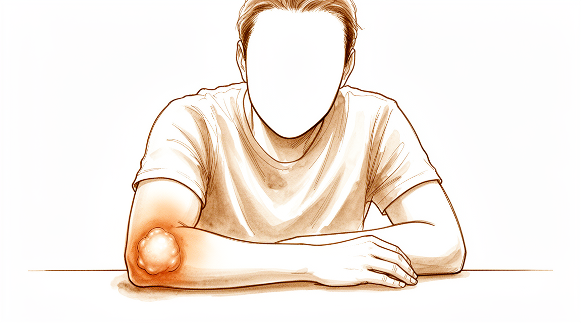
Bạn có thể nhận thấy một khối sưng ở đầu khuỷu tay. Sự sưng này là do tích tụ dịch trong bao hoạt dịch, một túi nhỏ chứa đầy dịch giúp đệm cho xương của bạn. Vùng này có thể cảm thấy nhạy cảm khi chạm vào. Bạn cũng có thể cảm thấy đau, đặc biệt nếu sự sưng là do nhiễm trùng hoặc nếu mô xương đã mất nguồn cung cấp máu. Sự kết hợp giữa sưng và đau này có thể gây khó chịu trong sinh hoạt hàng ngày.
Các cử động đơn giản trở nên khó khăn khi khuỷu tay của bạn bị sưng. Bạn có thể gặp khó khăn khi đặt khuỷu tay lên bàn hoặc mặt phẳng. Các công việc đòi hỏi phải gập cánh tay hoàn toàn, như với ra sau lưng để cài áo ngực, có thể gây đau. Ngay cả việc nhét áo vào quần hoặc nâng các vật nhẹ cũng có thể cảm thấy bất tiện hoặc khó chịu. Sự sưng có thể hạn chế khả năng duỗi hoặc gập cánh tay của bạn.
Cơn đau thường bùng phát sau khi bạn đã sử dụng cánh tay trong một thời gian. Nó cũng có thể nặng hơn vào ban đêm, khiến bạn khó tìm được tư thế ngủ thoải mái. Nằm nghiêng về phía bên bị ảnh hưởng tạo áp lực trực tiếp lên bao hoạt dịch bị sưng, điều này có thể gây đau đáng kể. Bạn có thể nhận thấy rằng nghỉ ngơi cánh tay giúp giảm bớt khó chịu, nhưng việc bất động kéo dài không phải lúc nào cũng làm giảm sự sưng. Nếu sự sưng kéo dài trong một thời gian dài, nó có thể cho thấy một vấn đề phức tạp hơn đòi hỏi sự chú ý cụ thể từ bác sĩ phẫu thuật của bạn.
Những gì thực sự đang xảy ra
Cùi chỏ của bạn có một túi nhỏ chứa dịch gọi là bao hoạt dịch cùi chỏ. Túi này nằm ngay trên đỉnh của xương khuỷu tay. Hãy tưởng tượng nó như một quả bóng nước nhỏ đóng vai trò làm đệm. Nó cho phép da của bạn trượt mượt mà trên xương khi bạn gập cánh tay.
Khi túi này bị kích thích, nó sẽ chứa quá nhiều dịch. Điều này gây ra sưng và đau ở đỉnh khuỷu tay. Tình trạng này được gọi là viêm bao hoạt dịch cùi chỏ. Nó có thể xảy ra do một cú va đập đột ngột, chẳng hạn như ngã xuống khuỷu tay. Nó cũng có thể xuất hiện do tỳ đè lên các bề mặt cứng trong thời gian dài. Đôi khi, một nhiễm trùng hoặc bệnh lý khác kích hoạt tình trạng này.
Trong một số trường hợp, tình trạng sưng không tự khỏi. Dịch có thể vẫn đặc hoặc chuyển thành mô sẹo. Điều này được gọi là viêm bao hoạt dịch mạn tính. Bạn có thể cảm thấy một khối cứng dưới da. Điều này được gọi là dây cùi chỏ. Nó có thể khiến khuỷu tay của bạn cảm thấy cứng hoặc căng.
Nếu tình trạng sưng do nhiễm trùng, nó được gọi là viêm bao hoạt dịch nhiễm trùng. Bác sĩ phẫu thuật của bạn cần điều trị tình trạng này nhanh chóng. Các nhiễm trùng không được điều trị có thể lan rộng. Trong một số trường hợp, cơ thể hình thành xương dư thừa trong mô mềm xung quanh khuỷu tay. Điều này được gọi là cốt hóa dị vị. Nó có thể hạn chế khả năng gập cánh tay của bạn. Việc loại bỏ xương dư thừa sớm thường giúp bạn phục hồi vận động nhanh hơn.
Bác sĩ phẫu thuật của bạn sẽ xem xét nguyên nhân gây ra tình trạng sưng. Hầu hết các trường hợp sẽ cải thiện với việc nghỉ ngơi, chườm đá và chèn ép. Bạn có thể không cần phẫu thuật. Nếu tình trạng sưng tiếp tục tái phát, bác sĩ phẫu thuật của bạn có thể dẫn lưu dịch. Họ cũng có thể sử dụng kim để tiêm thuốc làm teo túi bao hoạt dịch. Điều này được gọi là liệu pháp xơ hóa.
Trong các trường hợp hiếm, cần phải phẫu thuật. Bác sĩ phẫu thuật của bạn có thể loại bỏ toàn bộ túi bao hoạt dịch. Điều này được gọi là cắt bao hoạt dịch. Các kỹ thuật hiện đại cho phép thực hiện điều này với các vết rạch nhỏ. Quá trình phục hồi thường diễn ra nhanh chóng. Hầu hết mọi người trở lại các hoạt động bình thường ngay sau đó. Tuy nhiên, khoảng 11,5% bệnh nhân cần một thủ thuật khác sau phẫu thuật. Một số bệnh nhân có dây mô sẹo không hài lòng với kết quả. Bác sĩ phẫu thuật của bạn sẽ thảo luận về lựa chọn tốt nhất cho bạn dựa trên tình trạng cụ thể của bạn.
Những gì chúng tôi có thể làm về vấn đề này
Chúng tôi bắt đầu bằng việc tự chăm sóc và vật lý trị liệu. Bạn có thể nghỉ ngơi khuỷu tay và chườm đá để giảm sưng. Chuyên viên vật lý trị liệu của bạn sẽ hướng dẫn bạn thực hiện các cử động nhẹ nhàng để duy trì tính linh hoạt của khớp. Cách tiếp cận này tránh các thủ thuật xâm lấn. Tài liệu gần đây cho thấy rằng quản lý không xâm lấn thường là bước đầu tiên tốt nhất đối với viêm bao hoạt dịch không do nhiễm trùng. Nó giảm thiểu rủi ro so với tiêm hoặc phẫu thuật. Hầu hết các trường hợp viêm bao hoạt dịch do nhiễm trùng không biến chứng sẽ tự khỏi chỉ với điều trị kinh nghiệm. Bạn nên dành đủ thời gian cho phương pháp điều trị bảo tồn này trước khi xem xét các lựa chọn tích cực hơn.
Nếu tình trạng đau vẫn tiếp diễn, bác sĩ phẫu thuật của bạn có thể thảo luận về quản lý bằng thuốc. Đối với các nhiễm trùng do vi khuẩn, kháng sinh là phương pháp điều trị chính. Trong một số trường hợp viêm bao hoạt dịch tái phát không đáp ứng với điều trị tiêu chuẩn, bác sĩ phẫu thuật của bạn có thể đề nghị liệu pháp xơ hóa doxycycline trong bao hoạt dịch. Điều này liên quan đến việc tiêm một dung dịch vào bao hoạt dịch để làm co nhỏ nó. Đây là một lựa chọn thay thế hiệu quả cho phẫu thuật đối với các trường hợp cứng đầu. Đối với viêm bao hoạt dịch không do nhiễm trùng mạn tính hoặc tái phát, đốt nhiệt thủy nhiệt là một lựa chọn khác. Phương pháp này sử dụng nhiệt độ từ 50°C đến 52°C để điều trị vùng bị ảnh hưởng. Nó có ít biến chứng hơn phẫu thuật mở và hiệu quả tương đương. Trong khi các nghiên cứu cũ hỗ trợ việc tiêm cho viêm bao hoạt dịch không do nhiễm trùng, bằng chứng mới nhấn mạnh các tác dụng phụ tiềm ẩn. Do đó, chúng tôi chỉ dự trữ việc tiêm cho các tình huống cụ thể khi chúng mang lại lợi ích rõ ràng.
Phẫu thuật chỉ được xem xét khi điều trị bảo tồn thất bại. Nếu viêm bao hoạt dịch của bạn tái phát hoặc không cải thiện với thuốc và nghỉ ngơi, việc cắt bỏ bao hoạt dịch bằng phẫu thuật có thể được chỉ định. Thủ thuật này loại bỏ hoàn toàn bao hoạt dịch bị viêm. Đây là một giải pháp đáng tin cậy cho các trường hợp tái phát. Kết quả sau khi cắt bỏ bao hoạt dịch bằng phẫu thuật thường tốt cả về chức năng và thẩm mỹ. Trong một số trường hợp chọn lọc của viêm bao hoạt dịch do chấn thương mạn tính, khâu vá bao hoạt dịch là một lựa chọn thay thế khả thi cho việc loại bỏ hoàn toàn bao hoạt dịch. Phương pháp này kết hợp cả lợi ích về chức năng và thẩm mỹ. Gạt bỏ nội soi kết hợp với khâu ép là một lựa chọn ít xâm lấn khác. Nó cho phép phục hồi nhanh chóng với ít đau sau phẫu thuật và tỷ lệ tái phát thấp. Bác sĩ phẫu thuật của bạn sẽ chọn phương pháp tiếp cận tốt nhất dựa trên tình trạng và tiền sử cụ thể của bạn.
Những điều cần biết
Tiên lượng của bạn phụ thuộc phần lớn vào nguyên nhân gây sưng và cách bạn quản lý tình trạng này. Đối với hầu hết các trường hợp viêm bao hoạt dịch nhiễm khuẩn không biến chứng, bác sĩ phẫu thuật của bạn có thể điều trị nhiễm trùng bằng kháng sinh đơn thuần. Trong những trường hợp này, không cần phẫu thuật và tình trạng bệnh thường thuyên giảm khi được chăm sóc đúng cách. Nếu viêm bao hoạt dịch của bạn không do nhiễm trùng, các bằng chứng gần đây cho thấy rằng quản lý không xâm lấn thường an toàn và hiệu quả hơn so với tiêm hoặc phẫu thuật trong điều trị ban đầu.
Nếu bạn bị viêm bao hoạt dịch tái phát không cải thiện với điều trị bảo tồn, bác sĩ phẫu thuật của bạn có thể thảo luận về các lựa chọn thủ thuật. Một số bệnh nhân được thực hiện một thủ thuật gọi là cắt bao hoạt dịch, trong đó bao hoạt dịch bị sưng được loại bỏ. Khoảng 11,5% bệnh nhân cần một thủ thuật thứ hai sau phẫu thuật này. Nếu bạn có những thay đổi mô cụ thể được gọi là dây chằng khuỷu tay (olecranon cords), bạn có thể ít hài lòng hơn với kết quả của việc cắt bỏ phẫu thuật so với những người không có dây chằng này.
Trong một số trường hợp, tình trạng sưng có thể dai dẳng hoặc quay trở lại. Nếu viêm bao hoạt dịch của bạn tái phát nhiều lần, nó có thể báo hiệu một nguyên nhân ít phổ biến hơn, chẳng hạn như một loại nhiễm trùng vi khuẩn cụ thể. Bác sĩ phẫu thuật của bạn có thể sẽ lấy mẫu mô để kiểm tra các nguyên nhân ít phổ biến hơn này. Đối với các trường hợp mạn tính hoặc tái phát, các kỹ thuật mới như đốt nhiệt thủy lực (sử dụng nhiệt độ từ 50°C đến 52°C) cung cấp một lựa chọn an toàn với ít biến chứng hơn so với phẫu thuật mở. Một lựa chọn khác là nạo nội soi, liên quan đến tính xâm lấn tối thiểu và thời gian hồi phục nhanh với tỷ lệ tái phát thấp.
Trong khi các nghiên cứu cũ cho thấy rằng tiêm và phẫu thuật là các giải pháp tiêu chuẩn cho viêm bao hoạt dịch không nhiễm trùng, dữ liệu hiện tại nhấn mạnh các tác dụng phụ tiềm ẩn từ những phương pháp này. Do đó, bác sĩ phẫu thuật của bạn có thể sẽ ưu tiên các bước đơn giản, không xâm lấn trước tiên. Nếu phẫu thuật trở nên cần thiết, các phương pháp hiện đại như cắt bao hoạt dịch nội soi đã cho thấy không có tỷ lệ tái phát hoặc biến chứng lành vết thương đòi hỏi phải quay trở lại phòng mổ trong các nhóm được nghiên cứu. Quá trình hồi phục của bạn sẽ cảm thấy khác nhau tùy thuộc vào con đường đã chọn, nhưng mục tiêu luôn là giải quyết tình trạng sưng và khôi phục sự thoải mái với phương pháp điều trị hiệu quả xâm lấn tối thiểu.
Khi nào cần gặp bác sĩ
Hãy gặp bác sĩ đa khoa nếu bạn bị sưng khuỷu tay không cải thiện sau khi nghỉ ngơi. Hãy tìm kiếm sự đánh giá từ bác sĩ chuyên khoa nếu tình trạng sưng kéo dài, ngay cả khi bạn đang khỏe mạnh. Điều này có thể là dấu hiệu của một nhiễm trùng bất thường. Hãy tìm kiếm sự hỗ trợ nếu bạn bị sưng tái phát, vì điều này có thể cần các xét nghiệm đặc biệt. Rất khó để xác định xem tình trạng sưng là do nhiễm trùng hay các vấn đề khác. Hãy gặp bác sĩ nếu bạn xuất hiện các vết loét hở trên da trông giống như nhiễm trùng. Hãy đi kiểm tra nếu bạn cảm thấy đau hoặc yếu đột ngột ở khuỷu tay. Những dấu hiệu này cần được đánh giá đúng cách để tìm ra phương pháp điều trị phù hợp.
Evidence & references
Overview
- Non-operative treatment of olecranon fracture in patients aged ≥75 years provided excellent functional results at 6 months, without associated complications [1].
- Nonsurgical management of olecranon bursitis is significantly more effective and safer than surgical management [2].
- Nontuberculous mycobacterial olecranon bursitis should be considered in any patient with a swollen bursa and protracted course, regardless of immune status [3].
- Intrabursal doxycycline sclerotherapy may be an effective alternative to surgical bursectomy for patients with recurrent olecranon bursitis refractory to conservative management [4].
- Patients who underwent endoscopic olecranon bursectomy experienced no recurrences or wound-healing complications necessitating return to the operating room [5].
- Empirical management of uncomplicated septic olecranon bursitis was found to be effective with no patients requiring bursectomy, whereas 8 of 11 patients in the traditional aspiration group required bursectomy [6].
- More recent literature demonstrates adverse effects of intrabursal injections and surgery compared with noninvasive management for initial treatment of nonseptic olecranon bursitis [7].
- The revision rate after bursectomy for olecranon bursitis was 11.5% [8].
- Hydrothermal ablation at temperatures between 50C and 52C is a safe treatment option for recurrent or chronic olecranon bursitis with fewer complications than open bursectomy and a comparable efficacy [11].
- Patients with olecranon cords were less satisfied after surgical excision compared to those without cords [13].
- Low-profile double-plate osteosynthesis is a safe and effective alternative treatment of olecranon fractures with excellent subjective and objective clinical outcome measures [14].
- Endoscopic debridement combined with compression suture for the treatment of aseptic olecranon bursitis has several advantages: simple operation, minimal invasiveness, minimal postoperative pain, rapid recovery, a low recurrence rate, and satisfactory overall efficacy [16].
Anatomy & Pathophysiology
- Posteromedial elbow impingement is a source of disability in overhead throwing athletes [12].
- Boxers are prone to the development of anterior and posterior elbow impingement lesions involving the coronoid and olecranon process [33].
- In boxer's elbow, the lead arm is more vulnerable to impingement lesions than the non-lead arm [33].
- Evaluation and management of elbow injuries in young athletes requires knowledge of immature developing anatomy [25].
Classification
- Non-operative treatment of olecranon fracture in patients aged ≥75 years provided excellent functional results at 6 months, without associated complications [1].
- Nonsurgical management of olecranon bursitis is significantly more effective and safer than surgical management [2].
- Nontuberculous mycobacterial olecranon bursitis should be considered in any patient with a swollen bursa and protracted course, regardless of immune status [3].
- Intrabursal doxycycline sclerotherapy may be an effective alternative to surgical bursectomy for patients with recurrent olecranon bursitis refractory to conservative management [4].
- Patients who underwent endoscopic olecranon bursectomy for recalcitrant olecranon bursitis experienced no recurrences or wound-healing complications necessitating return to the operating room [5].
- Empirical management of uncomplicated septic olecranon bursitis was found to be effective with no patients requiring bursectomy, whereas 8 of 11 patients in the traditional aspiration group required bursectomy [6].
- More recent literature demonstrates adverse effects of intrabursal injections and surgery compared with noninvasive management for initial treatment of nonseptic olecranon bursitis [7].
- The revision rate after bursectomy for olecranon bursitis was 11.5% [8].
- Excision has been curative for all lesions of the olecranon bursa, whereas multiple medications have been tried for cutaneous and systemic infections without clear-cut success [9].
- Distinguishing between septic and aseptic olecranon bursitis can be difficult because the physical and laboratory data overlap [10].
- Posteromedial elbow impingement is a source of disability in the overhead throwing athlete [12].
- The first treatment line for olecranon bursitis is conservative, including ice, rest, anti-inflammatory and analgesic drugs and, occasionally, bursal fluid aspiration [15].
- Pyoderma gangrenosum must be considered in the differential diagnosis whenever a patient presents with ulcerative cutaneous lesions that resemble an infectious process such as olecranon bursitis [17].
- The available evidence did not support the central European concept of immediate bursectomy in cases of septic bursitis [22].
Clinical Presentation
- Non-operative treatment of olecranon fracture in patients aged ≥75 years provided excellent functional results at 6 months, without associated complications [1].
- Nonsurgical management of olecranon bursitis is significantly more effective and safer than surgical management [2].
- Nontuberculous mycobacterial olecranon bursitis should be considered in any patient with a swollen bursa and protracted course, regardless of immune status [3].
- Empirical management of uncomplicated septic olecranon bursitis was found to be effective with no patients requiring bursectomy, whereas 8 of 11 patients in the traditional aspiration group required bursectomy [6].
- Older studies showed resolution with injections and surgery, but more recent literature demonstrates adverse effects of intrabursal injections and surgery compared with noninvasive management for initial treatment of nonseptic olecranon bursitis [7].
- The revision rate after bursectomy for olecranon bursitis was 11.5% [8].
- Excision has been curative for all lesions of the olecranon bursa, whereas multiple medications have been tried for cutaneous and systemic infections without clear-cut success [9].
- Distinguishing between septic and aseptic olecranon bursitis can be difficult because the physical and laboratory data overlap [10].
- The first treatment line for olecranon bursitis is conservative, including ice, rest, anti-inflammatory and analgesic drugs and, occasionally, bursal fluid aspiration [15].
- PG must be considered in the differential diagnosis whenever a patient presents with ulcerative cutaneous lesions that resemble an infectious process such as olecranon bursitis [17].
- Diagnosis and proper management of the infected bursa and dermatitis have prevented recurrence [31].
- In the rheumatoid patient, septic arthritis of the elbow joint can mimic septic olecranon bursitis, and the fact that the elbow joint may also be involved should be suspected in the rheumatoid patient who has what appears to be a septic olecranon bursitis [34].
Investigations
- Distinguishing between septic and aseptic olecranon bursitis can be difficult because physical and laboratory data overlap [10].
- Nontuberculous mycobacterial olecranon bursitis should be considered in any patient with a swollen bursa and protracted course, regardless of immune status [3].
- Pyoderma gangrenosum must be considered in the differential diagnosis whenever a patient presents with ulcerative cutaneous lesions that resemble an infectious process such as olecranon bursitis [17].
- Early use of MRI and cautious interpretation of posterior elbow palpation signs are crucial parts of the diagnosis of osteochondral injury of the elbow trochlea [21].
Treatment
Non-Operative Management
- Nonsurgical management of olecranon bursitis is significantly more effective and safer than surgical management [2].
- Noninvasive management is preferred for the initial treatment of nonseptic olecranon bursitis due to adverse effects associated with intrabursal injections and surgery [7].
- The first treatment line for olecranon bursitis is conservative, including ice, rest, anti-inflammatory and analgesic drugs, and occasionally bursal fluid aspiration [15].
- Empirical management of uncomplicated septic olecranon bursitis without aspiration was effective, with no patients requiring bursectomy [6].
- In a comparison of empirical management versus traditional aspiration for uncomplicated septic olecranon bursitis, 8 of 11 patients in the traditional aspiration group required bursectomy [6].
Surgical and Interventional Management
- Intrabursal doxycycline sclerotherapy may be an effective alternative to surgical bursectomy for patients with recurrent olecranon bursitis refractory to conservative management [4].
- Hydrothermal ablation at temperatures between 50°C and 52°C is a safe treatment option for recurrent or chronic olecranon bursitis, offering fewer complications than open bursectomy and comparable efficacy [11].
- Endoscopic debridement combined with compression suture for recalcitrant aseptic olecranon bursitis offers minimal invasiveness, minimal postoperative pain, rapid recovery, a low recurrence rate, and satisfactory overall efficacy [16].
- Patients undergoing endoscopic olecranon bursectomy for recalcitrant olecranon bursitis experienced no recurrences or wound-healing complications necessitating return to the operating room [5].
- The revision rate after bursectomy for olecranon bursitis is 11.5% [8].
- Excision has been curative for lesions of the olecranon bursa, whereas multiple medications have been tried for cutaneous and systemic infections without clear-cut success [9].
Diagnostic Considerations Relevant to Treatment
- Distinguishing between septic and aseptic olecranon bursitis can be difficult because physical and laboratory data overlap [10].
- Nontuberculous mycobacterial olecranon bursitis should be considered in any patient with a swollen bursa and protracted course, regardless of immune status [3].
Complications
- Non-operative treatment of olecranon fracture in patients aged ≥75 years provided excellent functional results at 6 months without associated complications [1].
- Nonsurgical management of olecranon bursitis is significantly more effective and safer than surgical management [2].
- Intrabursal injections and surgery have adverse effects compared with noninvasive management for initial treatment of nonseptic olecranon bursitis [7].
- The revision rate after bursectomy for olecranon bursitis was 11.5% [8].
- Patients with olecranon cords were less satisfied after surgical excision compared to those without cords [13].
- Plating of the olecranon leads to predictable union, though the most common complication was lack of full extension in 39% of patients [19].
Recovery
Non-Operative Management
- Non-surgical management of olecranon bursitis is significantly more effective and safer than surgical management [2].
- More recent literature demonstrates adverse effects of intrabursal injections and surgery compared with noninvasive management for initial treatment of nonseptic olecranon bursitis [7].
- Empirical management of uncomplicated septic olecranon bursitis without aspiration was found to be effective, with no patients requiring bursectomy [6].
- In a comparison group, 8 of 11 patients in the traditional aspiration group required bursectomy for uncomplicated septic olecranon bursitis [6].
- The available evidence did not support the central European concept of immediate bursectomy in cases of septic bursitis [22].
Interventional and Operative Management
- Intrabursal doxycycline sclerotherapy may be an effective alternative to surgical bursectomy for patients with recurrent olecranon bursitis refractory to conservative management [4].
- Hydrothermal ablation at temperatures between 50C and 52C is a safe treatment option for recurrent or chronic olecranon bursitis with fewer complications than open bursectomy and comparable efficacy [11].
- Patients who underwent endoscopic olecranon bursectomy for recalcitrant olecranon bursitis experienced no recurrences or wound-healing complications necessitating return to the operating room [5].
- The revision rate after bursectomy for olecranon bursitis was 11.5% [8].
- Patients with olecranon cords were less satisfied after surgical excision compared to those without cords [13].
- Excision has been curative for all lesions of the olecranon bursa in cases of protothecal olecranon bursitis [9].
Specific Etiologies and Considerations
- Nontuberculous mycobacterial olecranon bursitis should be considered in any patient with a swollen bursa and protracted course, regardless of immune status [3].
Key Evidence
- [L4] Non-operative treatment of olecranon fracture in patients aged ≥75 years provided excellent functional results at 6 months, without associated complications. [1] (10.1016/j.otsr.2017.10.015)
- [L4] Based primarily on level IV evidence, nonsurgical management of olecranon bursitis is significantly more effective and safer than surgical management. [2] (10.1007/s00402-014-2088-3)
- [L4] Nontuberculous mycobacterial olecranon bursitis should be considered in any patient with a swollen bursa and protracted course, regardless of immune status. [3] (10.1016/j.jse.2008.07.009)
- [L4] This may be an effective alternative to surgical bursectomy for patients with recurrent olecranon bursitis refractory to conservative management. [4] (10.1016/j.jhsg.2024.03.006)
- [L4] In this population, patients who underwent endoscopic olecranon bursectomy experienced no recurrences or wound-healing complications necessitating return to the operating room. [5] (10.1016/j.asmr.2023.100832)
- [L4] Empirical management of uncomplicated septic olecranon bursitis was found to be effective with no patients requiring bursectomy, whereas 8 of 11 patients in the traditional aspiration group required bursectomy. [6] (10.1016/j.jhsa.2019.06.012)
- [L5] Older studies showed resolution with injections and surgery, but more recent literature demonstrates adverse effects of intrabursal injections and surgery compared with noninvasive management for initial treatment of nonseptic olecranon bursitis. [7] (10.1016/j.jhsa.2021.02.006)
- [L3] The revision rate after bursectomy for olecranon bursitis was 11.5%. [8] (10.1016/j.jse.2020.09.033)
- [Case_report] Excision has been curative for all lesions of the olecranon bursa, whereas multiple medications have been tried for cutaneous and systemic infections without clear-cut success. [9] (10.2106/00004623-198062050-00024)
- [L5] Distinguishing between septic and aseptic olecranon bursitis can be difficult because the physical and laboratory data overlap. [10] (10.1016/j.jse.2015.08.032)
- [L4] Hydrothermal ablation at temperatures between 50C and 52C is a safe treatment option for recurrent or chronic olecranon bursitis with fewer complications than open bursectomy and a comparable efficacy. [11] (10.1016/j.jse.2024.03.021)
- [L4] Posteromedial elbow impingement is a source of disability in the overhead throwing athlete. [12] (10.1016/j.arthro.2011.06.012)
- [L4] Patients with olecranon cords were less satisfied after surgical excision compared to those without cords. [13] (10.1016/j.jse.2015.04.016)
- [L3] Low-profile double-plate osteosynthesis is a safe and effective alternative treatment of olecranon fractures with excellent subjective and objective clinical outcome measures. [14] (10.1016/j.otsr.2019.08.019)
- [L4] The first treatment line for olecranon bursitis is conservative, including ice, rest, anti-inflammatory and analgesic drugs and, occasionally, bursal fluid aspiration. [15] (10.1016/j.surge.2012.02.002)
- [L4] Endoscopic debridement combined with compression suture for the treatment of aseptic olecranon bursitis has several advantages: simple operation, minimal invasiveness, minimal postoperative pain, rapid recovery, a low recurrence rate, and satisfactory overall efficacy. [16] (10.1186/s13018-024-05090-3)
- [Case_report] PG must be considered in the differential diagnosis whenever a patient presents with ulcerative cutaneous lesions that resemble an infectious process such as olecranon bursitis. [17] (10.1016/j.jse.2014.06.032)
- [L3] Plating of the olecranon leads to predictable union, though the most common complication was lack of full extension in 39% of patients. [19] (10.1016/j.injury.2016.04.015)
- [Case_report] It also emphasizes the early use of MRI and the cautious interpretation of posterior elbow palpation signs as a crucial part of the diagnosis of this lesion. [21] (10.1016/j.jse.2010.09.015)
- [L1] The available evidence did not support the central European concept of immediate bursectomy in cases of septic bursitis. [22] (10.1007/s00402-013-1882-7)
- [L5] Evaluation and management of elbow injuries in young athletes requires knowledge of the immature developing anatomy, injury pathophysiology, and established treatment algorithms for each diagnosis. [25] (10.1016/j.csm.2010.06.010)
- [L4] Diagnosis and proper management of the infected bursa and dermatitis have prevented recurrence. [31] (10.1016/j.jse.2011.10.013)
- [L4] Boxers are prone to development of anterior and posterior elbow impingement lesions, with the lead arm being more vulnerable. [33] (10.1016/j.jse.2016.09.035)
- [L4] In the rheumatoid patient, septic arthritis of the elbow joint can mimic septic olecranon bursitis, and the fact that the elbow joint may also be involved should be suspected in the rheumatoid patient who has what appears to be a septic olecranon bursitis. [34] (10.2106/00004623-198062060-00022)
References
[1] Results of non-operative treatment of olecranon fracture in over 75-year-olds. Orthopaedics & Traumatology: Surgery & Research. 2018. DOI: 10.1016/j.otsr.2017.10.015 [2] Treatment of olecranon bursitis: a systematic review. Archives of Orthopaedic and Trauma Surgery. 2014. DOI: 10.1007/s00402-014-2088-3 [3] Nontuberculous mycobacterial olecranon bursitis: Case reports and literature review. Journal of Shoulder and Elbow Surgery. 2009. DOI: 10.1016/j.jse.2008.07.009 [4] Intrabursal Doxycycline Sclerotherapy for Recurrent Olecranon Bursitis of the Elbow: A Case Control Study. Journal of Hand Surgery Global Online. 2024. DOI: 10.1016/j.jhsg.2024.03.006 [5] No Wound Healing Complications or Recurrences Were Seen and a High Level of Satisfaction Was Reported in Patients Who Underwent Endoscopic Olecranon Bursectomy for Recalcitrant Olecranon Bursitis. Arthroscopy, Sports Medicine, and Rehabilitation. 2024. DOI: 10.1016/j.asmr.2023.100832 [6] Empirical Treatment of Uncomplicated Septic Olecranon Bursitis Without Aspiration. The Journal of Hand Surgery. 2020. DOI: 10.1016/j.jhsa.2019.06.012 [7] Clinical Management of Olecranon Bursitis: A Review. The Journal of Hand Surgery. 2021. DOI: 10.1016/j.jhsa.2021.02.006 [8] Factors associated with revision surgery for olecranon bursitis after bursectomy. Journal of Shoulder and Elbow Surgery. 2021. DOI: 10.1016/j.jse.2020.09.033 [9] Protothecal olecranon bursitis. A case report and review of the literature.. The Journal of Bone & Joint Surgery. 1980. DOI: 10.2106/00004623-198062050-00024 [10] Olecranon bursitis. Journal of Shoulder and Elbow Surgery. 2016. DOI: 10.1016/j.jse.2015.08.032 [11] Hydrothermal ablation in recurrent or chronic olecranon bursitis: a prospective study. Journal of Shoulder and Elbow Surgery. 2024. DOI: 10.1016/j.jse.2024.03.021 [12] Posteromedial Elbow Impingement: Magnetic Resonance Imaging Findings in Overhead Throwing Athletes and Results of Arthroscopic Treatment. Arthroscopy. 2011. DOI: 10.1016/j.arthro.2011.06.012 [13] The existence of cords in olecranon bursae. Journal of Shoulder and Elbow Surgery. 2015. DOI: 10.1016/j.jse.2015.04.016 [14] Clinical evaluation of double-plate osteosynthesis for olecranon fractures: A retrospective case-control study. Orthopaedics & Traumatology: Surgery & Research. 2019. DOI: 10.1016/j.otsr.2019.08.019 [15] Diagnosis and management of olecranon bursitis. The Surgeon. 2012. DOI: 10.1016/j.surge.2012.02.002 [16] Clinical efficacy of endoscopic debridement combined with compression suture in the treatment of recalcitrant aseptic olecranon bursitis. Journal of Orthopaedic Surgery and Research. 2024. DOI: 10.1186/s13018-024-05090-3 [17] Case report: misdiagnosed olecranon bursitis: pyoderma gangrenosum. Journal of Shoulder and Elbow Surgery. 2014. DOI: 10.1016/j.jse.2014.06.032 [19] Outcomes after plating of olecranon fractures: A multicenter evaluation. Injury. 2016. DOI: 10.1016/j.injury.2016.04.015 [21] Arthroscopic debridement for osteochondral injury of the elbow trochlea: a case report with a long-term follow-up. Journal of Shoulder and Elbow Surgery. 2011. DOI: 10.1016/j.jse.2010.09.015 [22] Prepatellar and olecranon bursitis: literature review and development of a treatment algorithm. Archives of Orthopaedic and Trauma Surgery. 2013. DOI: 10.1007/s00402-013-1882-7 [25] Pediatric Sports Elbow Injuries. Clinics in Sports Medicine. 2010. DOI: 10.1016/j.csm.2010.06.010 [31] Septic olecranon bursitis, contact dermatitis, and pneumonitis in a gas turbine engine mechanic. Journal of Shoulder and Elbow Surgery. 2012. DOI: 10.1016/j.jse.2011.10.013 [33] Boxer's elbow: internal impingement of the coronoid and olecranon process. A report of seven cases. Journal of Shoulder and Elbow Surgery. 2017. DOI: 10.1016/j.jse.2016.09.035 [34] Septic arthritis presenting as olecranon bursitis in patients with rheumatoid arthritis. A report of three cases.. The Journal of Bone & Joint Surgery. 1980. DOI: 10.2106/00004623-198062060-00022


