டிஸ்டல் கிளாவிகல் எக்ஸிஷன்
Patients › Rehabilitation
Rehabilitation after isolated arthroscopic distal clavicle excision; combined cases follow the rotator cuff repair protocol.
மேட்டர் தனியார் மருத்துவமனை ராக்ஹாம்ப்டனில் உள்ள டாக்டர் கியரன் ஹிர்பாராவுடன் ஆர்த்ரோஸ்கோபிக் டிஸ்டல் கிளவிகல் வெட்டுதலுக்குப் பிறகு (ஏசி கூட்டு வெட்டுதல் அல்லது மம்ஃபோர்ட் நடைமுறை என்றும் அழைக்கப்படுகிறது) இந்த நெறிமுறை உங்கள் மீட்புக்கு வழிகாட்டுகிறது. கீழே உள்ள ஒவ்வொரு கட்டமும் உங்கள் பிசியோதெரபிஸ்ட்டுக்காக எழுதப்பட்ட கட்டமைக்கப்பட்ட நெறிமுறையால் பின்பற்றப்படும் மிக முக்கியமானவற்றின் எளிய ஆங்கில விளக்கத்துடன் தொடங்குகிறது; இந்த பக்கத்தை அல்லது அதன் PDF ஐ உங்கள் முதல் பிசியோதெரபி வருகைக்கு கொண்டு வாருங்கள், இதனால் உங்கள் மறுவாழ்வு ஒருங்கிணைக்கப்படும். உங்கள் பிசியோதெரபிஸ்ட் உங்கள் தோள்பட்டை எவ்வாறு மீண்டு வருகிறார் என்பதன் அடிப்படையில் கட்டங்கள் மூலம் முன்னேறுவார், காலண்டரில் அல்ல.
இந்த நெறிமுறை ஒரு தனிமைப்படுத்தப்பட்ட தூர விலா எலும்பு அறுவை சிகிச்சைக்கானது. உங்கள் அறுவை சிகிச்சையில் ஒரு சுழற்சி மஞ்ச் பழுதுபார்ப்பு அடங்கும் என்றால், ரோட்டேட்டர் கஃப்ட் பழுதுபார்க்கும் நெறிமுறை அதற்கு பதிலாக, சரிசெய்யப்பட்ட இடுப்பு மெதுவான வேகத்தை அமைக்கிறது. (தொலைவான clavicle excision பொதுவாக ஒரு subacromial decompression உடன் இணைக்கப்படுகிறது; கீழே உள்ள நெறிமுறை அந்த வழக்கில் சமமாக பொருந்தும்).
அறுவை சிகிச்சைக்குப் பிறகு உங்கள் காயத்தைப் பற்றி ஏதேனும் கவலைகள் இருந்தால், அறைகளைத் தொடர்பு கொள்ளுங்கள். காயத்தின் புகைப்படத்தை எடுத்து அதை மதிப்பாய்வு செய்ய மின்னஞ்சல் அனுப்புவது பெரும்பாலும் உதவியாக இருக்கும்.
எதிர்பார்ப்பது என்ன
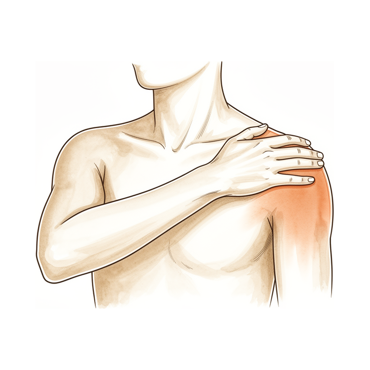
விளிம்பு விலா எலும்பு வெட்டுதல் விலா எலும்பின் முடிவில் இருந்து சில மில்லிமீட்டர் எலும்புகளை நீக்குகிறது, எனவே ஏசி மூட்டு (உங்கள் தோள்பட்டைக்கு மேலே உள்ள சிறிய மூட்டு) அணிந்த மேற்பரப்புகள் இனி ஒருவருக்கொருவர் அரைக்காது. அது குணமடையும் போது பாதுகாக்க வேண்டிய எதுவும் சரி செய்யப்படவில்லை, எனவே மறுவாழ்வு விரைவாக நகர்கிறது: ஸ்லிங் ஆறுதலுக்கு மட்டுமே மற்றும் பெரும்பாலான மக்கள் முதல் வாரம் அல்லது இரண்டு நாட்களுக்குள் வெளியேறுகிறார்கள், இயக்கம் உடனடியாக தொடங்குகிறது, மேலும் இந்த செயல்பாட்டிற்கான வெளியிடப்பட்ட நெறிமுறைகள் அனைத்தும் ஓய்வெடுப்பதை விட ஆரம்ப இயக்கத்தை நோக்கி தள்ளுகின்றன.
இரண்டு விஷயங்கள் இந்த நடவடிக்கைக்கு குறிப்பிட்டவை மற்றும் நெறிமுறையை வடிவமைக்கின்றன:
- உங்கள் உடல் முழுவதும் அடையும் மெதுவாக நகரும். குறுக்கு-உடல் (அடிக்சன்) இயக்கங்கள் எலும்பு அகற்றப்பட்ட பகுதியை அழுத்துகின்றன, எனவே அவை பெரும்பாலும் வசதியாக உணரக்கூடிய கடைசி இயக்கங்களாகும். ஆரம்பத்தில் அவை வேண்டுமென்றே தவிர்க்கப்படுகின்றன, பின்னர் படிப்படியாக மீண்டும் அறிமுகப்படுத்தப்படுகின்றன.
- கனமான அழுத்துதல் கடைசியாக வருகிறது. ஏசி கூட்டு கடினமாக சுமக்கும் உடற்பயிற்சிகள் (பெஞ்ச் பிரஸ், டிப்ஸ் மற்றும் புஷ்-அப்கள்) திரும்புவதற்கான கடைசி விஷயங்கள், பொதுவாக மூன்று முதல் நான்கு மாதங்களுக்கு மேல். பெரும்பாலான பிற செயல்பாடுகள் அதற்கு முன்பே திரும்பி வருகின்றன.
ஒரு பார்வையில் பயணம்:
- கட்டம் I ஆரம்பகால மீட்பு மற்றும் இயக்கம் வாரம் 02
- கட்டம் II உங்கள் வரம்பை மீட்டெடுப்பது வாரம் 26
- கட்டம் III வலுவூட்டல் மற்றும் செயல்பாட்டுக்கு திரும்புதல் வாரம் 612
- கட்டம் IV முழு செயல்பாட்டுக்கு திரும்புதல் 12 வாரங்கள் முதல்
பெரும்பாலான மக்கள் முதல் இரண்டு வாரங்களுக்குள் இலகுவான அன்றாட பணிகளுக்கு கையை பயன்படுத்துகிறார்கள், நான்கு முதல் ஆறு வாரங்களுக்குள் பெரும்பாலான சாதாரண நடவடிக்கைகளுக்குத் திரும்புகிறார்கள், மேலும் எட்டு முதல் பன்னிரண்டு வாரங்களுக்கு இடையில் விளையாட்டு மற்றும் கனமான வேலைகளுக்குத் திரும்புகிறார்கள். மீண்டும் மீண்டும் நிகழும் ஓவர்ஹெட் விளையாட்டுகளில் (எறிதல், நீச்சல், டென்னிஸ்) விளையாட்டு வீரர்கள் வழக்கமாக இரண்டு முதல் நான்கு மாதங்கள் வரை சிறிது நேரம் எடுத்துக்கொள்கிறார்கள்.
கட்டம் I ஆரம்பகால மீட்பு மற்றும் இயக்கம் (வாரம் 02)
முதல் இரண்டு வாரங்கள் தோள்பட்டை நகரும் போது வலி மற்றும் வீக்கத்தை நிவர்த்தி செய்வதைப் பற்றியது. வழக்கமாக பனியைப் பயன்படுத்துங்கள்: முதல் வாரம் அல்லது இரண்டில் ஒரு நாளைக்கு மூன்று முதல் நான்கு முறை பொதுவானது. ஸ்லிங் ஆறுதலுக்கு மட்டுமேஃ அது உதவும் அளவுக்கு அல்லது குறைவாக அணியுங்கள், கையை அடிக்கடி வெளியே எடுத்து, அது இல்லாமல் நீங்கள் வசதியாக இருக்கும்போது அதைப் பயன்படுத்துவதை நிறுத்துங்கள். வீட்டிற்கு வெளியே இருக்கும்போது, மக்கள் கையைத் தாக்காமல் தடுக்க இது மிகவும் பயனுள்ளதாக இருப்பதாக பலர் கருதுகின்றனர். நீங்கள் ஸ்லிங் அணிந்திருக்கும்போது வாகனம் ஓட்ட வேண்டாம்.
உடனடியாக நகர்த்தத் தொடங்குங்கள்: உங்கள் முழங்கை, மணிக்கட்டு மற்றும் கை ஆகியவை முதல் நாளிலிருந்து நகர்த்தப்பட வேண்டும், இதனுடன் மென்மையான ஊசலாட்ட பயிற்சிகள் மற்றும் உதவப்பட்ட தோள்பட்டை இயக்கங்கள் (உங்கள் மற்ற கையை, ஒரு குச்சி அல்லது ஒரு புல்லியைப் பயன்படுத்தி அறுவை சிகிச்சையளிக்கப்பட்ட கையை மேலே மற்றும் வெளியே உதவவும்). உங்கள் பயிற்சிகளுக்கு முன்னும் பிசியோதெரபி சந்திப்புகளுக்கு முன்னும் உங்கள் வலி நிவாரணிகளை எடுத்துக் கொள்ளுங்கள். இப்போது தனியாக விட்டுவிட வேண்டிய ஒரே இயக்கம் உங்கள் உடல் முழுவதும் எதிர் தோள்பட்டை நோக்கி அடைய வேண்டும்.
உங்கள் பிசியோதெரபிஸ்ட்டுக்கு:
இலக்குகள்
- வலி மற்றும் வீக்கத்தை குறைக்க
- ஆரம்பகால இயக்க வரம்புஃ ஏறக்குறைய 140° முன்னோக்கி வளைவு மற்றும் 40° பக்கவாட்டில் வெளிப்புற சுழற்சி கட்டத்தின் முடிவில்
- வீட்டு உடற்பயிற்சி திட்டத்துடன் சுதந்திரம்
நிர்வாகம்
- ஆறுதலுக்காக மட்டுமே ஸ்லிங்:
- குளிர்பதன சிகிச்சை தினமும் 3-4 முறை, குறிப்பாக உடற்பயிற்சிக்குப் பிறகு
- கழுத்து, முழங்கை, முதுகெலும்பு, மணிக்கட்டு மற்றும் கை ஆகியவற்றின் உடனடி செயலில் உள்ள இயக்க வரம்பு
- முதல் நாளிலிருந்து ஊசலாட்டம் பயிற்சிகள்
- அனுமதிக்கப்பட்டபடி செயலற்ற மற்றும் செயலில் உள்ள தோள்பட்டை இயக்கம் வரம்புஃ முன்னோக்கி உயர்வு, வெளிப்புற சுழற்சி மற்றும் பின்புற உள் சுழற்சி, காலிகள், ஒரு தடி அல்லது தடி பயன்படுத்தி, மற்றும் லேசான ஈர்ப்பு-உதவி நிலைகள்
- Scapular அமைவு மற்றும் periscapular இயக்கம் வரம்பில்; தொடக்கத்தில் இருந்து scapulohumeral ரிதம் கவனம்
- பிடியை வலுப்படுத்துதல்; மென்மையான தோள்பட்டை ஐசோமெட்ரிக்ஸ் வலி அனுமதிக்கிறது
- உடற்பயிற்சிகள் மற்றும் இயற்பியல் சிகிச்சை அமர்வுகளுக்கு முன் வலி நிவாரணம்
- தினமும் 35 முறை செய்யப்படும் வீட்டுத் திட்டம்
எச்சரிக்கைகள்
- குறுக்கு-உடல் (அளிமட்ட) சேர்க்கையைத் தவிர்க்கவும்
- மென்மையான ஐசோமெட்ரிக்குகளுக்கு அப்பால் தோள்பட்டைக்கு எதிரான உடற்பயிற்சி இல்லை
- இலகுவான அன்றாட பொருட்களைத் தவிர வேறு எதையும் தூக்கக் கூடாது; கை வழியாக எடை சுமக்கக் கூடாது (ஒரு நாற்காலியில் இருந்து அல்லது படுக்கையில் இருந்து தள்ளுதல்)
- ஸ்லிங் அணிந்திருக்கும் போது வாகனம் ஓட்டக்கூடாது
முன்னேற்றத்திற்கான அளவுகோல்கள்
- கயிறு அகற்றப்பட்டது, வலி குறைந்தது
- ஏறக்குறைய 140 ° மற்றும் வெளிப்புற சுழற்சிக்கு ஏறக்குறைய 40 ° வரை வசதியான உதவியுடன் உயர்வு
கட்டம் II உங்கள் வரம்பை மீட்டெடுப்பது (வாரம் 26)
இந்த கட்டத்தில் உங்கள் தோள்பட்டை அனைத்து திசைகளிலும் முழு இயக்கத்தை நோக்கி செயல்படுகிறது: முதலில் உதவி இயக்கங்கள், பின்னர் கையின் சொந்த சக்தியின் கீழ். உங்கள் இயக்கம் முழுமையாக நெருங்கியவுடன், ரோட்டேட்டர் கஃப் மற்றும் தோள்பட்டை தசைகளை மெதுவாக வலுப்படுத்துவது நெகிழ்வான பட்டைகள் மற்றும் லேசான எடைகளுடன் தொடங்குகிறது. அன்றாட நடவடிக்கைகளுக்கு சாதாரணமாக கையைப் பயன்படுத்துங்கள், ஆனால் மிதமான (5 கிலோவுக்கு மேல் இல்லை) தூக்குங்கள் மற்றும் கையை அழுத்தும் அல்லது கடினமாக ஏற்றும் எதையும் நிறுத்துங்கள். இந்த கட்டத்தில் ஆறுதல் அனுமதிப்பதைப் போல உங்கள் உடல் முழுவதும் அடைவது படிப்படியாக மீண்டும் அறிமுகப்படுத்தப்படுகிறது; அந்த இயக்கத்தின் முடிவில் சில பிஞ்சிங் விழிப்புணர்வு பொதுவானது மற்றும் வெப்பத்துடன் அமைகிறது. பலர் நீட்டிப்பு மற்றும் பனிக்கு முன் உதவியாக இருப்பதைக் காண்கிறார்கள், மேலும் நடைபயிற்சி, பைக் பயிற்சி அல்லது ஜாகிங் போன்ற இலகுவான கீழ்-உடல் உடற்பயிற்சி பொதுவாக நான்கு வாரங்களில் இருந்து தொடங்குகிறது.
உங்கள் பிசியோதெரபிஸ்ட்டுக்கு:
இலக்குகள்
- கட்டத்தின் முடிவில் அனைத்து விமானங்களிலும் முழு, அல்லது கிட்டத்தட்ட முழு, செயலில் உள்ள இயக்க வரம்பு
- இயல்பான scapulohumeral இயக்கவியல்
- சுழற்சி மஞ்ச் மற்றும் periscapular வலுவூட்டல் தொடங்க ஒருமுறை செயலில் வரம்பில் கிட்டத்தட்ட முழு உள்ளது
நிர்வாகம்
- அனைத்து விமானங்களிலும் செயலற்ற மற்றும் செயலில்-உதவி பெற்ற இயக்க வரம்பை செயலில் உள்ள வரம்பிற்கு முன்னேற்றம்ஃ வளைவு மற்றும் ஸ்கேப்புலர் விமானம் முழுமையானது, பக்கவாட்டில் வெளிப்புற சுழற்சி மற்றும் அனுமதிக்கப்பட்ட 90 ° கடத்தலில், மென்மையான பின்புற காப்ஸ்யூலர் நீட்டிப்புடன் முதுகுக்குப் பின்னால் உள் சுழற்சி
- அறிகுறிகளின் வழிகாட்டுதலின்படி படிப்படியாக குறுக்கு-உடல் சேர்க்கை வரம்பை மீண்டும் அறிமுகப்படுத்துங்கள்
- கையேடு சிகிச்சை மற்றும் glenohumeral அணிதிரட்டுதல்
- ஐசோமெட்ரிக்ஸிலிருந்து எலாஸ்டிக்-பேண்ட் ரோட்டேட்டர் மஞ்ச் மற்றும் ஸ்கேப்புலர் ஸ்டேபிலைசர் ஆகியவற்றின் முன்னேற்றம் இயக்கத்தின் செயல்பாட்டு வரம்பு கிட்டத்தட்ட முழுமையாக இருக்கும்போது வலுவூட்டுகிறதுஃ அதிக மறுபடியும், குறைந்த சுமை
- உடற்பயிற்சிக்கு முன்னர் வலி நிவாரணம்
- இயல்பான அன்றாட நடவடிக்கைகளுக்கு படிப்படியாக திரும்புதல்; லேசான கீழ்-உடல் நிலைப்படுத்தல் (நடைபயிற்சி, நிலையான சைக்கிள், ஜாகிங்) ஏறக்குறைய வாரம் 4 முதல்
எச்சரிக்கைகள்
- லேசான உயர்த்தி வைத்துஃ சுமார் 5 கிலோ அல்லது குறைவாக இயக்கப்படும் பக்கத்தில்
- ஏற்றப்பட்ட இறுதி வரம்பில் கிடைமட்ட சேர்க்கை மற்றும் மோதல் நிலைகளைத் தவிர்க்கவும்
- அழுத்தும் பயிற்சிகள் இல்லைஃ பெஞ்ச் பிரஸ், டிப்ஸ், புஷ்-அப்கள்
- தோள்பட்டை எரிச்சலடைந்தால் இணைந்த பிணைப்பு சுழற்சி (90/90) நீட்டிப்பு
முன்னேற்றத்திற்கான அளவுகோல்கள்
- கணிசமான அசௌகரியம் இல்லாமல் முழு அல்லது கிட்டத்தட்ட முழுமையான செயல்பாட்டு இயக்கம்
- வலி அதிகரிப்பு இல்லாமல் சகிப்புத்தன்மை கொண்ட பட்டை வலுவடைதல்
கட்டம் III வலுவூட்டல் மற்றும் செயல்பாட்டுக்கு திரும்புதல் (வாரம் 612)
முழு இயக்கம் மீண்டும் வரும்போது, வலிமையை மீண்டும் உருவாக்குவதற்கும், நீங்கள் என்ன செய்கிறீர்கள் என்பதில் கவனம் செலுத்துவதற்கும் கவனம் செலுத்துகிறது. ரோட்டேட்டர் கஃப், டெல்டாய்டு மற்றும் தோள்பட்டை தசைகளுக்கான எதிர்ப்பு வேலைகள் இசைக்குழுக்களிலிருந்து டம்பல்ஸுக்கு முன்னேறுகின்றன, மேலும் உடற்பயிற்சி மிகவும் செயல்பாட்டுக்கு வருகிறதுஃ உடற்பயிற்சி, நீச்சல், வீசுபவர்களுக்கு ஒரு பட்டப்படிப்பு வீசுதல் திட்டம், மற்றும் வேலை கடமைகள் மற்றும் விளையாட்டுக்கு படிப்படியாக திரும்புதல். இந்த கட்டத்தில், தோராயமாக எட்டு முதல் பன்னிரண்டு வாரங்களுக்கு இடையில், பெரும்பாலான மக்கள் விளையாட்டு மற்றும் கனமான வேலைகளுக்குத் திரும்புகிறார்கள், காலண்டரைக் காட்டிலும் ஆறுதல் மற்றும் வலிமையால் வழிநடத்தப்படுகிறார்கள். ஏசி மூட்டுக்கு அதிக சுமை கொடுக்கும் அழுத்தும் பயிற்சிகள் (பெஞ்ச் பிரஸ், டைப்ஸ் மற்றும் புஷ்-அப்கள்) மீண்டும் அறிமுகப்படுத்தப்படுவது கடைசியாக, இலகுவாகவும், ஆழமாகவும் தொடங்குகின்றன.
உங்கள் பிசியோதெரபிஸ்ட்டுக்கு:
இலக்குகள்
- வலிமை, சகிப்புத்தன்மை மற்றும் தோள்பட்டைப் பட்டைகளின் நரம்பு தசைக் கட்டுப்பாட்டை மீட்டெடுக்கவும்
- படிப்படியாக வேலைக்கு திரும்புதல், பொழுதுபோக்கு மற்றும் விளையாட்டு சார்ந்த பயிற்சி
நிர்வாகம்
- ரோட்டேட்டர் மஞ்ச், டெல்டோயிட் மற்றும் ஸ்கேப்புலர் ஸ்டேபிலைசர்களுக்கான முற்போக்கான எதிர்ப்புஃ பட்டைகள் முதல் ஒளி டம்பல்ஸ் வரை, பொதுவாக 2 × 3 செட் 8 × 15 மறுபடியும்
- எக்ஸென்ட்ரிக் வேலை, மூடிய சங்கிலி உடற்பயிற்சி மற்றும், கட்டத்தின் பிற்பகுதியில், நோயாளியின் விளையாட்டுக்கு பொருத்தமான பிளையோமெட்ரிக் பயிற்சிகளைச் சேர்க்கவும்
- செயல்பாட்டு மற்றும் விளையாட்டு-குறிப்பிட்ட பயிற்சிஃ நீச்சல், இடைவெளி வீசுதல் திட்டம் வீசுபவர்களுக்கு, கட்டுப்படுத்தப்பட்ட நிலைமைகளின் கீழ் விளையாட்டு பயிற்சிகள்
- முழு வேலை மற்றும் விளையாட்டுக்கு படிப்படியாக திரும்புதல், அளவுகோல் அடிப்படையில்
எச்சரிக்கைகள்
- ஏசி-லோடிங் பிரஸ்களை மீண்டும் அறிமுகப்படுத்துங்கள் (பெஞ்ச் பிரஸ், டிப்ஸ், புஷ்-அப்ஸ்) கடைசியாகஃ இலகுவான, குறைக்கப்பட்ட வரம்புடன் தொடங்குங்கள், மற்றும் முழங்கைகள் உடலின் கோட்டின் கீழ் அல்லது பின்னால் இறங்குவதைத் தவிர்க்கவும்
- முன்னேற்றம் அறிகுறி வழிகாட்டியாகவே உள்ளது: எரிச்சலூட்டும் ஏசி மூட்டு சுமைக்கு ஒரு குறுகிய படி பின்னால் செல்கிறது, தள்ளுவதன் மூலம் அல்ல
முன்னேற்றத்திற்கான அளவுகோல்கள்
- முழுமையான, வலி இல்லாத செயல்பாட்டு இயக்கம்
- சுமைகளுடன் அறுவை சிகிச்சை செய்யப்பட்ட பகுதியில் வலி அல்லது உணர்திறன் இல்லை
- சோதனையில் மற்ற பக்கத்தை நெருங்கும் வலிமை
கட்டம் IV முழு செயல்பாட்டுக்கு திரும்புதல் (12 வது வாரம் முதல்)
இறுதி கட்டம் என்பது கட்டுப்பாடற்ற செயல்பாட்டிற்கு திரும்புவதாகும். அன்றாட வாழ்க்கை மற்றும் பெரும்பாலான விளையாட்டுகள் பொதுவாக இந்த கட்டத்திற்கு முன்பே திரும்பி வருகின்றன; எஞ்சியிருப்பது சுமைகளின் கனமான முடிவாகும். ஜிம்மில் அழுத்தும் வலிமை படிப்படியாக மீண்டும் கட்டமைக்கப்படுகிறது, மேலும் முந்தைய பெஞ்ச் பிரஸ் செயல்திறனுக்கு திரும்புவது சுமார் நான்கு மாதங்கள் வரை ஆகலாம். மீண்டும் மீண்டும் ஓவர்ஹெட் விளையாட்டுகளில் உள்ள விளையாட்டு வீரர்கள் அறுவை சிகிச்சையிலிருந்து சுமார் இரண்டு முதல் நான்கு மாதங்களுக்குள் முன்னேறி வருகின்றனர். முழுமையான இயக்கம், வசதியான சுமை மற்றும் திருப்திகரமான பரிசோதனையின் அடிப்படையில் உங்கள் பின்தொடர்தல் மதிப்பாய்வில் விளையாட்டு மற்றும் கனமான வேலைக்கு முழுமையான திரும்புதல் உறுதிப்படுத்தப்படுகிறது.
உங்கள் பிசியோதெரபிஸ்ட்டுக்கு:
இலக்குகள்
- வேலை, உடற்பயிற்சி மற்றும் விளையாட்டுக்கு வரம்பற்ற திரும்புதல்
- தோள்பட்டை பெல்ட் வலிமை மற்றும் இயந்திரத்தை நீண்ட காலத்திற்கு பராமரிக்கவும்
நிர்வாகம்
- உடற்பயிற்சி நிலையம் மற்றும் விளையாட்டு சார்ந்த உடற்பயிற்சிகளை முன்னெடுத்துச் செல்லுதல், உடற்பயிற்சியில் அதிகப்படியான அழுத்தம் கொடுப்பதை உள்ளடக்கியது, AC கூட்டு அழுத்தத்தை கட்டுப்படுத்தும் நுட்பத்திற்கு கவனம் செலுத்துதல் (கட்டுப்படுத்தப்பட்ட ஆழம், முழங்கைகள் உடல் கோட்டின் பின்னால் செல்லாது)
- முழுமையான இடைவெளி வீசுதல் மற்றும் தேவைப்பட்டால் மேல்நிலை விளையாட்டு முன்னேற்றங்கள்
எச்சரிக்கைகள்
- முன்னேற்றம் அறிகுறிகளால் வழிநடத்தப்படுகிறது: ஒரு குறிப்பிட்ட லிஃப்ட் மூலம் ஏசி பகுதியில் மீண்டும் மீண்டும் வலி ஏற்படுவது முன்னேற்றத்திற்கு முன்னர் சுமை அல்லது நுட்பத்தை சரிசெய்ய ஒரு சமிக்ஞையாகும்
உங்கள் நெறிமுறை பிறகு
மேலே உள்ள கட்டங்கள் வெளியிடப்பட்ட மறுவாழ்வு நெறிமுறைகளிலிருந்து மாற்றியமைக்கப்பட்டுள்ளன (செயின்ட் லூயிஸ் பல்கலைக்கழக எலும்பியல் அறுவை சிகிச்சை துறை, உட்டா பல்கலைக்கழகம் விளையாட்டு மருத்துவம், விளையாட்டு அறுவை சிகிச்சை நியூயார்க், பால்ம் பீச் எலும்பியல் நிறுவனம், எலும்பியல் வர்ஜீனியா மற்றும் இல்லினாய்ஸ் சிறப்பு மருத்துவர்கள்), வார வரம்புகள் நிலையானவை அல்ல, மாறாக வழக்கமானதாக வெளிப்படுத்தப்படுகின்றன. உங்கள் மறுவாழ்வு உங்கள் பிசியோதெரபிஸ்டால் தனித்தனியாக வழிநடத்தப்படுகிறது, உங்கள் தோள்பட்டை எவ்வாறு மீண்டு வருகிறது என்பதை அடிப்படையாகக் கொண்டு நடைமுறையில் செயல்படுகிறது. இந்த பக்கம் நடைமுறையின் பொது மீட்பு ஆலோசனையுடன் இணைந்து செயல்படுகிறது; பார்க்கவும் அறுவை சிகிச்சைக்குப் பிந்தைய வலியை நிர்வகித்தல் மற்றும் காயம் பராமரிப்புஅறுவை சிகிச்சை தொடர்பாக, காண்க விண்மீன்களின் விளிம்பு பகுதியை அகற்றுதல்.
இந்த அறுவை சிகிச்சை மற்றும் அதன் புனர்வாழ்வின் பின்னணியில் உள்ள மருத்துவ சான்றுகள் (திறந்த-எதிராக-ஆர்த்ரோஸ்கோபிக் ஒப்பீடு, எவ்வளவு எலும்பு அகற்றப்படுகிறது மற்றும் ஏன், மற்றும் இந்த பக்கத்தை அடிப்படையாகக் கொண்ட வெளியிடப்பட்ட நெறிமுறைகள்) சான்றுகள் பிரிவில் முழு குறிப்புகளுடன் சுருக்கமாகக் கூறப்படுகின்றன, இந்த பக்கத்தின் மேலே இருந்து PDF ஆக கிடைக்கிறது.
Evidence & references
Distal Clavicle Excision (Mumford) — Surgical Rationale & Post-operative Rehabilitation
Topic scope: the evidence underpinning distal clavicle excision (DCE) — also called distal clavicle resection, AC joint excision, or the Mumford procedure — for the two main indications (acromioclavicular [AC] joint osteoarthritis and distal clavicular osteolysis, the "weightlifter's shoulder"), and the post-operative rehabilitation that follows. Covers open vs arthroscopic technique, direct vs indirect (bursal) approach, how much bone to resect, the iatrogenic-instability risk of over-resection, and the (consensus-based) phased rehab timeline.
Defining principle of the rehab here: DCE removes a few millimetres of worn bone from the end of the collarbone and repairs nothing that needs months of protection — provided the AC ligaments and superior/posterior capsule are preserved. So (like a debridement or subacromial decompression, and unlike a cuff repair, labral repair or AC-joint stabilisation) this is an early-motion pathway: a short sling for comfort only, movement from day one, strengthening as range returns. The single important caveat runs the other way: if the surgeon resects too much bone, or the stabilising AC ligaments/capsule are violated, the joint can become iatrogenically unstable — which is why technique (resection amount, ligament preservation) matters more here than the rehab calendar. The two operation-specific quirks the rehab respects are that cross-body (horizontal adduction) movement compresses the resected area and is the slowest to settle, and heavy pressing (bench press, dips, push-ups) loads the AC joint hardest and returns last.
A. THE OPERATION & ITS INDICATIONS
DCE removes the worn or eroded outer end of the clavicle so the acromion and clavicle no longer grind at the AC joint. Two indications dominate:
- AC joint osteoarthritis — degenerative wear, often with a history of prior AC injury or simply age-related change. Surgery follows failed non-operative care (activity modification, analgesia, AC joint corticosteroid injection).
- Distal clavicular osteolysis ("weightlifter's shoulder") — atraumatic, repetitive-microtrauma resorption of the distal clavicle seen in weightlifters and overhead athletes. Activity modification and rehabilitation are first-line; injection or surgery is reserved for refractory cases or athletes unwilling to stop loading [StatPearls 2023; Charron 1998].
DCE is frequently combined with subacromial decompression (for room/co-existing impingement) and the rehab is unchanged by that addition. If a rotator cuff repair is also performed, the slower protected cuff-repair pathway takes over.
B. EVIDENCE BY THEME
1. Open vs arthroscopic — equivalent long-term outcome, faster arthroscopic recovery
A randomised, prospective trial (Robertson et al., corpus, DOI 10.1016/j.jse.2006.10.006) and a systematic review (Pensak et al., Arthroscopy 2010) found similar long-term functional outcomes for open and arthroscopic DCE, with both arthroscopic techniques exceeding 90% good/excellent results. Across measures there was a trend toward earlier or better outcomes after arthroscopic treatment — less post-operative pain and a quicker return to daily activities — without a difference in the final result [Robertson RCT; Pensak SR 2010]. A second comparative cohort reached the same conclusion (corpus, DOI 10.1177/0363546511419633). Moderate (RCT + SR + cohort).
2. Direct vs indirect (bursal) arthroscopic approach — direct is faster
Among arthroscopic techniques, the direct (superior, top-down) approach permits a quicker return to athletic activity than the indirect/bursal approach, with equivalent long-term results — one comparison reporting a mean return to sport of ~21 days (direct) vs ~42 days (indirect) [Pensak SR 2010; arthroscopic-approach comparison]. Moderate.
3. How much bone to resect — and why over-resection is dangerous
This is the central technical controversy and the reason DCE rehab cannot be reduced to a calendar:
- The stabilising superior and posterior AC ligament/capsule runs from the anterior acromion to the posterior distal clavicle, and the coracoclavicular (trapezoid) ligament inserts on the clavicle undersurface roughly 22–25 mm from the tip [Renfree; capsule/ligament-insertion cadaveric study, corpus DOI 10.1016/j.arthro.2009.04.072; clavicular-strut cadaveric study, corpus DOI 10.1016/j.jse.2013.01.004].
- Anatomic work suggests as little as ~2.3–2.6 mm of resection can begin to violate the superior AC ligament [Renfree, via PMC6930955]; biomechanical models show AC joint anteroposterior translation increases after either open or arthroscopic excision, and stability falls as the resection lengthens [Blazar; resection-length biomechanical model, corpus DOI 10.1016/j.arthro.2007.07.004; DCE-vs-symmetric-resection biomechanics, corpus DOI 10.1177/0363546512469873].
- The practical consensus is to resect enough to abolish bony contact but no more — commonly quoted as ~5 mm (sufficient to clear contact in cadaveric models) up to ~8 mm, and not beyond ~8 mm, preserving the posterior/superior capsule and AC ligaments [PMC6930955; StatPearls 2023; resection-length biomechanics, corpus DOI 10.1016/j.arthro.2007.07.004].
- Over-resection or capsular violation can produce iatrogenic AC instability — a recognised (though uncommon) cause of persistent pain after DCE that may itself require ligament reconstruction [iatrogenic-instability case, PMC6930955; Painful Conditions of the AC Joint, corpus DOI 10.5435/00124635-199905000-00004].
Moderate (cadaveric/biomechanical + anatomic + expert consensus); exact safe threshold is debated.
4. Outcomes & return to activity
DCE is a reliable, high-satisfaction operation for the right indication — arthroscopic series report >90% good/excellent results [Robertson RCT; Pensak SR 2010]. For osteolysis in weightlifters, an arthroscopic-resection series reported return to sport at a mean of ~3 days and to the preoperative weight-training program at ~9 days, remaining asymptomatic and able to progress load beyond pre-operative levels at ~19-month follow-up [Charron, Am J Sports Med 1998, PMID 9548111]. Across the literature, most everyday activity returns within weeks and heavy pressing/overhead sport over ~3–4 months, consistent with the published rehab protocols. Moderate (cohort). A noted exception: worse ("poor") results cluster in patients with pre-existing post-traumatic AC instability or Workers'-Compensation claims [Pensak SR 2010] — DCE alone does not fix an unstable joint.
C. PHASED POST-OPERATIVE TIMELINE (isolated DCE ± subacromial decompression)
Week ranges are typical, not fixed — progression is criteria-based, guided by the physiotherapist.
| Phase | Window | Sling | ROM / use | Strengthening | Operation-specific notes |
|---|---|---|---|---|---|
| I — Early recovery & movement | Week 0–2 | Comfort only; weaned/discarded within 1–2 weeks | Elbow/wrist/hand + pendulums from day 1; passive + active-assisted shoulder elevation/ER/behind-the-back IR; scapular setting | Grip; gentle isometrics as pain allows | Avoid cross-body (horizontal) adduction — it compresses the resected area. No driving in the sling; no weight-bearing through the arm |
| II — Restoring range | Week 2–6 | Off | Progress assisted → active ROM to full in all planes; reintroduce cross-body adduction gradually (end-range pinching is common, settles) | Begin cuff + scapular band work once active range near full; lifting ≤ ~5 kg | Light lower-body conditioning (walk/bike/jog) from ~wk 4 |
| III — Strengthening & return | Week 6–12 | Off | Full functional ROM | Bands → dumbbells; functional + sport-specific; most return to sport/heavier work ~8–12 wk | Reintroduce AC-loading presses (bench/dips/push-ups) last — light, shallow depth, elbows not behind the body line |
| IV — Return to full activity | Week 12+ | Off | Full | Progressive heavy pressing; return to previous bench performance can take ~4 months; overhead athletes progressed over ~2–4 months | Symptom-guided — recurrent AC ache with a lift = adjust load/technique before progressing |
The phased structure is drawn from published surgeon/physiotherapy protocols (Saint Louis University; University of Utah Sports Medicine; Sports Surgery New York; Palm Beach Orthopaedic Institute; OrthoVirginia; Specialty Physicians of Illinois — see Citations). These are consensus/expert documents; no rehab RCT defines the optimal post-DCE regimen.
D. KEY CONTROVERSIES / EVIDENCE QUALITY
- How much bone to resect is the real debate, not the rehab. Too little leaves residual bony contact; too much risks iatrogenic instability. The safe window (~5–8 mm, capsule preserved) is supported by cadaveric/biomechanical and anatomic work, not by an RCT — exact thresholds vary by source. Moderate, technique-dependent.
- Open vs arthroscopic, direct vs indirect. Long-term outcomes converge; arthroscopic (and specifically the direct approach) recovers faster. Moderate (RCT + SR).
- Patient selection matters more than approach. Pre-existing AC instability and Workers'-Compensation status predict poorer results — DCE treats a worn/eroded joint, not an unstable one. Moderate.
- The rehab protocol itself is consensus. Phase timings come from surgeon patient-guidance protocols, not a rehab trial. Weak/consensus.
E. EVIDENCE STRENGTH FLAGS (summary)
- MODERATE (RCT / SR): open vs arthroscopic equivalence with faster arthroscopic recovery (Robertson RCT; Pensak SR 2010, >90% good/excellent); direct > indirect for return speed.
- MODERATE (cadaveric / biomechanical / anatomic): resection-length vs stability relationship and the iatrogenic-instability mechanism (resection-length model, corpus DOI 10.1016/j.arthro.2007.07.004; DCE-vs-symmetric biomechanics, corpus DOI 10.1177/0363546512469873; capsule/ligament insertions, corpus DOI 10.1016/j.arthro.2009.04.072).
- MODERATE (cohort): osteolysis-in-weightlifters return-to-sport (Charron 1998); high overall satisfaction; poorer results with pre-existing instability / WorkCover.
- WEAK / CONSENSUS: the post-operative rehabilitation protocol (surgeon patient-guidance documents; no defining rehab RCT); the exact "safe" resection threshold.
CITATIONS
RAG corpus (180,000+ Orthopaedic articles)
- Robertson WJ, et al. Arthroscopic versus open distal clavicle excision: comparative results at six months and one year from a randomized, prospective clinical trial. J Shoulder Elbow Surg. 2007. DOI: 10.1016/j.jse.2006.10.006
- Arthroscopic versus open distal clavicle excision (comparative cohort). Am J Sports Med. 2011. DOI: 10.1177/0363546511419633
- The biomechanical stability of distal clavicle excision versus symmetric acromioclavicular joint resection. Am J Sports Med. 2013. DOI: 10.1177/0363546512469873
- Arthroscopic distal clavicle resection: a biomechanical analysis of resection length and joint compliance in a cadaveric model. Arthroscopy. 2007. DOI: 10.1016/j.arthro.2007.07.004
- Analysis of the capsule and ligament insertions about the acromioclavicular joint: a cadaveric study. Arthroscopy. 2009. DOI: 10.1016/j.arthro.2009.04.072
- Acromioclavicular joint ligamentous system contributing to clavicular strut function: a cadaveric study. J Shoulder Elbow Surg. 2013. DOI: 10.1016/j.jse.2013.01.004
- Painful conditions of the acromioclavicular joint. J Am Acad Orthop Surg (JAAOS). 1999. DOI: 10.5435/00124635-199905000-00004
Literature (URLs)
- Pensak M, et al. Open versus arthroscopic distal clavicle resection (systematic review; >90% good/excellent, direct > indirect return). Arthroscopy. 2010. PMID 20434670. https://pubmed.ncbi.nlm.nih.gov/20434670/
- Charron KM, et al. Arthroscopic distal clavicle resection for isolated atraumatic osteolysis in weight lifters (return to sport ~3 d, training ~9 d). Am J Sports Med. 1998. PMID 9548111. https://pubmed.ncbi.nlm.nih.gov/9548111/
- Distal clavicular osteolysis (cause, activity-modification first line, ~8 mm resection preserving AC ligaments). StatPearls. 2023. https://www.ncbi.nlm.nih.gov/books/NBK582148/
- Distal clavicular augmentation with AC and CC ligament reconstruction in iatrogenic AC instability (~5 mm safe-resection guidance; Renfree ~2.3–2.6 mm violates superior AC ligament; trapezoid 22–25 mm from tip; Blazar AP-translation increase). PMC6930955. https://pmc.ncbi.nlm.nih.gov/articles/PMC6930955/
- A sports medicine clinician's guide to the diagnosis and management of distal clavicular osteolysis. PubMed. https://pubmed.ncbi.nlm.nih.gov/37294199/
- Distal clavicular osteolysis (review). Physiopedia. https://www.physio-pedia.com/Distal_Clavicular_Osteolysis
Published rehab protocols (patient-guidance — basis for the phase structure)
- Saint Louis University Dept of Orthopaedic Surgery. Subacromial Decompression / Distal Clavicle Excision Rehab Protocol. https://www.slu.edu/medicine/orthopaedic-surgery/sports-medicine/-pdf/shoulder-subacromial-decompression-and-distal-clavicle-excision.pdf
- Burks RT. Distal Clavicle Resection/Mumford Post-Op Protocol. University of Utah Sports Medicine. https://www.robertburksmd.med.utah.edu/pdfs/distal-clavicle-resection-mumford-protocol.pdf
- Sports Surgery New York. Arthroscopic Subacromial Decompression / Distal Clavicle Excision Rehab Protocol. https://www.sportssurgerynewyork.com/pdf/arthroscopic-subacromial-decompression-distal-clavicle-excision-rehab-protocol.pdf
- Hill B. Rehabilitation Protocol: Distal Clavicle Excision. Palm Beach Orthopaedic Institute. https://www.pboi.com/pdf/hill-pt-distal-clavicle-excision.pdf
- Eastwood D. Distal Clavicle Resection Therapy Protocol. OrthoVirginia. https://www.orthovirginia.com/wp-content/uploads/2022/09/Eastwood-distal-clavicle-resection-PT-protocol.pdf
- Mahylis JM. Distal Clavicle Excision Rehabilitation Protocol. Specialty Physicians of Illinois. https://jaredmahylismd.com/pdfs/distal-clavicle-excision-rehabilitation-protocol.pdf


