远端锁骨切除术
Patients › Rehabilitation
Rehabilitation after isolated arthroscopic distal clavicle excision; combined cases follow the rotator cuff repair protocol.
本方案由基兰·希尔帕拉(Kieran Hirpara)医生在罗克汉普顿 Mater 私人医院为您制定,用于指导您接受关节镜下锁骨远端切除术(也称为肩锁关节切除术,或 Mumford 手术)后的康复过程。以下每个阶段首先以通俗易懂的语言说明最关键的事项,随后附上为您物理治疗师编写的结构化方案;请在首次物理治疗就诊时携带此页面或其 PDF 版本,以确保您的康复过程协调一致。您的物理治疗师将根据您肩部的恢复情况推进各阶段,而非依据日历时间。
本方案适用于单纯的锁骨远端切除术。如果您的手术同时进行了肩袖修复,请遵循 肩袖修复方案,因为修复后的肌腱会要求更慢的康复节奏。(锁骨远端切除术通常也与肩峰下减压术联合进行;本方案同样适用于该情况。)
如果您对术后伤口有任何疑虑,请联系诊所。拍摄伤口照片并通过电子邮件发送以供审查通常很有帮助。
预期情况
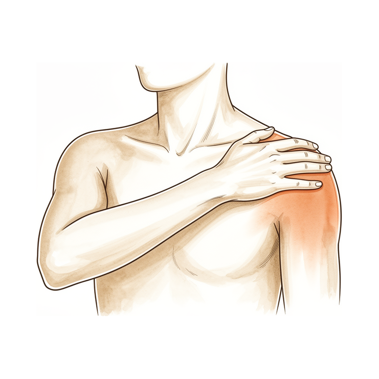
锁骨远端切除术从锁骨末端切除几毫米的骨骼,从而使肩锁关节(位于肩膀顶部的小关节)磨损的表面不再相互研磨。无需修复需要愈合期保护的结构,因此康复进程较快:悬吊带仅用于舒适目的,大多数人会在第一或第二周内停止使用;活动立即开始,且该手术的已发表方案均倾向于早期活动而非休息。
有两点与该手术相关,并决定了康复方案:
- 过体前伸动作是进展最慢的。 过体前伸(水平内收)动作会压迫骨骼被切除的区域,因此这些动作通常是最后才感到舒适的。早期应刻意避免,随后再逐渐重新引入。
- 大重量推举动作最后进行。 对肩锁关节施加较大负荷的动作(如卧推、双杠臂屈伸和俯卧撑)是最后恢复的项目,通常需要三到四个月。大多数其他活动在此之前的恢复情况良好。
整体进程概览:
- 第一阶段 — 早期恢复与活动 — 第 0–2 周
- 第二阶段 — 恢复活动范围 — 第 2–6 周
- 第三阶段 — 强化与恢复活动 — 第 6–12 周
- 第四阶段 — 完全恢复活动 — 第 12 周及以后
大多数人在前几周内即可用患肢进行轻度日常活动,四到六周内可恢复大多数正常活动,八到十二周之间可恢复运动及较重的工作。从事重复性过顶运动(如投掷、游泳、网球)的运动员通常需要更长的时间,约为两到四个月。
第一阶段——早期恢复与活动(第0–2周)
前两周的重点是控制疼痛和肿胀,同时开始肩部活动。请规律使用冰敷:在第一或前两周内,通常每天冰敷三至四次。护臂带仅用于提供舒适感:根据舒适度决定佩戴时间,经常将手臂取出,一旦在不佩戴护臂带的情况下感到舒适,即可停止使用。许多人发现,在外出时佩戴护臂带最为有用,可防止他人碰撞手臂。佩戴护臂带期间请勿驾驶。
立即开始活动:从第一天起,您的肘部、手腕和手部应开始活动,并进行轻柔的钟摆练习及辅助性肩部活动(使用另一只手臂、棍棒或滑轮帮助患侧手臂向上和向外活动)。在进行锻炼和物理治疗预约前,请服用止痛药。目前应避免的一个动作是向对侧肩膀方向跨越身体进行伸展。
致您的物理治疗师:
目标
- 控制疼痛和肿胀
- 早期活动范围:至本阶段结束时,前屈活动度约为140°,侧方外旋活动度约为40°
- 独立完成家庭锻炼计划
管理措施
- 护臂带仅用于提供舒适感:根据舒适度,在第一至第二周内逐渐减少使用并停用
- 冷疗:每日3–4次,尤其在锻炼后
- 颈部、肘部、前臂、手腕和手部的即刻主动活动范围训练
- 从第一天开始进行钟摆练习
- 根据耐受情况,进行被动及辅助性肩部活动范围训练:前屈、外旋及背后内旋,使用滑轮、棍棒或手杖,以及仰卧位重力辅助体位
- 肩胛骨定位及肩周活动范围训练;从一开始即关注肩肱节律
- 握力强化;根据疼痛情况,进行轻柔的肩部等长收缩练习
- 锻炼和物理治疗会话前使用镇痛药
- 家庭锻炼计划每日执行3–5次
注意事项
- 避免交叉身体(水平)内收
- 除轻柔的等长收缩练习外,禁止进行抗阻肩部练习
- 禁止提举超过日常轻物的物品;禁止通过手臂承重(如从椅子或床上撑起身体)
- 佩戴护臂带期间请勿驾驶
进阶标准
- 停用护臂带且疼痛得到控制
- 辅助性前屈活动度舒适达到约140°,外旋活动度舒适达到约40°
第二阶段——恢复活动范围(第2–6周)
去除吊带后,此阶段旨在使肩关节在所有方向上恢复完全活动度:首先进行辅助活动,随后依靠手臂自身力量活动。当活动度接近完全时,开始使用弹力带和轻重量对肩袖和肩胛骨肌肉进行温和的力量训练。在日常活动中正常使用手臂,但保持提举负荷适度(不超过约5公斤),并避免任何对手臂施加较大压力或负荷的动作。在此阶段,根据舒适度逐渐 reintroduce(重新引入)过体前屈活动;在该动作末端出现一些夹挤感是常见的,并会随时间消退。许多人发现拉伸前热敷和拉伸后冰敷有帮助,且从大约第4周开始可恢复较轻的下肢运动,如步行、健身车或慢跑。
致物理治疗师:
目标
- 阶段结束时,在所有平面达到完全或接近完全的主动活动范围
- 肩肱节律正常化
- 一旦主动活动范围接近完全,开始肩袖和肩周肌群的力量训练
管理
- 在所有平面将被动和辅助主动活动范围进展为主动活动范围:屈曲和肩胛平面抬举向完全活动度进展,侧方外旋和90°外展时的外旋按耐受情况进展,背后内旋配合温和的后关节囊拉伸
- 根据症状逐渐重新引入过体前屈活动范围
- 根据需要实施手法治疗和盂肱关节松动术
- 一旦主动活动范围接近完全,从等长收缩进展为使用弹力带的肩袖和肩胛稳定肌力量训练:高重复次数,低负荷
- 根据个人偏好,训练前热敷,训练后冰敷;运动前使用镇痛药
- 逐步恢复正常日常活动;从大约第4周开始进行轻度下肢体能训练(步行、固定自行车、慢跑)
注意事项
- 保持提举负荷轻:患侧约5公斤或以下
- 避免负荷下的终末水平内收和撞击体位
- 禁止推举类动作:卧推、双杠臂屈伸、俯卧撑
- 若肩关节易激惹,推迟90/90外展-旋转拉伸
进展标准
- 完全或接近完全的主动活动范围,且无明显不适
- 弹力带力量训练耐受良好,无疼痛加剧
第三阶段——强化与恢复活动(第6–12周)
随着活动范围完全恢复,重点转向重建力量并帮助您回归日常活动。针对肩袖、三角肌和肩胛骨肌肉的抗阻训练从弹力带进展至哑铃,训练内容更具功能性:包括健身房训练、游泳、投掷运动员的渐进式投掷计划,以及分阶段回归工作职能和体育运动。大多数人在此阶段(大约8至12周)回归体育运动和重体力工作,这一进程由舒适度和力量而非日历时间决定。对肩锁关节负荷最大的推举类动作(卧推、双杠臂屈伸和俯卧撑)最后 reintroduce,起始时负荷轻且幅度小。
致您的物理治疗师:
目标
- 恢复肩带肌群的力量、耐力和神经肌肉控制
- 分阶段回归工作职能、休闲活动及专项运动训练
管理措施
- 针对肩袖、三角肌和肩胛稳定肌的渐进性抗阻训练:从弹力带过渡至轻哑铃,通常为2–3组,每组8–15次重复
- 增加离心训练、闭链运动,并在本阶段后期根据患者运动项目需要加入增强式训练(plyometric drills)
- 功能性与专项运动训练:游泳、投掷运动员的间歇性投掷计划、受控条件下的专项运动 drills
- 基于标准分阶段回归全面工作职能和体育运动
注意事项
- 最后 reintroduce 增加肩锁关节负荷的推举动作(卧推、双杠臂屈伸、俯卧撑):起始时负荷轻、活动范围减小,并避免肘部低于或与身体后侧平齐
- 进展以症状为导向:若肩锁关节出现激惹,应通过短暂减少负荷来缓解,而非强行推进
进阶标准
- 功能性活动范围完全恢复且无痛
- 负荷状态下手术区域无疼痛或压痛
- 测试时力量接近对侧
第四阶段——恢复全面活动(第12周起)
最终阶段是恢复无限制活动。日常生活和大多数运动通常在此阶段之前已完全恢复;剩余的是最大负荷的恢复。健身房中的推举力量逐步重建,恢复至术前的卧推水平可能需要长达约四个月。从事重复性过头运动项目的运动员,从手术开始计算,大约需要两到四个月逐步恢复训练。全面恢复运动和重体力工作需经随访复查确认,依据是关节活动度完全恢复、负荷舒适且检查满意。
致您的物理治疗师:
目标
- 无限制地恢复工作、健身和运动
- 长期维持肩带力量与生物力学功能
管理
- 根据耐受情况推进健身房及运动专项训练,包括渐进式大重量推举训练,注意采用限制肩锁关节应力的技术(控制下放深度,肘部不超越身体中线)
- 在适用情况下,完成间歇性投掷训练及过头运动项目的渐进式训练
注意事项
- 进展仍以症状为导向:若特定动作后肩锁关节区域反复出现酸痛,应在进一步进展前调整负荷或技术
术后康复方案
上述阶段改编自已发表的锁骨远端切除术康复方案(圣路易斯大学骨科系、犹他大学运动医学、纽约运动外科、棕榈滩骨科研究所、OrthoVirginia 和伊利诺伊专科医生),其中周数范围表示为典型情况而非固定值。您的康复由物理治疗师根据您肩部的恢复情况,与诊所合作进行个体化指导。本页面与诊所的一般康复建议配合使用;请参阅术后疼痛管理和伤口护理。关于手术本身,请参阅锁骨远端切除术。
本手术及其康复(包括开放与关节镜手术的对比、切除骨量的多少及原因,以及本页面所参考的已发表方案)的临床证据,在证据部分进行了总结,并附有完整参考文献,可从本页面顶部下载 PDF 版本。
Evidence & references
Distal Clavicle Excision (Mumford) — Surgical Rationale & Post-operative Rehabilitation
Topic scope: the evidence underpinning distal clavicle excision (DCE) — also called distal clavicle resection, AC joint excision, or the Mumford procedure — for the two main indications (acromioclavicular [AC] joint osteoarthritis and distal clavicular osteolysis, the "weightlifter's shoulder"), and the post-operative rehabilitation that follows. Covers open vs arthroscopic technique, direct vs indirect (bursal) approach, how much bone to resect, the iatrogenic-instability risk of over-resection, and the (consensus-based) phased rehab timeline.
Defining principle of the rehab here: DCE removes a few millimetres of worn bone from the end of the collarbone and repairs nothing that needs months of protection — provided the AC ligaments and superior/posterior capsule are preserved. So (like a debridement or subacromial decompression, and unlike a cuff repair, labral repair or AC-joint stabilisation) this is an early-motion pathway: a short sling for comfort only, movement from day one, strengthening as range returns. The single important caveat runs the other way: if the surgeon resects too much bone, or the stabilising AC ligaments/capsule are violated, the joint can become iatrogenically unstable — which is why technique (resection amount, ligament preservation) matters more here than the rehab calendar. The two operation-specific quirks the rehab respects are that cross-body (horizontal adduction) movement compresses the resected area and is the slowest to settle, and heavy pressing (bench press, dips, push-ups) loads the AC joint hardest and returns last.
A. THE OPERATION & ITS INDICATIONS
DCE removes the worn or eroded outer end of the clavicle so the acromion and clavicle no longer grind at the AC joint. Two indications dominate:
- AC joint osteoarthritis — degenerative wear, often with a history of prior AC injury or simply age-related change. Surgery follows failed non-operative care (activity modification, analgesia, AC joint corticosteroid injection).
- Distal clavicular osteolysis ("weightlifter's shoulder") — atraumatic, repetitive-microtrauma resorption of the distal clavicle seen in weightlifters and overhead athletes. Activity modification and rehabilitation are first-line; injection or surgery is reserved for refractory cases or athletes unwilling to stop loading [StatPearls 2023; Charron 1998].
DCE is frequently combined with subacromial decompression (for room/co-existing impingement) and the rehab is unchanged by that addition. If a rotator cuff repair is also performed, the slower protected cuff-repair pathway takes over.
B. EVIDENCE BY THEME
1. Open vs arthroscopic — equivalent long-term outcome, faster arthroscopic recovery
A randomised, prospective trial (Robertson et al., corpus, DOI 10.1016/j.jse.2006.10.006) and a systematic review (Pensak et al., Arthroscopy 2010) found similar long-term functional outcomes for open and arthroscopic DCE, with both arthroscopic techniques exceeding 90% good/excellent results. Across measures there was a trend toward earlier or better outcomes after arthroscopic treatment — less post-operative pain and a quicker return to daily activities — without a difference in the final result [Robertson RCT; Pensak SR 2010]. A second comparative cohort reached the same conclusion (corpus, DOI 10.1177/0363546511419633). Moderate (RCT + SR + cohort).
2. Direct vs indirect (bursal) arthroscopic approach — direct is faster
Among arthroscopic techniques, the direct (superior, top-down) approach permits a quicker return to athletic activity than the indirect/bursal approach, with equivalent long-term results — one comparison reporting a mean return to sport of ~21 days (direct) vs ~42 days (indirect) [Pensak SR 2010; arthroscopic-approach comparison]. Moderate.
3. How much bone to resect — and why over-resection is dangerous
This is the central technical controversy and the reason DCE rehab cannot be reduced to a calendar:
- The stabilising superior and posterior AC ligament/capsule runs from the anterior acromion to the posterior distal clavicle, and the coracoclavicular (trapezoid) ligament inserts on the clavicle undersurface roughly 22–25 mm from the tip [Renfree; capsule/ligament-insertion cadaveric study, corpus DOI 10.1016/j.arthro.2009.04.072; clavicular-strut cadaveric study, corpus DOI 10.1016/j.jse.2013.01.004].
- Anatomic work suggests as little as ~2.3–2.6 mm of resection can begin to violate the superior AC ligament [Renfree, via PMC6930955]; biomechanical models show AC joint anteroposterior translation increases after either open or arthroscopic excision, and stability falls as the resection lengthens [Blazar; resection-length biomechanical model, corpus DOI 10.1016/j.arthro.2007.07.004; DCE-vs-symmetric-resection biomechanics, corpus DOI 10.1177/0363546512469873].
- The practical consensus is to resect enough to abolish bony contact but no more — commonly quoted as ~5 mm (sufficient to clear contact in cadaveric models) up to ~8 mm, and not beyond ~8 mm, preserving the posterior/superior capsule and AC ligaments [PMC6930955; StatPearls 2023; resection-length biomechanics, corpus DOI 10.1016/j.arthro.2007.07.004].
- Over-resection or capsular violation can produce iatrogenic AC instability — a recognised (though uncommon) cause of persistent pain after DCE that may itself require ligament reconstruction [iatrogenic-instability case, PMC6930955; Painful Conditions of the AC Joint, corpus DOI 10.5435/00124635-199905000-00004].
Moderate (cadaveric/biomechanical + anatomic + expert consensus); exact safe threshold is debated.
4. Outcomes & return to activity
DCE is a reliable, high-satisfaction operation for the right indication — arthroscopic series report >90% good/excellent results [Robertson RCT; Pensak SR 2010]. For osteolysis in weightlifters, an arthroscopic-resection series reported return to sport at a mean of ~3 days and to the preoperative weight-training program at ~9 days, remaining asymptomatic and able to progress load beyond pre-operative levels at ~19-month follow-up [Charron, Am J Sports Med 1998, PMID 9548111]. Across the literature, most everyday activity returns within weeks and heavy pressing/overhead sport over ~3–4 months, consistent with the published rehab protocols. Moderate (cohort). A noted exception: worse ("poor") results cluster in patients with pre-existing post-traumatic AC instability or Workers'-Compensation claims [Pensak SR 2010] — DCE alone does not fix an unstable joint.
C. PHASED POST-OPERATIVE TIMELINE (isolated DCE ± subacromial decompression)
Week ranges are typical, not fixed — progression is criteria-based, guided by the physiotherapist.
| Phase | Window | Sling | ROM / use | Strengthening | Operation-specific notes |
|---|---|---|---|---|---|
| I — Early recovery & movement | Week 0–2 | Comfort only; weaned/discarded within 1–2 weeks | Elbow/wrist/hand + pendulums from day 1; passive + active-assisted shoulder elevation/ER/behind-the-back IR; scapular setting | Grip; gentle isometrics as pain allows | Avoid cross-body (horizontal) adduction — it compresses the resected area. No driving in the sling; no weight-bearing through the arm |
| II — Restoring range | Week 2–6 | Off | Progress assisted → active ROM to full in all planes; reintroduce cross-body adduction gradually (end-range pinching is common, settles) | Begin cuff + scapular band work once active range near full; lifting ≤ ~5 kg | Light lower-body conditioning (walk/bike/jog) from ~wk 4 |
| III — Strengthening & return | Week 6–12 | Off | Full functional ROM | Bands → dumbbells; functional + sport-specific; most return to sport/heavier work ~8–12 wk | Reintroduce AC-loading presses (bench/dips/push-ups) last — light, shallow depth, elbows not behind the body line |
| IV — Return to full activity | Week 12+ | Off | Full | Progressive heavy pressing; return to previous bench performance can take ~4 months; overhead athletes progressed over ~2–4 months | Symptom-guided — recurrent AC ache with a lift = adjust load/technique before progressing |
The phased structure is drawn from published surgeon/physiotherapy protocols (Saint Louis University; University of Utah Sports Medicine; Sports Surgery New York; Palm Beach Orthopaedic Institute; OrthoVirginia; Specialty Physicians of Illinois — see Citations). These are consensus/expert documents; no rehab RCT defines the optimal post-DCE regimen.
D. KEY CONTROVERSIES / EVIDENCE QUALITY
- How much bone to resect is the real debate, not the rehab. Too little leaves residual bony contact; too much risks iatrogenic instability. The safe window (~5–8 mm, capsule preserved) is supported by cadaveric/biomechanical and anatomic work, not by an RCT — exact thresholds vary by source. Moderate, technique-dependent.
- Open vs arthroscopic, direct vs indirect. Long-term outcomes converge; arthroscopic (and specifically the direct approach) recovers faster. Moderate (RCT + SR).
- Patient selection matters more than approach. Pre-existing AC instability and Workers'-Compensation status predict poorer results — DCE treats a worn/eroded joint, not an unstable one. Moderate.
- The rehab protocol itself is consensus. Phase timings come from surgeon patient-guidance protocols, not a rehab trial. Weak/consensus.
E. EVIDENCE STRENGTH FLAGS (summary)
- MODERATE (RCT / SR): open vs arthroscopic equivalence with faster arthroscopic recovery (Robertson RCT; Pensak SR 2010, >90% good/excellent); direct > indirect for return speed.
- MODERATE (cadaveric / biomechanical / anatomic): resection-length vs stability relationship and the iatrogenic-instability mechanism (resection-length model, corpus DOI 10.1016/j.arthro.2007.07.004; DCE-vs-symmetric biomechanics, corpus DOI 10.1177/0363546512469873; capsule/ligament insertions, corpus DOI 10.1016/j.arthro.2009.04.072).
- MODERATE (cohort): osteolysis-in-weightlifters return-to-sport (Charron 1998); high overall satisfaction; poorer results with pre-existing instability / WorkCover.
- WEAK / CONSENSUS: the post-operative rehabilitation protocol (surgeon patient-guidance documents; no defining rehab RCT); the exact "safe" resection threshold.
CITATIONS
RAG corpus (180,000+ Orthopaedic articles)
- Robertson WJ, et al. Arthroscopic versus open distal clavicle excision: comparative results at six months and one year from a randomized, prospective clinical trial. J Shoulder Elbow Surg. 2007. DOI: 10.1016/j.jse.2006.10.006
- Arthroscopic versus open distal clavicle excision (comparative cohort). Am J Sports Med. 2011. DOI: 10.1177/0363546511419633
- The biomechanical stability of distal clavicle excision versus symmetric acromioclavicular joint resection. Am J Sports Med. 2013. DOI: 10.1177/0363546512469873
- Arthroscopic distal clavicle resection: a biomechanical analysis of resection length and joint compliance in a cadaveric model. Arthroscopy. 2007. DOI: 10.1016/j.arthro.2007.07.004
- Analysis of the capsule and ligament insertions about the acromioclavicular joint: a cadaveric study. Arthroscopy. 2009. DOI: 10.1016/j.arthro.2009.04.072
- Acromioclavicular joint ligamentous system contributing to clavicular strut function: a cadaveric study. J Shoulder Elbow Surg. 2013. DOI: 10.1016/j.jse.2013.01.004
- Painful conditions of the acromioclavicular joint. J Am Acad Orthop Surg (JAAOS). 1999. DOI: 10.5435/00124635-199905000-00004
Literature (URLs)
- Pensak M, et al. Open versus arthroscopic distal clavicle resection (systematic review; >90% good/excellent, direct > indirect return). Arthroscopy. 2010. PMID 20434670. https://pubmed.ncbi.nlm.nih.gov/20434670/
- Charron KM, et al. Arthroscopic distal clavicle resection for isolated atraumatic osteolysis in weight lifters (return to sport ~3 d, training ~9 d). Am J Sports Med. 1998. PMID 9548111. https://pubmed.ncbi.nlm.nih.gov/9548111/
- Distal clavicular osteolysis (cause, activity-modification first line, ~8 mm resection preserving AC ligaments). StatPearls. 2023. https://www.ncbi.nlm.nih.gov/books/NBK582148/
- Distal clavicular augmentation with AC and CC ligament reconstruction in iatrogenic AC instability (~5 mm safe-resection guidance; Renfree ~2.3–2.6 mm violates superior AC ligament; trapezoid 22–25 mm from tip; Blazar AP-translation increase). PMC6930955. https://pmc.ncbi.nlm.nih.gov/articles/PMC6930955/
- A sports medicine clinician's guide to the diagnosis and management of distal clavicular osteolysis. PubMed. https://pubmed.ncbi.nlm.nih.gov/37294199/
- Distal clavicular osteolysis (review). Physiopedia. https://www.physio-pedia.com/Distal_Clavicular_Osteolysis
Published rehab protocols (patient-guidance — basis for the phase structure)
- Saint Louis University Dept of Orthopaedic Surgery. Subacromial Decompression / Distal Clavicle Excision Rehab Protocol. https://www.slu.edu/medicine/orthopaedic-surgery/sports-medicine/-pdf/shoulder-subacromial-decompression-and-distal-clavicle-excision.pdf
- Burks RT. Distal Clavicle Resection/Mumford Post-Op Protocol. University of Utah Sports Medicine. https://www.robertburksmd.med.utah.edu/pdfs/distal-clavicle-resection-mumford-protocol.pdf
- Sports Surgery New York. Arthroscopic Subacromial Decompression / Distal Clavicle Excision Rehab Protocol. https://www.sportssurgerynewyork.com/pdf/arthroscopic-subacromial-decompression-distal-clavicle-excision-rehab-protocol.pdf
- Hill B. Rehabilitation Protocol: Distal Clavicle Excision. Palm Beach Orthopaedic Institute. https://www.pboi.com/pdf/hill-pt-distal-clavicle-excision.pdf
- Eastwood D. Distal Clavicle Resection Therapy Protocol. OrthoVirginia. https://www.orthovirginia.com/wp-content/uploads/2022/09/Eastwood-distal-clavicle-resection-PT-protocol.pdf
- Mahylis JM. Distal Clavicle Excision Rehabilitation Protocol. Specialty Physicians of Illinois. https://jaredmahylismd.com/pdfs/distal-clavicle-excision-rehabilitation-protocol.pdf


