PIP கூட்டு இணைப்பு
Patients › Rehabilitation
நடுத்தர விரல் மூட்டு இணைந்த பிறகு (ஆர்த்ரோடெசிஸ்) பாதுகாக்கப்பட்ட மீட்பு திட்டம், உருகிய மூட்டு இன்னும் ஒரு ஸ்பிளெண்டில் வைத்திருப்பதுடன், மற்ற அனைத்து மூட்டுகளையும் முதல் நாளிலிருந்து நகர்த்துவது, பின்னர் எலும்பு ஒன்றுபட்டவுடன் பிடியை மீட்டெடுப்பது மற்றும் பிஞ்ச் செய்வது.
இந்த நெறிமுறை உங்கள் மீட்பு வழிகாட்டுகிறது பிஐபி மூட்டு உருகுதல் (ஆர்த்ரோடெசிஸ்) இது உங்கள் வீட்டு உடற்பயிற்சி திட்டத்துடன் தொடங்குகிறது, அதைத் தொடர்ந்து கட்டமைக்கப்பட்ட மருத்துவ நெறிமுறை எழுதப்பட்டுள்ளது உங்கள் கை சிகிச்சையாளர்: இந்த பக்கத்தை அல்லது அதன் PDF ஐ உங்கள் முதல் சிகிச்சை வருகைக்கு எடுத்துச் செல்லுங்கள், இதனால் உங்கள் மறுவாழ்வு ஒருங்கிணைந்ததாக இருக்கும். உங்கள் கை சிகிச்சையாளர் உங்கள் மீட்பு எவ்வாறு முன்னேறுகிறது என்பதைப் பொறுத்து திட்டத்தை சரிசெய்யலாம்.
அறுவை சிகிச்சைக்குப் பிறகு உங்கள் காயத்தைப் பற்றி ஏதேனும் கவலைகள் இருந்தால், அறைகளைத் தொடர்பு கொள்ளுங்கள். காயத்தின் புகைப்படத்தை எடுத்து அதை மதிப்பாய்வு செய்ய மின்னஞ்சல் அனுப்புவது பெரும்பாலும் உதவியாக இருக்கும்.
எதிர்பார்ப்பது என்ன
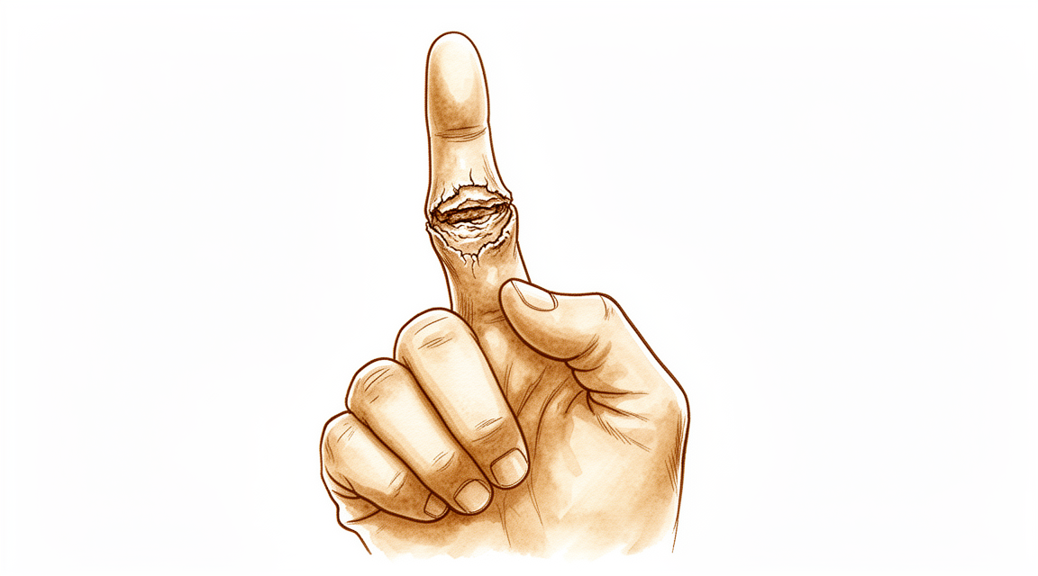
ஒரு PIP கூட்டு இணைப்பு ஒரு அணிந்த அல்லது நிலையற்ற நடுத்தர விரல் கூட்டு எடுத்து அதை திடமாக இணைக்கிறது, எனவே அது இனி நகராது. செயல்பாட்டு, லேசாக வளைந்த நிலை நேராக நேராக இல்லை: குறியீட்டு மற்றும் நடுத்தர விரல்களில் வளைவு மென்மையானது (சுமார் 15 20 °) மற்றும் கையின் சிறிய விரல் பக்கத்தை நோக்கி அதிகரிக்கிறது (சுமார் 25 40 ° வளையல் மற்றும் சிறிய விரல்களில்), நீங்கள் அவற்றை சுருக்கும்போது உங்கள் விரல்கள் இயற்கையான நீர்வீழ்ச்சியைப் பின்பற்றுகின்றன. உருகிய மூட்டு ஒரு சிறிய உள்வைப்புடன் (ஒரு பதற்றம்-பேண்ட் கம்பி, ஒரு தலை இல்லாத திருகு, கே-கம்பிகள் அல்லது ஒரு சிறிய தட்டு) இடத்தில் வைக்கப்படுகிறது, இது எலும்பு இணைப்பு முழுவதும் இணைக்கும் வரை அந்த கோணத்தை சீராக வைத்திருக்கும்.
இந்த செயல்பாடு பெரும்பாலும் தேர்வு செய்யப்படுகிறது காட்டி மற்றும் நடுத்தர விரல்கள், அங்கு ஒரு நிலையான கூட்டு பிஞ்ச் பிஐபி மூட்டு பின்னர் இறுக்கமாக இருக்க வேண்டும்: அதுதான் அறுவை சிகிச்சையின் நோக்கம். எனவே, ஒரு இடுப்பு அல்லது இழை சீரமைப்பு போலல்லாமல், மீட்பு என்பது அந்த மூட்டுக்கான இயக்கத்தை மீட்டெடுப்பதில்லை; அது எலும்பு இணைக்கும் வரை இணைப்பைப் பாதுகாக்கும், கையின் மற்ற அனைத்து மூட்டுகளும் சுதந்திரமாக நகரும் போது.
இந்த திட்டம் நான்கு யோசனைகளைச் சுற்றி கட்டமைக்கப்பட்டுள்ளது:
- எலும்பு ஒன்றிணைகிறது வரை இணைப்பு பாதுகாக்க. எலும்பு இணைப்பு பொதுவாக ஆறு வாரங்கள் ஆகும், சில நேரங்களில் ஒன்பது முதல் பன்னிரண்டு வாரங்கள் வரை ஆகும்.
- முதல் நாளிலிருந்து எல்லாவற்றையும் நகர்த்துவது: விரல்களின் கூட்டு, முழங்கை, அருகிலுள்ள விரல்கள், கட்டைவிரல் மற்றும் மணிக்கட்டு, அதனால் தசைகள் கீழே ஒட்டிக்கொள்ளாமல், மீதமுள்ள கை இறுக்கமடையாமல் இருக்கும்.
- வீக்கம் மற்றும் வடுக்களை நிர்வகிக்கவும் ஆரம்ப வாரங்களில்.
- இணைப்பு இணைந்தவுடன் பிடியை மீட்டெடுக்கவும் மற்றும் பிசைந்து கொள்ளவும். புகைக்க வேண்டாம்: புகைபிடிப்பது எலும்பு குணமடைவதை மெதுவாக்குகிறது மற்றும் ஒரு இணைப்பு ஒருங்கிணைப்பை தாமதப்படுத்துகிறது.
முன்னெச்சரிக்கைகள் மற்றும் வரம்புகள்
- அறுவை சிகிச்சை செய்யப்படும் விரலின் வழியாக வலுவாக சுமக்கவோ, பிடிக்கவோ அல்லது பிடுங்கவோ கூடாது எலும்பு ஒன்று சேரும் வரை (வழக்கமாக சுமார் ஆறு வாரங்கள், சில நேரங்களில் நீண்ட நேரம்): எலும்பு ஒன்று சேரும் முன் சுமைகளை ஏற்றுவது இணைப்பு ஏற்படாத அபாயங்களை ஏற்படுத்துகிறது.
- உருகிய கூட்டு முற்றிலும் இன்னும் வைத்து "சோதனை" செய்யவோ அல்லது வளைக்கவோ முயற்சிக்க வேண்டாம்.
- ஒவ்வொரு மற்ற கூட்டு நகரும் வைத்து முதல் நாட்களிலிருந்து: விரல் நுனியின் மூட்டு, முழங்கை, மற்ற விரல்கள், கட்டைவிரல் மற்றும் மணிக்கட்டு.
- ஸ்கிளின்ட் வைத்து சுத்தமான மற்றும் உலர்ந்த, அதை வழிகாட்டுதலின்படி அணியுங்கள், மற்றும் காயம் மற்றும் எந்த ஊசி இடங்களையும் பார்த்துக் கொள்ளுங்கள்.
- செய் இல்லை நீங்கள் சக்கரத்தை பாதுகாப்பாகக் கட்டுப்படுத்த முடியாத போது வாகனம் ஓட்டுதல், பொதுவாக நீங்கள் ஆறு வாரங்களில் ஸ்பிளின்ட் வெளியே இருக்கும் வரை.
- புகைக்காதே: இது எலும்புகள் இணைவதை தாமதப்படுத்துகிறது.
காயம், வீக்கம் மற்றும் வடுக்கள் மேலாண்மை, நடைமுறையில் பார்க்க காயம் பராமரிப்பு வழிகாட்டல்.
உங்கள் பயிற்சிகள்
இவை உங்கள் கையேட்டில் உள்ள பயிற்சிகள். டாக்டர் ஹிர்பரா மற்றும் உங்கள் கை சிகிச்சையாளரின் வழிகாட்டுதலின்படி மட்டுமே அவற்றைத் தொடங்குங்கள், உங்களுக்கு வழங்கப்பட்ட வரம்புகளுக்குள் தங்குங்கள். ஆரம்ப பயிற்சிகள் விரல் நுனி மூட்டு, முழங்கை, மற்றும் உங்கள் மற்ற விரல்கள், கட்டைவிரல் மற்றும் மணிக்கட்டு ஆகியவற்றை நகர்த்துகின்றன, உருகிய கூட்டு தன்னை நகர்த்தி அல்லது சுமை இல்லாமல்காயம் குணமடைந்தவுடன் வடு மசாஜ் தொடங்குகிறது. பிஞ்ச் மற்றும் பிடியை வலுப்படுத்துவது ஒரு பிற்கால கட்டத்திற்கு சொந்தமானது, மேலும் இணைவு ஒன்றிணைந்து நீங்கள் குறிப்பாக அழிக்கப்படும் வரை தொடங்கக்கூடாது. உருகிய மூட்டுக்கு மேல் கூர்மையான வலியை ஏற்படுத்தும் எதையும் நிறுத்துங்கள்.
உங்கள் மருத்துவ நெறிமுறை
இந்த பக்கத்தின் மீதமுள்ள பகுதி PIP கூட்டு ஆர்த்ரோடெசிஸுக்குப் பிறகு மறுவாழ்வுக்கான படிநிலை மருத்துவ நெறிமுறை ஆகும். இந்த பிரிவு உங்கள் கை சிகிச்சையாளருக்கு வழங்கப்பட வேண்டும், மேலும் ஒவ்வொரு கட்டமும் என்ன நடக்கிறது என்பதற்கான எளிய ஆங்கில விளக்கத்துடன் தொடங்குகிறது. எலும்புக் கூட்டு (வழக்கமாக 6 வாரங்கள், 9-12 வாரங்கள் வரை) "இணைந்த மூட்டு பாதுகாக்க, எல்லாவற்றையும் செயல்படுத்த": டிஐபி, எம்சிபி, அடுத்தடுத்த விரல்கள், கட்டைவிரல் மற்றும் மணிக்கட்டு ஆகியவை முதல் நாளிலிருந்து இடுப்பு ஒட்டுதல் மற்றும் விறைப்புத்தன்மையைத் தடுக்க நகர்கின்றன, வீக்கம் மற்றும் வடுக்கள் ஆரம்பத்தில் நிர்வகிக்கப்படுகின்றன, மேலும் இணைந்த பிறகு மட்டுமே பிணைப்பு / பிஞ்ச் மீட்டமைக்கப்படுகிறது.
சிகிச்சைக்கு முன்னர், நோயாளியின் செயல்பாட்டு அறிக்கை மற்றும் கடந்தகால மருத்துவ வரலாற்றை சரிபார்த்து, சிகிச்சையளிக்கும் அறுவை சிகிச்சையாளருடன் பயன்படுத்தப்படும் உறுதிப்படுத்தல் (டென்ஷன்-பேண்ட் கம்பி, தலை இல்லாத இன்ட்ராமெடுலரி திருகு, கே-கம்பிகள் அல்லது தட்டு), செட் ஃப்யூஷன் கோணம், மற்றும் கே-கம்பிகள் புதைக்கப்பட்டுள்ளதா அல்லது அகற்றப்பட வேண்டுமா என்பது குறித்து தொடர்பு கொள்ளுங்கள். டாக்டர் ஹிர்பாரா PIP ஐ ஒரு செயல்பாட்டு வளைந்த நிலையில் உருகுகிறார், இது கை முழுவதும் அல்னார் நோக்கி அதிகரிக்கிறது (குறியீட்டு / நடுத்தர ≈ 1520 °, மோதிரம் / சிறிய ≈ 25 40 °), பெரும்பாலும் குறியீட்டு / நடுத்தர விரத்திற்கு, பக்கவாச பிஞ்ச் ஸ்திரத்தன்மை PIP இயக்கத்தை விட அதிகமாக இருக்கும். உருகிய மூட்டு ஒரு ஸ்பிளண்டில் ஒன்றாக வைக்கப்படுகிறது; ரேடியோகிராஃபிக் யூனியன் வரை சான்றுகள் அடிப்படை குறைந்தவை (நிலை -4 வழக்கு தொடர் மற்றும் நிபுணத்துவ நிலை ஒருமிகு
கட்டம் I பாதுகாக்க மற்றும் குடியேற (வாரங்கள் 0 முதல் 2)
முதல் இரண்டு வாரங்கள் இணைவைப் பாதுகாத்து வீக்கத்தையும் காயத்தையும் சரிசெய்கிறது, அதே நேரத்தில் கையின் மற்ற அனைத்து மூட்டுகளும் உடனடியாக இயங்கத் தொடங்குகின்றன.
உங்கள் கை சிகிச்சையாளருக்கு:
இயலாமை - கடுமையான விரல் அடுக்கு அல்லது அடுக்கு விரிவடைகிறது MCP மற்றும் PIP, ஆனால் டிஐபி இலவசமாக விட்டு - உருகிய PIP இன்னும் நடைபெற்றது; oedema க்கான உயர்வு
கல்வி மற்றும் முன்னெச்சரிக்கைகள் - பிடிப்பு, பிஞ்ச் அல்லது சுமை இல்லை அறுவை சிகிச்சை செய்யப்பட்ட விரல் வழியாக - அடுப்பை சுத்தமாகவும் உலரமாகவும் வைத்திருங்கள்; காயத்தையும், எந்த ஊசி இடங்களையும் பாதுகாக்கவும்
நிர்வாகம் - காயம்ஃ அறுவை சிகிச்சை பாண்டேஜ்கள் வழிகாட்டுதலின்படி; தொற்றுநோய் மற்றும் ஊசி இடங்களை கண்காணிக்க K- கம்பிகள் பயன்படுத்தப்பட்டால் - வீக்கம்: உயர்வு, இலவச மூட்டுகளின் மென்மையான பம்ப், தேவைக்கேற்ப பனி - பயிற்சிகள்: இருந்து முதல் நாள், அனைத்து செயலில் இயக்கம் உருகாத மூட்டுகள்ஃ அருகிலுள்ள விரல்கள் (முழு கை / நீட்டிப்பு), கட்டைவிரல், மணிக்கட்டு; தொடக்கம் டிஐபி அறுவை சிகிச்சை செய்யப்படும் விரலின் செயலில் இயக்கம் நாட்களுக்குள்; அருகிலுள்ள விரல்களின் இடுப்பு சறுக்கல்கள்; உருகிய PIP இன் இயக்கம் அல்லது சுமை இல்லை
முன்னேற்றத்திற்கான அளவுகோல்கள் - காயம் குறையும்; வீக்கம் கட்டுப்படுத்தப்படும்; சுமார் 12 வாரங்களில் இறுதி தனிப்பயனாக்கப்பட்ட அடுக்குக்கு தயாராக உள்ளது
கட்டம் II தனிப்பயனாக்கப்பட்ட தெர்மோபிளாஸ்டிக் அடுக்கு மற்றும் இலவச மூட்டுகளின் செயலில் இயக்கம் (வாரங்கள் 2 முதல் 6)
அறுவை சிகிச்சைக்கு உட்படுத்தப்பட்ட விரலின் சுறுசுறுப்பான செயல்பாடு DIP மற்றும் MCP பிணைக்கப்பட்ட பிஐபி பாதுகாக்கப்பட்டு வருகிறது. எதிர்ப்பு, பிஞ்ச் அல்லது சுமை இன்னும் இல்லை.
உங்கள் கை சிகிச்சையாளருக்கு:
மதிப்பீடுகள் - அருகிலுள்ள-இணைப்பு ROM, வீக்கம், காயம்/படு மதிப்பாய்வு; மருத்துவ அடிப்படையில் மற்றும் அறுவை சிகிச்சையாளருக்கு ஏற்ப உறுதிப்படுத்தல் நிலையானது
இயலாமை - மாற்றம் தனிப்பயனாக்கப்பட்ட தெர்மோபிளாஸ்டிக் ஸ்பிளிண்ட் அருகிலுள்ள இணைப்புகளை விடுவிக்கும்போது உருகிய PIP ஐ ஆதரித்தல்; தொடர்ச்சியான பாதுகாப்பு அடுக்குகள் ~ 6 வாரங்கள்
கல்வி மற்றும் முன்னெச்சரிக்கைகள் - பிடிப்பு, பிஞ்ச் அல்லது சுமைக்கு எதிர்ப்பு இல்லை அறுவை சிகிச்சை செய்த விரலின் - உடற்பயிற்சிக்காக மட்டுமே ஸ்பிளின்ட் வெளியே
நிர்வாகம் - பயிற்சிகள்: அறுவை சிகிச்சை செய்யப்பட்ட விரலின் செயலில் உள்ள டிஐபி மற்றும் எம்சிபி இயக்கம் (டிஐபி சில நாட்களில் தொடங்கியது, எம்சிபி இங்கே சேர்க்கப்பட்டது); அருகிலுள்ள விரல்களின் இடுப்பு சறுக்கல்கள்; தொடர்ச்சியான கட்டைவிரல் / மணிக்கட்டு / அருகிலுள்ள விரல் இயக்கம்; தொடங்கவும் வடு மற்றும் வீக்கம் மேலாண்மை காயம் குணமடைந்தவுடன் - இல்லை பிடிப்பு/பின்ச்/சுமைக்கு எதிரான எதிர்ப்பு
முன்னேற்றத்திற்கான அளவுகோல்கள் - ரேடியோகிராபிக் யூனியன் (வழக்கமாக சுமார் 6 வாரங்கள், 912 வரை); எந்தவொரு சுமைக்கும் முன் உருகிய கூட்டு மருத்துவ ரீதியாகவும் கதிரியக்க ரீதியாகவும் நிலையானது
கட்டம் III ஸ்பிளெண்டை களைந்து, முன்னேற்றம் லேசான பயன்பாடு (~6 வாரங்களில், ஒருமுறை இணைக்கப்பட்டது)
இணைவு ஒன்றுபட்டவுடன் (பொதுவாக ஆறு வாரங்களுக்குள்), ஸ்பிளின்ட் தூக்கி எறியப்பட்டு படிப்படியாக வெட்டப்படுகிறது, ஒளி செயல்பாட்டு பயன்பாடு அறிமுகப்படுத்தப்படுகிறது, மற்றும் பிஞ்ச், எதிர்ப்பு மற்றும் பிடிப்பு படிப்படியாக மீண்டும் கட்டமைக்கப்படுகின்றன. கே-கம்பிகள், பயன்படுத்தப்பட்டால், பொதுவாக ஆறு வாரங்களுக்குள் அகற்றப்படுகின்றன.
உங்கள் கை சிகிச்சையாளருக்கு:
மதிப்பீடுகள் - அறுவை சிகிச்சையாளருடன் ரேடியோகிராஃபிக் இணைப்பை உறுதிப்படுத்துதல்; மற்ற கைக்கு எதிராக பிடிப்பு / குத்துவிசை வலிமை; இலவச மூட்டுகளின் ROM; ஸ்கார்
கல்வி மற்றும் முன்னெச்சரிக்கைகள் - தாய்ப்பால் மற்றும் அடுக்குகளை வெட்டுதல் இணைப்பு உறுதி செய்யப்பட்டதும்; பயன்படுத்தப்பட்டால் K-கம்பி ~6 வாரங்கள் அகற்றவும் - படிப்படியாக நிரப்புதல்: முதலில் லேசான பயன்பாடு, பின்னர் மாறுபட்ட பிஞ்ச் / பிடியில்
நிர்வாகம் - பயிற்சிகள்: முன்னேற்றம் ஒளி செயல்பாட்டு பயன்பாடு → பிஞ்ச், எதிர்ப்பு மற்றும் பிடிப்புதொடங்குகிறது பிடிப்பு/பின்ச் வலுவூட்டல் (பந்து / புட்டு, பக்கவாட்டு பிஞ்ச்) மற்றும் படிப்படியாக உருவாக்க; வடு வேலை தொடர - ரேடியோகிராஃபிக் இணைப்புக்குப் பிறகு விதைக்கப்பட்ட ஸ்பிளின்ட்
முன்னேற்றத்திற்கான அளவுகோல்கள் - ஒன்றிணைந்த உருகுதல் லேசான சுமை தாங்கும் வலி இல்லாதது; ஸ்பிளண்ட் நிறுத்தப்பட்டது; படிப்படியான வலுவூட்டலுக்கு தயாராக உள்ளது
கட்டம் IV படிப்படியான வலுவடைதல் மற்றும் திரும்புதல் (~ 812 வாரங்களில்)
ஒற்றுமை மற்றும் ஒளி பயன்பாட்டை மீட்டெடுப்பதன் மூலம், வலுவூட்டல் மற்றும் சுமை முன்னேற்றம் அடைகிறது, மேலும் விளையாட்டு, கனமான அல்லது கையேடு நடவடிக்கைக்கு திரும்புவது கட்டமைக்கப்படுகிறது. இறுதி நிலையான முடிவு சுமார் ஒன்பது முதல் பன்னிரண்டு மாதங்களில் அடையப்படுகிறது.
உங்கள் கை சிகிச்சையாளருக்கு:
மதிப்பீடுகள் - பிடிப்பு மற்றும் பிஞ்ச் வலிமை மற்ற பக்கத்துடன் ஒப்பிடும்போது; செயல்பாட்டு மற்றும் வேலை-/விளையாட்டு-குறிப்பிட்ட சோதனைகள்
கல்வி மற்றும் முன்னெச்சரிக்கைகள் - படிப்படியாக சுமை வரை எதிர்ப்பு கட்ட; உருகிய கூட்டு வடிவமைப்பு மூலம் நிரந்தரமாக இறுக்கமாக உள்ளது; பிடியில் வலிமை மற்றும் பக்கவாட்டு பிஞ்ச் கவனம்
நிர்வாகம் - பயிற்சிகள்: படிப்படியான வலுவூட்டல் மற்றும் சுமை பிடிப்பு மற்றும் பிஞ்ச்; படிப்படியாக திரும்ப விளையாட்டு, கனமான மற்றும் கை வேலைகள் - வலிமை செயல்பாட்டு மற்றும் கிட்டத்தட்ட சமச்சீரற்றதாக இருக்கும்போது வெளியேற்றத்தை கருத்தில் கொள்ளுங்கள்; மீட்பு மேடைகள் இருந்தால் சிகிச்சையளிக்கும் மருத்துவரை மீண்டும் பார்க்கவும்
திருப்பி அனுப்புவதற்கான அளவுகோல்கள் - வலி இல்லாத, சுமை கீழ் நிலையான உருகிய கூட்டு; வேலைக்கு போதுமான பிடியில் / பிஞ்ச் வலிமை, காலண்டர் மூலம் அல்லாமல் மருத்துவ ரீதியாக தீர்மானிக்கப்படுகிறது; 912 மாதங்களில் இறுதி தீர்வு முடிவு
வேலை மற்றும் செயற்பாட்டிற்கு திரும்புதல்
எளிய அன்றாட கை பயன்பாடு மீதமுள்ள கையுடன் தொடக்கத்தில் இருந்து, வசதியுடன் ஊக்குவிக்கப்படுகிறது; முக்கிய கட்டுப்பாடு இணைப்பு சேரும் வரை செயல்படுத்தப்பட்ட விரல் வழியாக பிடிப்பது, பிடுங்குவது அல்லது சுமப்பது இல்லை. நீங்கள் சக்கரத்தை பாதுகாப்பாக கட்டுப்படுத்த முடியாத வரை வாகனம் ஓட்டக் கூடாது என்பதால், ஆரம்ப வாரங்களில் போக்குவரத்து உதவிக்கு திட்டமிடுங்கள்; வழக்கமாக ஆறு வாரங்களில் வாகனம் ஓட்டுதல் தொடங்குகிறது, நீங்கள் ஸ்பிளின்ட் வெளியே மற்றும் பாதுகாப்பாக கார் கட்டுப்படுத்த முடியும் ஒருமுறை.
இணைந்த பிறகு (சுமார் ஆறு வாரங்கள்) நீங்கள் ஆரம்பிக்கலாம் லேசான பயன்பாடு மற்றும் மென்மையான பிடிப்பு. தூக்குதல், பிடித்தல் மற்றும் அழுத்துதல் பற்றி இருந்து உருவாக்க எட்டு வாரங்கள், மற்றும் முழுமையான செயல்பாடு அல்லது விளையாட்டு சுமார் பன்னிரண்டு வாரங்கள்பல மாதங்களாக இந்த இணைப்பு நிலைத்து நிற்கிறது, எனவே ஒன்பது முதல் பன்னிரண்டு மாதங்களுக்குள் முழுமையாக தீர்வு காணப்படும்.இந்த நேரங்கள் நிபுணர்-ஒருமித்த ஒற்றை மருத்துவமனை வழிகாட்டிகள் (வழக்கமான மற்றும் தனிப்பயனாக்கப்பட்ட, தரப்படுத்தப்படாத வாசல்கள்) மற்றும் உங்கள் முன்னேற்றம் டாக்டர் ஹிர்பாரா மற்றும் உங்கள் கை சிகிச்சையாளரால் மதிப்பீடு செய்யப்படுகிறது, கலவை குணமடைவது எப்படி, காலண்டர் மூலம் மட்டும் அல்ல.
உங்கள் நெறிமுறை பிறகு
இந்த நெறிமுறை நடைமுறையின் பொது மீட்பு ஆலோசனையுடன் இணைந்து செயல்படுகிறது. அறுவை சிகிச்சைக்குப் பிந்தைய வலியை நிர்வகித்தல், காயம் பராமரிப்பு மற்றும் வடு மேலாண்மைமேலே உள்ள படிப்படியான திட்டம் பிஐபி கூட்டு ஆர்த்ரோடெசிஸுக்குப் பிறகு மறுவாழ்வு குறித்த வெளியிடப்பட்ட வழிகாட்டுதல்களை பிரதிபலிக்கிறது, மேலும் உங்கள் விரல் எவ்வாறு குணமடைகிறது என்பதைப் பொறுத்து உங்கள் தற்போதைய மீட்பு டாக்டர் ஹிர்பாரா மற்றும் உங்கள் கை சிகிச்சையாளரால் தனித்தனியாக வழிநடத்தப்படுகிறது.
Evidence & references
PIP Joint Fusion — Procedure Outcomes & Post-operative Rehabilitation (Proximal Interphalangeal Arthrodesis)
Topic scope: post-operative rehabilitation after arthrodesis (fusion) of the proximal interphalangeal (PIP) joint of a finger — a worn, painful or unstable PIP joint is fused solid in a functional flexed position. This is a fusion, not a reconstruction or a motion- preserving procedure: the PIP is deliberately made stiff to trade motion for a stable, pain-free, load-bearing digit. The rehab is therefore not about regaining PIP motion but about protecting the construct until bony union while keeping every other joint of the hand moving, then restoring grip and pinch.
Defining principle of the rehab here: PIP arthrodesis eliminates motion at one joint by design to gain stability for pinch and grip. The fused joint is set in a functional flexed position that increases ulnar-ward across the hand (index/middle ≈ 15–20°, ring/little ≈ 25–40°, following the digital cascade) and held by internal fixation (tension-band wire, headless intramedullary screw, K-wires, or plate) until union. Because nothing here needs to move to heal — it needs to unite — the single governing rule is "protect the fused joint, mobilise everything else." DIP, MCP, adjacent digits, thumb and wrist move from day one to prevent tendon adhesion and stiffness; oedema and scar are managed early; grip and pinch are restored only after radiographic union (~6 weeks, up to 9–12). The branch point is the indication — primary degenerative/post-traumatic fusion versus salvage of a failed PIP arthroplasty, where union is slower and the construct more demanding.
A. PROCEDURE OUTCOMES (PIP arthrodesis)
PIP arthrodesis is a reliable, well-established salvage and reconstructive operation, but its evidence base is uniformly low-level — predominantly retrospective level-4 case series and expert opinion, with no randomised controlled trials. Outcomes are reported as union, complication and reoperation rates rather than from comparative trials.
- The evidence base is low-level and consensus-driven. A systematic review of PIP arthrodesis found the literature is overwhelmingly level-4 (~94%) with no RCTs; conclusions on fixation choice and outcomes rest on case series and expert consensus [EFORT Open Rev 2021, DOI 10.1530/eor-21-0102]. Low (level-4 SR, no RCTs).
- Fixation holds the angle to union; nonunion and reoperation are the principal concerns. Series reporting nonunion and reoperation identify patient factors (including smoking and comorbidity) as drivers of failure, underscoring that the rehab job is to protect the construct until the bone joins [HAND 2020, DOI 10.1177/1558944720939196]. Low–moderate (case series).
- The fusion angle is chosen for function, especially pinch. A biomechanical/kinematic study of index PIP fusion (simulated 30–50°) shows the set angle is a functional trade-off: fusing the index/middle PIP stabilises lateral (key) pinch at the cost of PIP motion, which is why these digits are common fusion sites [J Hand Surg Am 2011, DOI 10.1016/j.jhsa.2011.09.010]. Mechanistic / cadaveric.
- Arthrodesis is a dependable salvage for failed PIP arthroplasty, but union is slow. A series of arthrodesis for failed PIP joint replacement reported a mean time to union of 5.8 months, illustrating that salvage fusions unite more slowly than primary fusions and need correspondingly extended protection [J Hand Surg Am 2011, DOI 10.1016/j.jhsa.2010.10.030]. Low (case series).
- The biomechanics of digital loss/fusion frame the functional cost. Reviews of the biomechanics of digital amputation and fusion describe how eliminating an IP joint redistributes grip and pinch mechanics — the rationale for accepting a stiff joint when it buys stability [Hand Clin 2016, DOI 10.1016/j.hcl.2016.07.003]. Mechanistic / narrative.
B. REHABILITATION / THERAPY EVIDENCE
There are no trials of rehab regimens after PIP arthrodesis; the programme is built on sound surgical principle and expert consensus. The two evidence-anchored levers are the union timeline (which sets when load may be applied) and the modifiable risk factor of smoking (which delays union).
- Protect-until-union, mobilise-everything-else is the consensus regimen. The fused PIP is splinted continuously until radiographic union (~6 weeks, up to 9–12); from day one the DIP, MCP, adjacent digits, thumb and wrist are actively moved to prevent tendon adhesion and global hand stiffness. This is stable across sources (surgeon protocols, hand-therapy guidance and patient-education material) even though it is not trial-tested [Melbourne Arm Clinic protocol; OrthOracle PIPJ arthrodesis; OrthoInfo finger IP fusion]. Consensus / expert.
- Smoking is an evidence-supported delayed-union risk. A study of hand and wrist arthrodesis found smoking delays union, making smoking cessation the one rehab-adjacent intervention with direct supporting evidence in this setting [J Hand Surg Am 2022, DOI 10.1016/j.jhsa.2022.05.016]. Moderate (cohort, modifiable risk factor).
- Union timing governs progression — and is slower in salvage fusions. Primary fusions are typically protected to ~6 weeks; salvage of failed arthroplasty unites far more slowly (mean 5.8 months), so loading must be union-led rather than calendar-led [J Hand Surg Am 2011, DOI 10.1016/j.jhsa.2010.10.030]. Low (case series).
- The set fusion angle is the functional anchor of the rehab goal. Because the index/middle PIP is fused at ~15–20° (and ring/little at ~25–40°) specifically to stabilise lateral pinch, the Phase III–IV strengthening rightly targets pinch and grip rather than any attempt at PIP motion [J Hand Surg Am 2011, DOI 10.1016/j.jhsa.2011.09.010]. Mechanistic.
Recovery trajectory (expected, evidence-anchored)
| Phase | Window | Restraint | Hand use / therapy focus | Strength / load | Notes |
|---|---|---|---|---|---|
| I — Protect & settle | Week 0–2 | Volar finger splint/cast spanning MCP + PIP, DIP left free | Elevation, wound/pin-site care, oedema control; active DIP within days + full motion of all non-fused joints (adjacent digits, thumb, wrist) | No grip / pinch / loading | Fused PIP kept still; everything else mobilised from day one |
| II — Custom splint & free-joint motion | Week 2–6 | Custom thermoplastic splint supporting the fused PIP, freeing adjacent joints; continuous splinting to ~6 wk | Active DIP + MCP of operated finger out of splint; tendon glides of adjacent digits; scar/oedema once healed | No resisted grip / pinch / loading | Union typically at ~6 wk (up to 9–12); load only after radiographic union |
| III — Wean splint & light use | From ~6 wk (united) | Splint weaned/cut down after union; K-wire out ~6 wk if used | Progress light use → pinch, opposition, gripping; begin grip/pinch strengthening | Graded grip/pinch, build gradually | Restraints lifted only once union confirmed |
| IV — Strengthen & return | ~8–12 wk+ | Restrictions lifted | Progressive strengthening/loading; return to sport/heavy/manual work | Build load progressively; target lateral pinch | Final settled result 9–12 months |
(Phase windows mirror the precautions in the patient protocol; they are expert-consensus, single-clinic guides — typical and individualised, not graded or trial-derived thresholds. Return milestones: driving ~6 wk, light use/gentle grip ~6 wk after union, lifting/gripping/pinch ~8 wk, full activity/sport ~12 wk, final result 9–12 months.)
C. KEY CONTROVERSIES / EVIDENCE QUALITY
- Whole topic is low-level evidence. PIP arthrodesis rests on level-4 case series and expert consensus with no RCTs (~94% level-4 in systematic review). All outcome and timing figures should be read as typical guides, not trial-validated thresholds [EFORT 2021]. Low.
- Fixation choice is unsettled. Tension-band wire, headless intramedullary screw, K-wires and plate all achieve union; comparative data are weak and selection is largely surgeon preference and bone/soft-tissue quality [EFORT 2021; HAND 2020]. Low.
- Fusion angle is a functional trade-off, not a fixed number. The ~15–20° (index/middle) to ~25–40° (ring/little) cascade is consensus-stable but individualised to the digit and the demands of pinch [J Hand Surg Am 2011 kinematics]. Mechanistic / consensus.
- Union timing is variable and indication-dependent. Primary fusions ~6 weeks; salvage of failed arthroplasty far slower (mean 5.8 months). Loading must be union-led [J Hand Surg Am 2011 salvage series]. Low.
- Smoking and patient factors drive nonunion/reoperation. Smoking is an evidence-supported delayed-union risk and a modifiable target [J Hand Surg Am 2022; HAND 2020]. Moderate (for the smoking association).
D. EVIDENCE STRENGTH FLAGS (summary)
- STRONG (RCT / SR): none — there are no RCTs in PIP arthrodesis; the best synthesis is a level-4 systematic review (~94% level-4 studies).
- MODERATE: smoking as a delayed-union risk after hand/wrist arthrodesis; patient factors driving nonunion/reoperation; cadaveric/kinematic basis for the functional fusion angle and pinch rationale.
- WEAK / CONSENSUS: the protect-until-union, mobilise-everything-else rehab regimen (mechanistically sound, not trial-tested); the specific fusion angles (consensus-stable); exact timelines (single-clinic, expert-consensus guides — typical, not graded thresholds); fixation choice (surgeon preference).
CITATIONS
RAG corpus (180,000+ Orthopaedic articles)
- Proximal interphalangeal joint arthrodesis: a systematic review (predominantly level-4 evidence; no RCTs). EFORT Open Rev. 2021. DOI: 10.1530/eor-21-0102
- Nonunion and reoperation after proximal interphalangeal joint arthrodesis: patient factors and outcomes. HAND. 2020. DOI: 10.1177/1558944720939196
- Index finger proximal interphalangeal joint arthrodesis and pinch kinematics (simulated 30–50° fusion). J Hand Surg Am. 2011. DOI: 10.1016/j.jhsa.2011.09.010
- Arthrodesis as salvage for failed proximal interphalangeal joint arthroplasty (mean time to union 5.8 months). J Hand Surg Am. 2011. DOI: 10.1016/j.jhsa.2010.10.030
- Smoking delays union after hand and wrist arthrodesis. J Hand Surg Am. 2022. DOI: 10.1016/j.jhsa.2022.05.016
- Biomechanics of digital loss and fusion. Hand Clin. 2016. DOI: 10.1016/j.hcl.2016.07.003
PIP arthrodesis rehabilitation & procedure literature (URLs)
- Melbourne Arm Clinic. PIP / DIP arthrodesis rehabilitation protocol. https://melbournearmclinic.com.au/orthopaedic-rehabilitation/shoulder-rehabilitation/pip-dip-arthrodesis-protocol/
- OrthOracle. Proximal interphalangeal joint (PIPJ) arthrodesis in the hand using the Apex system (Extremity Medical). https://www.orthoracle.com/library/proximal-interphalangeal-joint-pipj-arthrodesis-in-the-hand-using-the-apex-system-extremity-medical/
- EFORT Open Reviews. Proximal interphalangeal joint review (PMC). https://pmc.ncbi.nlm.nih.gov/articles/PMC6598614/
- American Academy of Orthopaedic Surgeons (OrthoInfo). Finger (interphalangeal) joint fusion. https://orthoinfo.aaos.org/en/treatment/finger-ip-joint-fusion/


