

近端指间关节融合术 资料
本方案指导您在 Mater Private Hospital Rockhampton 接受 Kieran Hirpara 医生进行的近端指间关节(PIP 关节)融合术(关节固定术)(手指的中部关节)后的康复过程。方案首先介绍您的居家锻炼计划,随后是专为您的手部治疗师撰写的结构化临床方案:请在首次治疗就诊时携带此页面或其 PDF 文件,以确保您的康复过程协调一致。您的手部治疗师可能会根据您的康复进展调整计划。
如果您对术后伤口有任何疑虑,请联系诊室。通常,拍摄伤口照片并通过电子邮件发送以供查看会很有帮助。
预期情况
近端指间关节(PIP)融合术针对磨损或不稳定的中间关节,将其牢固连接,使其不再活动。关节被固定在功能性、轻微弯曲的位置,而非完全伸直:食指和中指的弯曲度较缓(约 15–20°),向手掌小指侧逐渐增加(无名指和小指约 25–40°),以顺应手指屈曲时形成的自然弧度。融合关节通过小型植入物(张力带钢丝、无头螺钉、克氏针或小钢板)固定,以维持该角度稳定,直至骨性融合完成。
该手术通常用于食指和中指,因为对于捏握功能而言,稳定的关节比中间关节的活动度更为重要。PIP关节术后预期会僵硬:这正是手术的目的。因此,与肌腱或韧带修复不同,恢复过程并非旨在恢复该关节的活动度,而是保护融合部位直至骨性愈合,同时保持手部其他所有关节的自由活动。
康复计划围绕以下四个核心原则构建:
- 在骨性愈合前保护融合部位。 骨性愈合通常需要约六周,有时需九至十二周。在此之前,融合关节需由支具支撑。
- 从第一天起保持其他部位活动: 包括指尖关节、掌指关节、邻近手指、拇指和手腕,以防止肌腱粘连和手部其他部位僵硬。
- 在早期数周内管理肿胀和瘢痕。
- 融合愈合后恢复抓握和捏握功能。 请勿吸烟:吸烟已知会延缓骨愈合并推迟融合部位的愈合。
注意事项与限制
- 在骨融合愈合之前(通常为六周左右,有时更长),切勿通过手术手指进行负重、抓握或用力捏取:在骨骼连接之前负重可能导致融合失败。
- 按照指示,保持融合关节在支具中完全静止;不要试图“测试”或弯曲它。
- 从最初几天起,保持其他所有关节活动:指尖关节、掌指关节、其他手指、拇指和手腕。
- 保持支具清洁干燥,按指示佩戴,并护理伤口及任何钢针部位。
- 在无法安全控制方向盘期间,切勿驾驶,通常需等到约四至六周拆除支具后。
- 不要吸烟:吸烟会延缓骨骼愈合。
关于伤口、肿胀和瘢痕管理,请参阅本诊所的伤口护理指南。
您的锻炼
Kieran Hirpara 4.0
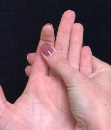
手术手指的远端指间关节活动
在获得您的手部治疗师许可后,轻轻弯曲并伸直手术手指最末端(指尖)的关节。用另一只手固定手指中部,使仅指尖活动。融合关节必须保持完全静止——仅指尖弯曲。保持该关节活动可防止肌腱在融合愈合过程中粘连。
10次,每日3次,仅指尖关节
Kieran Hirpara 4.0
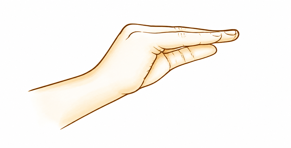
手术手指的掌指关节(MCP)活动
轻轻弯曲并伸直手术手指的掌指关节(大指关节,即手指与手掌连接处)。融合的中间关节在整个过程中保持静止——仅掌指关节活动。这有助于保持掌指关节的灵活性,并防止在融合稳定期间手指僵硬。
10次,每日3次,仅限掌指关节
Kieran Hirpara 4.0
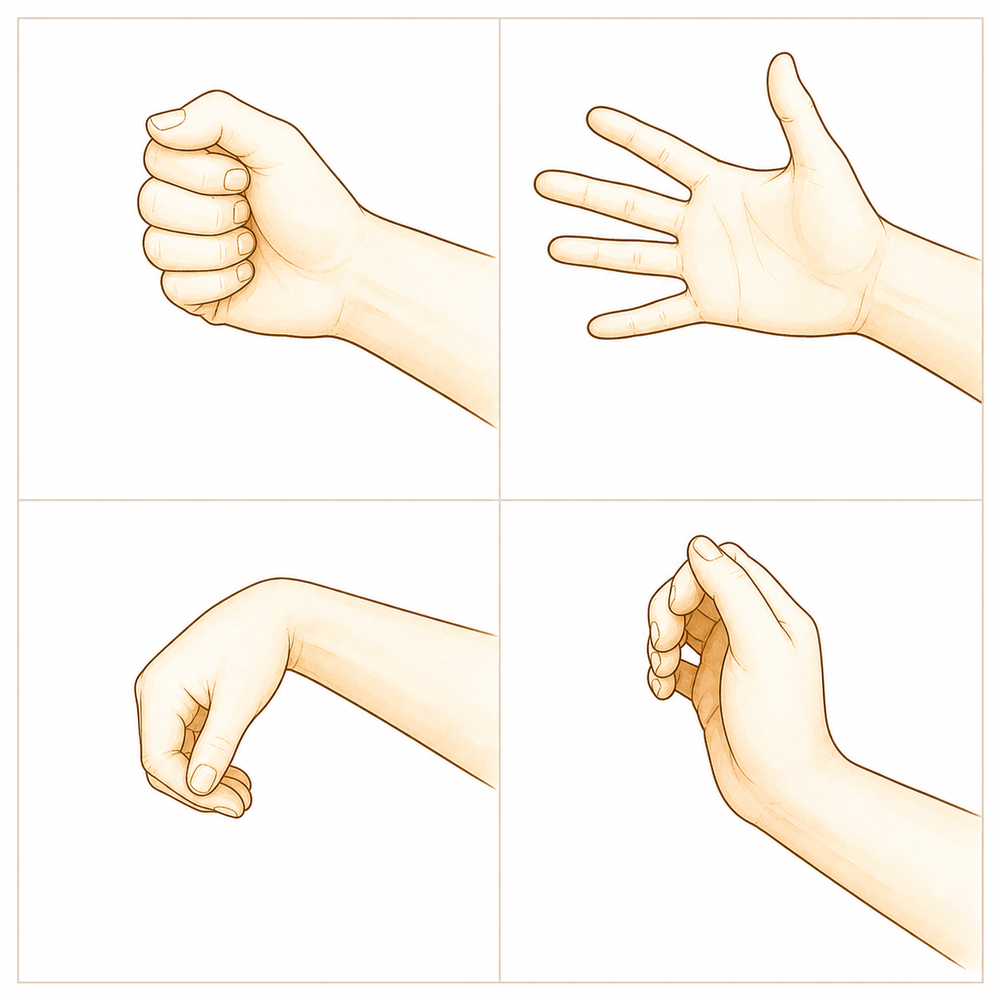
活动您的其他手指、拇指和手腕
从最初几天起,请保持所有未手术关节自由活动——用其他手指完全握拳并完全张开,拇指向各个方向活动,以及屈曲、伸直和环转手腕。这是保护融合关节的同时,保持手部柔韧并防止僵硬的最重要事项。
每次10次,每天数次
Kieran Hirpara 4.0
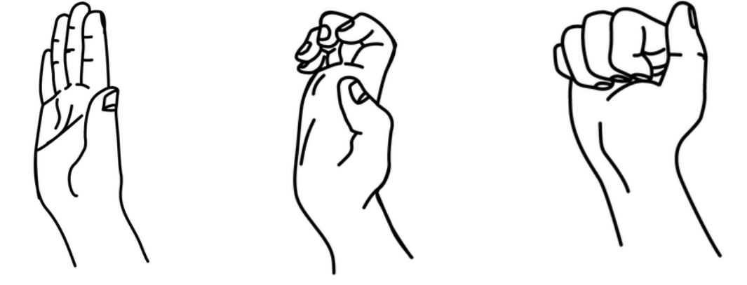
腱滑动(钩状、握拳、伸直)
缓慢地通过三种手型活动手部,以保持肌腱的滑动:钩状(弯曲指尖关节和中间关节,掌指关节伸直)、全握拳,然后是完全伸直的手掌。请用未手术的手指完全执行这些动作,并让手术手指仅在未融合(自由)关节允许的范围内活动。这有助于保持肌腱顺畅滑动,并防止其在手术部位附近粘连。
每种形状5个,每日3次
Kieran Hirpara 4.0
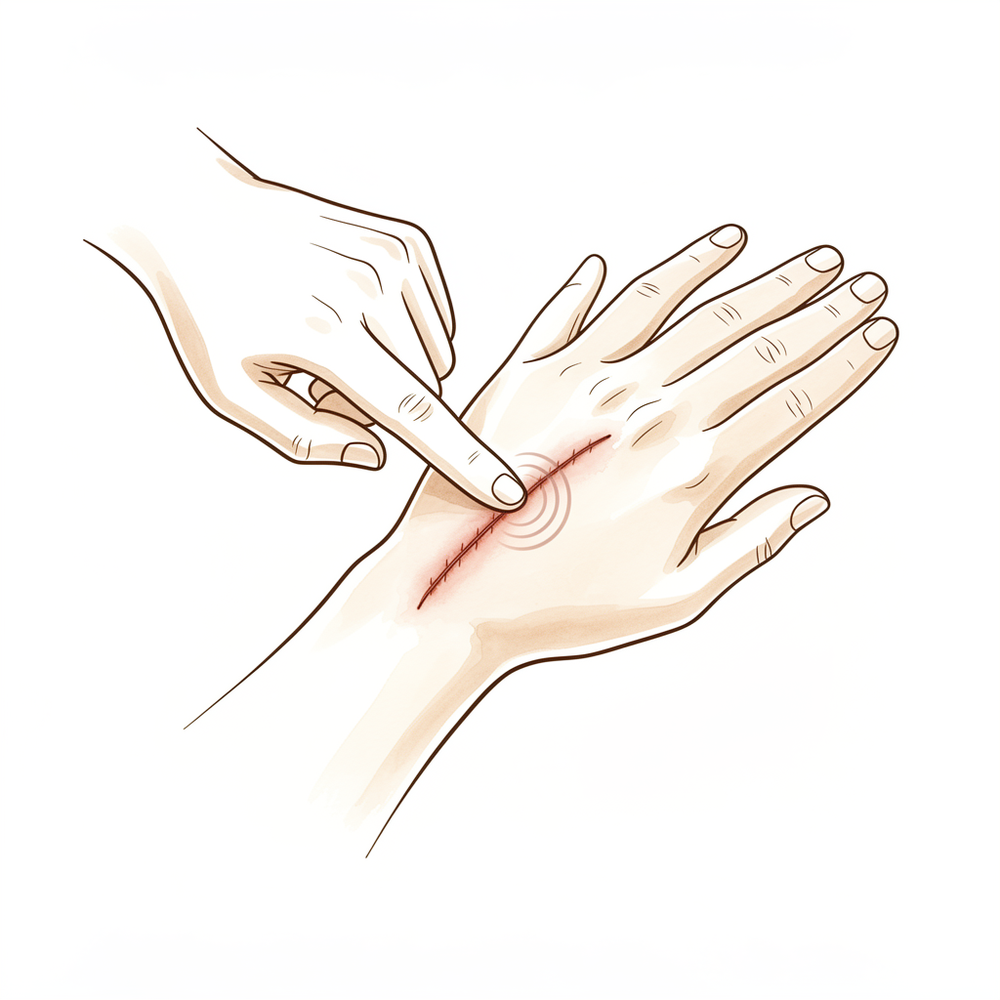
瘢痕按摩
待伤口完全愈合且手部治疗师检查确认后,用坚定的小圆圈动作在疤痕上涂抹少量润肤霜,持续几分钟。这能软化疤痕、缓解不适感,并防止其与下方组织粘连。在皮肤完全闭合前请勿开始此操作。
2–3 分钟,每天 2–3 次(伤口愈合后)
Kieran Hirpara 4.0
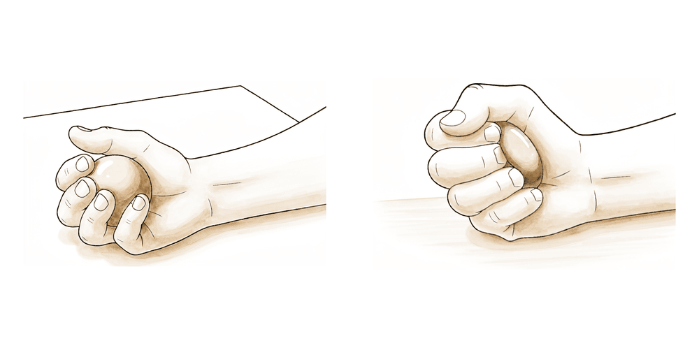
捏握与抓握力量强化(骨性愈合后)
后期锻炼——仅在骨融合愈合且手部治疗师开始指导您进行肌力训练(通常在约六周后)时进行。通过挤压软球或橡皮泥来锻炼抓握力,并用拇指与手术手指捏住小物体,以增强该融合手术旨在使其稳定的侧方捏力。逐渐增加用力程度。在获得许可前,切勿进行任何抗阻抓握或捏持动作——在骨骼愈合前,这些动作会对融合部位施加负荷。
10次,2–3 次/天,遵医嘱(仅在骨愈合后)
这些是您的手册中列出的锻炼项目。仅在Hirpara医生和您的手部治疗师的指导下开始这些锻炼,并严格保持在您被允许的范围内。早期的锻炼旨在保持指尖关节、指关节以及您其他所有手指、拇指和手腕的活动,但不移动或负重已融合的关节本身,该关节在夹板中保持静止。一旦伤口愈合,即可开始瘢痕按摩。捏握和抓握力量训练属于后期阶段,在融合骨愈合且您获得明确许可之前,不应开始。如果任何动作导致融合关节处出现剧烈疼痛,请立即停止。
您的临床方案
本页其余部分为掌指关节(PIP)关节融合术后康复的分阶段临床方案。本节内容应提供给您的手部治疗师,每个阶段均以通俗易懂的语言解释当前发生的情况。在骨性愈合(通常约6周,最长可达 9–12 周)之前,必须保护融合关节免受负荷;核心原则是“保护融合关节,活动其余所有部位”:从第一天起即活动远端指间关节(DIP)、掌指关节(MCP)、相邻手指、拇指和手腕,以防止肌腱粘连和僵硬;早期管理水肿和瘢痕,仅在骨性愈合后恢复抓握和捏持功能。
在治疗前,请查阅患者的手术报告及既往病史,并与主刀医生沟通所使用的内固定方式(张力带钢丝、无头髓内螺钉、克氏针或钢板)、设定的融合角度,以及克氏针是埋入皮下还是需取出。Hirpara 医生在功能屈曲位融合掌指关节,且该角度沿手部尺侧逐渐增大(食指/中指 ≈ 15–20°,环指/小指 ≈ 25–40°),最常用于食指/中指,因为侧方捏持的稳定性优于掌指关节的活动度。融合关节需佩戴支具直至影像学显示骨性愈合;由于证据等级较低(4级病例系列及专家共识),时间安排应个体化,而非采用分级阈值。
第一阶段 — 保护与稳定(第 0 至 2 周)
最初两周旨在保护融合部位,并控制肿胀及伤口情况,同时手部其他所有关节立即开始活动。
致您的手部治疗师:
固定 - 使用急性期掌侧手指夹板或石膏,跨越掌指关节(MCP)和近端指间关节(PIP),但保持远端指间关节(DIP)自由活动 - 融合后的 PIP 关节保持静止;抬高患肢以减轻水肿
宣教与注意事项 - 禁止通过手术手指进行抓握、捏持或负重 - 保持夹板清洁干燥;保护伤口及任何钢针针眼(如有)
管理 - 伤口:按医嘱使用外科敷料;监测感染迹象,若使用克氏针(K-wires)则需监测针眼 - 水肿:抬高患肢,对自由活动关节进行轻柔的泵式活动,必要时冰敷 - 练习:从第一天起,进行所有未融合关节的主动活动:相邻手指(完全握拳/伸展)、拇指、腕关节;在数天内开始手术手指的DIP关节主动活动;相邻手指的肌腱滑动练习;融合 PIP 关节禁止活动或负重
晋级标准 - 伤口趋于稳定;肿胀得到控制;约在 2 周时准备好定制定型夹板
第二阶段 — 定制热塑性夹板及游离关节的主动活动(第2至6周)
大约从两周开始,定制热塑性夹板支撑融合关节,同时释放相邻关节以进行主动活动。手术手指的远指间关节(DIP)和掌指关节(MCP)在脱离夹板状态下进行锻炼;融合的指间关节(PIP)保持保护状态。尚不允许进行抗阻抓握、捏持或负重。
致您的手部治疗师:
评估 - 相邻关节活动度(ROM)、水肿、伤口/瘢痕检查;根据临床依据及外科医生意见确认固定稳定
制动 - 过渡到定制热塑性夹板,支撑融合的指间关节(PIP)同时释放相邻关节;持续保护性夹板固定至约6周
教育与注意事项 - 在骨性愈合前,禁止对手术手指进行抗阻抓握、捏持或负重 - 仅在锻炼时脱离夹板
管理 - 锻炼:手术手指脱离夹板状态下的主动远指间关节(DIP)和掌指关节(MCP)活动(远指间关节在术后数天内开始,掌指关节在此阶段加入);相邻手指的肌腱滑动;继续拇指/腕部/相邻手指活动;伤口愈合后开始瘢痕和水肿管理 - 禁止抗阻抓握/捏持/负重
进展标准 - 影像学骨性愈合(通常在第6周左右,最长可达 9–12);在进行任何负重之前,融合关节在临床和影像学上必须稳定
第三阶段 — 逐步停用支具并过渡至轻度使用(约6周起,骨性愈合后)
一旦融合骨愈合(通常在6周左右),支具将逐步停用并逐渐裁剪,引入轻度功能性使用,并逐步重建捏持、对掌和抓握功能。如果使用了克氏针,通常在6周左右取出。
致您的手部治疗师:
评估 - 与外科医生确认影像学上的骨性愈合;与健侧手对比的握力/捏力;活动关节的活动范围;瘢痕
宣教与注意事项 - 确认骨性愈合后,逐步停用并裁剪支具;如果使用了克氏针,约6周时取出 - 逐步增加负荷:先进行轻度使用,然后进行分级捏持/抓握
管理 - 练习:逐步过渡至轻度功能性使用 → 捏持、对掌和抓握;开始握力/捏力强化训练(球/治疗泥,侧捏),并逐步增加强度;继续瘢痕处理 - 影像学确认骨性愈合后停用支具
进展标准 - 融合骨愈合,能无痛耐受轻度负荷;已停用支具;准备好进行渐进性强化训练
第四阶段 — 渐进性强化与恢复(约从 8–12 周开始)
随着骨融合愈合及轻度使用功能的恢复,强化训练和负重将逐步推进,并逐步建立重返运动、重体力或手工活动的能力。最终稳定的结果通常在九至十二个月时达到。
致您的手部治疗师:
评估 - 握力与捏力对比健侧;根据情况适当进行功能性及工作/运动特异性测试
宣教与注意事项 - 逐渐增加抗阻负重;融合关节在设计上永久僵硬;将强化重点放在握力和侧捏力上
管理 - 练习:握力和捏力的渐进性强化与负重;分级恢复运动、重体力及手工任务 - 一旦力量达到功能性且接近对称,可考虑出院;若恢复停滞,请转回主治医生
恢复标准 - 负重下无痛且稳定的融合关节;握力/捏力足以应对任务需求,依据临床判断而非日历时间;最终稳定结果于 9–12 个月时达到
恢复工作与活动
从术后初期起,即在舒适范围内鼓励进行轻度的日常手部活动;关键限制是在融合骨愈合之前,禁止对手术手指进行抓握、捏持或负重。由于在无法安全控制方向盘时不得驾驶,请在术后早期几周内安排交通协助;通常在术后四至六周左右恢复驾驶,此时您已拆除支具且能安全控制车辆。
骨愈合后(约六周),您可以开始轻度使用和轻柔抓握。提举、抓握和捏持功能从约八周开始逐步恢复,完全活动或运动从约十二周开始。融合部位会在数月内继续稳定,因此最终完全稳定的结果大约在九至十二个月时达到。这些时间点是专家共识的单中心指南(具有典型性和个体化特征,而非分级阈值),您的康复进度将由Hirpara医生和您的手部治疗师根据融合愈合情况评估,而非仅依据日历时间。
您的方案之后
本方案与诊所的总体康复建议配合使用;另请参阅 术后疼痛管理、伤口护理 和 瘢痕管理。上述分阶段计划反映了关于近指间关节融合术后康复的已发表指南,您的持续康复将由 Hirpara 医生和您的手部治疗师根据您手指的愈合情况进行个体化指导。
Evidence & references
This is the clinical evidence summary written for health professionals. It is technical, and it lists the research this page was built from. You do not need to read it to understand your treatment or to make a decision about it.
Topic scope: post-operative rehabilitation after arthrodesis (fusion) of the proximal interphalangeal (PIP) joint of a finger — a worn, painful or unstable PIP joint is fused solid in a functional flexed position. This is a fusion, not a reconstruction or a motion- preserving procedure: the PIP is deliberately made stiff to trade motion for a stable, pain-free, load-bearing digit. The rehab is therefore not about regaining PIP motion but about protecting the construct until bony union while keeping every other joint of the hand moving, then restoring grip and pinch.
Defining principle of the rehab here: PIP arthrodesis eliminates motion at one joint by design to gain stability for pinch and grip. The fused joint is set in a functional flexed position that increases ulnar-ward across the hand (index/middle ≈ 15–20°, ring/little ≈ 25–40°, following the digital cascade) and held by internal fixation (tension-band wire, headless intramedullary screw, K-wires, or plate) until union. Because nothing here needs to move to heal — it needs to unite — the single governing rule is "protect the fused joint, mobilise everything else." DIP, MCP, adjacent digits, thumb and wrist move from day one to prevent tendon adhesion and stiffness; oedema and scar are managed early; grip and pinch are restored only after radiographic union (~6 weeks, up to 9–12). The branch point is the indication — primary degenerative/post-traumatic fusion versus salvage of a failed PIP arthroplasty, where union is slower and the construct more demanding.
A. PROCEDURE OUTCOMES (PIP arthrodesis)
PIP arthrodesis is a reliable, well-established salvage and reconstructive operation, but its evidence base is uniformly low-level — predominantly retrospective level-4 case series and expert opinion, with no randomised controlled trials. Outcomes are reported as union, complication and reoperation rates rather than from comparative trials.
- The evidence base is low-level and consensus-driven. A systematic review of PIP arthrodesis found the literature is overwhelmingly level-4 (~94%) with no RCTs; conclusions on fixation choice and outcomes rest on case series and expert consensus [EFORT Open Rev 2021, DOI 10.1530/eor-21-0102]. Low (level-4 SR, no RCTs).
- Fixation holds the angle to union; nonunion and reoperation are the principal concerns. Series reporting nonunion and reoperation identify patient factors (including smoking and comorbidity) as drivers of failure, underscoring that the rehab job is to protect the construct until the bone joins [HAND 2020, DOI 10.1177/1558944720939196]. Low–moderate (case series).
- The fusion angle is chosen for function, especially pinch. A biomechanical/kinematic study of index PIP fusion (simulated 30–50°) shows the set angle is a functional trade-off: fusing the index/middle PIP stabilises lateral (key) pinch at the cost of PIP motion, which is why these digits are common fusion sites [J Hand Surg Am 2011, DOI 10.1016/j.jhsa.2011.09.010]. Mechanistic / cadaveric.
- Arthrodesis is a dependable salvage for failed PIP arthroplasty, but union is slow. A series of arthrodesis for failed PIP joint replacement reported a mean time to union of 5.8 months, illustrating that salvage fusions unite more slowly than primary fusions and need correspondingly extended protection [J Hand Surg Am 2011, DOI 10.1016/j.jhsa.2010.10.030]. Low (case series).
- The biomechanics of digital loss/fusion frame the functional cost. Reviews of the biomechanics of digital amputation and fusion describe how eliminating an IP joint redistributes grip and pinch mechanics — the rationale for accepting a stiff joint when it buys stability [Hand Clin 2016, DOI 10.1016/j.hcl.2016.07.003]. Mechanistic / narrative.
B. REHABILITATION / THERAPY EVIDENCE
There are no trials of rehab regimens after PIP arthrodesis; the programme is built on sound surgical principle and expert consensus. The two evidence-anchored levers are the union timeline (which sets when load may be applied) and the modifiable risk factor of smoking (which delays union).
- Protect-until-union, mobilise-everything-else is the consensus regimen. The fused PIP is splinted continuously until radiographic union (~6 weeks, up to 9–12); from day one the DIP, MCP, adjacent digits, thumb and wrist are actively moved to prevent tendon adhesion and global hand stiffness. This is stable across sources (surgeon protocols, hand-therapy guidance and patient-education material) even though it is not trial-tested [Melbourne Arm Clinic protocol; OrthOracle PIPJ arthrodesis; OrthoInfo finger IP fusion]. Consensus / expert.
- Smoking is an evidence-supported delayed-union risk. A study of hand and wrist arthrodesis found smoking delays union, making smoking cessation the one rehab-adjacent intervention with direct supporting evidence in this setting [J Hand Surg Am 2022, DOI 10.1016/j.jhsa.2022.05.016]. Moderate (cohort, modifiable risk factor).
- Union timing governs progression — and is slower in salvage fusions. Primary fusions are typically protected to ~6 weeks; salvage of failed arthroplasty unites far more slowly (mean 5.8 months), so loading must be union-led rather than calendar-led [J Hand Surg Am 2011, DOI 10.1016/j.jhsa.2010.10.030]. Low (case series).
- The set fusion angle is the functional anchor of the rehab goal. Because the index/middle PIP is fused at ~15–20° (and ring/little at ~25–40°) specifically to stabilise lateral pinch, the Phase III–IV strengthening rightly targets pinch and grip rather than any attempt at PIP motion [J Hand Surg Am 2011, DOI 10.1016/j.jhsa.2011.09.010]. Mechanistic.
Recovery trajectory (expected, evidence-anchored)
| Phase | Window | Restraint | Hand use / therapy focus | Strength / load | Notes |
|---|---|---|---|---|---|
| I — Protect & settle | Week 0–2 | Volar finger splint/cast spanning MCP + PIP, DIP left free | Elevation, wound/pin-site care, oedema control; active DIP within days + full motion of all non-fused joints (adjacent digits, thumb, wrist) | No grip / pinch / loading | Fused PIP kept still; everything else mobilised from day one |
| II — Custom splint & free-joint motion | Week 2–6 | Custom thermoplastic splint supporting the fused PIP, freeing adjacent joints; continuous splinting to ~6 wk | Active DIP + MCP of operated finger out of splint; tendon glides of adjacent digits; scar/oedema once healed | No resisted grip / pinch / loading | Union typically at ~6 wk (up to 9–12); load only after radiographic union |
| III — Wean splint & light use | From ~6 wk (united) | Splint weaned/cut down after union; K-wire out ~6 wk if used | Progress light use → pinch, opposition, gripping; begin grip/pinch strengthening | Graded grip/pinch, build gradually | Restraints lifted only once union confirmed |
| IV — Strengthen & return | ~8–12 wk+ | Restrictions lifted | Progressive strengthening/loading; return to sport/heavy/manual work | Build load progressively; target lateral pinch | Final settled result 9–12 months |
(Phase windows mirror the precautions in the patient protocol; they are expert-consensus, single-clinic guides — typical and individualised, not graded or trial-derived thresholds. Return milestones: driving ~6 wk, light use/gentle grip ~6 wk after union, lifting/gripping/pinch ~8 wk, full activity/sport ~12 wk, final result 9–12 months.)
C. KEY CONTROVERSIES / EVIDENCE QUALITY
- Whole topic is low-level evidence. PIP arthrodesis rests on level-4 case series and expert consensus with no RCTs (~94% level-4 in systematic review). All outcome and timing figures should be read as typical guides, not trial-validated thresholds [EFORT 2021]. Low.
- Fixation choice is unsettled. Tension-band wire, headless intramedullary screw, K-wires and plate all achieve union; comparative data are weak and selection is largely surgeon preference and bone/soft-tissue quality [EFORT 2021; HAND 2020]. Low.
- Fusion angle is a functional trade-off, not a fixed number. The ~15–20° (index/middle) to ~25–40° (ring/little) cascade is consensus-stable but individualised to the digit and the demands of pinch [J Hand Surg Am 2011 kinematics]. Mechanistic / consensus.
- Union timing is variable and indication-dependent. Primary fusions ~6 weeks; salvage of failed arthroplasty far slower (mean 5.8 months). Loading must be union-led [J Hand Surg Am 2011 salvage series]. Low.
- Smoking and patient factors drive nonunion/reoperation. Smoking is an evidence-supported delayed-union risk and a modifiable target [J Hand Surg Am 2022; HAND 2020]. Moderate (for the smoking association).
D. EVIDENCE STRENGTH FLAGS (summary)
- STRONG (RCT / SR): none — there are no RCTs in PIP arthrodesis; the best synthesis is a level-4 systematic review (~94% level-4 studies).
- MODERATE: smoking as a delayed-union risk after hand/wrist arthrodesis; patient factors driving nonunion/reoperation; cadaveric/kinematic basis for the functional fusion angle and pinch rationale.
- WEAK / CONSENSUS: the protect-until-union, mobilise-everything-else rehab regimen (mechanistically sound, not trial-tested); the specific fusion angles (consensus-stable); exact timelines (single-clinic, expert-consensus guides — typical, not graded thresholds); fixation choice (surgeon preference).
CITATIONS
RAG corpus (180,000+ Orthopaedic articles)
- Proximal interphalangeal joint arthrodesis: a systematic review (predominantly level-4 evidence; no RCTs). EFORT Open Rev. 2021. DOI: 10.1530/eor-21-0102
- Nonunion and reoperation after proximal interphalangeal joint arthrodesis: patient factors and outcomes. HAND. 2020. DOI: 10.1177/1558944720939196
- Index finger proximal interphalangeal joint arthrodesis and pinch kinematics (simulated 30–50° fusion). J Hand Surg Am. 2011. DOI: 10.1016/j.jhsa.2011.09.010
- Arthrodesis as salvage for failed proximal interphalangeal joint arthroplasty (mean time to union 5.8 months). J Hand Surg Am. 2011. DOI: 10.1016/j.jhsa.2010.10.030
- Smoking delays union after hand and wrist arthrodesis. J Hand Surg Am. 2022. DOI: 10.1016/j.jhsa.2022.05.016
- Biomechanics of digital loss and fusion. Hand Clin. 2016. DOI: 10.1016/j.hcl.2016.07.003
PIP arthrodesis rehabilitation & procedure literature (URLs)
- Melbourne Arm Clinic. PIP / DIP arthrodesis rehabilitation protocol. https://melbournearmclinic.com.au/orthopaedic-rehabilitation/shoulder-rehabilitation/pip-dip-arthrodesis-protocol/
- OrthOracle. Proximal interphalangeal joint (PIPJ) arthrodesis in the hand using the Apex system (Extremity Medical). https://www.orthoracle.com/library/proximal-interphalangeal-joint-pipj-arthrodesis-in-the-hand-using-the-apex-system-extremity-medical/
- EFORT Open Reviews. Proximal interphalangeal joint review (PMC). https://pmc.ncbi.nlm.nih.gov/articles/PMC6598614/
- American Academy of Orthopaedic Surgeons (OrthoInfo). Finger (interphalangeal) joint fusion. https://orthoinfo.aaos.org/en/treatment/finger-ip-joint-fusion/





