எலும்பு முறிவுக்கான தோள்பட்டை மாற்று
Patients › Rehabilitation
Rehabilitation after hemiarthroplasty or reverse shoulder replacement for a proximal humerus fracture — slower than elective replacement while the tuberosities heal.
இந்த நெறிமுறை ஒரு உடைந்த தோள்பட்டை (ஒரு அருகிலுள்ள இடுப்பு முறிவு) க்கான டாக்டர் கீரன் ஹிர்பாராவுடன் மேட்டர் தனியார் மருத்துவமனை ராக்ஹாம்ப்டனில் தோள்பட்டை மாற்றுதலுக்குப் பிறகு உங்கள் மீட்புக்கு வழிகாட்டுகிறது. முறிவைப் பொறுத்து, மாற்றுவது ஒரு தலைகீழ் தோள்பட்டை மாற்றுதல் அல்லது ஒரு ஹெமியார்ட்ரோபிளாஸ்டி (இணைப்பின் பந்து மட்டும் மாற்றுவது) இருக்கலாம். மறுவாழ்வு பயணம் இரண்டிற்கும் ஒரே வடிவத்தைக் கொண்டுள்ளது, ஏனென்றால் வேகத்தை நிர்ணயிப்பது எலும்பு துண்டுகளின் குணமாகும், உள்வைப்பு வகை அல்ல. கீழே உள்ள ஒவ்வொரு கட்டமும் என்ன நடக்கிறது என்பதற்கான எளிய ஆங்கில விளக்கத்துடன் தொடங்குகிறது மற்றும் மிகவும் முக்கியமானது, அதைத் தொடர்ந்து எழுதப்பட்ட கட்டமைக்கப்பட்ட நெறிமுறை உங்கள் பிசியோதெரபிஸ்ட்டுக்கு: உங்கள் முதல் பிசியோதெரபி வருகைக்கு இந்த பக்கத்தை அல்லது அதன் PDF ஐ எடுத்துச் செல்லுங்கள், இதனால் உங்கள் மறுவாழ்வு ஒருங்கிணைந்ததாக இருக்கும். உங்கள் பிசியோதெரபி உங்கள் மீட்பு எவ்வாறு முன்னேறுகிறது என்பதைப் பொறுத்து திட்டத்தை சரிசெய்யலாம்.
அறுவை சிகிச்சைக்குப் பிறகு உங்கள் காயத்தைப் பற்றி ஏதேனும் கவலைகள் இருந்தால், அறைகளைத் தொடர்பு கொள்ளுங்கள். காயத்தின் புகைப்படத்தை எடுத்து அதை மதிப்பாய்வு செய்ய மின்னஞ்சல் அனுப்புவது பெரும்பாலும் உதவியாக இருக்கும்.
எலும்பு முறிவுக்குப் பிறகு குணமடைவது ஏன் மெதுவாக இருக்கிறது
ஒரு தோள்பட்டை கீல்வாதம் அல்லது அணிந்த ரோட்டேட்டர் கம்பளத்திற்காக தேர்ந்தெடுக்கப்பட்ட முறையில் மாற்றப்படும்போது, புதிய மூட்டுக்கு சுற்றியுள்ள எலும்பு அப்படியே இருக்கும், மற்றும் மறுவாழ்வு மென்மையான திசு குணமடைதல் வேகத்தில் நகர்த்த முடியும். முறிவுக்குப் பிறகு இது வேறுபட்டது. கை எலும்பின் மேற்புறத்தில் உள்ள இரண்டு எலும்பு முனைகள் (பெரிய மற்றும் சிறிய தண்டுகள், அங்கு ரோட்டேட்டர் கம்பள தசைகள் இணைக்கப்படுகின்றன) பொதுவாக உடைந்து விடுகின்றன, மேலும் உங்கள் அறுவை சிகிச்சையின் போது அவை புதிய புரோதெஸிஸைச் சுற்றி மீண்டும் பொருத்தப்படுகின்றன. அந்த துண்டுகள் இப்போது எலும்பு மற்றும் உள்வைப்பில் குணமடைய வேண்டும், வேறு எந்த முறிவு போலவே, இது பொதுவாக பல வாரங்கள் முதல் மாதங்கள் வரை ஆகும்.
எலும்பு முறிவுக்காக செய்யப்படும் தோள்பட்டை மாற்றத்திற்கான வெளியிடப்பட்ட மறுவாழ்வு நெறிமுறைகள் இந்த புள்ளியில் சீரானவைஃ தோள்பட்டை எவ்வளவு நன்றாக வேலை செய்கிறது என்பது கட்டிகள் நிலைக்கு குணமடைகிறதா என்பதைப் பொறுத்தது, மற்றும் ஆரம்ப அல்லது வலுவான இயக்கம் (செயலற்ற இயக்கம் கூட) பழுதுபார்ப்பை இழுக்கலாம் மற்றும் அந்த குணமடைவதை ஆபத்தில் ஆழ்த்தலாம். தலைகீழ் தோள்பட்டை மாற்று நெறிமுறைஒரு எலும்பு முறிவுக்குப் பிறகு, அதே பயணம் மெதுவாக செல்கிறது, ஏனென்றால் எலும்பு துண்டுகள் முதலில் குணமடைய வேண்டும். உங்கள் மறுவாழ்வின் ஒவ்வொரு அடியும் இரண்டு விஷயங்களில் ஒன்றாக மூடப்பட்டிருக்கும்ஃ போதுமான நேரம் கடந்து, எக்ஸ்-கதிர்கள் எலும்பு குணமடைவதைக் காட்டுகின்றன, டாக்டர் ஹிர்பாராவுடன் உங்கள் மதிப்பாய்வில் உறுதிப்படுத்தப்பட்டுள்ளது.
எதிர்பார்ப்பது என்ன
நீங்கள் எழுந்திருக்கும்போது உங்கள் கையில் ஒரு மயக்கம் இருக்கும், சுமார் 24 மணி நேரத்திற்குப் பிறகு அந்த உணர்வு திரும்பத் தொடங்க வேண்டும். ஒரு வாரம் வரை சில மயக்கம் அல்லது பலவீனம் இருக்கலாம்.
உங்கள் அறுவை சிகிச்சையிலிருந்து நீங்கள் எழுந்திருக்கும்போது, உங்கள் தோள்பட்டைக்கு மேல் ஒரு பெரிய பேட் உடன் ஒரு ஸ்லிங்கில் இருப்பீர்கள். இந்த பேட் வெளியேற்றப்படுவதற்கு முன்பு அகற்றப்படும். அடியில் ஒரு அறுவை சிகிச்சை பசை நாடாவை மூடும் நீர்ப்புகா பேண்டேஜ் இருக்கும், இது 2 வாரங்களுக்கு தனியாக விடப்படலாம். உங்கள் தையல்கள் கரைக்கக்கூடியவை மற்றும் அகற்றத் தேவையில்லை, ஆனால் காயத்தின் ஒவ்வொரு முனையிலும் சில தையல் வால் இருக்கலாம், அவை 2 வாரங்களுக்குப் பிறகு சருமத்துடன் வெட்டப்படலாம். உங்கள் அறுவை சிகிச்சைக்குப் பிறகு 1 2 வாரங்களுக்கு காயத்தை சரிபார்க்க எங்கள் செவிலியரைப் பார்க்க நீங்கள் முன்பதிவு செய்யப்படுவீர்கள். பேண்டேஜ் சோதனைக்கு நீங்கள் கலந்து கொள்ள முடியாவிட்டால், 2 வாரங்களுக்குப் பிறகு உங்கள் பேண்டேஜை நீங்களே அகற்றலாம்.
செயல்பாடுகளுக்குத் திரும்புவதற்கான தோராயமான கால அளவுகள் (தேர்ந்தெடுக்கப்பட்ட மாற்றீட்டைக் காட்டிலும் மெதுவாக, எப்போதும் உங்கள் எலும்பு எவ்வாறு குணமடைகிறது என்பதைப் பொறுத்து):
- வாகனம் ஓட்டுதல் பெரும்பாலான மக்கள் 6 முதல் 12 வாரங்களுக்கு இடையில் திரும்பி வருகிறார்கள், இயக்கம் மற்றும் கட்டுப்பாடு திரும்பியவுடன்; இதை உங்கள் மதிப்பாய்வில் விவாதிக்கவும்.
- நீச்சல்: உங்கள் மதிப்பாய்வில் வழிகாட்டப்பட்டபடி சுமார் 4 மாதங்களிலிருந்து மார்பகப் பயிற்சி; பின்னர் ஃப்ரீஸ்டைல்.
- தூக்குதல்: சுமார் 12 வாரங்கள் வரை ஒரு கப் காபியை விட கனமான எதையும் செய்யக்கூடாது; 12 வாரங்களில் இருந்து படிப்படியாக லேசான தூக்குதல் கட்டமைக்கப்படுகிறது; 6 மாதங்களுக்கு கனமான பொருட்களை தூக்குவதை தவிர்க்கவும்.
- வேலை: உட்கார்ந்திருக்கும் வேலை: ஆறு வாரங்களில் இருந்து, வசதிக்கு ஏற்ப; கைத்தொழில்: டாக்டர் ஹிர்பராவின் வழிகாட்டுதலின் கீழ்.
முறிவுக்கான மாற்றத்திற்குப் பிறகு, தோள்பட்டை பொதுவாக காயமடையாத தோளின் முழு அளவையும் மீட்டெடுக்காது. வெளியிடப்பட்ட முடிவுகள் இடுப்பு மற்றும் தோள்பட்டை உயரத்தில் (மற்றும் பெரும்பாலும் மேல்) கையை வசதியாக, வலி கட்டுப்படுத்தப்பட்ட பயன்பாட்டை வழக்கமான விளைவாக விவரிக்கிறது, அறுவை சிகிச்சைக்குப் பிறகு 1224 மாதங்களுக்கு இயக்கம் மற்றும் வலிமை தொடர்ந்து மேம்படுகிறது.
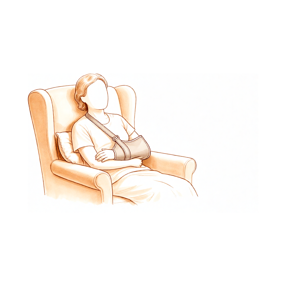
உங்கள் ஸ்லிங் அணிந்து
எலும்பு துண்டுகள் குணமடையும் வரை உங்கள் தோள்பட்டைக்கு ஆதரவாக உங்கள் ஸ்லிங் (தோள்பட்டை இம்போலைசர்) உள்ளது. விதிகள் எளிமையானவை:
- அதை அணியுங்கள் 6 வாரங்கள், தூங்கும்போது கூட.
- குளியல் மற்றும் உங்கள் உடற்பயிற்சிகளுக்கு மட்டுமே அதை அவிழ்த்து விடுங்கள், எப்படி செய்வது என்று உங்களுக்குக் காண்பிக்கப்பட்டவுடன்; கயிறு அவிழ்த்து விடப்பட்டிருக்கும் போதெல்லாம், உங்கள் கையை உங்கள் பக்கத்தில் வைத்திருங்கள்.
- வீட்டில் ஓய்வெடுக்கும்போது, நீங்கள் அதைப் பற்றி புத்திசாலித்தனமாக இருந்தால், அது வெளியே வரலாம்ஃ உட்கார்ந்திருக்கும்போது ஒரு தலையணையில் கை வைக்கப்படுகிறது.
- சட்டைப் பெட்டியை அணிந்து கொண்டு வாகனம் ஓட்டாதீர்கள்.
- குறிப்பாக உடற்பயிற்சி செய்த பிறகு, தோள்பட்டை வீங்கியிருந்தால் அல்லது வலி இருந்தால், பனியைப் பயன்படுத்துங்கள்.
உங்கள் பிசியோதெரபிஸ்ட் ஆரம்பத்தில் ஸ்லிங்கை வைக்க உங்களுக்கு உதவுவார், நீங்கள் வீட்டிற்குச் செல்வதற்கு முன்பு அதை சுயாதீனமாக நிர்வகிக்க உங்களுக்குக் கற்பிப்பார். அதை சரியாக பொருத்துவது முக்கியம்ஃ ஒரு தளர்வான ஸ்லிங்க் உங்களை சரியாக ஆதரிக்காது.
- கயிறு பொருத்தும்போது, உங்கள் முழங்கை கயிறு மூலையில் வைக்கப்பட்டு, நன்கு ஆதரிக்கப்படுவதை எப்போதும் உறுதிப்படுத்திக் கொள்ளுங்கள்.
- [அடிக்குறிப்புகள்] [அடிக்குறிப்புகள்]
- உங்கள் ஸ்லிங்கில் இரண்டு வெல்க்ரோ கயிறுகள் உள்ளன: ஒன்று உங்கள் கழுத்தில், மற்றொன்று உங்கள் இடுப்பில்.
- உங்கள் முழங்கை மற்றும் முதுகெலும்புகளை சரியாக நிலைநிறுத்தியவுடன், உங்கள் அறுவை சிகிச்சை செய்யப்படாத கையை பயன்படுத்தி உங்கள் கழுத்தை சுற்றி மேல் பட்டையை முன்னால் சுழற்றவும், அதை ஸ்லிங்கின் மேல் வளையத்தின் வழியாக இணைக்கவும்.
- உங்கள் இடுப்பை சுற்றி கீழ் பட்டை இணைக்க அதே முறையை பயன்படுத்தவும், கயிற்றின் கீழ் வளையத்தின் மூலம் பாதுகாக்கவும்.
ஒரு நல்ல தோரணை நிலையை அடைய, உங்கள் காதுகள், தோள்பட்டைகள் மற்றும் இடுப்புகளை வரிசையில் வைக்க முயற்சிக்கவும். உங்கள் முதுகுக்கு நல்ல தோரணை முக்கியமானது மற்றும் உங்கள் தோள்பட்டை மூட்டு இறுக்கம் ஏற்படுவதைத் தடுக்க உதவும். உட்கார்ந்திருக்கும் போது உங்கள் முதுகின் சிறிய பகுதியில் வைக்கப்படும் ஒரு சுருட்டப்பட்ட துண்டு ஒரு நட்பு நினைவூட்டலாக செயல்படலாம்.
மருத்துவமனையில் உங்கள் முதல் நாட்கள்
நீங்கள் வீட்டிற்குச் செல்வதற்கு முன், மருத்துவமனை உடற்கூறியல் சிகிச்சையாளர்கள் ஒரு எளிய திட்டத்தில் உங்களைத் தொடங்குவார்கள். அவர்கள் பயன்படுத்தும் மூன்று சொற்களைப் புரிந்துகொள்ள இது உதவுகிறது. செயலில் இயக்கத்தின் வரம்பு என்பது நீங்களே செய்யும் இயக்கம், உதவி அல்லது உதவியின்றி. செயலில்-உதவி இயக்கத்தின் வரம்பு என்பது உங்கள் மற்ற கையை (அல்லது ஒரு பொருளை, உதாரணமாக ஒரு கைத்தடியை) பயன்படுத்தி கையை நகர்த்த உதவுகிறது. செயலற்றது எலும்பு முறிவுக்கான மாற்று அறுவை சிகிச்சைக்குப் பிறகு, அறுவை சிகிச்சைக்கு உட்படுத்தப்பட்ட தோள்பட்டை முதல் ஆறு வாரங்களுக்கு மட்டும், மென்மையான வரம்புகளுக்குள் மட்டுமே நகர்த்தப்படுகிறது; நீங்கள் தீவிரமாக நகர்த்தும் மூட்டுகள் உங்கள் முழங்கை, மணிக்கட்டு மற்றும் கை.
இந்த முதல் நாட்களுக்கு சில நடைமுறை குறிப்புகள்:
- நீங்கள் ஸ்லிங் தூங்க வேண்டும்.
- தேவைப்பட்டால் வலி நிவாரணத்திற்கு பனியைப் பயன்படுத்துங்கள்.
- [பக்கம் 3-ன் படம்]
- உடற்பயிற்சி செய்வதற்கு முன்பும், உடற்பயிற்சி செய்வதற்கு முன்பும் உங்கள் வலி நிவாரணி மருந்துகளை எடுத்துக் கொள்ளுங்கள்.
- உங்கள் உடற்பயிற்சிகள் மற்றும் குளியல் ஆகியவற்றிற்காக உங்கள் கையை ஸ்லிங்கில் இருந்து வெளியே எடுக்க அனுமதிக்கப்படுகிறீர்கள்.
- நீங்கள் 6 வாரங்களுக்கு உங்கள் ஸ்லிங் அணிய வேண்டும், குறிப்பாக வீட்டை விட்டு வெளியேறும் போது.
- அறுவை சிகிச்சை செய்யப்படும் பக்கத்தில் உள்ள உங்கள் கையை மெதுவாக பிஸியாக வைத்திருங்கள்: எழுதுவது, சாப்பிடுவது மற்றும் கைப்பிடியில் கை தங்கியிருக்கும் போது தொலைபேசியைப் பயன்படுத்துவது ஆகியவை ஊக்குவிக்கப்படுகின்றன.
- உங்கள் சொந்த உடல்நல சிகிச்சையை ஏற்பாடு செய்ய நீங்கள் தேர்வு செய்யாவிட்டால், உங்களுக்காக ஒரு சந்திப்பு ஏற்பாடு செய்யப்பட்டு, உங்கள் வெளியேற்றப் பொட்டலத்தில் விரிவாகக் கூறப்பட்டுள்ளது.
- உங்களுக்கு ஏதேனும் பிரச்சினைகள் இருந்தால், அலுவலகத்தை தொடர்பு கொள்ளவும் அல்லது உங்கள் பிசியோதெரபிஸ்ட்டிடம் தெரிவிக்கவும்.
கட்டம் I எலும்பு குணமடையும் போது பாதுகாப்பு (வாரங்கள் 06)
முதல் ஆறு வாரங்கள் ஒரு விஷயத்தைப் பற்றியது: எலும்பு மற்றும் புரோதெசிஸில் குணமடையும் துண்டுகளை குறுக்கிடாமல் விட்டுவிடுங்கள். நீங்கள் இரவும் பகலும் ஸ்லிங்கில் இருக்கிறீர்கள், உங்கள் முழங்கை, மணிக்கட்டு மற்றும் கையை நகர்த்திக் கொள்ளுங்கள், மற்றும் தோள்பட்டை தன்னை கீழே உள்ள வரம்புகளுக்குள் (மெதுவாக, உங்கள் உடற்கூறியல் நிபுணரால் அல்லது முற்றிலும் நிதானமாக) நகர்த்த அனுமதிக்கவும். மிக முக்கியமான விதிகள்ஃ தோள்பட்டை தீவிரமாக நகர்த்த வேண்டாம், எல்லைகளைத் தாண்டி கையை வெளியே சுழற்ற வேண்டாம், உங்கள் முதுகுக்குப் பின்னால் அடைய வேண்டாம், எதையும் தூக்க வேண்டாம், உங்கள் கைகளால் தள்ள வேண்டாம். உங்களுக்கு ஒரு தலைகீழ் மாற்று இருந்தால், இந்த விதிகள் மென்மையான திசுக்கள் குணமடையும் போது புதிய மூட்டு இடப்பெயர்வுக்கு எதிராகவும் பாதுகாக்கின்றன. உங்கள் முதுகில் படுத்துக் கொண்டிருக்கும்போது, ஒரு சிறிய தலையணியை அல்லது உங்கள் முழங்கையின் கீழ் சுருட்டிக்கொண்டு, உங்கள் தோள்பட்டை பின்புறமாக நீட்டக்கூடாது.
உங்கள் பிசியோதெரபிஸ்ட்டுக்கு:
இலக்குகள்
- குடல் சீரமைப்பு மற்றும் புரோதெசிஸை பாதுகாக்கவும்
- வீக்கத்தைக் குறைத்தல், வலியைக் குறைத்தல்
- முழங்கை, மணிக்கட்டு மற்றும் கையில் செயல்பாட்டு இயக்கம் (ROM) பராமரிக்கவும்
- பாதுகாப்பான மண்டலத்திற்குள் மென்மையான பாதுகாப்பு தோள்பட்டை செயலற்ற இயக்க வரம்பு (PROM)
- விழிப்புணர்வை பராமரித்தல்
- நோயாளி கல்வி
கயிறு
- 6 வாரங்கள், தூங்கும் போது உட்பட
- கழுவுதல் மற்றும் உடற்பயிற்சிகளுக்காக மட்டுமே அகற்றப்படுகிறது; கை கயிற்றிலிருந்து வெளியேறும்போது பக்கத்தில் இருக்கும்
நிர்வாகம்
- வீக்கக் கட்டுப்பாடுஃ பனி, அழுத்தம்
- இயக்க வரம்பு / இயக்கம்:
- PROM மட்டுமே, பாதுகாப்பான மண்டலத்திற்குள்ஃ ஸ்கேப்புலர் விமானத்தில் உயர்வு ≤ 90 டிகிரி; வெளிப்புற சுழற்சி (ER) முதல் 4 வாரங்களுக்கு நடுநிலைக்கு (0 டிகிரி), பின்னர் ≤ 30 டிகிரி; பின்புறத்தில் உள் சுழற்சி (IR) இல்லை
- ஊசலாட்டம், ஒரு செயலற்ற உடற்பயிற்சியாக முழுமையாக நிதானமாக செய்யப்படுகிறது
- சுறுசுறுப்பான உதவியுடன் இயக்க வரம்பு (AAROM): தோள்பட்டைக்கு எதுவும் இல்லை
- செயலில் உள்ள இயக்க வரம்பு (AROM): முழங்கை, மணிக்கட்டு, கை மற்றும் விரல்கள்; கர்ப்பப்பை முதுகெலும்பு
- தோள்பட்டை அமைத்தல், தோள்பட்டை குலுக்கல் மற்றும் தோரணை சரிசெய்தல்
- ஸ்லிங்கில் இருக்கும்போது கையை லேசாகப் பயன்படுத்துவதை ஊக்குவிக்கவும் (எழுதுதல், சாப்பிடுதல், தொலைபேசி)
- உடற்பயிற்சிகள் மற்றும் இயற்பியல் சிகிச்சை அமர்வுகளுக்கு முன் வலி நிவாரணம்
எச்சரிக்கைகள்
- தோள்பட்டை AROM அல்லது AAROM இல்லை
- மேற்கூறிய வரம்புகளைத் தாண்டிய பாசிவ் அல்லது ஆக்டிவ் ஈ.ஆர். இல்லை (அதிக எலும்புக்கூடு சரிசெய்தல் வெளிப்புற சுழற்சியால் சுமை செய்யப்படுகிறது)
- உள் சுழற்சிக்கு எதிர்ப்பு இல்லை (குறைந்த tuberosity/subscapularis பழுது உள் சுழற்சியால் சுமை செய்யப்படுகிறது)
- முதுகுக்குப் பின்னால் எட்டிப் பார்க்கக் கூடாது; ஒருங்கிணைந்த ஊடுருவல், உள் சுழற்சி மற்றும் நீட்டிப்பு இல்லை (திருப்பு மாற்று இடப்பெயர்ச்சி நிலை)
- பொருட்களை தூக்கக்கூடாது; உடல் எடையை கைகளால் ஆதரிக்கக்கூடாது (எடுத்துக்காட்டாக, ஒரு நாற்காலியில் இருந்து அல்லது படுக்கையில் இருந்து தள்ளுவது)
- முதுகில் படுத்துக் கொண்டிருக்கும்போது ஒரு சிறிய தலையணை அல்லது துண்டுத் துணியை முழங்காலின் கீழ் வைக்கவும், இதனால் தோள்பட்டை நீட்டிப்பில் விழாது
- வலியை ஏற்படுத்தும் எந்த இயக்கத்தையும் கட்டாயப்படுத்த வேண்டாம்
முன்னேற்றத்திற்கான அளவுகோல்கள்
- 6 வாரங்கள் கடந்துவிட்டன மற்றும் எக்ஸ்-கதிர்கள் குடலிறக்கங்கள் குணமடைவதைக் காட்டுகின்றன, டாக்டர் ஹிர்பாராவுடன் உங்கள் மதிப்பாய்வில் உறுதிப்படுத்தப்பட்டபடி
- வாய்வழி அனல்ஜீசியா மூலம் வலியைக் கட்டுப்படுத்துதல்
- காயங்கள் பற்றிய கவலைகளும் நிலையற்ற தன்மையின் அறிகுறிகளும் இல்லை
கட்டம் II இயக்கத்தை மீட்டெடுப்பது (வாரங்கள் 612)
உங்கள் ஆறு வார மதிப்பாய்வில், எலும்பு துண்டுகள் நிலைக்கு குணமடைகின்றனவா என்பதை எக்ஸ்-கதிர்கள் சரிபார்க்கின்றன. அவை இருந்தால், ஸ்லிங் விலகி, தோள்பட்டை தானாகவே நகர்த்தத் தொடங்குகிறது, முதலில் உங்கள் மற்ற கை, ஒரு கைத்தடி அல்லது ஒரு புல்லியைப் பயன்படுத்தி உதவுகிறது, பின்னர் தீவிரமாக. இந்த வாரங்களில் செயலற்ற வரம்பு முழுமையாக முன்னேறுகிறது, மற்றும் சுழற்சி வரம்புகள் எளிதாகின்றன. முதல் மென்மையான தசை-செயல்படுத்தும் வேலை டெல்டாய்டு மற்றும் தோள்பட்டை-பிளேட் (பெரிஸ்காப்புலர்) தசைகளுக்கு தொடங்குகிறது. இன்னும் தொடங்காதது வலுவடைகிறதுஃ பழுதுபார்ப்பு இன்னும் ஒருங்கிணைக்கப்படுகிறது, எனவே தூக்கும் விதி ஒரு கப் காபியை விட கனமாக இல்லை, இன்னும் கைகள் மூலம் தள்ளுவது இல்லை.
உங்கள் பிசியோதெரபிஸ்ட்டுக்கு:
இலக்குகள்
- ஆறு வார மறுபரிசீலனைக்குப் பிறகு கயிற்றிலிருந்து விடுபடுதல்
- ஆறுதல் அனுமதிக்கிறது என முழு வீச்சு நோக்கி முன்னேற்றம் தோள்பட்டை PROM
- தோள்பட்டை AAROM துவக்க, AROM முன்னேறும்
- மென்மையான டெல்டோயிட் மற்றும் பெரிஸ்காப்புலர் செயல்பாட்டைத் தொடங்கவும்
- இடுப்பு மற்றும் மார்பு உயரத்தில் அன்றாட வாழ்க்கையின் இலகுவான செயல்பாடுகளுக்கு கையின் பயன்பாட்டை மீட்டெடுக்கவும்
- நோயாளி கல்வி
கயிறு
- ஆறு வார மறுபரிசீலனைக்குப் பிறகு நிறுத்தவும்; தாய்ப்பால் குடித்த முதல் வாரம் அல்லது இரண்டு வாரங்களில் கூட்டத்தில் பாதுகாப்பிற்காக வீட்டை விட்டு வெளியே அணியலாம்
நிர்வாகம்
- கட்டம் I தலையீடுகளை தேவைக்கேற்ப தொடரவும்
- இயக்க வரம்பு / இயக்கம்:
- PROM: சுமார் 810 வாரங்களுக்குள் அனைத்து விமானங்களிலும் முழு அளவிலான முன்னேற்றம், வலியைப் பொறுத்து
- AAROM: மேஜை ஸ்லைடுகள், சுவர் ஏறுதல், துருவிகள், தண்டு உதவியுடன் வளைத்தல் மற்றும் வெளிப்புற சுழற்சி, உட்கார்ந்திருக்கும் நிலைக்கு முன்னேறுதல்
- AROM: AAROM வசதியாகவும், இயக்கத் தரம் நன்றாகவும் இருக்கும்போது தொடங்குங்கள், தலைகீழ் வளைவோடு தொடங்கி நேராக வளைவோடு தொடரவும்.
- ER: 30 டிகிரிக்கு அப்பால் முன்னேற்றம் குணமடைந்தவுடன் ஆறுதல் அனுமதிக்கிறது
- மென்மையான செயல்பாட்டு கை-பின்புறம் மற்றும் நீட்டிப்பு இந்த கட்டத்தில் தாமதமாக அறிமுகப்படுத்தப்பட்டது, தெளிவுபடுத்தப்பட்டது. இந்த நிலைகளில் நீட்ட வேண்டாம்.
- பலப்படுத்துதல்:
- வலி இல்லாத சப்-மேக்ஸிமல் ஐசோமெட்ரிக்ஸ் மட்டுமேஃ ஸ்கேப்புலர் விமானத்தில் டெல்டாய்டு, பெரிஸ்கேப்புலர் செட்டிங், குறைந்த வரிசை, ஸ்கேப்புலர் இழுப்பு
- இந்த கட்டத்தில் மென்மையான வலி இல்லாத ரோட்டேட்டர் கஃப்ட் ஐசோமெட்ரிக்ஸ்; மறுபரிசீலனையில் அழிக்கப்பட்டதும் மட்டுமே எதிர்ப்பு உள் சுழற்சியை அறிமுகப்படுத்துங்கள்
- மோட்டார் கட்டுப்பாடுஃ ஸ்கேப்புலர் கட்டுப்பாட்டை வலியுறுத்துங்கள் மற்றும் இழுத்தல் அல்லது பிற ஈடுசெய்யும் வடிவங்களைத் தவிர்க்கவும்
எச்சரிக்கைகள்
- ஒரு கப் காபியை விட கனமான பொருட்களை தூக்கக் கூடாது
- கைகள் மூலம் உடல் எடையை ஆதரிப்பது இல்லை
- மென்மையான ஐசோமெட்ரிக்குகளுக்கு அப்பால் எதிர்ப்பு அல்லது வலுவூட்டல் வேலை இல்லை
- கட்டாயப்படுத்தும் வரம்பைத் தவிர்க்கவும்ஃ உறுதியான அச om கரியத்திற்கு மட்டுமே நீட்டவும், கூர்மையான வலி இல்லை
முன்னேற்றத்திற்கான அளவுகோல்கள்
- தோள்பட்டை உயரத்திற்குக் கீழே வசதியான செயலில் இயக்கம், நல்ல ஸ்கேப்புலர் கட்டுப்பாடு மற்றும் குறைந்தபட்ச இழப்பீடு
- டாக்டர் ஹிர்பாராவுடன் நீங்கள் பரிசீலித்தபோது உறுதிப்படுத்தப்பட்டபடி, எக்ஸ்-ரேவில் கட்டிகள் தொடர்ந்து குணமடைகின்றன.
- எதிர்ப்பு வேலை தொடங்க போதுமான வலி அமைக்க
கட்டம் III வலுவூட்டல் (வாரங்கள் 1224)
சுமார் பன்னிரண்டு வாரங்களுக்குள், குடலிறக்கங்கள் பொதுவாக ஒற்றுமைக்கான பாதையில் உள்ளன, மேலும் டாக்டர் ஹிர்பாராவுடன் உங்கள் மதிப்பாய்வு தோள்பட்டை சுமைக்கு தயாராக இருக்கிறதா என்பதை உறுதிப்படுத்துகிறது. பின்னர் வலுவூட்டல் மெதுவாக தொடங்குகிறது, நெகிழ்வான பட்டைகள் மற்றும் ஒளி எடைகளுடன் தொடங்கி, தோளை இயக்கும் டெல்டாய்டு மற்றும் தோள்பட்டை-பிளேட் தசைகளில் கவனம் செலுத்துகிறது (ஒரு தலைகீழ் மாற்றத்திற்குப் பிறகு, டெல்டாய்டு ரோட்டேட்டர் கஃப் பயன்படுத்திய வேலையில் பெரும்பகுதியைச் செய்கிறது). சுழலும் கஃப் குணமடைந்த குடலிறக்கங்கள் அனுமதிப்பதைப் போலவே வலுவூட்டப்படுகிறது. இந்த கட்டத்தில் லேசான தூக்குதல் படிப்படியாக உருவாகிறது, இடுப்பு மற்றும் தோள்பட்டை உயரத்தில் தினசரி நடவடிக்கைகள் இயல்பு நிலைக்கு நெருக்கமாக உணரப்பட வேண்டும், மேலும் இது ஏற்கனவே தொடரவில்லை என்றால் இந்த கட்டத்தில் வழக்கமாக மீண்டும் ஓட்டுகிறது.
உங்கள் பிசியோதெரபிஸ்ட்டுக்கு:
இலக்குகள்
- அனைத்து விமானங்களில் வலி இல்லாத PROM மற்றும் முன்னேற்றம் AROM பராமரிக்கவும்
- வலிமை மற்றும் சகிப்புத்தன்மையின் படிப்படியான மீட்புஃ டெல்டோயிட், பெரிஸ்காப்புலர் தசைகள், பின்னர் ரோட்டேட்டர் கம்பளி
- இயக்க நிலைத்தன்மை, மோட்டார் கட்டுப்பாடு மற்றும் சுய உணர்வை மேம்படுத்துதல்
- லேசான தூக்குதல் உள்ளிட்ட பெரும்பாலான அன்றாட நடவடிக்கைகளுக்கு திரும்புதல்
நிர்வாகம்
- அனைத்து விமானங்களிலும் range-of-motion மற்றும் mobility பணிகளை தொடரவும்
- வலுவூட்டல், ஐசோமெட்ரிக்ஸிலிருந்து நெகிழ்வான பட்டைகள் மற்றும் பின்னர் இலகுரக எடைகள் (குறைந்த சுமை, அதிக மறுபடியும்):
- பெரிஸ்காப்புலர்ஃ வரிசைகள், செராடஸ் பஞ்ச்ஸ், எதிர்ப்புடன் ஸ்காப்புலர் இழுப்பு
- டெல்டோயிட்ஃ செயல்பாட்டு நிலைகளில் வளைவு மற்றும் ஸ்கேப்ஷனை எதிர்த்தது, கட்டுப்படுத்தப்பட்ட எக்ஸென்ட்ரிக் குறைப்புடன்
- ரோட்டேட்டர் மஞ்ச்ஃ ER மற்றும் IR நெகிழ்வு எதிர்ப்புடன், பக்கவாட்டு ER, குணமடைந்த tuberosities அனுமதிக்கும் போது முன்னேறுகிறது
- மோட்டார் கட்டுப்பாடு: தாள நிலைப்படுத்தல், சுயநல நரம்பியல் தசை வசதி (PNF) முனை வடிவங்கள், பந்து நிலைப்படுத்தல் வேலை
- படிப்படியாக உயர்த்தல்ஃ ஆரம்ப கட்டத்தில் சுமார் 2 கிலோ முதல் இறுதிக்குள் சுமார் 5 கிலோ வரை, கட்டுப்பாடு அனுமதிக்கிறது
- இந்த கட்டத்தில் படிப்படியாக கை வழியாக எடை தாங்கி (எடுத்துக்காட்டாக ஒரு நாற்காலியில் இருந்து தள்ளுதல்) மீண்டும் அறிமுகப்படுத்தப்படுகிறது.
எச்சரிக்கைகள்
- கனமான பொருட்களை தூக்கக் கூடாது; இந்த கட்டத்தில் சுமைகளை 5 கிலோவுக்குக் குறைவாக வைத்திருக்க வேண்டும்
- வலுவான இறுதியில் வரம்பில் நீட்டிப்பு தவிர்க்க, மற்றும் சுமை இணைந்த கடத்தல் மற்றும் வெளிப்புற சுழற்சி தவிர்க்க
- நோயின் முன்னேற்றம் அறிகுறிகளால் வழிநடத்தப்படுகிறதுஃ வலி அல்லது வீக்கம் அதிகரித்தால், ஒரு நிலைக்கு பின்வாங்கவும்
முன்னேற்றத்திற்கான அளவுகோல்கள்
- டாக்டர் ஹிர்பாராவுடன் மறுஆய்வில் புற்றுநோய் சங்கம் உறுதிப்படுத்தப்பட்டது
- வலி இல்லாத அன்றாட வாழ்க்கை நடவடிக்கைகள், வலுவூட்டல் எரியாமல் தாங்கப்படுகிறது
- கிடைக்கக்கூடிய வரம்பில் நல்ல இயக்கம் தரம்
கட்டம் IV முழுமையான செயல்பாட்டிற்கு திரும்புதல் (6 மாதங்கள் மற்றும் அதற்கு மேல்)
கடைசி கட்டம் கனமான பணிகள், கையேடு வேலை மற்றும் பொழுதுபோக்குக்கு படிப்படியாக திரும்புவது, காலண்டரை விட உங்கள் வலிமை மற்றும் கட்டுப்பாட்டால் வழிநடத்தப்படுகிறது. கனமான வீட்டு மற்றும் தோட்டப் பணிகள் படிப்படியாகத் திரும்பும்; கையேடு வேலை டாக்டர் ஹிர்பராவால் வழிநடத்தப்படுகிறது; நீச்சல் மற்றும் கோல்ஃப் பொதுவாக நான்கு முதல் ஆறு மாதங்கள் வரை தொடர்கின்றன. தோள்பட்டை மாற்றுதலுக்குப் பிறகு மிக கனமான தூக்குதல் நீண்ட காலத்திற்குத் தவிர்க்கப்படுகிறது; வெளியிடப்பட்ட நெறிமுறைகள் வழக்கமான தூக்குதலை சுமார் 10 கிலோவுக்குக் குறைவாக வைத்திருக்க பரிந்துரைக்கின்றன. இந்த கட்டத்திற்கு அப்பால் தோள் தொடர்ந்து மேம்படுகிறதுஃ பெரும்பாலான மக்கள் அறுவை சிகிச்சைக்குப் பிறகு 1224 மாதங்களுக்கு இயக்கம், வலிமை மற்றும் நம்பிக்கையை தொடர்ந்து பெறுகிறார்கள், எனவே முறையான உடற்பயிற்சி சிகிச்சை முடிந்தபின் உங்கள் வீட்டு உடற்பயிற்சி திட்டத்தை பராமரிப்பது மதிப்பு.
உங்கள் பிசியோதெரபிஸ்ட்டுக்கு:
இலக்குகள்
- நோயாளியின் அன்றாட தேவைகளுக்கான செயல்பாட்டு வலிமை மற்றும் சகிப்புத்தன்மையை மேம்படுத்துதல்
- வேலை, ஓய்வு மற்றும் விளையாட்டுக்கு படிப்படியாக திரும்புதல்
- நீண்டகால கூட்டு-பாதுகாப்பு கல்வி மற்றும் சுயாதீனமான வீட்டு உடற்பயிற்சி திட்டம்
நிர்வாகம்
- சுழற்சி மஞ்ச் மற்றும் பெரிஸ்காப்புலர் வலுவூட்டலை வலியுறுத்தி, குறைந்த எடை மற்றும் அதிக மறுபரிசீலனைகளில் படிப்படியான எதிர்ப்பு வேலையைத் தொடரவும்
- நரம்பு தசை மற்றும் சுயநல பயிற்சி; தேவைக்கேற்ப செயல்பாட்டு மற்றும் வேலை-குறிப்பிட்ட பணிகள்
- நான்கு முதல் ஆறு மாதங்கள் வரை நீச்சல், கோல்ஃப் மற்றும் பிற பொழுதுபோக்குகளுக்கு திரும்புதல், மறுஆய்வில் அழிக்கப்பட்டபடி
- முறையான உடற்பயிற்சி சிகிச்சை முடிந்ததும் தொடர தனிப்பயனாக்கப்பட்ட வீட்டு உடற்பயிற்சி திட்டம்
எச்சரிக்கைகள்
- அறுவை சிகிச்சையிலிருந்து 6 மாதங்களுக்கு கனமான பொருட்களை தூக்குவதைத் தவிர்க்கவும்; நீண்ட காலத்திற்கு 10 கிலோவுக்குக் குறைவான எடையை தொடர்ந்து தூக்குங்கள்.
- டாக்டர் ஹிர்பாராவின் ஒப்புதலின்படி கையேடு வேலை மற்றும் தொடர்பு அல்லது சுமை தாங்கும் விளையாட்டுகள் மட்டுமே
நெறிமுறையை நிறைவு செய்வதற்கான அளவுகோல்கள்
- டாக்டர் ஹிர்பாராவுடன் பரிசீலனையில் அனுமதி, குடலிறக்க சங்கம் உறுதிப்படுத்தப்பட்டது
- நோயாளியின் அன்றாட தேவைகளை பூர்த்தி செய்யும் வலி கட்டுப்படுத்தப்பட்ட, செயல்பாட்டு இயக்கம்
- நீண்டகால வீட்டு உடற்பயிற்சி திட்டத்துடன் சுயாதீனமாக
உங்கள் நெறிமுறை பிறகு
மேலே உள்ள கட்டங்கள் அறுவை சிகிச்சை இலக்கியத்திலிருந்து எடுக்கப்பட்ட தொப்புள் குணப்படுத்துதலுக்கு முக்கியத்துவம் அளித்து, அருகிலுள்ள இடுப்பு எலும்பு முறிவுக்கான தோள்பட்டை மாற்றுவதற்கான வெளியிடப்பட்ட மறுவாழ்வு நெறிமுறைகளிலிருந்து (டெக்சாஸ் ஹெல்த் ஆர்த்தோபடிக் ஸ்பெஷலிஸ்ட்ஸ், நார்த் டீஸ் மற்றும் ஹார்ட்ல்பூல் என்ஹெச்எஸ் அறக்கட்டளை அறக்கட்டளை காயம் மற்றும் தலைகீழ் தோள்பட்டை ஆர்த்தோபிளாஸ்டி, மற்றும் ராபர்ட் ஜோன்ஸ் மற்றும் அக்னஸ் ஹன்ட் ஆர்த்தோபடிக் மருத்துவமனை), அறுவை சிகிச்சை இலக்கியத்திலிருந்து எடுக்கப்பட்டது. வார வரம்புகள் நிலையானவை அல்ல, உங்கள் மறுவாழ்வு தனித்தனியாக உங்கள் இயற்பியல் சிகிச்சையாளரால் முன்னேறப்படுகிறது, நடைமுறையில் வேலை செய்கிறது, டாக்டர் ஹிர்பாராவுடன் உங்கள் மதிப்புரைகள். இந்த பக்கம் நடைமுறையின் பொதுவான மீட்பு ஆலோசனையுடன் இணைந்து செயல்படுகிறது; பார்க்கவும் அறுவை சிகிச்சைக்குப் பிந்தைய வலியை நிர்வகித்தல் மற்றும் காயம் பராமரிப்புஅறுவை சிகிச்சை தொடர்பாக, காண்க எலும்பு முறிவுக்கான தோள்பட்டை மாற்றுஇந்த பயணத்தின் தேர்ந்தெடுக்கப்பட்ட பதிப்பைப் பார்க்கவும் தலைகீழ் தோள்பட்டை மாற்று நெறிமுறைஇந்த நெறிமுறையின் பின்னணியில் உள்ள ஆதாரங்கள் (ஏன் குடலிறக்க குணப்படுத்துதல் வேகத்தை அமைக்கிறது, ஹெமிஆர்த்ரோபிளாஸ்டி-வெர்சஸ்-ரிவர்ஸ் ஒப்பீடு, மற்றும் வெளியிடப்பட்ட மறுவாழ்வு வழிகாட்டுதல்கள்) ஆதாரப் பிரிவில் சுருக்கமாகக் கூறப்படுகின்றன, இந்த பக்கத்தின் மேலே இருந்து PDF ஆக கிடைக்கிறது.
Evidence & references
Shoulder Replacement for Proximal Humerus Fracture — Evidence Behind the Rehabilitation Protocol
Topic scope: The evidence underpinning rehabilitation after shoulder arthroplasty performed for an acute proximal humerus fracture — covering both the implant options used in this setting: reverse total shoulder arthroplasty (rTSA) for fracture and stemmed hemiarthroplasty for fracture. The defining feature of both, and the reason this is a slow, protected pathway, is that the greater and lesser tuberosities are broken free and stitched back around the prosthesis at operation — they must heal as fractures before the shoulder can be actively moved or loaded. This contrasts with elective arthroplasty (for arthritis or cuff arthropathy), where the bone is intact and rehabilitation can move at the pace of soft-tissue healing.
Defining principle of this rehab: Replacement-for-fracture is a PROTECT pathway driven by TUBEROSITY HEALING, not by implant type. At operation the greater tuberosity (with supraspinatus/infraspinatus) and lesser tuberosity (with subscapularis) are reattached around the stem. Until those bone fragments unite — typically the first 6 weeks, confirmed on X-ray — the shoulder is moved passively only and within a restricted arc: forward elevation kept low, external rotation limited (it loads the greater-tuberosity repair), resisted/active internal rotation avoided (it loads the lesser-tuberosity/subscapularis repair), and no reaching behind the back. Active range and strengthening are deferred, not just reduced. This is the opposite of an early-motion pathway such as capsular release (where immediate aggressive motion is the treatment) or calcific excision (early movement below shoulder height from day 1). The whole pace is set by the fractured tuberosities, not the new joint surface.
The operation, and why the tuberosities matter
A displaced three- or four-part proximal humerus fracture in an older patient may be unreconstructable with plate-and-screw fixation, and arthroplasty is then offered. Two implants are used:
- Hemiarthroplasty (HA) — replaces the ball (humeral head) only. It relies on the surgeon reconstructing the head–tuberosity–shaft relationship and, critically, on the tuberosities healing back in anatomical position so the rotator cuff can pull on them. Where the tuberosities fail to heal, function is poor.
- Reverse total shoulder arthroplasty (rTSA) — reverses the ball-and-socket so the deltoid, not the rotator cuff, drives elevation. This makes rTSA less dependent on tuberosity healing than HA, which is the main reason it has largely supplanted hemiarthroplasty for fracture in the elderly. Tuberosity healing still improves the result (it adds rotation), but a workable shoulder is achievable even when the greater tuberosity does not unite.
In both operations the surgeon repairs the tuberosities, and in both the rehabilitation protects that repair. That shared dependence — not the choice of implant — is why the protocol has one shape for both.
Evidence by theme
1. Tuberosity healing drives the functional result (the rationale for protection)
This is the best-supported principle in the topic, and the reason rehabilitation protects the repair for ~6 weeks.
- A large systematic review and meta-analysis of 21 studies (1,616 reverse arthroplasties) found a pooled greater-tuberosity non-healing rate of ~32% (anatomic healing in roughly two-thirds). Healed tuberosities had significantly better active abduction, anterior elevation and external rotation, and better functional scores, without any increase in pain — confirming that attempting and protecting the repair is worthwhile [meta-analysis, 2025; Schmalzl 2020].
- In the corpus, a dedicated study asked "can a healed tuberosity improve the functional outcomes?" after reverse arthroplasty for four-part fracture and answered yes — healing was associated with better rotation and outcome scores. DOI: 10.1016/j.jse.2016.11.034.
- For hemiarthroplasty, the dependence is even stronger: reported greater-tuberosity healing rates range widely (~30–91%), and tuberosity malunion/nonunion/migration is the dominant cause of a poor result. This is precisely why HA is now reserved and rTSA preferred in older patients [web review literature].
Strength: MODERATE–STRONG for "healing predicts outcome" (large pooled cohorts/meta-analysis); the link is consistent and direction-of-effect is not in dispute. It is observational, not randomised.
2. Hemiarthroplasty vs reverse arthroplasty for fracture
- A corpus multicentre randomised controlled trial (the SHeRPA trial) found superior functional outcome with reverse arthroplasty compared with hemiarthroplasty for displaced three- and four-part fractures in patients 65 and older. DOI: 10.1016/j.jse.2024.05.016.
- An earlier corpus comparative study (JBJS) likewise compared HA and rTSA for elderly proximal humeral fractures. DOI: 10.2106/jbjs.l.01637.
- Reverse-for-fracture does carry a caveat: one corpus study asked whether rTSA for fracture does worse than rTSA for elective indications, reflecting that the fracture setting (broken tuberosities, frailer patients) is more demanding than an elective replacement — the same reason this rehab runs more slowly than the elective pathway. DOI: 10.1016/j.jse.2020.03.053.
Strength: MODERATE–STRONG (an RCT plus comparative cohorts) that rTSA generally outperforms HA for fracture in the elderly. This is a surgeon's intra-operative decision, made per patient and per fracture pattern.
3. Early vs delayed motion, and the rehabilitation protocol itself
This is where the evidence is thinnest and least standardised — honestly, the phase timings below are consensus and protocol-derived, not RCT-derived.
- A JOSPT systematic review of proposed rehabilitation guidelines after anatomic and reverse arthroplasty found substantial heterogeneity between published protocols — sling use ranged from "comfort only" to a full 6 weeks; permitted motion ranged from none to precautionary limits; early external rotation was commonly capped (e.g. to ~30° in the scapular plane) to protect the repair. There is no single agreed regimen.
- A corpus randomised single-blinded trial of early rehabilitation vs immobilisation after reverse arthroplasty exists (DOI: 10.1016/j.jse.2019.10.005), and the 2025 Neer Award SHORT trial compared surgeon-directed home therapy with formal outpatient physiotherapy after rTSA (DOI: 10.1016/j.jse.2025.10.005) — but these are largely elective rTSA populations, where the tuberosities are not broken. They do not licence early active motion in the fracture setting, where the repair must be protected first.
- A corpus network meta-analysis explicitly examined the optimal combination of arthroplasty type, fixation method and post-operative rehabilitation protocol for complex proximal humerus fractures (DOI: 10.1016/j.jse.2024.03.040), and a corpus survey found that trauma and shoulder surgeons differ in their rehabilitation preferences (DOI: 10.1016/j.jse.2021.12.045) — both confirm that the protocol is an area of genuine, unresolved variation rather than settled science.
Strength: WEAK / CONSENSUS for the specific phase structure and week ranges. The principle "protect the tuberosity repair, defer active motion and load until union" is well grounded; the exact timings are typical, not trial-proven.
Phased post-op timeline (consistent with the patient protocol)
| Phase | Window | Sling | Shoulder ROM | Strengthening | What's healing / why |
|---|---|---|---|---|---|
| I — Protection | Weeks 0–6 | Full-time, including sleep | Passive only, within a safe zone: scapular-plane elevation ≤ 90°; external rotation to neutral (0°) for ~4 weeks then ≤ 30°; no internal rotation behind the back; pendulums (fully relaxed). Active elbow/wrist/hand. | None for the shoulder | Tuberosity fragments uniting onto bone + prosthesis; ER loads the greater-tuberosity repair, resisted IR loads the lesser-tuberosity (subscapularis) repair. In rTSA this window also protects against early dislocation. |
| II — Restoring movement | Weeks 6–12 | Wean after the 6-week X-ray review | Progress passive → active-assisted → active; ease the rotation limits as healing is confirmed | Gentle sub-maximal isometrics (deltoid, periscapular) late in phase; resisted IR only once cleared | Tuberosities consolidating; lifting still limited to ~a cup of coffee; no weight-bearing through the hands |
| III — Strengthening | Weeks 12–24 | Off | Maintain/progress active range in all planes | Banded → light weights; deltoid + periscapular first, rotator cuff as the healed tuberosities allow | Tuberosity union usually well advanced and confirmed at review before loading begins |
| IV — Return to activity | 6 months onwards | Off | Functional range for daily demands | Progressive, guided by control not the calendar; lifting kept moderate long-term | Strength/movement keep improving for 12–24 months; full uninjured-shoulder range is not expected |
The pacing is gated on two things together at each step: enough time, AND X-rays showing the tuberosities healing, confirmed at review with Dr Hirpara. Approximate functional milestones (subject to healing): driving ~6–12 weeks (never in the sling); breaststroke swimming ~4 months; nothing heavier than a cup of coffee until ~12 weeks; heavy lifting avoided for 6 months.
Key controversies / evidence quality
- Implant choice (HA vs reverse). An RCT and comparative cohorts favour reverse over hemiarthroplasty for displaced fractures in older patients, chiefly because rTSA is less hostage to tuberosity healing. This is a per-patient surgical decision — and either way the rehabilitation protects the tuberosity repair. Moderate–strong.
- Does the tuberosity actually need to heal? For hemiarthroplasty, effectively yes — non-union/malunion is the main failure mode. For reverse, healing is beneficial but not essential (the deltoid drives elevation), though healed tuberosities add rotation and reduce complications. The protection strategy is justified for both because attempting healing is worthwhile and forced early motion can disrupt it. Moderate.
- Early vs delayed motion / the protocol itself. The single weakest area. Published protocols vary widely (JOSPT review), the trials that test early motion are mostly in elective reverse populations, and surgeons genuinely disagree on the optimum. The conservative, tuberosity-led timeline used here reflects the fracture-specific protocols and the healing biology rather than a defining RCT. Weak / consensus.
Evidence-strength flags (summary)
- MODERATE–STRONG: tuberosity healing predicts better range/function after reverse arthroplasty for fracture (21-study meta-analysis, 1,616 cases; ~32% non-healing); reverse > hemiarthroplasty for elderly displaced fractures (SHeRPA RCT + comparative cohorts).
- MODERATE: hemiarthroplasty outcome is strongly tuberosity-dependent (healing ~30–91%; malunion the dominant failure mode); reverse-for-fracture more demanding than elective reverse.
- WEAK / CONSENSUS: the specific phased rehabilitation protocol and its week ranges — published guidelines are heterogeneous (JOSPT review); early-motion trials are mostly elective populations; the tuberosity-protective timeline is derived from fracture-specific protocols and healing biology, not a defining rehab RCT.
Citations
RAG corpus (180,000+ Orthopaedic articles) — real DOIs, returned by search
- Reverse shoulder arthroplasty for four-part proximal humerus fracture in elderly patients: can a healed tuberosity improve the functional outcomes? J Shoulder Elbow Surg. DOI: 10.1016/j.jse.2016.11.034
- Superior functional outcome following reverse shoulder arthroplasty compared to hemiarthroplasty for displaced three- and four-part fractures in patients 65 and older — the SHeRPA randomized controlled trial. J Shoulder Elbow Surg. 2024. DOI: 10.1016/j.jse.2024.05.016
- Comparison of Hemiarthroplasty and Reverse Shoulder Arthroplasty for the Treatment of Proximal Humeral Fractures in Elderly Patients. J Bone Joint Surg Am. DOI: 10.2106/jbjs.l.01637
- Does reverse total shoulder arthroplasty for proximal humeral fracture portend poorer outcomes than for elective indications? J Shoulder Elbow Surg. DOI: 10.1016/j.jse.2020.03.053
- A randomized single-blinded trial of early rehabilitation versus immobilization after reverse total shoulder arthroplasty. J Shoulder Elbow Surg. 2019. DOI: 10.1016/j.jse.2019.10.005
- Optimal combination of arthroplasty type, fixation method, and postoperative rehabilitation protocol for complex proximal humerus fractures in the elderly: a network meta-analysis. J Shoulder Elbow Surg. 2024. DOI: 10.1016/j.jse.2024.03.040
- Understanding postoperative rehabilitation preferences in operatively managed proximal humerus fractures: do trauma and shoulder surgeons differ? J Shoulder Elbow Surg. DOI: 10.1016/j.jse.2021.12.045
- 2025 Neer Award Part 1: The SHORT trial — surgeon-directed home therapy vs. outpatient rehabilitation after reverse total shoulder arthroplasty. J Shoulder Elbow Surg. 2025. DOI: 10.1016/j.jse.2025.10.005
- Acute versus delayed reverse total shoulder arthroplasty for proximal humeral fractures in the elderly: a systematic review and meta-analysis. J Shoulder Elbow Surg. DOI: 10.1016/j.jse.2018.10.004
- Ten-year follow-up of stemmed hemiarthroplasty for acute proximal humeral fractures. Bone Joint J. DOI: 10.1302/0301-620x.103b6.bjj-2020-1753.r1
Literature (URLs)
- Anatomic healing of greater tuberosity improves range of motion and functional outcomes after reverse total shoulder arthroplasty for proximal humerus fractures: an updated systematic review and meta-analysis on 21 studies (1,616 rTSAs; ~32% GT non-healing). PubMed. https://pubmed.ncbi.nlm.nih.gov/39914739/
- Schmalzl J, et al. Tuberosity healing improves functional outcome following primary reverse shoulder arthroplasty for proximal humeral fractures with a 135° prosthesis. Eur J Orthop Surg Traumatol. 2020. https://pubmed.ncbi.nlm.nih.gov/32162048/
- Tuberosity healing after reverse shoulder arthroplasty for proximal humerus fractures: is there clinical improvement? PubMed. https://pubmed.ncbi.nlm.nih.gov/33364654/
- Prosthesis designs and tuberosity fixation techniques in reverse total shoulder arthroplasty: influence on tuberosity healing in proximal humerus fractures. PMC. https://pmc.ncbi.nlm.nih.gov/articles/PMC8468418/
- A Systematic Review of Proposed Rehabilitation Guidelines Following Anatomic and Reverse Shoulder Arthroplasty. JOSPT 2019. https://www.jospt.org/doi/10.2519/jospt.2019.8616
Published rehab protocols (patient-guidance — basis for the phase structure)
- Frantz T. Proximal Humerus Fracture Rehabilitation Protocol (reverse shoulder arthroplasty). Texas Health Orthopedic Specialists. https://www.frantzorthopedics.com/pdf/proximal-humerus-fracture-reverse-shoulder-arthroplasty-updnew.pdf
- North Tees and Hartlepool NHS Foundation Trust. Shoulder Hemi-Arthroplasty (trauma) — post-operative protocol. Reviewed April 2023. https://www.nth.nhs.uk/resources/shoulder-hemi-arthroplasty-trauma/
- North Tees and Hartlepool NHS Foundation Trust. Reverse shoulder arthroplasty — post-operative protocol. Reviewed April 2023. https://www.nth.nhs.uk/resources/reverse-shoulder-arthroplasty/
- Maddocks C, Lloyd Evans J. Rehabilitation guide following reverse total shoulder replacement. Robert Jones and Agnes Hunt Orthopaedic Hospital NHS Foundation Trust. February 2024. https://www.rjah.nhs.uk/media/qctjsd1w/reverse-shoulder-replacement-guideline-2024.pdf


