骨折的肩关节置换术
Patients › Rehabilitation
Rehabilitation after hemiarthroplasty or reverse shoulder replacement for a proximal humerus fracture — slower than elective replacement while the tuberosities heal.
本方案指导您在基兰·希尔帕拉(Kieran Hirpara)医生于洛克汉普顿 Mater 私立医院接受因肩关节骨折(肱骨近端骨折)而行肩关节置换术后的康复过程。根据骨折类型,置换术可能为反式肩关节置换术或半肩关节置换术(仅置换关节的肱骨头)。两种术式的康复进程基本一致,因为决定康复节奏的是骨折块的愈合情况,而非植入物的类型。以下每个阶段均以通俗易懂的语言解释当前情况及最关键事项,随后附上供您物理治疗师使用的结构化方案:请将此页面或其 PDF 文件带给您的首次物理治疗就诊,以确保康复协调一致。您的物理治疗师可能会根据您的康复进展调整该计划。
如果您对术后伤口有任何疑虑,请联系诊室。拍摄伤口照片并通过电子邮件发送以供审查通常很有帮助。
为什么骨折后的恢复速度较慢
当因关节炎或磨损的肩袖而择期进行肩关节置换时,新关节周围的骨骼是完整的,康复可以按照软组织愈合的速度进行。骨折后的情况则不同。肱骨顶端的两个骨性突起(大结节和小结节,即肩袖肌肉附着处)通常已经断裂,并在手术中被缝合回新假体周围的位置。这些骨片现在必须像其他任何骨折一样,与骨骼和植入物愈合,这通常需要数周至数月的时间。
针对因骨折而进行的肩关节置换术的已发表康复方案在这一观点上是一致的:肩关节术后的功能在很大程度上取决于结节是否愈合在正确的位置,早期或剧烈的运动(即使是被动运动)可能会牵拉修复部位,从而危及愈合。因此,康复方案遵循与择期反式肩关节置换方案相同的路径。骨折后的康复进程之所以更慢,是因为骨片必须先愈合。康复的每一步进展都取决于两个条件:经过足够的时间,以及X光片显示骨骼正在愈合,这一点将在您与Hirpara医生的复查中得到确认。
预期情况
您醒来时手臂会麻木,感觉通常在约 24 小时后开始恢复。麻木或无力感可能会持续长达一周。
手术后醒来时,您将佩戴悬吊带,肩部覆盖有大块敷料。该敷料将在出院前移除。其下方为防水敷料,覆盖手术胶条,可保持 2 周不动。您的缝线为可吸收线,无需拆除,但伤口两端可能会有线头,可在 2 周后与皮肤齐平剪除。我们将为您预约术后 1–2 周的伤口检查护士随访。如果您无法参加敷料检查,可在 2 周后自行移除敷料。
恢复活动的预计时间框架(比择期置换术恢复慢,且始终取决于骨骼愈合情况):
- 驾驶: 佩戴悬吊带期间不得驾驶。大多数人通常在 6 至 12 周后,当活动度和控制力恢复时重新驾驶;请在复查时讨论此事。
- 游泳: 蛙泳约从 4 个月开始;自由泳稍后,具体听从复查时的指导。
- 提重物: 约 12 周内不得提起比一杯咖啡更重的物品;12 周后逐渐增加轻负荷;6 个月内避免提重物。
- 工作: 久坐工作:约 6 周起,以舒适度为准;体力劳动:由 Hirpara 医生指导。
此外,建议尽早设定合理预期:骨折置换术后,肩部通常无法恢复至未受伤肩关节的完全活动范围。文献报道的结果表明,手臂在腰部及肩部高度(以及经常性的过头动作)下舒适、无痛使用为常见结果,且术后 12–24 个月内活动度和力量会持续改善。
佩戴您的悬吊带
您的悬吊带(肩部固定器)可在骨碎片愈合期间支撑您的肩部。规则很简单:
- 佩戴 6 周,包括睡眠时。
- 仅在淋浴以及在接受指导后进行锻炼时取下;只要悬吊带未佩戴,手臂应保持在身体侧面。
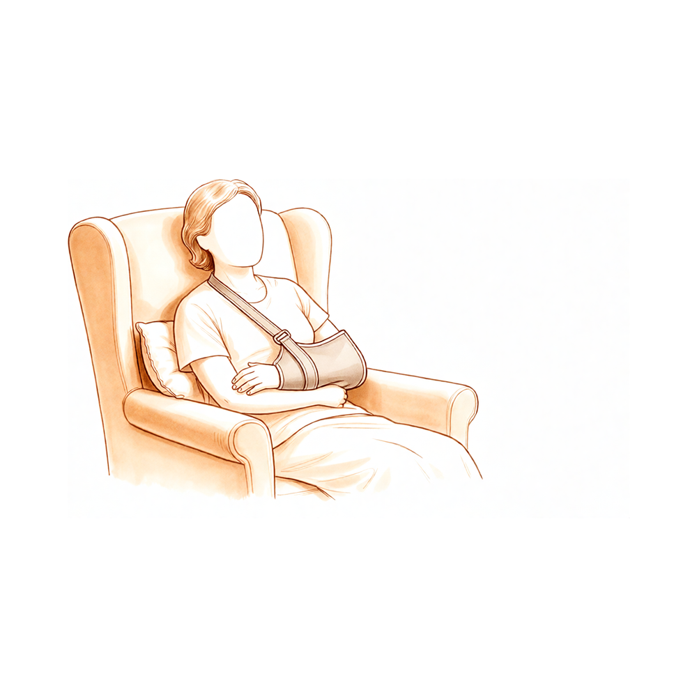
- 在家休息时,如果注意得当,可以取下:坐着时手臂用枕头支撑。
- 佩戴悬吊带期间请勿驾驶。
- 如果肩部肿胀或疼痛,请使用冰敷,尤其是在锻炼后。
您的物理治疗师最初会帮助您佩戴悬吊带,并在您出院前教会您独立管理。正确佩戴至关重要:松垮的悬吊带无法提供适当的支撑。
- 始终确保在佩戴悬吊带时,您的肘部位于悬带角的角落并得到良好支撑。
- 佩戴合适的悬吊带,其末端应舒适地置于小指的指关节处。如果您的手伸出悬吊带太远,将无法提供足够的支撑。
- 您的悬吊带有两条魔术贴带:一条系在颈部,一条系在腰部。
- 正确放置肘部和前臂后,使用未手术侧的手臂将上部带子绕过头部至前方,并通过悬吊带上的上部环扣固定。
- 使用相同的方法将下部带子系在腰部,并通过悬吊带上的下部环扣固定。
在佩戴悬吊带期间,请时刻注意您的姿势,避免肩部塌陷。为了保持良好的姿势,请尽量保持耳朵、肩部和髋部在一条直线上。良好的姿势对您的背部很重要,并有助于防止肩关节僵硬。坐着时,在腰部下方放置一条卷起的毛巾,可以作为友好的提醒。
您住院的最初几天
在您出院回家之前,医院的物理治疗师将开始为您制定一个简单的康复计划。理解以下三个术语将有助于您更好地配合:主动活动范围是指您在不借助外力或帮助的情况下自行完成的活动。主动辅助活动范围是指使用您的另一只手臂(或拐杖等物体)来帮助移动患侧手臂。被动活动范围是指患侧手臂完全放松,由另一只手臂(或他人)完成100%的活动。在骨折置换术后,术后六周内,手术侧肩关节仅进行被动活动,且活动幅度需轻柔受限;您可主动活动的关节为肘关节、腕关节和手部。
关于最初几天的几个实用要点:
- 您需要佩戴悬吊带睡觉。
- 如有需要,可使用冰敷缓解疼痛。
- 佩戴悬吊带时,请放松肩部,让悬吊带承担手臂的重量。
- 在进行锻炼和物理治疗预约前,请服用止痛药。
- 您可以将手臂从悬吊带中取出,以便进行锻炼和淋浴。
- 您需要佩戴悬吊带6周,尤其是在外出时。
- 保持手术侧手部适度活动:鼓励在手臂置于悬吊带内时进行写字、进食和使用手机等活动。
- 除非您选择自行安排物理治疗,否则已为您预约了物理治疗,具体详情见您的出院资料包。
- 如有任何问题,请联系办公室或告知您的物理治疗师。
第一阶段 — 骨折愈合期间的保护(第0–6周)
前六周的目标只有一个:让大结节骨块在不受干扰的情况下愈合到骨骼和假体上。您需全天佩戴支具(包括夜间),保持肘部、手腕和手部的活动,而肩关节本身仅进行被动活动(由您的物理治疗师轻柔操作,或在手臂完全放松的状态下),并严格限制在以下范围内。最重要的规则是:不要主动活动肩关节;不要将手臂外旋超过规定限度;不要将手伸到背后;不要提举任何物品;不要通过双手支撑身体发力。如果您接受的是反式肩关节置换术,这些规则还能在软组织愈合期间保护新关节防止脱位。仰卧时,请在肘部下方垫一个小枕头或卷起的毛巾,以防止肩关节过度后伸。
致您的物理治疗师:
目标
- 保护大结节修复及假体
- 减轻肿胀,最小化疼痛
- 维持肘部、手腕和手部的主动活动范围(ROM)
- 在安全范围内进行轻柔的保护性肩关节被动活动范围(PROM)
- 维持肩胛骨及姿势意识
- 患者教育
支具
- 佩戴6周,包括睡眠期间
- 仅在洗澡和进行锻炼时取下;取出支具时手臂应保持在身体侧方
管理措施
- 肿胀管理:冰敷,加压
- 活动范围/灵活性:
- 仅进行PROM,且在安全范围内:肩胛平面内上举 ≤ 90度;前4周外旋(ER)至中立位(0度),之后 ≤ 30度;禁止内旋(IR)至背后
- 钟摆运动,作为被动锻炼在完全放松状态下进行
- 主动辅助活动范围(AAROM):肩关节禁止
- 主动活动范围(AROM):肘部、手腕、手部及手指;颈椎
- 肩胛骨定位,耸肩及姿势矫正
- 鼓励在佩戴支具期间进行手部轻度活动(如写字、进食、使用手机)
- 在锻炼和物理治疗 session 前给予镇痛药
注意事项
- 禁止肩关节主动活动(AROM)或主动辅助活动(AAROM)
- 禁止被动或主动外旋超过上述限度(大结节修复会受到外旋负荷)
- 禁止抗阻内旋(小结节/肩胛下肌修复会受到内旋负荷)
- 禁止将手伸到背后;禁止同时执行内收、内旋和后伸(这是反式置换术脱位的位置)
- 禁止提举物品;禁止通过双手支撑体重(例如从椅子或床上撑起身体)
- 仰卧时,在肘部下方放置小枕头或毛巾卷,以防止肩关节陷入过伸
- 切勿强行进行任何引起疼痛的动作
进阶标准
- 已满6周,且X光片显示大结节在正确位置愈合,并经Hirpara医生复查确认
- 疼痛通过口服镇痛药得到控制
- 伤口无异常,且无不稳定迹象
第二阶段——恢复活动度(第6–12周)
在六周复查时,X线检查确认骨碎片正在原位愈合。如果愈合情况良好,则去除悬吊带,肩关节开始自主活动,初期由健侧手臂、手杖或滑轮辅助,随后转为主动活动。被动活动度(PROM)在此期间逐渐恢复至全范围,旋转受限情况逐步改善。开始进行三角肌和肩胛骨周围(肩胛带)肌肉的轻柔激活训练。此时尚不进行力量训练:因为修复仍在巩固中,提重物限制仍为不超过一杯咖啡的重量,且仍禁止通过手部支撑身体向上推起。
致物理治疗师:
目标
- 六周复查后逐步停用悬吊带
- 根据舒适度进展肩关节被动活动度(PROM)至全范围
- 启动肩关节辅助主动活动度(AAROM),并进展至主动活动度(AROM)
- 启动轻柔的三角肌和肩胛带肌肉激活训练
- 恢复手臂在腰部及胸部高度进行轻度日常生活活动的能力
- 患者教育
悬吊带
- 六周复查后停用;在停用后的第一或第二周外出时,可在人群密集处佩戴以提供保护
管理
- 根据需要继续第一阶段的干预措施
- 活动度/灵活性:
- 被动活动度(PROM):在约第8–10周,在尊重疼痛反应的前提下,进展至所有平面的全范围活动
- 辅助主动活动度(AAROM):桌面滑动、爬墙、滑轮、手杖辅助屈曲和外旋,从仰卧位逐渐过渡到坐位
- 主动活动度(AROM):当AAROM舒适且动作质量良好时开始,从仰卧位屈曲开始,逐渐过渡到直立位屈曲和侧平举(scaption)
- 外旋(ER):一旦确认愈合,根据舒适度进展至超过30度
- 在阶段末期,经许可后引入轻柔的功能性手背后及伸展动作。切勿拉伸至这些体位。
- 力量训练:
- 仅限无痛的次最大等长收缩:肩胛平面内的三角肌、肩胛带稳定训练、低拉训练、肩胛骨后缩
- 在阶段末期引入轻柔无痛的肩袖等长收缩;仅在复查获准后引入抗阻内旋
- 运动控制:强调肩胛骨控制,避免耸肩或其他代偿模式
注意事项
- 禁止提举超过一杯咖啡重量的物体
- 禁止通过手部支撑身体重量
- 禁止进行除轻柔等长收缩以外的抗阻或力量训练
- 避免强行增加活动度:仅拉伸至紧绷的不适感,绝不可出现锐痛
进展标准
- 肩高以下的主动活动舒适,肩胛骨控制良好且代偿最小
- X线显示结节继续愈合,并经与Hirpara医生的复查确认
- 疼痛已缓解至足以开始抗阻训练
第三阶段——强化训练(第12–24周)
大约在12周时,大结节通常已接近愈合,您与Hirpara医生的复查将确认肩部是否已准备好开始承重。随后开始强化训练,从弹力带和轻重量开始,轻柔地进行,重点锻炼驱动肩部的三角肌和肩胛骨周围肌肉(在反式肩关节置换术后,三角肌承担了原来肩袖的大部分功能)。随着愈合的大结节允许,肩袖本身也逐渐得到强化。在此阶段,轻负荷举重逐渐增加,日常活动在腰部及肩部高度应接近正常水平,如果此前尚未恢复,驾驶功能通常在此阶段恢复。
给物理治疗师的信息:
目标
- 维持无痛的被动活动范围(PROM),并在所有平面内进展主动活动范围(AROM)
- 逐步恢复力量和耐力:三角肌、肩胛周围肌肉,然后是肩袖
- 改善动态稳定性、运动控制和本体感觉
- 恢复大多数日常活动,包括轻负荷举重
管理
- 继续所有平面的活动范围和灵活性训练
- 强化训练,从等长收缩进展到弹力带,再到轻重量(低负荷,高重复次数):
- 肩胛周围肌肉:划船、前锯肌冲拳、抗阻肩胛骨后缩
- 三角肌:在功能性位置进行抗阻屈曲和侧平举,配合受控的离心下降
- 肩袖:使用弹力阻力进行外旋(ER)和内旋(IR)、侧卧位外旋,随着愈合的大结节允许而逐步进展
- 运动控制:节律性稳定训练、本体感觉神经肌肉促进法(PNF)对角线模式、球类稳定训练
- 渐进式举重:在此阶段早期从约2公斤开始,随着控制能力的提高,到阶段结束时达到约5公斤
- 在此阶段,随着获得许可,逐步重新通过手臂进行负重(例如从椅子上撑起)
注意事项
- 禁止重负荷举重;在此阶段保持负荷低于约5公斤
- 避免用力进行终末端拉伸,避免在负重情况下进行外展和外旋的组合动作
- 进展以症状为指导:如果出现疼痛或肿胀加剧,需退回到上一级别
进展标准
- 复查时由Hirpara医生确认大结节愈合
- 日常活动无痛,强化训练耐受良好且无症状加剧
- 在可用活动范围内具有良好的运动质量
第四阶段 — 恢复全面活动(术后6个月起)
最终阶段是在力量和控制的指导下,逐步恢复较重的任务、手工劳动和娱乐活动,而非依据日历时间。家务和园艺等较重任务将逐步恢复;手工劳动需遵循Hirpara医生的指导;游泳和高尔夫运动通常在术后四至六个月恢复。肩关节置换术后,长期应避免提举重物;已发表的指南建议日常提举重量保持在10公斤以下。肩关节在此阶段之后仍会持续改善:大多数患者在术后12至24个月内,活动度、力量和信心仍会继续提高,因此在正式物理治疗结束后,坚持家庭锻炼计划仍十分有益。
致您的物理治疗师:
目标
- 优化患者日常需求所需的功能性力量和耐力
- 逐步恢复工作、娱乐和运动
- 长期的关节保护教育及独立的家庭锻炼计划
管理
- 继续进行轻重量、高次数的渐进性抗阻训练,强调肩袖和肩胛周围肌群的强化
- 神经肌肉和本体感觉训练;根据需要进行功能性和工作特异性任务训练
- 在复查确认合格后,在术后四至六个月左右安排恢复游泳、高尔夫及其他娱乐活动
- 制定定制化的家庭锻炼计划,以便在正式物理治疗结束后继续执行
注意事项
- 术后6个月内避免提举重物;长期日常提举重量应保持在10公斤以下
- 手工劳动以及接触性或负重运动仅在Hirpara医生批准后进行
完成本方案的指征
- 经Hirpara医生复查确认,且大结节愈合
- 疼痛得到控制,活动度满足患者日常需求的功能性活动范围
- 能够独立执行长期家庭锻炼计划
您的方案之后
上述阶段改编自已发表的针对肱骨近端骨折行肩关节置换术的康复方案(Texas Health Orthopedic Specialists、North Tees and Hartlepool NHS Foundation Trust 针对创伤性半肩关节置换术及反式肩关节置换术,以及 Robert Jones and Agnes Hunt Orthopaedic Hospital),并参考外科文献重点强调大结节愈合。周数范围为典型值而非固定值,您的康复进程由物理治疗师与诊所合作,并根据您与 Hirpara 医生的复诊情况个体化推进。本页面与诊所的一般术后恢复建议配合使用;请参阅 管理术后疼痛 和 伤口护理。关于手术本身,请参阅 骨折行肩关节置换术;关于此治疗路径的择期版本,请参阅 反式肩关节置换术方案。本方案背后的证据(为何大结节愈合决定进度、半肩关节置换术与反式肩关节置换术的比较,以及已发表的康复指南)总结在证据部分,可从本页面顶部下载 PDF 版本。
Evidence & references
Shoulder Replacement for Proximal Humerus Fracture — Evidence Behind the Rehabilitation Protocol
Topic scope: The evidence underpinning rehabilitation after shoulder arthroplasty performed for an acute proximal humerus fracture — covering both the implant options used in this setting: reverse total shoulder arthroplasty (rTSA) for fracture and stemmed hemiarthroplasty for fracture. The defining feature of both, and the reason this is a slow, protected pathway, is that the greater and lesser tuberosities are broken free and stitched back around the prosthesis at operation — they must heal as fractures before the shoulder can be actively moved or loaded. This contrasts with elective arthroplasty (for arthritis or cuff arthropathy), where the bone is intact and rehabilitation can move at the pace of soft-tissue healing.
Defining principle of this rehab: Replacement-for-fracture is a PROTECT pathway driven by TUBEROSITY HEALING, not by implant type. At operation the greater tuberosity (with supraspinatus/infraspinatus) and lesser tuberosity (with subscapularis) are reattached around the stem. Until those bone fragments unite — typically the first 6 weeks, confirmed on X-ray — the shoulder is moved passively only and within a restricted arc: forward elevation kept low, external rotation limited (it loads the greater-tuberosity repair), resisted/active internal rotation avoided (it loads the lesser-tuberosity/subscapularis repair), and no reaching behind the back. Active range and strengthening are deferred, not just reduced. This is the opposite of an early-motion pathway such as capsular release (where immediate aggressive motion is the treatment) or calcific excision (early movement below shoulder height from day 1). The whole pace is set by the fractured tuberosities, not the new joint surface.
The operation, and why the tuberosities matter
A displaced three- or four-part proximal humerus fracture in an older patient may be unreconstructable with plate-and-screw fixation, and arthroplasty is then offered. Two implants are used:
- Hemiarthroplasty (HA) — replaces the ball (humeral head) only. It relies on the surgeon reconstructing the head–tuberosity–shaft relationship and, critically, on the tuberosities healing back in anatomical position so the rotator cuff can pull on them. Where the tuberosities fail to heal, function is poor.
- Reverse total shoulder arthroplasty (rTSA) — reverses the ball-and-socket so the deltoid, not the rotator cuff, drives elevation. This makes rTSA less dependent on tuberosity healing than HA, which is the main reason it has largely supplanted hemiarthroplasty for fracture in the elderly. Tuberosity healing still improves the result (it adds rotation), but a workable shoulder is achievable even when the greater tuberosity does not unite.
In both operations the surgeon repairs the tuberosities, and in both the rehabilitation protects that repair. That shared dependence — not the choice of implant — is why the protocol has one shape for both.
Evidence by theme
1. Tuberosity healing drives the functional result (the rationale for protection)
This is the best-supported principle in the topic, and the reason rehabilitation protects the repair for ~6 weeks.
- A large systematic review and meta-analysis of 21 studies (1,616 reverse arthroplasties) found a pooled greater-tuberosity non-healing rate of ~32% (anatomic healing in roughly two-thirds). Healed tuberosities had significantly better active abduction, anterior elevation and external rotation, and better functional scores, without any increase in pain — confirming that attempting and protecting the repair is worthwhile [meta-analysis, 2025; Schmalzl 2020].
- In the corpus, a dedicated study asked "can a healed tuberosity improve the functional outcomes?" after reverse arthroplasty for four-part fracture and answered yes — healing was associated with better rotation and outcome scores. DOI: 10.1016/j.jse.2016.11.034.
- For hemiarthroplasty, the dependence is even stronger: reported greater-tuberosity healing rates range widely (~30–91%), and tuberosity malunion/nonunion/migration is the dominant cause of a poor result. This is precisely why HA is now reserved and rTSA preferred in older patients [web review literature].
Strength: MODERATE–STRONG for "healing predicts outcome" (large pooled cohorts/meta-analysis); the link is consistent and direction-of-effect is not in dispute. It is observational, not randomised.
2. Hemiarthroplasty vs reverse arthroplasty for fracture
- A corpus multicentre randomised controlled trial (the SHeRPA trial) found superior functional outcome with reverse arthroplasty compared with hemiarthroplasty for displaced three- and four-part fractures in patients 65 and older. DOI: 10.1016/j.jse.2024.05.016.
- An earlier corpus comparative study (JBJS) likewise compared HA and rTSA for elderly proximal humeral fractures. DOI: 10.2106/jbjs.l.01637.
- Reverse-for-fracture does carry a caveat: one corpus study asked whether rTSA for fracture does worse than rTSA for elective indications, reflecting that the fracture setting (broken tuberosities, frailer patients) is more demanding than an elective replacement — the same reason this rehab runs more slowly than the elective pathway. DOI: 10.1016/j.jse.2020.03.053.
Strength: MODERATE–STRONG (an RCT plus comparative cohorts) that rTSA generally outperforms HA for fracture in the elderly. This is a surgeon's intra-operative decision, made per patient and per fracture pattern.
3. Early vs delayed motion, and the rehabilitation protocol itself
This is where the evidence is thinnest and least standardised — honestly, the phase timings below are consensus and protocol-derived, not RCT-derived.
- A JOSPT systematic review of proposed rehabilitation guidelines after anatomic and reverse arthroplasty found substantial heterogeneity between published protocols — sling use ranged from "comfort only" to a full 6 weeks; permitted motion ranged from none to precautionary limits; early external rotation was commonly capped (e.g. to ~30° in the scapular plane) to protect the repair. There is no single agreed regimen.
- A corpus randomised single-blinded trial of early rehabilitation vs immobilisation after reverse arthroplasty exists (DOI: 10.1016/j.jse.2019.10.005), and the 2025 Neer Award SHORT trial compared surgeon-directed home therapy with formal outpatient physiotherapy after rTSA (DOI: 10.1016/j.jse.2025.10.005) — but these are largely elective rTSA populations, where the tuberosities are not broken. They do not licence early active motion in the fracture setting, where the repair must be protected first.
- A corpus network meta-analysis explicitly examined the optimal combination of arthroplasty type, fixation method and post-operative rehabilitation protocol for complex proximal humerus fractures (DOI: 10.1016/j.jse.2024.03.040), and a corpus survey found that trauma and shoulder surgeons differ in their rehabilitation preferences (DOI: 10.1016/j.jse.2021.12.045) — both confirm that the protocol is an area of genuine, unresolved variation rather than settled science.
Strength: WEAK / CONSENSUS for the specific phase structure and week ranges. The principle "protect the tuberosity repair, defer active motion and load until union" is well grounded; the exact timings are typical, not trial-proven.
Phased post-op timeline (consistent with the patient protocol)
| Phase | Window | Sling | Shoulder ROM | Strengthening | What's healing / why |
|---|---|---|---|---|---|
| I — Protection | Weeks 0–6 | Full-time, including sleep | Passive only, within a safe zone: scapular-plane elevation ≤ 90°; external rotation to neutral (0°) for ~4 weeks then ≤ 30°; no internal rotation behind the back; pendulums (fully relaxed). Active elbow/wrist/hand. | None for the shoulder | Tuberosity fragments uniting onto bone + prosthesis; ER loads the greater-tuberosity repair, resisted IR loads the lesser-tuberosity (subscapularis) repair. In rTSA this window also protects against early dislocation. |
| II — Restoring movement | Weeks 6–12 | Wean after the 6-week X-ray review | Progress passive → active-assisted → active; ease the rotation limits as healing is confirmed | Gentle sub-maximal isometrics (deltoid, periscapular) late in phase; resisted IR only once cleared | Tuberosities consolidating; lifting still limited to ~a cup of coffee; no weight-bearing through the hands |
| III — Strengthening | Weeks 12–24 | Off | Maintain/progress active range in all planes | Banded → light weights; deltoid + periscapular first, rotator cuff as the healed tuberosities allow | Tuberosity union usually well advanced and confirmed at review before loading begins |
| IV — Return to activity | 6 months onwards | Off | Functional range for daily demands | Progressive, guided by control not the calendar; lifting kept moderate long-term | Strength/movement keep improving for 12–24 months; full uninjured-shoulder range is not expected |
The pacing is gated on two things together at each step: enough time, AND X-rays showing the tuberosities healing, confirmed at review with Dr Hirpara. Approximate functional milestones (subject to healing): driving ~6–12 weeks (never in the sling); breaststroke swimming ~4 months; nothing heavier than a cup of coffee until ~12 weeks; heavy lifting avoided for 6 months.
Key controversies / evidence quality
- Implant choice (HA vs reverse). An RCT and comparative cohorts favour reverse over hemiarthroplasty for displaced fractures in older patients, chiefly because rTSA is less hostage to tuberosity healing. This is a per-patient surgical decision — and either way the rehabilitation protects the tuberosity repair. Moderate–strong.
- Does the tuberosity actually need to heal? For hemiarthroplasty, effectively yes — non-union/malunion is the main failure mode. For reverse, healing is beneficial but not essential (the deltoid drives elevation), though healed tuberosities add rotation and reduce complications. The protection strategy is justified for both because attempting healing is worthwhile and forced early motion can disrupt it. Moderate.
- Early vs delayed motion / the protocol itself. The single weakest area. Published protocols vary widely (JOSPT review), the trials that test early motion are mostly in elective reverse populations, and surgeons genuinely disagree on the optimum. The conservative, tuberosity-led timeline used here reflects the fracture-specific protocols and the healing biology rather than a defining RCT. Weak / consensus.
Evidence-strength flags (summary)
- MODERATE–STRONG: tuberosity healing predicts better range/function after reverse arthroplasty for fracture (21-study meta-analysis, 1,616 cases; ~32% non-healing); reverse > hemiarthroplasty for elderly displaced fractures (SHeRPA RCT + comparative cohorts).
- MODERATE: hemiarthroplasty outcome is strongly tuberosity-dependent (healing ~30–91%; malunion the dominant failure mode); reverse-for-fracture more demanding than elective reverse.
- WEAK / CONSENSUS: the specific phased rehabilitation protocol and its week ranges — published guidelines are heterogeneous (JOSPT review); early-motion trials are mostly elective populations; the tuberosity-protective timeline is derived from fracture-specific protocols and healing biology, not a defining rehab RCT.
Citations
RAG corpus (180,000+ Orthopaedic articles) — real DOIs, returned by search
- Reverse shoulder arthroplasty for four-part proximal humerus fracture in elderly patients: can a healed tuberosity improve the functional outcomes? J Shoulder Elbow Surg. DOI: 10.1016/j.jse.2016.11.034
- Superior functional outcome following reverse shoulder arthroplasty compared to hemiarthroplasty for displaced three- and four-part fractures in patients 65 and older — the SHeRPA randomized controlled trial. J Shoulder Elbow Surg. 2024. DOI: 10.1016/j.jse.2024.05.016
- Comparison of Hemiarthroplasty and Reverse Shoulder Arthroplasty for the Treatment of Proximal Humeral Fractures in Elderly Patients. J Bone Joint Surg Am. DOI: 10.2106/jbjs.l.01637
- Does reverse total shoulder arthroplasty for proximal humeral fracture portend poorer outcomes than for elective indications? J Shoulder Elbow Surg. DOI: 10.1016/j.jse.2020.03.053
- A randomized single-blinded trial of early rehabilitation versus immobilization after reverse total shoulder arthroplasty. J Shoulder Elbow Surg. 2019. DOI: 10.1016/j.jse.2019.10.005
- Optimal combination of arthroplasty type, fixation method, and postoperative rehabilitation protocol for complex proximal humerus fractures in the elderly: a network meta-analysis. J Shoulder Elbow Surg. 2024. DOI: 10.1016/j.jse.2024.03.040
- Understanding postoperative rehabilitation preferences in operatively managed proximal humerus fractures: do trauma and shoulder surgeons differ? J Shoulder Elbow Surg. DOI: 10.1016/j.jse.2021.12.045
- 2025 Neer Award Part 1: The SHORT trial — surgeon-directed home therapy vs. outpatient rehabilitation after reverse total shoulder arthroplasty. J Shoulder Elbow Surg. 2025. DOI: 10.1016/j.jse.2025.10.005
- Acute versus delayed reverse total shoulder arthroplasty for proximal humeral fractures in the elderly: a systematic review and meta-analysis. J Shoulder Elbow Surg. DOI: 10.1016/j.jse.2018.10.004
- Ten-year follow-up of stemmed hemiarthroplasty for acute proximal humeral fractures. Bone Joint J. DOI: 10.1302/0301-620x.103b6.bjj-2020-1753.r1
Literature (URLs)
- Anatomic healing of greater tuberosity improves range of motion and functional outcomes after reverse total shoulder arthroplasty for proximal humerus fractures: an updated systematic review and meta-analysis on 21 studies (1,616 rTSAs; ~32% GT non-healing). PubMed. https://pubmed.ncbi.nlm.nih.gov/39914739/
- Schmalzl J, et al. Tuberosity healing improves functional outcome following primary reverse shoulder arthroplasty for proximal humeral fractures with a 135° prosthesis. Eur J Orthop Surg Traumatol. 2020. https://pubmed.ncbi.nlm.nih.gov/32162048/
- Tuberosity healing after reverse shoulder arthroplasty for proximal humerus fractures: is there clinical improvement? PubMed. https://pubmed.ncbi.nlm.nih.gov/33364654/
- Prosthesis designs and tuberosity fixation techniques in reverse total shoulder arthroplasty: influence on tuberosity healing in proximal humerus fractures. PMC. https://pmc.ncbi.nlm.nih.gov/articles/PMC8468418/
- A Systematic Review of Proposed Rehabilitation Guidelines Following Anatomic and Reverse Shoulder Arthroplasty. JOSPT 2019. https://www.jospt.org/doi/10.2519/jospt.2019.8616
Published rehab protocols (patient-guidance — basis for the phase structure)
- Frantz T. Proximal Humerus Fracture Rehabilitation Protocol (reverse shoulder arthroplasty). Texas Health Orthopedic Specialists. https://www.frantzorthopedics.com/pdf/proximal-humerus-fracture-reverse-shoulder-arthroplasty-updnew.pdf
- North Tees and Hartlepool NHS Foundation Trust. Shoulder Hemi-Arthroplasty (trauma) — post-operative protocol. Reviewed April 2023. https://www.nth.nhs.uk/resources/shoulder-hemi-arthroplasty-trauma/
- North Tees and Hartlepool NHS Foundation Trust. Reverse shoulder arthroplasty — post-operative protocol. Reviewed April 2023. https://www.nth.nhs.uk/resources/reverse-shoulder-arthroplasty/
- Maddocks C, Lloyd Evans J. Rehabilitation guide following reverse total shoulder replacement. Robert Jones and Agnes Hunt Orthopaedic Hospital NHS Foundation Trust. February 2024. https://www.rjah.nhs.uk/media/qctjsd1w/reverse-shoulder-replacement-guideline-2024.pdf


