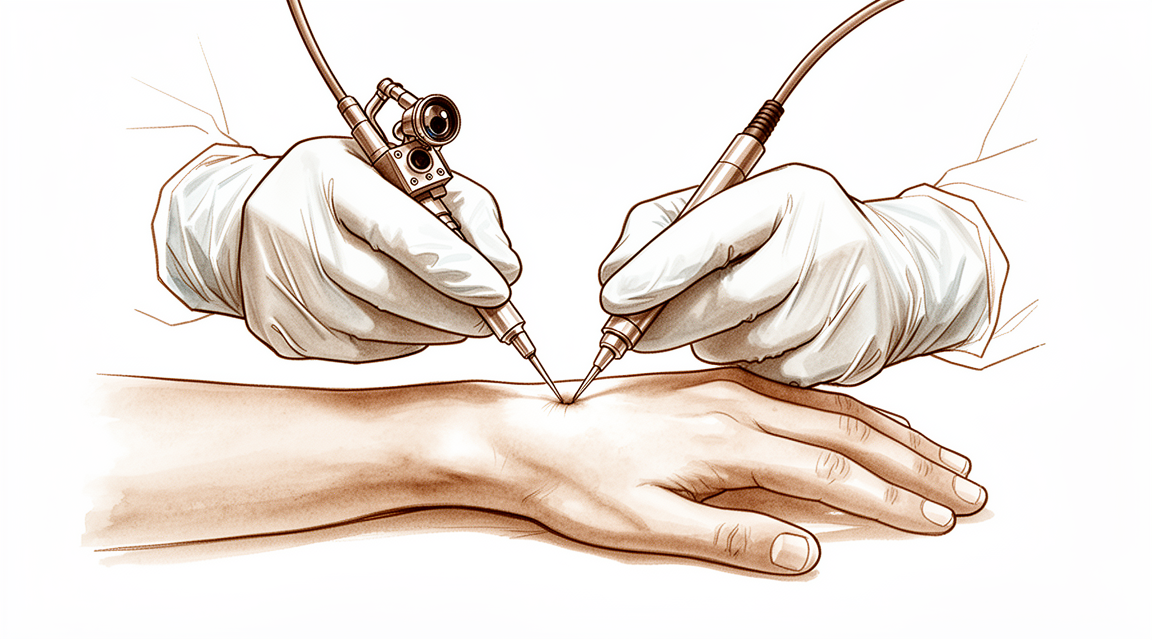
மணிக்கட்டு ஆர்த்ரோஸ்கோபி
Patients › Rehabilitation
கீஹோல் (ஆர்த்ரோஸ்கோபிக்) மணிக்கட்டு அறுவை சிகிச்சைக்குப் பிறகு ஒரு மீட்பு திட்டம், அங்கு வேகம் உள்ளே என்ன செய்யப்பட்டது என்பதைப் பொறுத்ததுஃ ஒரு சுத்தம் (டிப்ரிட்மென்ட், சினோவெக்டோமி, கேங்க்லியன் அல்லது மத்திய டி.எஃப்.சி.சி.
இந்த நெறிமுறை உங்கள் மீட்பு வழிகாட்டுகிறது மணிக்கட்டில் கீஹோல் (ஆர்த்ரோஸ்கோபிக்) அறுவை சிகிச்சை மேட்டர் தனியார் மருத்துவமனை ராக்ஹாம்ப்டனில் டாக்டர் கீரன் ஹிர்பாராவுடன். மணிக்கட்டு ஆர்த்தோஸ்கோபி மணிக்கட்டின் பின்புறத்தில் சில சிறிய வெட்டுக்கள் (போர்டல்கள்) மூலம் செய்யப்படுகிறது, எனவே தோல் விரைவாக குணமடைகிறது, ஆனால் உங்கள் மீட்பு வேகம் மூட்டுக்குள் என்ன செய்யப்பட்டது என்பதைப் பொறுத்தது. இது உங்கள் வீட்டு உடற்பயிற்சி திட்டத்துடன் தொடங்குகிறது, அதைத் தொடர்ந்து உங்கள் கை சிகிச்சையாளருக்கு எழுதப்பட்ட கட்டமைக்கப்பட்ட மருத்துவ நெறிமுறை. உங்கள் முதல் சிகிச்சை வருகைக்கு இந்த பக்கம் அல்லது அதன் PDF ஐ எடுத்துச் செல்லுங்கள், இதனால் உங்கள் மறுவாழ்வு ஒருங்கிணைக்கப்படுகிறது, மேலும் உங்கள் சிகிச்சையாளர் உங்கள் அறுவை சிகிச்சையுடன் பொருந்தக்கூடிய திட்டத்தைப் பின்பற்றுகிறார். உங்கள் சிகிச்சையாளர் உங்கள் மீட்பு எவ்வாறு முன்னேறுகிறது என்பதைப் பொறுத்து திட்டத்தை சரிசெய்யலாம்.
அறுவை சிகிச்சைக்குப் பிறகு உங்கள் காயங்கள் குறித்து உங்களுக்கு ஏதேனும் கவலைகள் இருந்தால், அறைகளைத் தொடர்பு கொள்ளுங்கள். காயத்தின் புகைப்படத்தை எடுத்து அதை மறுஆய்வு செய்ய மின்னஞ்சல் அனுப்புவது பெரும்பாலும் உதவியாக இருக்கும்.
எதிர்பார்ப்பது என்ன
மணிக்கட்டு ஆர்த்ரோஸ்கோபி என்பது ஒரு சிறிய கேமரா மற்றும் நுட்பமான கருவிகளைப் பயன்படுத்தி ஒரு சிறிய கீல்ஹோல் போர்ட்டல்கள் வழியாக உங்கள் மணிக்கட்டுக்குள் அறுவை சிகிச்சையாளர் வேலை செய்கிறார். வெட்டுக்கள் சிறியவை என்பதால், தோல் மற்றும் மென்மையான திசுக்கள் விரைவாக குணமடைகின்றன, ஆனால் மணிக்கட்டின் உட்புறம் கால அட்டவணையை அமைக்கிறது, மேலும் இரண்டு வெவ்வேறு மீட்பு பாதைகள் உள்ளன:
-
ஒரு தூய்மைப்படுத்தல் (debridement, synovectomy, dorsal ganglion அகற்றுதல், அல்லது ஒரு மத்திய TFCC தூய்மைப்படுத்தல்). இங்கே, பாதுகாக்க வேண்டிய எதுவும் மீண்டும் இணைக்கப்படவில்லை; சேதமடைந்த அல்லது வீங்கிய திசு வெறுமனே வெட்டப்பட்டு அல்லது அகற்றப்படுகிறது. எனவே இயலாமை சுருக்கமானது (பெரும்பாலும் மென்மையான உடை அல்லது ஆறுதலுக்காக குறுகிய அடுக்கு), மணிக்கட்டு இயக்கம் சில நாட்களுக்குள் தொடங்குகிறது, மேலும் சில வாரங்களில் நீங்கள் பெரும்பாலான செயல்பாடுகளுக்குத் திரும்புவீர்கள். TFCC இன் மையப் பகுதிக்கு (நிறைய விரல் பக்கத்தில் உள்ள தலையணை) இரத்த விநியோகம் இல்லை, அது தைக்கப்பட்டால் குணமடைய முடியாது, எனவே அது கிழிந்தபோது அது சரிசெய்யப்படுவதற்குப் பதிலாக வெட்டப்படுகிறது, மேலும் அந்த வெட்டுதல் ஒரு சுத்தம் போலவே செயல்படுகிறது.
-
ஒரு TFCC பழுது (ஒரு புற அல்லது foveal கண்ணீர் மீண்டும் கீழே தைத்து). TFCC இன் வெளிப்புற விளிம்பு செய்கிறது இரத்த சப்ளை மற்றும் முடியும் குணமடைய, அதனால் அது கிழிந்த போது அங்கு அது சரி செய்யப்படுகிறது, மற்றும் அந்த பழுது பாதுகாக்கப்பட வேண்டும். ஒரு குணமடைய TFCC பழுது மீது ஒற்றை மிகப்பெரிய மன அழுத்தம் உள்ளது முதுகெலும்பை திருப்புதல் கைத்தடி மேலே மற்றும் கீழே சுழலும். இதன் காரணமாக, மணிக்கட்டு மற்றும் முதுகெலும்பு சுமார் நான்கு முதல் ஆறு வாரங்களுக்கு ஒரு அடுக்கு அல்லது மியூன்ஸ்டர் (முழங்கைக்கு மேலே) நடிப்பில் ஓய்வெடுக்கின்றன; இது முழங்கை வளைக்க அனுமதிக்கிறது, ஆனால் முதுகெலும்பு சுழற்சியைத் தடுக்கிறது. இயக்கம், பின்னர் வலிமை, பின்னர் கவனமாக கட்டங்களாக மீண்டும் கட்டமைக்கப்படுகிறது, மேலும் முழுமையான மீட்பு சுமார் மூன்று மாதங்கள் அல்லது அதற்கு மேல் ஆகும்.
உங்கள் கை சிகிச்சையாளர் உங்கள் மணிக்கட்டில் உண்மையில் என்ன செய்யப்பட்டது என்பதற்கான திட்டத்தைப் பின்பற்றுவார்; நீங்கள் எந்த பாதையில் இருக்கிறீர்கள் என்று உறுதியாக தெரியாவிட்டால், டாக்டர் ஹிர்பாராவிடம் கேளுங்கள் அல்லது உங்கள் அறுவை சிகிச்சை குறிப்பை சரிபார்க்கவும்.
முன்னெச்சரிக்கைகள் மற்றும் வரம்புகள்
- உங்கள் விரல்கள் தொடக்கத்தில் இருந்து நகரும் வைத்து: ஒரு நாளைக்கு பல முறை முழங்கால் மற்றும் முழுமையாக நேராக. இது எப்போதும் அனுமதிக்கப்படுகிறது மற்றும் விறைப்பு மற்றும் வீக்கத்தைத் தடுக்கிறது.
- திட்டத்தை பின்பற்றுங்கள் என்ன செய்யப்பட்டது. பிறகு ஒரு சுத்தம் செய்தல், மென்மையான மணிக்கட்டு இயக்கம் சில நாட்களில் தொடங்குகிறது. TFCC பழுது, மணிக்கட்டு மற்றும் முதுகெலும்பு பாதுகாக்கப்படுகின்றன ஒரு அடுக்கு அல்லது நடிகர்கள் மற்றும் நீங்கள் செய்ய இல்லை உங்கள் கை சிகிச்சையாளர் அதை அகற்றும் வரை (நான்கு முதல் ஆறு வாரங்கள் வரை) முதுகெலும்பை (கைப்பிடி மேலே / கைப்பிடி கீழே) திருப்புங்கள்.
- பிறகு ஒரு TFCC பழுது, முதுகெலும்பு சுழற்சியை கட்டாயப்படுத்தவோ அல்லது சுமக்கவோ வேண்டாம், அதை அகற்றும் வரை இறுக்கமாகப் பிடிக்கவோ அல்லது தூக்கவோ வேண்டாம்; சுழற்சி மற்றும் சுமை என்பது பழுதுபார்ப்புக்கு அழுத்தம் கொடுக்கும்.
- பூட்டு துளை இணையதளங்கள் சுத்தமான மற்றும் வறண்ட வரை குணமடையும் வரை வைத்து; செய்ய இல்லை சிறிய வடுக்கள் குணமடைந்தவுடன் அவற்றைப் பார்த்துக் கொள்ளுங்கள்.
- செய் இல்லை உங்கள் மணிக்கட்டில் ஸ்பிளென்ட், காஸ்ட் அல்லது சக்கரத்தை பாதுகாப்பாகக் கட்டுப்படுத்த முடியாத நிலையில் வாகனம் ஓட்டுங்கள்; ஆரம்ப வாரங்களில் போக்குவரத்துக்கு உதவியை ஏற்பாடு செய்யுங்கள்.
- சுலபமான அன்றாட பணிகளைச் செய்ய, நீங்கள் தவிர்க்க வேண்டும் என்று கூறப்பட்ட இயக்கங்கள் அல்லது சுமைகளை உள்ளடக்காத வரை, கையை பயன்படுத்தவும்.
காயம், வீக்கம் மற்றும் வடுக்கள் மேலாண்மை, நடைமுறையில் பார்க்க காயம் பராமரிப்பு வழிகாட்டல்.
உங்கள் பயிற்சிகள்
இவை உங்கள் கையேட்டில் உள்ள பயிற்சிகள். டாக்டர் ஹிர்பாரா மற்றும் உங்கள் கை சிகிச்சையாளரின் வழிகாட்டுதலின்படி மட்டுமே அவற்றைத் தொடங்குங்கள், உங்களுக்கு கொடுக்கப்பட்ட வரம்புகள் மற்றும் வரம்புகளுக்குள் இருங்கள். விரல் இயக்கம் மற்றும் வீக்கக் கட்டுப்பாடு அனைவருக்கும் ஆரம்பத்தில் இருந்து தொடங்குங்கள். மணிக்கட்டு இயக்கம், முதுகெலும்பு சுழற்சி மற்றும் பிடிப்பு சுத்திகரிப்புக்குப் பிறகு ஆரம்பத்தில் செய்யப்பட்டது, ஆனால் TFCC பழுதுபார்ப்புக்குப் பிறகு நிறுத்தப்பட்டது (முன்கை சுழற்சி குறிப்பாக, இது விடுவிக்கப்பட வேண்டிய கடைசி விஷயம்). சிறிய காயங்கள் குணமடைந்தவுடன் ஸ்கார் மற்றும் போர்டல் பராமரிப்பு தொடங்குகிறது. கூர்மையான வலியை ஏற்படுத்தும் எதையும் நிறுத்துங்கள்.
உங்கள் மருத்துவ நெறிமுறை
இந்த பக்கத்தின் மீதமுள்ள பகுதி மணிக்கட்டு ஆர்த்தோஸ்கோபியாவுக்குப் பிறகு மறுவாழ்வுக்கான மருத்துவ நெறிமுறை ஆகும். இந்த பிரிவு கை சிகிச்சையாளருக்கு வழங்கப்பட வேண்டும், மேலும் ஒவ்வொரு கட்டமும் என்ன நடக்கிறது என்பதற்கான எளிய ஆங்கில விளக்கத்துடன் தொடங்குகிறது. நடைமுறை என்ன செய்யப்பட்டது மீது கிளைகள். ஒரு நோயறிதல் அல்லது சிகிச்சை தூய்மைப்படுத்தல் (debridement, synovectomy, dorsal ganglion excision, chondral அல்லது scapholunate debridement, central TFCC debridement) ஆரம்ப இயக்கப் பாதை. ஒரு புறநிலை / foveal TFCC பழுது பின்வருமாறு பாதுகாக்கப்பட்ட சுழற்சி பாதைஅறுவை சிகிச்சை குறிப்பு மற்றும் சிகிச்சையளிக்கும் அறுவை சிகிச்சை நிபுணருடன் எந்த பாதை பொருந்தும் என்பதை எப்போதும் உறுதிப்படுத்தவும்.
சிகிச்சைக்கு முன்னர், நோயாளியின் அறுவை சிகிச்சை அறிக்கை மற்றும் கடந்தகால மருத்துவ வரலாற்றை சரிபார்த்து, மேற்கொள்ளப்பட்ட நடைமுறை குறித்து சிகிச்சையளிக்கும் அறுவை சிகிச்சை நிபுணருடன் தொடர்பு கொள்ளுங்கள் (தூய டிப்ரிட்மென்ட் / சினோவெக்டோமி / கேங்க்லியன் / சென்ட்ரல்-டிஎஃப்சிசி டிப்ரிட்மென்ட் Vs புற / ஃபோவல் டிஎஃப்சிசி பழுது), எந்தவொரு தொடர்புடைய டி.ஆர்.யு.ஜே ஸ்திரமின்மை மற்றும் பரிந்துரைக்கப்பட்ட இயலாமை. கீழே உள்ள இரண்டு பாதைகள் முக்கியமாக முன்கை சுழற்சி எவ்வளவு காலம் பாதுகாக்கப்படுகிறது என்பதில் வேறுபடுகின்றன.
பாதை A சுத்தம் செய்தல் (டிப்ரிட்மென்ட் / சினோவெக்டோமி / கேங்க்லியன் / மத்திய-TFCC டிப்ரிட்மென்ட்): ஆரம்ப இயக்கம்
சுத்தம் செய்யும் பாதை பாதுகாக்கப்பட வேண்டிய ஒரு கட்டமைப்பை உருவாக்காமல் திசுக்களை அகற்றுகிறது அல்லது வெட்டுகிறது, எனவே நோக்கம் ஆரம்பத்தில் இயக்கத்தை மீட்டெடுப்பது மற்றும் கடினத்தன்மையைத் தவிர்ப்பது ஆகும். இயக்கம் குறுகியது மற்றும் ஆறுதலுக்கு மட்டுமே.
கட்டம் I ஆரம்ப இயக்கம் (0 முதல் 2 வாரங்கள் வரை)
முதல் இரண்டு வாரங்களில் வீக்கம் மற்றும் வலி குறையும், உடனடியாக இயக்கம் தொடங்குகிறது.
உங்கள் கை சிகிச்சையாளருக்கு:
கல்வி மற்றும் முன்னெச்சரிக்கைகள் - இயலாமை என்பது ஆறுதலுக்காக மட்டுமே மென்மையான உறை அல்லது குறுகிய அடுக்கு, வழக்கமாக ~ 2 வாரங்கள் வரை; சுத்தமான மைய debridement/synovectomy/ganglion க்கு சுழற்சி கட்டுப்பாடு இல்லை - விரல்கள், கட்டைவிரல் மற்றும் (சௌகரியமான இடங்களில்) மணிக்கட்டு முதல் நாளிலிருந்து நகரும் - போர்ட்டல்கள் குணமாகும் வரை சுத்தமாகவும் உலர்ந்ததாகவும் வைத்திருங்கள்
நிர்வாகம் - வீக்கம்: உயர்வு, மென்மையான விரல் பம்ப், தேவைக்கேற்ப பனி - பயிற்சிகள்: முழுமையாக செயலில் விரல் மற்றும் கட்டைவிரல் ROM ஆரம்பத்தில் இருந்து; செயலில் உள்ள மணிக்கட்டு வளைவு/நீட்டிப்பு மற்றும் ரேடியல்/அல்னார் விலகல் ஆறுதல் முதல் நாட்களில் அனுமதிக்கிறது; மென்மையான முதுகெலும்பு உச்சரிப்பு/சூபினேஷன் வசதியளிக்கும் அளவு - காயம்: வழிகாட்டுதலின்படி போர்டல் பாண்டேஜ்கள்; தொற்றுநோய்க்கான கண்காணிப்பு
முன்னேற்றத்திற்கான அளவுகோல்கள் - போர்டல்ஸ் குணமடைந்தது; வீக்கம் அமைகிறது; வசதியான ஆரம்ப ROM
கட்டம் II இயக்கம் மற்றும் தொடக்க வலிமையை மீட்டெடுப்பது (2 முதல் 6 வாரங்கள் வரை)
இயக்கம் இயல்பு நிலைக்கு கொண்டுவரப்பட்டு, சற்று வலுவூட்டல் சேர்க்கப்படுகிறது.
உங்கள் கை சிகிச்சையாளருக்கு:
மதிப்பீடுகள் - செயலில் மற்றும் செயலற்ற மணிக்கட்டு ROM மற்றும் முதுகெலும்பு சுழற்சி; பிடிப்பு; வலி மற்றும் வீக்கம்; வடு / போர்டல் ஆய்வு
நிர்வாகம் - பயிற்சிகள்: முன்னேற்றம் முழு மணிக்கட்டு மற்றும் முன்கை ROM; தொடங்குகிறது லேசான பிடிப்பு மற்றும் புட்டி வலுவூட்டல் சுமார் 2 வாரங்களில்; தொடங்குங்கள் வடுக்கள்/போர்டல் உணர்திறன் குறைப்பு மற்றும் மசாஜ் குணமடைந்த பின்பு - முன்னேற்றம் செயல்பாட்டு கை பயன்பாடு ஆறுதல் அனுமதிக்கிறது என
முன்னேற்றத்திற்கான அளவுகோல்கள் - கிட்டத்தட்ட முழு வலியற்ற ROM; சரிந்த வீக்கம்; பிடிப்பு கட்டிடம்
கட்டம் III வலுவூட்டல் மற்றும் திரும்புதல் (4 முதல் 6 வாரங்கள் மற்றும் அதற்கு அப்பால்)
வலி மற்றும் பணி சகிப்புத்தன்மை மீண்டும் கட்டமைக்கப்படுகின்றன; பெரும்பாலான நோயாளிகள் சில வாரங்களுக்குள் இயல்பான செயல்பாட்டிற்குத் திரும்புகிறார்கள். சுத்தம் செய்வது நம்பகமான அறிகுறி நிவாரணத்தை அளிக்கிறது, ஆனால் பரவலான அல்லது மறுக்கமுடியாத அல்னார் பக்க வலிக்கு உத்தரவாதம் அளிக்கப்படவில்லை என்பதை நினைவில் கொள்க; வலி மையமற்றதாக இருக்கும்போது எதிர்பார்ப்புகளை நிர்வகிக்கவும்.
உங்கள் கை சிகிச்சையாளருக்கு:
நிர்வாகம் - உடற்பயிற்சிகள்ஃ படிப்படியான பிடிப்பு மற்றும் முதுகெலும்பு / மணிக்கட்டு வலுவூட்டல்; பணி மற்றும் வேலை சார்ந்த சுமைகள் - வெளிச்சத்திற்கு திரும்புதல்/பெரும்பாலான செயல்பாடு பொதுவாக 26 வாரங்கள்; ஏற்றுக்கொள்ளப்பட்ட மற்றும் அளவுகோல் அடிப்படையிலான கனமான கையேடு அல்லது விளையாட்டு சுமை - வலிமை கிட்டத்தட்ட சமச்சீர் மற்றும் செயல்பாடு மீட்டெடுக்கப்பட்டதும் வெளியேற்றத்தை கருத்தில் கொள்ளுங்கள்
பாதை B புற / foveal TFCC பழுதுஃ பாதுகாக்கப்பட்ட சுழற்சி
பழுதுபார்ப்பு TFCC இன் வெளிப்புற (vascularised) விளிம்பை மீண்டும் கீழே தைக்கிறது. ஏனெனில் முதுகெலும்பு சுழற்சி திருத்தத்தை வலியுறுத்துகிறது, முதுகெலும்பு ஒரு ஸ்பிளின்ட் அல்லது மியூன்ஸ்டர் (முழங்கைக்கு மேலே) நடிப்பில் பாதுகாக்கப்படுகிறது (இது முழங்கை வளைவு / நீட்டிப்பை அனுமதிக்கிறது, ஆனால் ப்ரோனேஷன் / சப்னேஷனைத் தடுக்கிறது) சுமார் நான்கு முதல் ஆறு வாரங்களுக்கு. இயக்கம், பின்னர் வலிமை, பின்னர் பின்னோக்கி தரப்படுத்தப்படுகிறது.
கட்டம் I பாதுகாக்கப்பட்ட இம்மோபிலிசேஷன் (வாரங்கள் 0 முதல் 6)
சுழற்சி சுமைகளிலிருந்து பாதுகாக்கப்படுகிறது. விரல்கள் நகரும் நிலையில் இருக்கும்போது நடைமுறை மாறுபடுகிறது, ஆனால் மிகவும் பொதுவான முறை சுமார் ஆறு வாரங்களுக்கு நடுநிலை முதல் லேசான-சூப்பினேஷனில் முதுகெலும்பு இயலாமை ஆகும்; சுழற்சியை உறுதியாகக் கட்டுப்படுத்த வேண்டியிருக்கும் போது ஒரு மேலே-கட்டி (மியூன்ஸ்டர்) நடிப்பு அல்லது அடுக்கு பயன்படுத்தப்படுகிறது.
உங்கள் கை சிகிச்சையாளருக்கு:
கல்வி மற்றும் முன்னெச்சரிக்கைகள் - இயங்காத நிலைக்கு முதுகெலும்பு சுழற்சியை பாதுகாக்க: ஸ்பிளிண்ட் அல்லது மியூன்ஸ்டர் / முழங்கை மேலே நடிப்பு முழங்கை இலவசம், முதுகெலும்பு சுழற்சி தடுக்கப்பட்டது), முதுகெலும்பு நடுநிலை அல்லது லேசான உட்கார்ந்த நிலையில், ~46 வாரங்கள் (பொதுவாக 6) - செயலில் அல்லது செயலற்ற முதுகெலும்பு புரோனேஷன்/சூபினேஷன் இல்லை இந்த கட்டத்தில் - முதல் நாளிலிருந்து முழு விரல் மற்றும் கட்டைவிரல் ROM; மென்மையான தோள்பட்டை ROM - போர்ட்டல்களை சுத்தமாகவும் உலர்ந்ததாகவும் வைத்திருங்கள்; தொற்றுநோய்களைக் கண்காணிக்கவும்
நிர்வாகம் - வீக்கம்: உயர்வு, விரல் பம்ப், தேவைக்கேற்ப பனி - பயிற்சிகள்: விரல் / கட்டைவிரல் AROMதனிமைப்படுத்தப்பட்ட முழங்கை வளைவு/நீட்டிப்பு ஒரு மியூன்ஸ்டர் அனுமதித்தால்; மணிக்கட்டு அல்லது முன்கை சுழற்சி சுமை இல்லை - காயம்/காயம்: கதவு பராமரிப்பு; காயம் குணமடைந்தவுடன் சிகிச்சையைத் தொடங்குதல்
முன்னேற்றத்திற்கான அளவுகோல்கள் - ~46 வாரங்கள் கடந்துவிட்டன; பழுதுபார்ப்பு பாதுகாக்கப்பட்டது; வாயில்கள் குணமடைந்தன; விரல்கள் நெகிழ்வானவை
கட்டம் II படிப்படியான இயக்கம் (6 முதல் 8 வாரங்கள் வரை)
அச்சுப்பொறி/அடுக்கு துண்டிக்கப்பட்டு இயக்கம் மீண்டும் உருவாக்கப்படுகிறது, முதுகெலும்பு சுழற்சியை கடைசியாக மற்றும் படிப்படியாக அறிமுகப்படுத்துதல், இது திருத்தத்தை வலியுறுத்தியது.
உங்கள் கை சிகிச்சையாளருக்கு:
மதிப்பீடுகள் - மணிக்கட்டு மற்றும் முதுகெலும்பு ROM; வலி மற்றும் வீக்கம்; வடுக்கள் ஆய்வு
நிர்வாகம் - பயிற்சிகள்: துவக்கம் செயலில் உள்ள மணிக்கட்டு வளைவு/நீட்டிப்பு மற்றும் ரேடியல்/அல்னார் விலகல்; படிப்படியாக முதுகெலும்பு புரோனேஷன்/சூபினேஷனை மீண்டும் அறிமுகப்படுத்தவும் ஆறுதல் உள்ள, அடுத்த வாரங்களில் சுழற்சி வளைவு கட்டமைத்து அதை கட்டாயப்படுத்தி விட - ஸ்கார் / போர்டல் உணர்திறன் குறைப்பு தொடரவும்
முன்னேற்றத்திற்கான அளவுகோல்கள் - வசதியான, மணிக்கட்டு மற்றும் முன்கை ROM ஐ மேம்படுத்துதல்; வலி அமைதிப்படுத்தல்
கட்டம் III வலுவடைதல் மற்றும் திரும்புதல் (8 முதல் 12+ வாரங்கள் மற்றும் அதற்கு அப்பால்)
சுமார் 70100% மணிக்கட்டு மற்றும் முதுகெலும்பு இயக்கம் மீட்டெடுக்கப்பட்டவுடன் வலுவூட்டல் தொடங்குகிறது, பின்னர் சுமை மற்றும் பணி சகிப்புத்தன்மை மீண்டும் தரப்படுத்தப்படுகிறது.
உங்கள் கை சிகிச்சையாளருக்கு:
மதிப்பீடுகள் - மற்ற பக்கத்துடன் ஒப்பிடும்போது பிடிப்பு மற்றும் முதுகெலும்பு வலிமை; சுமைக்கு வலி / வீக்கம் பதில்; செயல்பாட்டு மற்றும் வேலை / விளையாட்டு-குறிப்பிட்ட சோதனை
நிர்வாகம் - பயிற்சிகள்: துவக்கம் சுமார் 8 வாரங்களில் கைப்பிடிப்பு மற்றும் முதுகெலும்பு / மணிக்கட்டு வலுவடைதல், 70100% ROM மீட்டெடுக்கப்பட்டதும்; படிப்படியான எதிர்ப்பு மற்றும் பணி-குறிப்பிட்ட சுமைக்கு முன்னேற்றம் - விளையாட்டு / கனமான வேலைக்கு திரும்புவது அளவுகோலை அடிப்படையாகக் கொண்டது, பொதுவாக சுமார் மூன்று மாதங்கள் (தேவையைப் பொறுத்து ~34+ மாதங்கள் வரம்பு) - வலிமை கிட்டத்தட்ட சமச்சீர் மற்றும் செயல்பாடு மீட்டெடுக்கப்பட்டவுடன் வெளியேற்றத்தை கருத்தில் கொள்ளுங்கள்; மீட்பு மேடை அல்லது DRUJ நிலையற்ற தன்மை மீண்டும் வந்தால் சிகிச்சையளிக்கும் மருத்துவரை மீண்டும் பார்க்கவும்
வேலை மற்றும் செயற்பாட்டிற்கு திரும்புதல்
சாதாரண தினசரி கை பயன்பாடு (உண்ணல், எழுதுதல், சாதாரண சுய-பராமரிப்பு) உங்களுக்கு வழங்கப்பட்ட வரம்புகளுக்குள் இருக்கும் வரை, ஆரம்பத்தில் இருந்தே வசதியாக ஊக்குவிக்கப்படுகிறது. எந்தவொரு அடுக்கு அல்லது வார்ப்படமும் இல்லாமல் நீங்கள் மீண்டும் வாகனம் ஓட்டுவதைத் தொடங்குங்கள் மற்றும் உங்கள் மதிப்பாய்வில் உறுதிப்படுத்தப்பட்டபடி சக்கரத்தை பாதுகாப்பாகக் கட்டுப்படுத்தலாம்; ஆரம்ப வாரங்களில் போக்குவரத்து உதவிக்கு திட்டமிடவும்.
நீங்கள் எவ்வளவு விரைவாக திரும்புகிறீர்கள் என்பது நீங்கள் செய்ததைப் பொறுத்தது. சுத்தம் செய்தல் (debridement, synovectomy, ganglion or central-TFCC tidy-up), பெரும்பாலான மக்கள் சாதாரண ஒளி செயல்பாட்டிற்குள் திரும்புகிறார்கள் இரண்டு முதல் ஆறு வாரங்கள், கனமான சுமை வசதி அனுமதிக்கிறது மீண்டும் கட்டப்பட்டது. TFCC பழுது, முதுகெலும்பு சுழற்சி சுமார் நான்கு முதல் ஆறு வாரங்களுக்கு பாதுகாக்கப்படுகிறது, வலுவூட்டல் சுமார் எட்டு வாரங்களில் தொடங்குகிறது, மற்றும் விளையாட்டு அல்லது கனமான கை வேலைக்கு திரும்புவது பொதுவாக மூன்று மாதங்கள் ஆகும், இயக்கத்தை மீட்டெடுப்பதன் மூலமும், போதுமான, சமச்சீர் வலிமையின் மூலமும் தீர்மானிக்கப்படுகிறது, காலண்டர் மூலம் மட்டும் அல்ல, மற்றும் டாக்டர் ஹிர்பாரா மற்றும் உங்கள் கை சிகிச்சையாளர் ஒன்றாக முடிவு செய்தனர்.
உங்கள் நெறிமுறை பிறகு
இந்த நெறிமுறை நடைமுறையின் பொதுவான மீட்பு ஆலோசனையுடன் இணைந்து செயல்படுகிறதுஃ அறுவை சிகிச்சைக்குப் பிந்தைய வலியை நிர்வகித்தல், காயம் பராமரிப்பு மற்றும் வடு மேலாண்மை. உங்கள் அறுவை சிகிச்சையானது தொலைதூர ரேடியோல்னார் மூட்டுடன் தொடர்புடையதாக இருந்தால் அல்லது எந்த பாதை பொருந்தும் என்று உங்களுக்குத் தெரியாவிட்டால், டிஸ்டல் ரேடியோல்னார் மூட்டு (DRUJ) ஹெமிசெக்ஷன் மேற்கண்ட படிப்படியான திட்டம், மணிக்கட்டு ஆர்த்த்ரோஸ்கோபி மற்றும் TFCC அறுவை சிகிச்சைக்குப் பிறகு வெளியிடப்பட்ட மறுவாழ்வு வழிகாட்டுதல்களை பிரதிபலிக்கிறது, மேலும் உங்கள் தொடர்ச்சியான மீட்பு உங்கள் மணிக்கட்டு முன்னேற்றத்திற்கு ஏற்ப டாக்டர் ஹிர்பாரா மற்றும் உங்கள் கை சிகிச்சையாளரால் தனித்தனியாக வழிநடத்தப்படுகிறது.
Evidence & references
Wrist Arthroscopy — Procedure Outcomes & Post-operative Rehabilitation (Diagnostic / Therapeutic Keyhole Wrist Surgery)
Topic scope: post-operative rehabilitation after arthroscopic wrist surgery through small dorsal portals — covering debridement (central TFCC, chondral, scapholunate), synovectomy, dorsal ganglion excision, and peripheral/foveal TFCC repair. The defining feature of this topic is that a single operative approach (keyhole access) covers procedures with opposite rehabilitation needs: a clean-up creates nothing to protect and follows an early-motion pathway, whereas a repair creates a construct loaded by forearm rotation and must be protected. The rehabilitation pathway is therefore gated by what was done, not by the fact that arthroscopy was used.
Defining principle of the rehab here: wrist arthroscopy is a route, not a single operation. Where tissue is only removed (debridement of an avascular central TFCC tear, synovectomy, a dorsal ganglion, a chondral or scapholunate tidy-up), nothing has been reconstructed — immobilisation is brief and for comfort, wrist motion begins within days, and return is measured in weeks. Where the vascularised peripheral or foveal TFCC is repaired, the dominant stress on the construct is forearm rotation (pronation/supination), so the forearm is protected — typically a splint or Muenster (above-elbow) cast that frees the elbow but blocks rotation — for about 4–6 weeks, after which motion and then strength are graded back over roughly three months. The single branch point a therapist must establish from the operation note is debridement-class vs repair-class, and within repair, how long rotation is to be protected. The rehabilitation evidence itself is low-level and heterogeneous — protocols rest on biology, surgeon preference and expert/therapist consensus more than on trials.
A. PROCEDURE OUTCOMES (debridement / synovectomy / ganglion vs TFCC repair)
Wrist arthroscopy is both the diagnostic gold standard and the therapeutic workhorse for intra-articular ulnar-sided wrist pathology. The principal outcome split is between clean-up procedures and repair.
- Wrist arthroscopy is a versatile, low-morbidity platform. Through small dorsal portals it permits direct inspection and treatment of TFCC tears, chondral lesions, synovitis and ganglia, with the keyhole approach giving fast soft-tissue healing and small scars [Gupta, Bozentka, Osterman — JAAOS 2001, DOI 10.5435/00124635-200105000-00006]. Narrative/mechanistic.
- Arthroscopic debridement of central (Palmer 1A) TFCC tears relieves symptoms in well-selected, focal cases. The central disc is avascular and cannot heal if sutured, so trimming is the rational treatment; classic and long-term series report good symptom relief and durable function in suitable patients [Osterman — Arthroscopy 1990, DOI 10.1016/0749-8063(90)90012-3; Soreide et al., 19-year follow-up — HAND 2017, DOI 10.1177/1558944717708029]. Moderate–weak (case series, long follow-up).
- Debridement is unreliable for diffuse, non-focal ulnar-sided wrist pain. Where pain is recalcitrant and not clearly localised to a focal central tear, simple arthroscopic debridement has little useful value on the clinical course — a caution against over-attributing diffuse ulnar wrist pain to a debridable lesion [Nishizuka et al. — Bone Joint J 2013, DOI 10.1302/0301-620x.95b12.31918]. Moderate (prospective cohort).
- Peripheral/foveal TFCC repair restores DRUJ stability and gives good outcomes when the rim is repairable. All-arthroscopic and arthroscopic-assisted repair techniques (e.g. FasT-Fix, all-inside suture) report reliable pain relief, return of grip and high return-to-activity rates in vascularised peripheral tears, especially with DRUJ instability [Yao, Dantuluri, Osterman — Arthroscopy 2007, DOI 10.1016/j.arthro.2007.02.010; Yao — Hand Clin 2011, DOI 10.1016/j.hcl.2011.05.004]. Moderate–weak (technique series).
- Procedure choice is driven by tear location and DRUJ stability. Central tears → debridement; peripheral/foveal tears, particularly with DRUJ instability → repair. In ulnar-positive wrists, peripheral repair may be combined with or weighed against ulnar shortening osteotomy [Papapetropoulos et al. — J Hand Surg Am 2010, DOI 10.1016/j.jhsa.2010.06.015]. Moderate.
- Arthroscopic dorsal ganglion excision gives recurrence rates comparable to (or, in some series, better than) open excision, with the keyhole advantage of faster recovery and smaller scars. Arthroscopic resection reliably removes the cyst and addresses the stalk at its capsular origin [Nishikawa et al. — J Hand Surg Br 2001, DOI 10.1054/jhsb.2001.0620; Luchetti et al. — J Hand Surg Br 2000, DOI 10.1054/jhsb.1999.0290; Konigsberg et al. — HAND 2021, DOI 10.1177/15589447211003184; Suen, Fung, Lung — ISRN Orthop 2013, DOI 10.1155/2013/940615]. Moderate–weak (retrospective comparison + series).
B. REHABILITATION / THERAPY EVIDENCE
The central rehab question is how long, and against what, to protect the wrist — and the answer is set entirely by the procedure. The evidence base for the rehabilitation (as opposed to the surgery) is low-level and markedly heterogeneous, with no level-1 protocol and wide variation in immobilisation and progression timings; recommendations are best regarded as biologically-rationalised, consensus-driven guides rather than trial-proven schedules.
- Clean-up procedures follow an early-motion pathway. After debridement, synovectomy or ganglion excision there is no construct to protect: immobilisation is a soft dressing or short splint for comfort (≈2 weeks at most), wrist motion begins within days, and light grip strengthening is added at around 2 weeks. Return to most activity is measured in weeks (≈2–6) [How-we-treat reviews and technique series; consistent across sources]. Weak–moderate (consensus + series).
- TFCC repair protects forearm rotation, not just the wrist. Because pronation/supination is the dominant load on a peripheral/foveal repair, the forearm is immobilised — commonly a splint or Muenster/above-elbow cast that frees the elbow but blocks rotation — for about 4–6 weeks (six is the most commonly reported figure), in neutral to slight supination [scoping review of arthroscopic peripheral TFCC repair rehabilitation, PMC12274733; Australian hand-therapist survey of foveal-repair rehabilitation, J Hand Ther 2024]. Weak–moderate (scoping review + survey of practice).
- Forearm rotation is re-introduced last and graded. After the protected phase, wrist flexion/extension and deviation are restored first, with pronation/supination re-introduced gradually because it is the motion that stressed the repair. Strengthening typically begins once 70–100% of wrist and forearm ROM is regained (around 8 weeks) [scoping review PMC12274733]. Weak (consensus from heterogeneous protocols).
- Rehabilitation protocols are heterogeneous and lack consensus. Across studies, complete immobilisation ranged 1–8 weeks (forearm most commonly 6), ROM commencement and strengthening start varied widely (strengthening 3–12 weeks), and authors explicitly call for level-1 evidence. The practical implication is to follow the operating surgeon's prescription for the specific repair rather than a fixed universal schedule [scoping review PMC12274733; Australian hand-therapist survey]. Weak (the evidence's own conclusion).
Recovery trajectory (expected, evidence-anchored)
| Phase | Window | Restraint | Hand use / therapy focus | Strength / load | Notes |
|---|---|---|---|---|---|
| Clean-up I — early motion | Week 0–2 | Soft dressing / short splint for comfort | Fingers move day 1; early wrist flexion/extension, deviation and gentle forearm rotation within days; portal care | Light functional use | No construct to protect; rotation not restricted for central debridement/synovectomy/ganglion |
| Clean-up II–III — restore & return | Week 2–6+ | Restrictions lifted | Full wrist/forearm ROM; scar/portal desensitisation once healed | Light grip/putty from ~2 wk, graded loading thereafter | Return to most activity 2–6 wk; debridement unreliable for diffuse (non-focal) ulnar pain |
| Repair I — protected immobilisation | Week 0–6 | Forearm rotation blocked (splint / Muenster cast, neutral–slight supination) | Full finger/thumb ROM day 1; elbow flexion/extension if Muenster permits; no pronation/supination | None to the repair | ~4–6 wk (commonly 6); rotation is the dominant repair load |
| Repair II — graded motion | Week 6–8 | Rotation re-introduced gradually | Active wrist flexion/extension & deviation; forearm rotation re-introduced last and built up | Light, no resisted load yet | Restore motion before strength |
| Repair III — strengthen & return | Week 8–12+ | Load progressed by criteria | Grip/forearm strengthening once 70–100% ROM regained; task-specific loading | Strengthening from ~8 wk; graded resisted load | Return to sport/heavy work ~3 months (range ~3–4+); grip ~85% of opposite side, ~87% return to pre-injury activity |
(Phase windows mirror the precautions and recovery structure in the patient protocol; they are typical, consensus-derived guides — not trial-derived deadlines, and the surgeon's prescription overrides them.)
C. KEY CONTROVERSIES / EVIDENCE QUALITY
- Debridement vs repair is decided by tear location and DRUJ stability, not access. Central (avascular) tears are trimmed; peripheral/foveal (vascularised) tears, especially with DRUJ instability, are repaired. The same keyhole approach therefore launches opposite rehab pathways — the therapist must establish which from the operation note. Strong biological rationale, moderate clinical evidence.
- Debridement is not a panacea for ulnar wrist pain. It relieves focal central-tear symptoms but has little value for diffuse, recalcitrant ulnar-sided pain — a key expectation-setting point [Nishizuka 2013]. Moderate.
- How long to protect forearm rotation after repair is unsettled. Reported immobilisation ranges 1–8 weeks (forearm most commonly 6); there is no level-1 consensus, and practice (including Australian hand-therapist practice) varies. The defensible position is to protect rotation ~4–6 weeks and follow the operating surgeon. Weak (heterogeneous).
- Arthroscopic vs open ganglion excision. Recurrence is broadly comparable, with arthroscopy offering faster recovery and smaller scars; the evidence is retrospective rather than randomised [Konigsberg 2021; Nishikawa 2001; Luchetti 2000]. Moderate–weak.
- The rehabilitation evidence base is the weak link, not the surgery. Outcome studies of the operations outnumber and outrank the rehabilitation studies; rehab timings are biologically and consensus-driven, and the literature itself calls for level-1 trials [scoping review PMC12274733]. Weak.
D. EVIDENCE STRENGTH FLAGS (summary)
- MODERATE: versatility and low morbidity of wrist arthroscopy as a diagnostic/therapeutic platform; symptom relief from debridement of focal central TFCC tears (with long-term series); limited value of debridement for diffuse ulnar wrist pain; comparable recurrence of arthroscopic vs open dorsal ganglion excision; good outcomes of peripheral/foveal TFCC repair when the rim is repairable.
- WEAK / CONSENSUS: the specific rehabilitation schedules — clean-up early-motion (~2-wk comfort splint, motion within days, return 2–6 wk) and repair protected-rotation (splint/Muenster ~4–6 wk, ROM then strength from ~8 wk, return ~3 months). Immobilisation duration and progression timings are heterogeneous across the literature with no level-1 consensus; figures are typical guides, and the operating surgeon's prescription governs.
- EXPECTATION-SETTING (natural history): grip recovers to ≈85% of the opposite side and ≈87% of patients return to pre-injury activity after TFCC repair; debridement of diffuse (non-focal) ulnar wrist pain may not relieve symptoms.
CITATIONS
RAG corpus (180,000+ Orthopaedic articles)
- Wrist arthroscopy: principles and clinical applications. J Am Acad Orthop Surg. 2001. DOI: 10.5435/00124635-200105000-00006
- Arthroscopic debridement of triangular fibrocartilage complex tears. Arthroscopy. 1990. DOI: 10.1016/0749-8063(90)90012-3
- Arthroscopic-assisted resection of triangular fibrocartilage complex lesions: a 19-year follow-up. HAND. 2017. DOI: 10.1177/1558944717708029
- Simple debridement has little useful value on the clinical course of recalcitrant ulnar wrist pain. Bone Joint J. 2013. DOI: 10.1302/0301-620x.95b12.31918
- A novel technique of all-inside arthroscopic triangular fibrocartilage complex repair. Arthroscopy. 2007. DOI: 10.1016/j.arthro.2007.02.010
- All-arthroscopic repair of peripheral triangular fibrocartilage complex tears using FasT-Fix. Hand Clin. 2011. DOI: 10.1016/j.hcl.2011.05.004
- Management of peripheral triangular fibrocartilage complex tears in the ulnar positive patient: arthroscopic repair versus ulnar shortening osteotomy. J Hand Surg Am. 2010. DOI: 10.1016/j.jhsa.2010.06.015
- Arthroscopic diagnosis and treatment of dorsal wrist ganglion. J Hand Surg Br. 2001. DOI: 10.1054/jhsb.2001.0620
- Arthroscopic resection of dorsal wrist ganglia and treatment of recurrences. J Hand Surg Br. 2000. DOI: 10.1054/jhsb.1999.0290
- Recurrence rates of dorsal wrist ganglion cysts after arthroscopic versus open surgical excision: a retrospective comparison. HAND. 2021. DOI: 10.1177/15589447211003184
- Treatment of ganglion cysts. ISRN Orthop. 2013. DOI: 10.1155/2013/940615
Wrist arthroscopy / TFCC rehabilitation literature (URLs)
- Clinical and functional outcomes of rehabilitation strategies following arthroscopic repair of chronic isolated peripheral TFCC tears: a scoping review. J Orthop. 2025. PMC. https://pmc.ncbi.nlm.nih.gov/articles/PMC12274733/
- Current rehabilitation recommendations following primary triangular fibrocartilage complex foveal repair surgery: a survey of Australian hand therapists. J Hand Ther. 2024. https://www.jhandtherapy.org/article/S0894-1130(23)00117-5/fulltext
- TFCC injuries: how we treat? J Clin Orthop Trauma / PMC. 2020. https://pmc.ncbi.nlm.nih.gov/articles/PMC7384326/
- Review and update on the management of triangular fibrocartilage complex injuries in professional athletes. World J Orthop. 2024. https://www.wjgnet.com/2218-5836/full/v15/i2/110.htm
- Arthroscopic-assisted repair of the triangular fibrocartilage complex. J Hand Surg Glob Online. 2024. https://www.jhsgo.org/article/S2589-5141(24)00066-5/fulltext


