

腕关节镜 资料
本方案指导您在 Mater Private Hospital Rockhampton 接受 Kieran Hirpara 医生进行的腕部关节镜(微创)手术后的康复。腕关节镜手术通过在腕背侧的几个微小切口(通道)进行,因此皮肤愈合较快,但您的康复速度取决于关节内所进行的具体操作。康复从您的家庭锻炼计划开始,随后是为您手部治疗师撰写的结构化临床方案。请在首次治疗就诊时携带此页面或其 PDF 文件,以确保您的康复过程协调一致,并使您的治疗师遵循与您手术相匹配的计划。您的治疗师可能会根据您的康复进展调整该计划。
如果您对术后伤口有任何疑虑,请联系诊所前台。通常,拍摄伤口照片并通过电子邮件发送以供审阅会很有帮助。
预期情况
腕关节镜手术是指外科医生通过腕部的小切口(钥匙孔通道),使用微型摄像头和精细器械在腕部内部进行操作。由于切口较小,皮肤和软组织愈合较快,但腕部内部的恢复情况决定了时间表,且存在两条截然不同的康复路径:
-
清理术(清创、滑膜切除、背侧腱鞘囊肿切除或中央三角纤维软骨复合体修整)。 在此情况下,无需保护以重新接合的组织;受损或发炎的组织仅被修剪或移除。因此,固定时间较短(通常仅为舒适起见使用软敷料或短期夹板),腕部活动可在数天内开始,并在几周内恢复大多数活动。三角纤维软骨复合体(TFCC)的中央部分(位于腕部小指侧的缓冲垫)没有血液供应,缝合后无法愈合,因此当该处撕裂时,会进行修剪而非修复,这种修剪的效果类似于清理术。
-
TFCC修复术(缝合修复外周或窝部撕裂)。 TFCC的外缘确实有血液供应且能够愈合,因此当该处撕裂时会进行修复,且该修复部位必须受到保护。对愈合中的TFCC修复部位最大的单一应力是前臂旋转(手掌向上和向下旋转)。因此,腕部和前臂需在夹板或孟斯特(肘上)石膏中休息约四到六周;这允许肘部弯曲,但阻止会牵拉修复部位的前臂旋转。随后,活动能力,然后是力量,将分阶段仔细重建,完全恢复大约需要三个月或更长时间。
在两条路径中,手指从一开始就保持活动(因为手指未接受手术),肿胀得到控制,并护理小的切口瘢痕。您的手部治疗师将根据您腕部实际接受的治疗方案执行计划;如果您不确定自己处于哪条路径,请咨询Hirpara医生或查看您的手术记录。
注意事项与限制
- 从一开始就保持手指活动:每天多次握拳并完全伸直。这始终是被允许的,可防止僵硬和肿胀。
- 遵循针对所实施手术制定的计划。 在清理术后,温和的手腕活动可在数天内开始。在TFCC修复术后,手腕和前臂需通过夹板或石膏保护,且在您的手部治疗师许可之前(约四至六周),您不得旋转前臂(掌心向上 / 掌心向下)。
- 在TFCC修复术后,不得强行或负重进行前臂旋转,且在获得许可前不得用力抓握或提重物;旋转和负重正是对修复部位产生应力的动作。
- 保持钥匙孔切口清洁干燥直至愈合;不得浸泡或擦洗这些切口。待小疤痕愈合后,请妥善护理。
- 当您的手腕处于夹板固定、石膏固定状态,或无法安全控制方向盘时,不得驾驶;在早期几周请安排他人协助交通出行。
- 在舒适范围内,将手用于轻微的日常生活任务,只要不涉及您被告知需避免的动作或负重。
关于伤口、肿胀和疤痕管理,请参阅本诊所的伤口护理指南。
您的锻炼
Kieran Hirpara 4.0
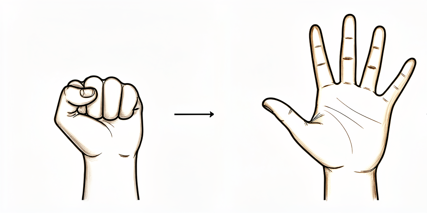
手指活动(早期)
从术后第一或第二天起,缓慢地握紧拳头,然后将手指完全伸直。手指并非修复部位的一部分,因此从一开始就鼓励保持手指活动——这有助于控制肿胀并防止手部僵硬。即使手腕在夹板或石膏中休息时,也应进行此练习。
10次,每天数次,从一开始即进行
Kieran Hirpara 4.0
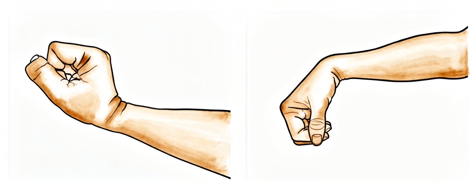
腕部活动(根据您的计划)
如果您接受了清理术(清创、滑膜切除或腱鞘囊肿切除),轻柔的腕关节上下屈伸——以及左右侧偏——通常在术后几天内即可开始,只要舒适度允许即可进行。如果您接受了三角纤维软骨复合体(TFCC)修复术,腕部需先置于夹板或石膏中休息,腕关节活动开始得较晚,仅在您的手部治疗师启动时才开始。请务必始终在您被允许的活动范围内活动。
每个方向10次,每天2–3次,开始后进行
Kieran Hirpara 4.0
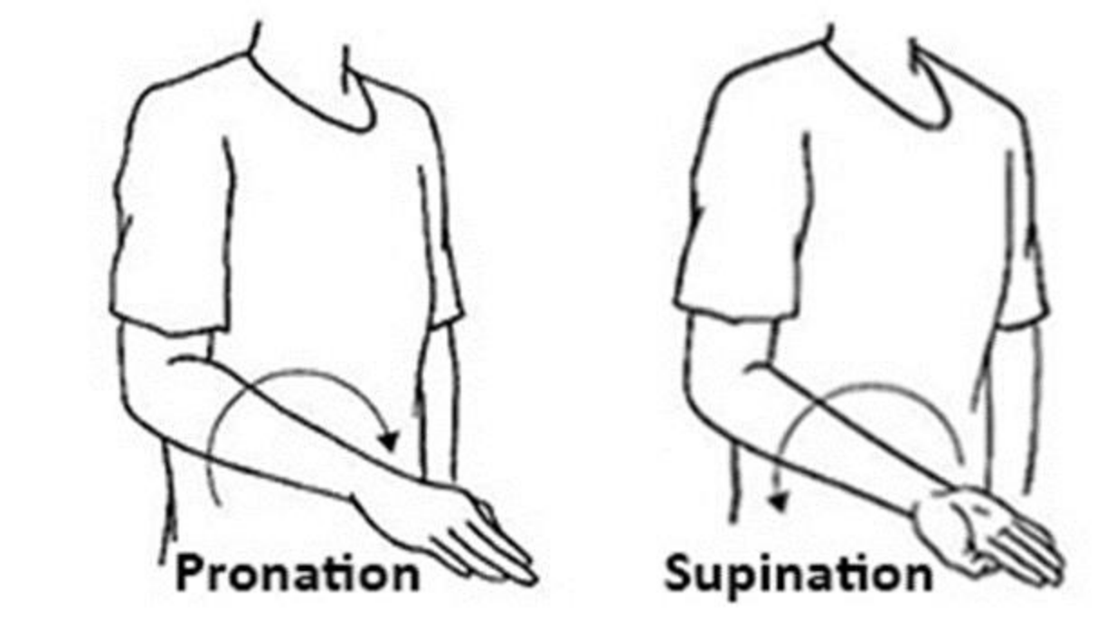
前臂旋转——掌心向上/掌心向下(根据您的计划)
将肘部贴紧身体一侧,轻轻将手掌向上转向天花板,然后向下转向地面。在清理术后,通常可以较早开始此动作。在三角纤维软骨复合体(TFCC)修复术后,这是最后恢复的动作——前臂旋转会牵拉修复部位,因此需用夹板或石膏固定约四至六周,随后再逐步引入该动作。仅在您的手部治疗师确认允许后,方可进行此动作。
每个方向10次,每天2–3次,获准后进行
Kieran Hirpara 4.0
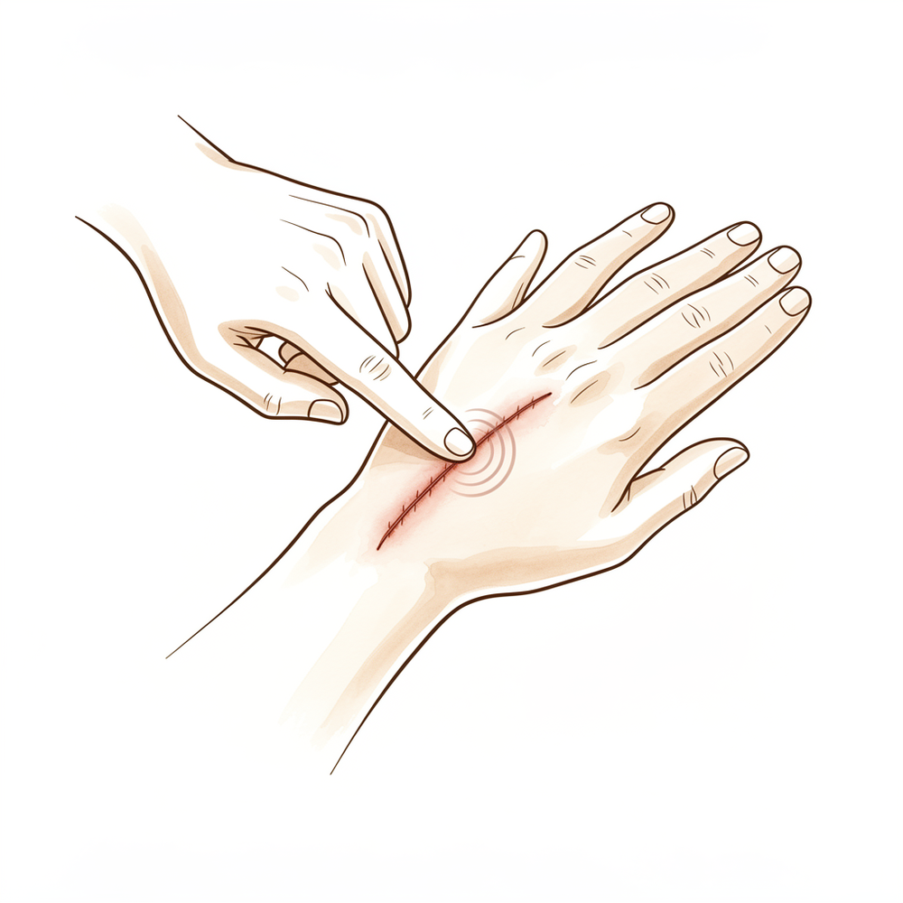
瘢痕与穿刺口护理
待小切口伤口完全愈合且干燥后,使用普通保湿霜,以指尖在每个小瘢痕上以小圆圈方式进行按摩。锁孔式切口虽小,但可能感觉紧绷或敏感——轻柔的按摩和叩击(脱敏)可保持皮肤柔韧,并缓解任何压痛。
每天2–3次,每次几分钟,待伤口愈合后
Kieran Hirpara 4.0
握力强化(后期)
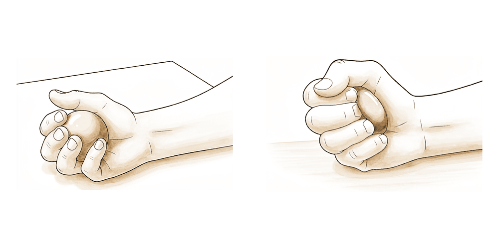
后期锻炼——仅在您的手部治疗师开始指导后进行。挤压软球或治疗用橡皮泥,短暂保持,然后放松。清理术后,轻度抓握通常在一两周内开始;三角纤维软骨复合体(TFCC)修复后,强化训练需等待至约八周,待活动度恢复后再进行。逐渐增加力度,若出现剧烈疼痛则立即停止。
挤压10–15次,每天2–3次,获准后进行
这些是您手册中的锻炼项目。仅在Hirpara医生和您的手部治疗师的指导下开始,并严格保持在您被允许的范围内和限制内。手指活动和肿胀控制从一开始就适用于所有人。手腕活动、前臂旋转和抓握取决于所进行的手术:在清理术后早期开始,但在三角纤维软骨复合体(TFCC)修复术后需推迟(尤其是前臂旋转,这是最后才解除限制的动作)。疤痕和关节镜切口护理在小伤口愈合后开始。停止任何引起剧烈疼痛的动作。
您的临床方案
本页其余部分为腕关节镜术后康复的分阶段临床方案。本节内容需提供给手部治疗师,每个阶段均以通俗易懂的语言解释当前正在发生的情况。该方案根据所实施的手术类型进行分支。 诊断性或治疗性清理术(清创术、滑膜切除术、背侧腱鞘囊肿切除术、软骨或舟月关节清创术、中央三角纤维软骨复合体清创术)遵循早期活动路径。外周/小凹三角纤维软骨复合体修复术遵循保护性旋转路径,因为前臂旋转会对修复部位产生负荷。务必根据手术记录并与主刀医生确认适用哪条路径。
在治疗前,请查阅患者的手术报告及既往病史,并与主刀医生沟通所实施的手术类型(纯清创术/滑膜切除术/腱鞘囊肿切除/中央三角纤维软骨复合体清创术 与 外周/小凹三角纤维软骨复合体修复术)、是否伴有远端桡尺关节不稳定,以及规定的固定方式。以下两条路径的主要区别在于前臂旋转的保护时长。
路径 A — 清理术(清创 / 滑膜切除 / 腱鞘囊肿 / 中央三角纤维软骨复合体清创):早期活动
清理术路径旨在切除或修整组织,而不形成需要保护的构建结构,因此目标是尽早恢复活动并避免僵硬。制动时间短暂,仅用于缓解不适。
第一阶段 — 早期活动(第 0 至 2 周)
最初几周旨在消退肿胀和疼痛,同时活动几乎立即开始。
致您的手部治疗师:
宣教与注意事项 - 制动仅为软敷料或短支具以提供舒适感,通常最长约 2 周;对于单纯的中央清理/滑膜切除/腱鞘囊肿切除,无需限制旋转 - 手指、拇指及(在舒适范围内)手腕从第一天起即可活动 - 保持关节镜入路(portals)清洁干燥直至愈合
管理 - 水肿:抬高患肢、轻柔的手指泵式运动、必要时冰敷 - 练习:从一开始即进行全范围主动手指和拇指活动度(ROM);在最初几天内,在舒适允许范围内进行主动腕关节屈曲/伸展及桡偏/尺偏;在舒适允许范围内进行轻柔的前臂旋前/旋后 - 伤口:按指示更换入路敷料;监测感染迹象
晋级标准 - 入路愈合;肿胀消退;早期活动度舒适
第二阶段——恢复活动度并开始力量训练(第2至6周)
当关节活动范围达到舒适水平后,活动度恢复正常,并加入轻度力量训练。
致您的手部治疗师:
评估 - 腕关节主动和被动活动度及前臂旋转;握力;疼痛与肿胀;瘢痕/切口检查
管理 - 练习:逐步过渡至腕关节和前臂全范围活动度;约从第2周开始进行轻度握力和橡皮泥力量训练;待愈合后开始瘢痕/切口脱敏和按摩 - 根据舒适度逐步增加手部功能性使用
晋级标准 - 接近全范围无痛活动度;肿胀消退;握力增强
第三阶段——强化与恢复(第6周及以后)
力量和任务耐受性逐步重建;大多数患者在数周内恢复正常活动。请注意,清理术可提供可靠的症状缓解,但对于弥漫性或顽固性的尺侧疼痛并非保证有效;当疼痛非局灶性时,应合理管理患者预期。
致您的手部治疗师:
管理 - 练习:分级握力及前臂/腕部强化训练;针对任务和工作的特异性负荷训练 - 通常 2–6 周 后可恢复轻/大部分活动;较重的体力劳动或运动负荷在可耐受且符合标准的情况下进行 - 当力量接近对称且功能恢复时,考虑出院
路径 B — 周边/窝部 TFCC 修复:保护性旋转
修复手术将 TFCC 的外缘(有血供部分)重新缝合固定。由于前臂旋转会对修复部位产生应力,前臂需使用夹板或 Muenster(肘上)石膏进行保护(允许肘关节屈伸,但限制前臂旋前/旋后),时长约为四至六周。随后,先逐步恢复活动度,再逐步恢复肌力。
第一阶段 — 保护性固定(第 0 至 6 周)
在手指保持活动能力的同时,修复部位受到保护,免受旋转负荷。临床实践存在差异,但最常见的模式是前臂固定于中立位至轻度旋后位,持续约六周;当需要严格控制旋转时,使用肘上(Muenster)石膏或支具。
致您的手部治疗师:
教育与注意事项 - 固定以保护前臂旋转:使用支具或Muenster/肘上石膏(肘部自由,前臂旋转被阻挡),前臂置于中立位至轻度旋后位,持续约4–6周(通常为 6 周) - 在此期间禁止主动或被动的前臂旋前/旋后 - 从第一天起即进行全范围的手指和拇指活动度(ROM);轻柔的肩部活动度训练 - 保持关节镜入路(portals)清洁干燥;监测感染迹象
管理 - 水肿:抬高患肢、手指泵式运动、必要时冰敷 - 练习:手指/拇指主动活动度(AROM);若 Muenster 支具允许,进行孤立的肘关节屈曲/伸展;禁止腕关节或前臂旋转负荷 - 伤口/瘢痕:入路护理;愈合后开始瘢痕处理
晋级标准 - 已过约4–6周;修复部位得到保护;入路愈合;手指灵活
第二阶段 — 分级运动(第6至8周)
拆除石膏/支具并重建运动功能,最后且逐步引入前臂旋转,因为该动作对修复部位施加了应力。
致您的手部治疗师:
评估 - 腕部及前臂活动度(ROM);疼痛与肿胀;瘢痕检查
管理 - 练习:开始主动腕关节屈曲/伸展及桡偏/尺偏;在舒适范围内逐步重新引入前臂旋前/旋后,在接下来的几周内逐渐增加旋转弧度,而非强行拉伸 - 继续进行瘢痕/切口脱敏治疗
晋级标准 - 腕部及前臂活动度舒适且持续改善;疼痛减轻
第三阶段 — 强化与恢复(第8至12+周及以后)
一旦腕部和前臂活动度恢复至约 70–100%,即开始强化训练,随后逐步恢复负荷和任务耐受性。
致您的手部治疗师:
评估 - 握力及前臂肌力与对侧对比;负荷下的疼痛/肿胀反应;功能性及工作/运动特异性测试
管理 - 练习:在恢复约 70–100% 的活动范围(ROM)后,从第8周左右开始进行握力及前臂/腕部强化训练;逐步过渡到分级抗阻及任务特异性负荷 - 重返运动/重体力工作基于标准判定,通常在三个月左右(视需求而定,范围约为 3–4+ 个月) - 当肌力接近对称且功能恢复时,可考虑出院;若恢复停滞或远端桡尺关节(DRUJ)不稳定复发,请转回主治医生处
恢复工作与活动
鼓励从初期开始进行轻度日常手部使用(如进食、书写、轻度自我护理),以舒适为度,且须保持在已告知您的限制范围内。一旦您不再佩戴任何夹板或石膏,并能安全操控方向盘(经复诊确认),即可恢复驾驶;建议在早期几周内安排他人协助交通出行。
恢复速度取决于所接受的手术类型。在清理术(包括清创、滑膜切除、腱鞘囊肿或中央型三角纤维软骨复合体修整)后,大多数人可在两至六周内恢复正常的轻度活动,并随着舒适度提高逐步增加负重。在三角纤维软骨复合体(TFCC)修复术后,前臂旋转功能需保护约四至六周,强化训练通常从第八周左右开始,恢复运动或较重的体力劳动通常在三个月左右,具体取决于是否恢复了活动度及足够且对称的力量,而非仅依据时间,并由Hirpara医生与您的手部治疗师共同决定。
您的方案之后
本方案与本诊所的一般康复建议配合使用:请参阅术后疼痛管理、伤口护理和瘢痕管理。如果您的手术涉及远端桡尺关节,或您不确定适用哪条路径,远端桡尺关节(DRUJ)半切除术方案是一个相关的姊妹方案。上述分阶段计划反映了腕关节镜检查和三角纤维软骨复合体(TFCC)手术后已发表的康复指南,您的持续康复将根据腕部进展情况,由Hirpara医生和您的手部治疗师进行个体化指导。
Evidence & references
This is the clinical evidence summary written for health professionals. It is technical, and it lists the research this page was built from. You do not need to read it to understand your treatment or to make a decision about it.
Topic scope: post-operative rehabilitation after arthroscopic wrist surgery through small dorsal portals — covering debridement (central TFCC, chondral, scapholunate), synovectomy, dorsal ganglion excision, and peripheral/foveal TFCC repair. The defining feature of this topic is that a single operative approach (keyhole access) covers procedures with opposite rehabilitation needs: a clean-up creates nothing to protect and follows an early-motion pathway, whereas a repair creates a construct loaded by forearm rotation and must be protected. The rehabilitation pathway is therefore gated by what was done, not by the fact that arthroscopy was used.
Defining principle of the rehab here: wrist arthroscopy is a route, not a single operation. Where tissue is only removed (debridement of an avascular central TFCC tear, synovectomy, a dorsal ganglion, a chondral or scapholunate tidy-up), nothing has been reconstructed — immobilisation is brief and for comfort, wrist motion begins within days, and return is measured in weeks. Where the vascularised peripheral or foveal TFCC is repaired, the dominant stress on the construct is forearm rotation (pronation/supination), so the forearm is protected — typically a splint or Muenster (above-elbow) cast that frees the elbow but blocks rotation — for about 4–6 weeks, after which motion and then strength are graded back over roughly three months. The single branch point a therapist must establish from the operation note is debridement-class vs repair-class, and within repair, how long rotation is to be protected. The rehabilitation evidence itself is low-level and heterogeneous — protocols rest on biology, surgeon preference and expert/therapist consensus more than on trials.
A. PROCEDURE OUTCOMES (debridement / synovectomy / ganglion vs TFCC repair)
Wrist arthroscopy is both the diagnostic gold standard and the therapeutic workhorse for intra-articular ulnar-sided wrist pathology. The principal outcome split is between clean-up procedures and repair.
- Wrist arthroscopy is a versatile, low-morbidity platform. Through small dorsal portals it permits direct inspection and treatment of TFCC tears, chondral lesions, synovitis and ganglia, with the keyhole approach giving fast soft-tissue healing and small scars [Gupta, Bozentka, Osterman — JAAOS 2001, DOI 10.5435/00124635-200105000-00006]. Narrative/mechanistic.
- Arthroscopic debridement of central (Palmer 1A) TFCC tears relieves symptoms in well-selected, focal cases. The central disc is avascular and cannot heal if sutured, so trimming is the rational treatment; classic and long-term series report good symptom relief and durable function in suitable patients [Osterman — Arthroscopy 1990, DOI 10.1016/0749-8063(90)90012-3; Soreide et al., 19-year follow-up — HAND 2017, DOI 10.1177/1558944717708029]. Moderate–weak (case series, long follow-up).
- Debridement is unreliable for diffuse, non-focal ulnar-sided wrist pain. Where pain is recalcitrant and not clearly localised to a focal central tear, simple arthroscopic debridement has little useful value on the clinical course — a caution against over-attributing diffuse ulnar wrist pain to a debridable lesion [Nishizuka et al. — Bone Joint J 2013, DOI 10.1302/0301-620x.95b12.31918]. Moderate (prospective cohort).
- Peripheral/foveal TFCC repair restores DRUJ stability and gives good outcomes when the rim is repairable. All-arthroscopic and arthroscopic-assisted repair techniques (e.g. FasT-Fix, all-inside suture) report reliable pain relief, return of grip and high return-to-activity rates in vascularised peripheral tears, especially with DRUJ instability [Yao, Dantuluri, Osterman — Arthroscopy 2007, DOI 10.1016/j.arthro.2007.02.010; Yao — Hand Clin 2011, DOI 10.1016/j.hcl.2011.05.004]. Moderate–weak (technique series).
- Procedure choice is driven by tear location and DRUJ stability. Central tears → debridement; peripheral/foveal tears, particularly with DRUJ instability → repair. In ulnar-positive wrists, peripheral repair may be combined with or weighed against ulnar shortening osteotomy [Papapetropoulos et al. — J Hand Surg Am 2010, DOI 10.1016/j.jhsa.2010.06.015]. Moderate.
- Arthroscopic dorsal ganglion excision gives recurrence rates comparable to (or, in some series, better than) open excision, with the keyhole advantage of faster recovery and smaller scars. Arthroscopic resection reliably removes the cyst and addresses the stalk at its capsular origin [Nishikawa et al. — J Hand Surg Br 2001, DOI 10.1054/jhsb.2001.0620; Luchetti et al. — J Hand Surg Br 2000, DOI 10.1054/jhsb.1999.0290; Konigsberg et al. — HAND 2021, DOI 10.1177/15589447211003184; Suen, Fung, Lung — ISRN Orthop 2013, DOI 10.1155/2013/940615]. Moderate–weak (retrospective comparison + series).
B. REHABILITATION / THERAPY EVIDENCE
The central rehab question is how long, and against what, to protect the wrist — and the answer is set entirely by the procedure. The evidence base for the rehabilitation (as opposed to the surgery) is low-level and markedly heterogeneous, with no level-1 protocol and wide variation in immobilisation and progression timings; recommendations are best regarded as biologically-rationalised, consensus-driven guides rather than trial-proven schedules.
- Clean-up procedures follow an early-motion pathway. After debridement, synovectomy or ganglion excision there is no construct to protect: immobilisation is a soft dressing or short splint for comfort (≈2 weeks at most), wrist motion begins within days, and light grip strengthening is added at around 2 weeks. Return to most activity is measured in weeks (≈2–6) [How-we-treat reviews and technique series; consistent across sources]. Weak–moderate (consensus + series).
- TFCC repair protects forearm rotation, not just the wrist. Because pronation/supination is the dominant load on a peripheral/foveal repair, the forearm is immobilised — commonly a splint or Muenster/above-elbow cast that frees the elbow but blocks rotation — for about 4–6 weeks (six is the most commonly reported figure), in neutral to slight supination [scoping review of arthroscopic peripheral TFCC repair rehabilitation, PMC12274733; Australian hand-therapist survey of foveal-repair rehabilitation, J Hand Ther 2024]. Weak–moderate (scoping review + survey of practice).
- Forearm rotation is re-introduced last and graded. After the protected phase, wrist flexion/extension and deviation are restored first, with pronation/supination re-introduced gradually because it is the motion that stressed the repair. Strengthening typically begins once 70–100% of wrist and forearm ROM is regained (around 8 weeks) [scoping review PMC12274733]. Weak (consensus from heterogeneous protocols).
- Rehabilitation protocols are heterogeneous and lack consensus. Across studies, complete immobilisation ranged 1–8 weeks (forearm most commonly 6), ROM commencement and strengthening start varied widely (strengthening 3–12 weeks), and authors explicitly call for level-1 evidence. The practical implication is to follow the operating surgeon's prescription for the specific repair rather than a fixed universal schedule [scoping review PMC12274733; Australian hand-therapist survey]. Weak (the evidence's own conclusion).
Recovery trajectory (expected, evidence-anchored)
| Phase | Window | Restraint | Hand use / therapy focus | Strength / load | Notes |
|---|---|---|---|---|---|
| Clean-up I — early motion | Week 0–2 | Soft dressing / short splint for comfort | Fingers move day 1; early wrist flexion/extension, deviation and gentle forearm rotation within days; portal care | Light functional use | No construct to protect; rotation not restricted for central debridement/synovectomy/ganglion |
| Clean-up II–III — restore & return | Week 2–6+ | Restrictions lifted | Full wrist/forearm ROM; scar/portal desensitisation once healed | Light grip/putty from ~2 wk, graded loading thereafter | Return to most activity 2–6 wk; debridement unreliable for diffuse (non-focal) ulnar pain |
| Repair I — protected immobilisation | Week 0–6 | Forearm rotation blocked (splint / Muenster cast, neutral–slight supination) | Full finger/thumb ROM day 1; elbow flexion/extension if Muenster permits; no pronation/supination | None to the repair | ~4–6 wk (commonly 6); rotation is the dominant repair load |
| Repair II — graded motion | Week 6–8 | Rotation re-introduced gradually | Active wrist flexion/extension & deviation; forearm rotation re-introduced last and built up | Light, no resisted load yet | Restore motion before strength |
| Repair III — strengthen & return | Week 8–12+ | Load progressed by criteria | Grip/forearm strengthening once 70–100% ROM regained; task-specific loading | Strengthening from ~8 wk; graded resisted load | Return to sport/heavy work ~3 months (range ~3–4+); grip ~85% of opposite side, ~87% return to pre-injury activity |
(Phase windows mirror the precautions and recovery structure in the patient protocol; they are typical, consensus-derived guides — not trial-derived deadlines, and the surgeon's prescription overrides them.)
C. KEY CONTROVERSIES / EVIDENCE QUALITY
- Debridement vs repair is decided by tear location and DRUJ stability, not access. Central (avascular) tears are trimmed; peripheral/foveal (vascularised) tears, especially with DRUJ instability, are repaired. The same keyhole approach therefore launches opposite rehab pathways — the therapist must establish which from the operation note. Strong biological rationale, moderate clinical evidence.
- Debridement is not a panacea for ulnar wrist pain. It relieves focal central-tear symptoms but has little value for diffuse, recalcitrant ulnar-sided pain — a key expectation-setting point [Nishizuka 2013]. Moderate.
- How long to protect forearm rotation after repair is unsettled. Reported immobilisation ranges 1–8 weeks (forearm most commonly 6); there is no level-1 consensus, and practice (including Australian hand-therapist practice) varies. The defensible position is to protect rotation ~4–6 weeks and follow the operating surgeon. Weak (heterogeneous).
- Arthroscopic vs open ganglion excision. Recurrence is broadly comparable, with arthroscopy offering faster recovery and smaller scars; the evidence is retrospective rather than randomised [Konigsberg 2021; Nishikawa 2001; Luchetti 2000]. Moderate–weak.
- The rehabilitation evidence base is the weak link, not the surgery. Outcome studies of the operations outnumber and outrank the rehabilitation studies; rehab timings are biologically and consensus-driven, and the literature itself calls for level-1 trials [scoping review PMC12274733]. Weak.
D. EVIDENCE STRENGTH FLAGS (summary)
- MODERATE: versatility and low morbidity of wrist arthroscopy as a diagnostic/therapeutic platform; symptom relief from debridement of focal central TFCC tears (with long-term series); limited value of debridement for diffuse ulnar wrist pain; comparable recurrence of arthroscopic vs open dorsal ganglion excision; good outcomes of peripheral/foveal TFCC repair when the rim is repairable.
- WEAK / CONSENSUS: the specific rehabilitation schedules — clean-up early-motion (~2-wk comfort splint, motion within days, return 2–6 wk) and repair protected-rotation (splint/Muenster ~4–6 wk, ROM then strength from ~8 wk, return ~3 months). Immobilisation duration and progression timings are heterogeneous across the literature with no level-1 consensus; figures are typical guides, and the operating surgeon's prescription governs.
- EXPECTATION-SETTING (natural history): grip recovers to ≈85% of the opposite side and ≈87% of patients return to pre-injury activity after TFCC repair; debridement of diffuse (non-focal) ulnar wrist pain may not relieve symptoms.
CITATIONS
RAG corpus (180,000+ Orthopaedic articles)
- Wrist arthroscopy: principles and clinical applications. J Am Acad Orthop Surg. 2001. DOI: 10.5435/00124635-200105000-00006
- Arthroscopic debridement of triangular fibrocartilage complex tears. Arthroscopy. 1990. DOI: 10.1016/0749-8063(90)90012-3
- Arthroscopic-assisted resection of triangular fibrocartilage complex lesions: a 19-year follow-up. HAND. 2017. DOI: 10.1177/1558944717708029
- Simple debridement has little useful value on the clinical course of recalcitrant ulnar wrist pain. Bone Joint J. 2013. DOI: 10.1302/0301-620x.95b12.31918
- A novel technique of all-inside arthroscopic triangular fibrocartilage complex repair. Arthroscopy. 2007. DOI: 10.1016/j.arthro.2007.02.010
- All-arthroscopic repair of peripheral triangular fibrocartilage complex tears using FasT-Fix. Hand Clin. 2011. DOI: 10.1016/j.hcl.2011.05.004
- Management of peripheral triangular fibrocartilage complex tears in the ulnar positive patient: arthroscopic repair versus ulnar shortening osteotomy. J Hand Surg Am. 2010. DOI: 10.1016/j.jhsa.2010.06.015
- Arthroscopic diagnosis and treatment of dorsal wrist ganglion. J Hand Surg Br. 2001. DOI: 10.1054/jhsb.2001.0620
- Arthroscopic resection of dorsal wrist ganglia and treatment of recurrences. J Hand Surg Br. 2000. DOI: 10.1054/jhsb.1999.0290
- Recurrence rates of dorsal wrist ganglion cysts after arthroscopic versus open surgical excision: a retrospective comparison. HAND. 2021. DOI: 10.1177/15589447211003184
- Treatment of ganglion cysts. ISRN Orthop. 2013. DOI: 10.1155/2013/940615
Wrist arthroscopy / TFCC rehabilitation literature (URLs)
- Clinical and functional outcomes of rehabilitation strategies following arthroscopic repair of chronic isolated peripheral TFCC tears: a scoping review. J Orthop. 2025. PMC. https://pmc.ncbi.nlm.nih.gov/articles/PMC12274733/
- Current rehabilitation recommendations following primary triangular fibrocartilage complex foveal repair surgery: a survey of Australian hand therapists. J Hand Ther. 2024. https://www.jhandtherapy.org/article/S0894-1130(23)00117-5/fulltext
- TFCC injuries: how we treat? J Clin Orthop Trauma / PMC. 2020. https://pmc.ncbi.nlm.nih.gov/articles/PMC7384326/
- Review and update on the management of triangular fibrocartilage complex injuries in professional athletes. World J Orthop. 2024. https://www.wjgnet.com/2218-5836/full/v15/i2/110.htm
- Arthroscopic-assisted repair of the triangular fibrocartilage complex. J Hand Surg Glob Online. 2024. https://www.jhsgo.org/article/S2589-5141(24)00066-5/fulltext





