கடுமையான அருகிலுள்ள இடுப்பு எலும்பு முறிவுக்கான தோள்பட்டை ஆர்த்தோபிளாஸ்டிசி
இந்த அறுவை சிகிச்சை ஏன் பரிந்துரைக்கப்பட்டது
உடைந்த மேல் கை எலும்புக்கான தோள்பட்டை மாற்று அறுவை சிகிச்சை பொதுவாக பிளேட்டுகள் அல்லது ஊசிகளால் மட்டுமே சரிசெய்ய முடியாத சிக்கலான எலும்பு முறிவுகளைக் கொண்ட வயதானவர்களுக்கு வழங்கப்படுகிறது. உங்கள் அறுவை சிகிச்சையாளர் இதை பரிந்துரைத்திருக்கலாம், ஏனென்றால் ஆரம்ப அறுவை சிகிச்சை அல்லாத பராமரிப்பு போதுமான முன்னேற்றத்தை வழங்கவில்லை, அல்லது உங்கள் குறிப்பிட்ட எலும்பு முறிவு முறை கூட்டு பாதுகாப்பை வெற்றிகரமாக செய்ய வாய்ப்பில்லை. எலும்பு துண்டுகள் மிகவும் சேதமடைந்திருக்கும்போது இந்த அணுகுமுறை பெரும்பாலும் பரிசீலிக்கப்படுகிறது.
முக்கிய நோக்கம் வலியைக் குறைத்து செயல்பாட்டை மீட்டெடுப்பதாகும். இந்த நடைமுறை திருப்திகரமான நீண்டகால வலி நிவாரணத்தை வழங்கக்கூடும் என்று சான்றுகள் காட்டுகின்றன, இருப்பினும் தோள்பட்டை இயக்கம் முடிவுகள் குறைவாக கணிக்கக்கூடியவை. வயதான நோயாளிகளுக்கு, உள்வைப்புக்கு இணைக்கப்பட்ட தசை தண்டுகளை (குழாய்கள்) குணப்படுத்துவது வலிமை மற்றும் தினசரி செயல்பாடு இரண்டையும் கணிசமாக மேம்படுத்துகிறது. அறுவை சிகிச்சை அல்லாத சிகிச்சை பொதுவானது என்றாலும், கடுமையான, இடம்பெயர்ந்த எலும்புகள் உள்ளவர்களுக்கு விரைவாக சுயாதீனத்தை மீட்டெடுக்க வேண்டியவர்களுக்கு அறுவை சிகிச்சை சிறந்த செயல்பாட்டு முடிவுகளையும் குறைந்த சிக்கல் விகிதங்களையும் வழங்கக்கூடும்.
அறுவை சிகிச்சைக்கு முன்
உங்கள் அறுவை சிகிச்சைக்கு முன் நீங்கள் உண்ணாவிரதம் இருக்க வேண்டும். வீட்டிற்கு ஒரு சவாரி ஏற்பாடு செய்து, உங்கள் தற்போதைய மருந்துகளின் பட்டியலைக் கொண்டு வாருங்கள். வசதியான ஆடைகளை அணியுங்கள். உங்கள் அறுவை சிகிச்சையாளர் எக்ஸ்-கதிர்கள், இரத்த பரிசோதனைகள் அல்லது எம்.ஆர்.ஐ ஆகியவற்றை ஆர்டர் செய்யலாம். இந்த சோதனைகள் உங்கள் பராமரிப்பைத் திட்டமிடவும், மயக்க மருந்துக்கு நீங்கள் பாதுகாப்பாக இருப்பதை உறுதிப்படுத்தவும் உதவுகின்றன. மயக்க பரிசோதனையும் பொதுவானது. இந்த எலும்பு முறிவு கொண்ட பெரும்பாலான நோயாளிகளுக்கு அறுவை சிகிச்சை தேவையில்லை, ஆனால் நீங்கள் செய்தால், தயாரிப்பு முக்கியமானது. குறிப்பிட்ட மருந்துகளை நிறுத்துவதில் உங்கள் குழு உங்களுக்கு வழிகாட்டும். இது உங்கள் மீட்புக்கு ஒரு மென்மையான தொடக்கத்தை உறுதி செய்கிறது.
அன்று
இந்த அறுவை சிகிச்சை பொது மயக்க மருந்தின் கீழ் ஒரு பிராந்திய நரம்புத் தடுப்புடன் இணைந்து செய்யப்படுகிறது. நீங்கள் அறுவை சிகிச்சைக்கு முற்றிலும் தூங்கிக் கொண்டிருப்பீர்கள், மேலும் நீங்கள் எழுந்திருக்கும் முன் கைக்கு உணவளிக்கும் நரம்புகளை மயக்கும் ஒரு ஊசி, அறுவை சிகிச்சைக்குப் பிறகு முதல் 12 முதல் 24 மணிநேரங்களுக்கு வலி நிவாரணத்தை அளிக்கிறது. அறுவை சிகிச்சைக்கு முன் மயக்க மருத்துவர் உங்களைச் சந்தித்து, இரண்டு பகுதிகளிலும் உங்களுடன் பேசுவார்.
பின்னர் நீங்கள் அறுவை சிகிச்சை அறைக்கு கொண்டு செல்லப்படுவீர்கள். உங்கள் அறுவை சிகிச்சையாளர் முறிவை அணுக தோள்பட்டைக்கு மேல் ஒரு வழக்கமான வெட்டு செய்கிறார். இந்த திறந்த அணுகுமுறை உடைந்த எலும்பு துண்டுகளை நேரடியாக சரிசெய்ய அனுமதிக்கிறது. செயல்முறை முடிந்ததும், நீங்கள் மீட்பு பகுதிக்கு நகர்த்தப்படுவீர்கள். நீங்கள் எழுந்தவுடன் செவிலியர்கள் உங்கள் முக்கிய அறிகுறிகளையும் வலி அளவையும் கண்காணிப்பார்கள். இந்த ஆரம்ப காலத்தில் நரம்புத் தடுப்பு உங்கள் கையை வசதியாக வைத்திருக்கும். உங்கள் மீட்புத் திட்டத்தைப் பொறுத்து, மருத்துவமனை அறைக்குத் திரும்புவதற்கு அல்லது வீட்டிற்குச் செல்லும் அளவுக்கு நீங்கள் நிலையானதாக இருக்கும் வரை இங்கே ஓய்வெடுப்பீர்கள்.
அறுவை சிகிச்சையின் உள்ளடக்கம்
உங்கள் அறுவை சிகிச்சையாளர் உங்கள் தோள்பட்டைக்கு முன்னால் சுமார் 8 முதல் 10 செ.மீ. நீளமுள்ள ஒரு வெட்டு செய்கிறார். இந்த திறந்த அணுகுமுறை உடைந்த எலும்புகளுக்கு தெளிவான அணுகலை வழங்குகிறது. நீங்கள் எந்த சிறிய சாவி துளை வெட்டுக்கள் அல்லது நோக்குநிலைகளையும் காண மாட்டீர்கள். அறுவை சிகிச்சை நிபுணர் காயம் சரிசெய்ய இந்த ஒரு திறப்பு மூலம் நேரடியாக வேலை செய்கிறார்.
உள்புறத்தில், உங்கள் அறுவை சிகிச்சையாளர் உங்கள் மேல் கை எலும்பின் சிக்கலான எலும்பு முறிவை நிவர்த்தி செய்கிறார். நீங்கள் ஒரு தலைகீழ் தோள்பட்டை மாற்றுதலைக் கொண்டிருந்தால், அறுவை சிகிச்சையாளர் தோள்பட்டை மூட்டு சேதமடைந்த பந்தை அகற்றுகிறார். அவர்கள் அதை ஒரு உலோக பந்து மற்றும் ஒரு பிளாஸ்டிக் சாக்கெட்டுடன் மாற்றுகிறார்கள். இந்த புதிய வடிவமைப்பு உங்கள் தோள்பட்டை தசைகள் உங்கள் கையை உயர்த்த உதவுகிறது எலும்பு துண்டுகள் நிலையற்றதாக இருந்தாலும் கூட.
இந்த அறுவை சிகிச்சையின் ஒரு முக்கியமான பகுதி மென்மையான திசு இணைப்புகளை சரிசெய்வதாகும். உங்கள் அறுவை சிகிச்சையாளர் உங்கள் தோள்பட்டை தசைகள் இணைக்கும் சிறிய எலும்பு குமிழ்களை கவனமாக மீண்டும் இணைக்கிறார். இந்த துண்டுகளை சரியான இடங்களில் வைத்திருக்க அவர்கள் திருகுகள் அல்லது கம்பிகளைப் பயன்படுத்துகிறார்கள். இந்த சீரமைப்பை சரியாகப் பெறுவது உங்கள் எதிர்கால இயக்கத்திற்கு இன்றியமையாதது.
சில சந்தர்ப்பங்களில், உடைந்த துண்டுகளுக்கு கூடுதல் ஆதரவை வழங்குவதற்காக எலும்பு தண்டுக்குள் ஒரு சிறப்பு ஆணி மற்றும் தகடு அமைப்பை அவர்கள் பயன்படுத்தலாம்.
எலும்புகள் மற்றும் தசைகள் இணைக்கப்பட்டவுடன், உங்கள் அறுவை சிகிச்சை நிபுணர் வெட்டுவை மூடுகிறார். அவர்கள் தையல் அல்லது கிராப்பர்களைப் பயன்படுத்தி தோல் விளிம்புகளை ஒன்றாக இணைக்கிறார்கள். பகுதியை பாதுகாக்க ஒரு மலட்டு உறை பயன்படுத்தப்படுகிறது. முழு செயல்முறையும் பொது மயக்க மருந்தின் கீழ் ஒரு அமர்வில் செய்யப்படுகிறது, உங்கள் அறுவை சிகிச்சை நிபுணர் நீங்கள் எழுவதற்கு முன்பு தேவையான அனைத்து பழுதுகளையும் முடிக்க அனுமதிக்கிறது.
அறுவை சிகிச்சைக்குப் பிறகு
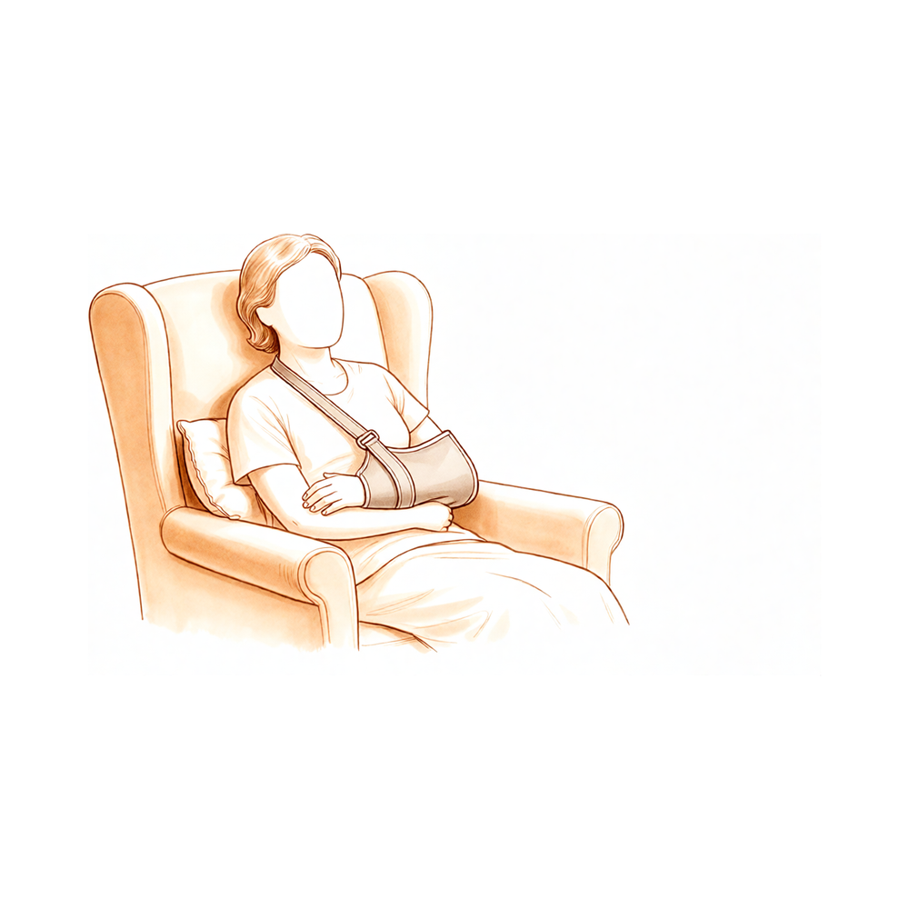
நீங்கள் மீட்புப் பிரிவில் எழுந்திருப்பீர்கள். உங்கள் அறுவை சிகிச்சை நிபுணர் நிலையான முறைகளைப் பயன்படுத்தி உங்கள் வலியை நிர்வகிப்பார். உங்கள் தோளில் ஒரு ஸ்லிங், பாண்டேஜ்கள் மற்றும் ஒருவேளை ஒரு பிரேஸ் இருக்கும். அறிவுறுத்தப்பட்டபடி பகுதியை உலர்ந்ததாகவும் சுத்தமாகவும் வைத்திருங்கள். உங்களுக்கு உதவ முதல் 24 மணிநேரங்களுக்கு யாராவது உங்களுடன் தங்க வேண்டும். இந்த அறுவை சிகிச்சைக்குப் பிறகு பெரும்பாலான நோயாளிகள் ஒரு இரவு மருத்துவமனையில் தங்கியிருக்கிறார்கள், இருப்பினும் சிலர் அதே நாளில் வீட்டிற்குச் செல்ல முடியும். இது தோளில் ஒரு வெட்டுடன் ஒரு திறந்த அறுவை சிகிச்சை. எந்தவொரு தோள் அறுவை சிகிச்சைக்குப் பிறகு குறைந்தது ஆறு வாரங்களுக்கு நீங்கள் வாகனம் ஓட்டக்கூடாது, எந்தக் கையில் அறுவை சிகிச்சை செய்யப்பட்டது என்பதைப் பொருட்படுத்தாமல். ஒரு ஸ்லிங்கில் உள்ள நோயாளிகள் வாகனம் ஓட்டக்கூடாது. உங்கள் அறுவை சிகிச்சை நிபுணர் உங்களை அனுமதித்தவுடன், வழக்கமாக ஆறு வார மதிப்பாய்வில், நீங்கள் மீண்டும் வாகனம் ஓட்டலாம். மேல் கால் அறுவை சிகிச்சைக்குப் பிறகு வாகனம் ஓட்டுதல் மேலும் விவரங்களுக்கு.
மீட்பு
உங்கள் தோள்பட்டைக்கு மேல் ஒரு வெட்டு இருக்கும். முதல் சில நாட்களில், வலி மற்றும் வீக்கம் இயல்பானது. உங்கள் கை கனமாகவும் இறுக்கமாகவும் உணரும். ஐஸ் பேக்குகள் மற்றும் பரிந்துரைக்கப்பட்ட மருந்துகள் இந்த அச om கரியத்தை குறைக்க உதவுகின்றன. உங்கள் அறுவை சிகிச்சையாளரின் அறிவுறுத்தலின்படி உங்கள் கையை ஸ்லிங்கில் வைத்திருங்கள். நீங்கள் ஓய்வெடுக்கும் போது இது குணமடைந்த திசுக்களைப் பாதுகாக்கிறது.
வீக்கம் குறையும்போது, நீங்கள் மென்மையான உடற்கூறியல் பயிற்சிகளைத் தொடங்குவீர்கள். இந்த இயக்கங்கள் பழுதுபார்ப்புக்கு சிரமமின்றி அடிப்படை தோள்பட்டை செயல்பாட்டை மீட்டெடுக்கும். ஒரு கையால் எப்படி உடை அணிவது மற்றும் அன்றாட பணிகளைச் செய்வது என்பதை நீங்கள் கற்றுக்கொள்வீர்கள். முதலில் தூங்குவது கடினமாக இருக்கலாம்; தலையணைகள் மூலம் உங்களை ஆதரிப்பது பெரும்பாலும் உதவுகிறது. உங்கள் அறுவை சிகிச்சையாளர் மற்றும் உடற்கூறியல் நிபுணர் உங்கள் வேகத்தை வழிநடத்துவார்கள். உங்கள் காலவரிசை மற்றவர்களிடமிருந்து வேறுபடலாம்.
நீங்கள் ஒரு ஸ்லிங்கில் இருக்கும்போது வாகனம் ஓட்டக்கூடாது. எந்தவொரு தோள்பட்டை அறுவை சிகிச்சைக்குப் பிறகு குறைந்தது ஆறு வாரங்களுக்கு நீங்கள் வாகனம் ஓட்டக்கூடாது என்று உங்கள் அறுவை சிகிச்சையாளரின் கொள்கை தேவைப்படுகிறது. எந்தக் கையில் அறுவை சிகிச்சை செய்யப்பட்டது என்பதைப் பொருட்படுத்தாமல். உங்கள் அறுவை சிகிச்சையாளர் உங்களை அனுமதித்தவுடன், பொதுவாக ஆறு வார மதிப்பாய்வில் நீங்கள் வாகனம் ஓட்டலாம். மேல் கால் அறுவை சிகிச்சைக்குப் பிறகு வாகனம் ஓட்டுதல் மேலும் விவரங்களுக்கு.
நீண்டகால மீட்பு வலிமை மற்றும் இயக்க வரம்பை மீட்டெடுப்பதில் கவனம் செலுத்துகிறது. பெரும்பாலான நோயாளிகள் காலப்போக்கில் திருப்திகரமான வலி நிவாரணத்தை அனுபவிக்கிறார்கள். இருப்பினும், முழு தோள்பட்டை இயக்கம் குறைவாக கணிக்கக்கூடியதாக இருக்கலாம். நிலையான உடற்பயிற்சி உங்கள் வெற்றிக்கு முக்கியமானது. செயல்முறையை நம்புங்கள் மற்றும் உங்கள் பராமரிப்புக் குழுவின் ஆலோசனையை நெருக்கமாகப் பின்பற்றுங்கள்.
என்ன தவறு நடக்கலாம்
பெரும்பாலான நோயாளிகள் நன்றாக இருக்கிறார்கள், ஆனால் எப்போதாவது பிரச்சினைகள் ஏற்படலாம். உங்கள் அறுவை சிகிச்சை நிபுணரும் குழுவும் எந்தவொரு பிரச்சினையையும் ஆரம்பத்தில் கண்டறிய உங்களை உன்னிப்பாகக் கண்காணிப்பார்கள்.
வலி மற்றும் குணப்படுத்தும் பிரச்சினைகள் காலப்போக்கில் தோள்பட்டை இயக்கம் இறுக்கமாகவோ அல்லது வரையறுக்கப்பட்டதாகவோ இருப்பதை நீங்கள் கவனிக்கலாம். இந்த அறுவை சிகிச்சைக்குப் பிறகு இயக்க முடிவுகளை கணிக்க முடியாததால் இது பொதுவானது. உங்களுக்கு வயதான நோயாளி சுயவிவரம் இருந்தால், உங்கள் வலிமையை மீண்டும் பெறுவதற்கு எலும்பு துண்டுகள் (குழாய்) குணமடைவது முக்கியம். இங்கு மோசமான குணமடைதல் தொடர்ச்சியான பலவீனம் அல்லது வலிக்கு வழிவகுக்கும். உங்கள் பின்தொடர்தல் வருகைகளின் போது தொடர்ச்சியான இறுக்கத்தன்மை அல்லது முன்னேற்றமின்மை குறித்து நீங்கள் தெரிவிக்க வேண்டும்.
பொது சுகாதார அபாயங்கள் இந்த காயம் பெரும்பாலும் வயதான பெரியவர்களை பாதிக்கிறது என்பதால், உங்கள் ஒட்டுமொத்த ஆரோக்கியம் மீட்பில் ஒரு பெரிய பாத்திரத்தை வகிக்கிறது. ஒரு வருடத்திற்குள் இறப்பு அதிக ஆபத்து உட்பட, காயம் ஏற்பட்ட பிறகு நீங்கள் கடுமையான மருத்துவ பிரச்சினைகளுக்கு அதிக ஆபத்தில் இருக்கலாம். இந்த ஆபத்து பொது மக்களில் இருப்பதை விட அதிகமாக உள்ளது மற்றும் பிற குறிப்பிட்ட சுகாதார காரணிகளைப் பொருட்படுத்தாமல் உள்ளது. காயம் ஏற்படுவதற்கு முன்பு நீங்கள் பலவீனமாக இருந்தால், உங்கள் வயதிற்கு சராசரியை விட அதிக இறப்பு அபாயத்தை நீங்கள் எதிர்கொள்ளலாம். உங்கள் பொது சுகாதார வரலாறு பற்றி உங்கள் குழுவுடன் நேர்மையாக இருங்கள், இதனால் அவர்கள் உங்களுக்கு சிறந்த ஆதரவை வழங்க முடியும்.
மருத்துவமனை மறுமலர்ச்சி வீட்டுக்குச் சென்றபின் நீங்கள் எதிர்பாராத விதமாக மீண்டும் மருத்துவமனைக்குச் செல்ல வேண்டியிருக்கும். இந்தத் திட்டமிடப்படாத மறுசீரமைப்புகளில் பெரும்பாலானவை தோள்பட்டையுடன் தொடர்புடையவை அல்ல, மாறாக மருத்துவப் பிரச்சினைகளுடன் தொடர்புடையவை. நீங்கள் பொதுவாக உடல்நிலை சரியில்லாமல் இருந்தால், காய்ச்சல் இருந்தால் அல்லது புதிய மருத்துவ அறிகுறிகள் இருந்தால், உடனடியாக உங்கள் மருத்துவரைத் தொடர்பு கொள்ளுங்கள். இது தோள்பட்டை வலி என்று கருத வேண்டாம்.
அறுவை சிகிச்சை சிக்கல்கள் நீங்கள் இன்னும் மருத்துவமனையில் இருக்கும்போது சிக்கல்கள் ஏற்படும் அபாயம் உள்ளது. மற்ற முறைகளுடன் ஒப்பிடும்போது நீங்கள் ஒரு தலைகீழ் தோள்பட்டை மாற்றீட்டைக் கொண்டிருந்தால் இவை அதிகமாக இருக்கும். சிக்கல்களில் நோய்த்தொற்று, இரத்தப்போக்கு அல்லது எலும்பு குணமடைவதில் சிக்கல்கள் இருக்கலாம். வெட்டுப் பகுதியில் அதிகரித்த வலி, சிவத்தல் அல்லது வீக்கம் ஆகியவற்றை நீங்கள் உணரலாம். இந்த அறிகுறிகளை நீங்கள் கவனித்தால், உடனடியாக உங்கள் செவிலியர் அல்லது மருத்துவரிடம் தெரிவிக்கவும். சிக்கலான எலும்பு முறிவுகளுக்கான அறுவை சிகிச்சை பலருக்கு நல்ல நீண்டகால முடிவுகளுக்கு வழிவகுக்கும் அதே வேளையில், இது மற்றொரு செயல்முறை தேவைப்படுவதற்கான அதிக வாய்ப்பைக் கொண்டுள்ளது. உங்கள் அறுவை சிகிச்சையாளர் இந்த அறிகுறிகளை ஆரம்பத்தில் சரிபார்க்க முடியும் என்பதால் உங்கள் மீட்பு சந்திப்புகளை வைத்திருங்கள்.
இந்த பக்கத்தில் உள்ள சிக்கல்கள் அட்டவணை வழக்கமான விகிதங்களை பட்டியலிடுகிறது நீங்கள் விவரங்களை விரும்பினால்.
எப்போது எங்களை அழைக்க வேண்டும்
உங்களுக்கு காய்ச்சல், அதிகரிக்கும் காயம் சிவத்தல், அல்லது வெளியேற்றம் இருந்தால் எங்களை அழைக்கவும். நீங்கள் திடீரென்று கடுமையான வலி, கன்றுக்குட்டி வீக்கம், அல்லது மூச்சுத் திணறல் ஆகியவற்றை உணர்ந்தால் அவசரநிலைக்குச் செல்லுங்கள். உணர்வு இழப்பு அல்லது உங்கள் உறுப்பை நகர்த்த முடியாமைக்கு அவசர சிகிச்சை பெறவும். இந்த அறிகுறிகளுக்கு உடனடி மதிப்பீடு தேவை.
Evidence & references
Overview
- Patients undergoing arthroplasty for acute proximal humeral fractures may achieve satisfactory long-term pain relief, though overall shoulder motion results are less predictable [1].
- In elderly patients undergoing reverse shoulder arthroplasty for acute proximal humeral fractures, anatomic tuberosity healing improves objective and subjective outcomes [2].
- Clinical results at 1-year follow-up confirm the advantage of applying a new intramedullary support nail and plate system to 3- or 4-part proximal humeral fractures in older patients [3].
- Elderly patients requiring admission after sustaining a proximal humeral fracture are frail and subject to a greater-than-average risk of mortality for their age [4].
- Patients who undergo initial nonoperative management have worse functional outcomes and higher complication rates than those who undergo acute reverse total shoulder arthroplasty (rTSA) for proximal humeral fractures [5].
- Patients with acute proximal humeral fractures who undergo reverse shoulder arthroplasty appear to achieve superior 5-year functional outcomes compared with patients who undergo hemiarthroplasty [6].
- The study cited represents the largest long-term follow-up of acute proximal humeral fractures treated with hemiarthroplasty [9].
- In most studies of proximal humeral fractures, only 1 or 2 patients experiencing an alternative outcome or lost to follow-up would change the conclusions for the dichotomous outcome studied [16].
- The increased in-hospital risk for major adverse events and surgical complications may moderate enthusiasm for reverse total shoulder arthroplasty (RTSA) for proximal humeral fractures in patients 65 years and older [21].
- Available literature suggests that reverse shoulder arthroplasty performed to address complex proximal humeral fractures might result in more favorable clinical outcomes than hemiarthroplasty performed for the same indication [26].
- Reverse total shoulder arthroplasty (RTSA) performed for acute 3- and 4-part proximal humeral fractures yields overall worse clinical outcomes and active range of motion compared with RTSA performed for elective indications [66].
- No clear benefits were observed in treating patients 65 years or older with four-part fractures of the proximal humerus with either hemiarthroplasty or nonoperative treatment [67].
Anatomy & Pathophysiology
- Shoulder rotational ability is improved by systematically repairing the tuberosities around the implant, provided their consolidation is anatomic [8].
- Greater tuberosity healing does not seem to impact reverse shoulder arthroplasty biomechanics during abduction or forward flexion [28].
- Greater tuberosity healing affects reverse shoulder arthroplasty biomechanics during external rotation [28].
- With minimal and moderate amounts of glenohumeral abduction, glenohumeral joint forces are significantly displaced superiorly [30].
- Varus and antecurvatum proximal humerus deformities as small as 15 degrees were associated with statistically significant alterations in glenohumeral joint mechanics [40].
- The control volume is an important anatomic and functional area of the proximal humerus [44].
- Vertical abduction has the greatest effect on axillary nerve position during the split lateral deltoid approach [43].
- Horizontal glenohumeral forward flexion and humeral rotation have little effect on axillary nerve position during the split lateral deltoid approach [43].
- The study demonstrates variability in the glenopolar angle with increased AP rotational offset of the shoulder radiograph [38].
- The study reveals inaccuracies in glenopolar angle measurement even at an institution with an established protocol [38].
- Range of motion and strength thresholds can identify subjects with normal shoulder function [29].
- The authors recommend performing the measurement of objective strength at the insertion of the deltoid muscle in a 90° abduction position in the scapula plane [35].
- Dominance of the affected shoulder has no influence on functional and quality of life outcome compared with the nondominant shoulder [37].
- Dominance of the affected shoulder should not be used to make treatment decisions [37].
Classification
- The Neer classification system covers 98% of all proximal humeral fractures and is appropriate for clinical practice [58].
- Classifications of proximal humeral fractures using the Neer system based on CT scans and plain radiographs are not very reliable or reproducible due to difficulty in determining which segments are fractured [60].
- The HGLS classification is a reliable method of describing fractures of the proximal humerus compared with the Neer and AO systems [56].
- A new classification system with emphasis on the qualitative aspects of proximal humeral fractures showed high reliability when based on a standardized imaging protocol including computed tomography scans [49].
- Consensus when managing proximal humerus fractures is limited to specific scenarios, whereas lack of consensus still exists in others [11].
Clinical Presentation
- Patients undergoing arthroplasty for acute proximal humerus fractures may achieve satisfactory long-term pain relief, though overall shoulder motion results are less predictable [1].
- In elderly patients undergoing reverse shoulder arthroplasty for acute proximal humeral fractures, anatomic tuberosity healing improves objective and subjective outcomes [2].
- Clinical results at 1-year follow-up confirm the advantage of applying a new intramedullary support nail and plate system to 3- or 4-part proximal humeral fractures in older patients [3].
- Elderly patients requiring admission after sustaining a proximal humeral fracture are frail and subject to a greater-than-average risk of mortality for their age [4].
- Patients undergoing initial nonoperative management have worse functional outcomes and higher complication rates than those undergoing acute reverse total shoulder arthroplasty for proximal humeral fractures [5].
- Fractures of the proximal humerus follow characteristic patterns [7].
- A majority of patients with proximal humeral fractures undergo non-operative treatment [10].
- Consensus on managing proximal humerus fractures is limited to specific scenarios, while a lack of consensus exists in others [11].
- There is significant heterogeneity in the terminology and definitions used to describe complications following non-surgical management of proximal humeral fractures [12].
- Nonoperative treatment of proximal humeral fractures produces considerable variation in shoulder-specific and general health outcomes at 1 year, with a substantial proportion of patients having poor perceived functional outcomes [13].
- Mortality at 1 year for fragility proximal humerus fractures is universally high regardless of risk factors [15].
- The majority of unplanned hospital readmissions after surgical treatment of proximal humerus fractures are associated with medical diagnoses [19].
- In patients presenting with a traumatic shoulder injury and normal radiographs, the anterior bruise sign (ABS) is a highly sensitive and specific clinical aid to identify occult greater tuberosity fractures [22].
- Most proximal humeral fractures in elderly patients can be treated nonoperatively with good functional outcomes [23].
- Patients sustaining a proximal humeral fracture have a significantly higher risk of mortality up to one year after the injury compared with the general population [25].
- Most pediatric patients with proximal humerus fractures have favorable results, and complications are infrequent [50].
- Reverse shoulder arthroplasty is a powerful tool for managing proximal humerus fracture sequelae when joint-preserving options are not optimal, provided there is careful management of the tuberosities and understanding of associated pearls and pitfalls [54].
- Prevention of local complications, particularly those leading to severe varus deviation, appears essential to improve shoulder function after a proximal humeral fracture [55].
- Factors associated with poor results after internal fixation of three-part and four-part proximal humerus fracture-dislocations include being a woman, four-part fracture dislocation, and absence of metaphyseal head extension [57].
- A wide range of outcome measures are used in proximal humeral fracture studies, but there is limited evidence regarding their psychometric properties in this specific population [59].
Investigations
- Elderly patients requiring admission after sustaining a proximal humeral fracture are frail and subject to a greater-than-average risk of mortality for their age [4].
- A majority of patients with proximal humeral fractures undergo non-operative treatment [10].
- Fractures of the proximal humerus follow characteristic patterns [7].
- Despite a delayed diagnosis of more than one year, osteotomy and realignment of a displaced lesser tuberosity fracture can be successful and enhance overall shoulder function [17].
- Undisplaced greater tuberosity fractures can be managed non-operatively with good results [72].
- Patients with persistent post-traumatic shoulder pain and limitation of function warrant MRI investigation to identify occult greater tuberosity fractures [72].
- In patients presenting with a traumatic shoulder injury with normal radiographs, the anterior bruise sign (ABS) is a highly sensitive and specific clinical aid to identify patients with an occult greater tuberosity fracture [22].
- There is relevant variability in displacement measurements between shoulder radiographs and CT scans in the coronal plane [73].
- Nearly 30% of cases suggesting surgical treatment on radiographs are reclassified for conservative treatment based on CT findings [73].
- The inherent nature of medial comminution of proximal humeral fracture may lead to inferior radiographic outcomes [71].
- Routine use of 3D-printed models may not be beneficial for classifying proximal humeral fracture patterns beyond the information gained from currently available imaging modalities [74].
- The routine use of 3D-printed models should be avoided as the sole determinant for recommending surgical intervention at this time [74].
- Convolutional neural networks (CNNs) proficiently rule out proximal humerus fractures on plain radiographs [76].
- Missed posterior dislocation of the shoulder after intramedullary fixation of proximal humeral fractures is an extremely rare injury that can be missed due to inadequate initial and postoperative x-ray images and incorrect interpretation [79].
- Consensus when managing proximal humerus fractures is limited to specific scenarios, whereas lack of consensus still exists in others [11].
Treatment
- Patients undergoing shoulder hemiarthroplasty for acute proximal humerus fractures may achieve satisfactory long-term pain relief, though overall shoulder motion results are less predictable [1].
- In elderly patients undergoing reverse shoulder arthroplasty for acute proximal humeral fractures, anatomic tuberosity healing improves objective and subjective outcomes [2].
- Clinical results at 1-year follow-up confirm the advantage of applying a new intramedullary support nail and plate system to 3- or 4-part proximal humeral fractures in older patients [3].
- Elderly patients requiring admission after sustaining a proximal humeral fracture are frail and subject to a greater-than-average risk of mortality for their age [4].
- Patients undergoing initial nonoperative management have worse functional outcomes and higher complication rates than those undergoing acute reverse total shoulder arthroplasty (rTSA) for proximal humeral fractures [5].
- Patients with acute proximal humeral fractures who undergo reverse shoulder arthroplasty appear to achieve superior 5-year functional outcomes compared with patients who undergo hemiarthroplasty [6].
- Shoulder rotational ability is improved by systematically repairing the tuberosities around the implant in complex shoulder fractures treated by reverse shoulder arthroplasty, provided their consolidation is anatomic [8].
- A majority of patients with proximal humeral fractures underwent non-operative treatment [10].
- Significant heterogeneity exists in the terminology and definitions used to describe complications following non-surgical management of proximal humeral fractures [12].
- Nonoperative treatment of proximal humeral fractures produces considerable variation in shoulder-specific and general health outcomes at 1 year, with a substantial proportion of patients having poor perceived functional outcomes [13].
- Primary shoulder hemiarthroplasty for proximal humeral fracture is associated with satisfactory prosthetic survival at an average of 6.3 years [14].
- Short and long periods of immobilization yield similar results for nonoperatively treated proximal humeral fractures, independent of the fracture pattern [20].
- Most proximal humeral fractures in elderly patients can be treated nonoperatively with good functional outcomes [23].
- Available literature suggests that reverse shoulder arthroplasty performed to address complex proximal humeral fractures might result in more favorable clinical outcomes than hemiarthroplasty performed for the same indication [26].
- Nonsurgical management of proximal humerus fractures decreased during the study period [46].
- Treatment with reverse shoulder arthroplasty provides superior functional outcomes compared with conservative treatment for patients presenting with an acute proximal humeral fracture [47].
- There is no significant difference in clinical outcomes at 2 years between surgery and non-operative treatment in patients 60 years of age or older with displaced 2-part fractures of the proximal humerus [48].
- Nonsurgical management of proximal humerus fractures demonstrates successful outcomes and union rates greater than 90% [51].
- Nonsurgical treatment should have a more prominent role in the treatment of proximal humeral fractures compared to locking plate fixation [52].
- Osteoporosis may not be regarded as a contraindication for open reduction and internal fixation of unilateral displaced 3- or 4-part fractures, as shoulder function was restored to preinjury levels for most patients at 12-month follow-up [53].
- Percutaneous treatment of selected proximal humeral fractures results in predictable union and good clinical results with a low rate of complications [62].
- With narrow indications, use of a specific fracture stem and adequate tuberosity management, successful radiographic and functional results are presented after a mean follow-up of 4.8 years after hemiarthroplasty for primary nonreconstructable humeral head fractures [65].
Complications
- Patients undergoing arthroplasty for acute proximal humeral fractures may achieve satisfactory long-term pain relief, but overall shoulder motion results are less predictable [1].
- In elderly patients undergoing reverse shoulder arthroplasty for acute proximal humeral fractures, anatomic tuberosity healing improves objective and subjective outcomes [2].
- Clinical results at 1-year follow-up confirm the advantage of applying a new intramedullary support nail and plate system to 3- or 4-part proximal humeral fractures in older patients [3].
- Elderly patients requiring admission after sustaining a proximal humeral fracture are frail and subject to a greater-than-average risk of mortality for their age [4].
- Patients undergoing initial nonoperative management have worse functional outcomes and higher complication rates than those undergoing acute reverse total shoulder arthroplasty (rTSA) for proximal humeral fractures [5].
- Patients with acute proximal humeral fractures who undergo reverse shoulder arthroplasty (RSA) appear to achieve superior 5-year functional outcomes compared with patients who undergo hemiarthroplasty [6].
- Fractures of the proximal humerus follow characteristic patterns [7].
- Primary shoulder hemiarthroplasty for proximal humeral fracture is associated with satisfactory prosthetic survival at an average of 6.3 years [14].
- Mortality at 1 year for fragility proximal humerus fractures is universally high regardless of risk factors [15].
- The majority of unplanned hospital readmissions after surgical treatment of proximal humerus fractures are associated with medical diagnoses [19].
- In-hospital complications are more likely to occur after reverse shoulder arthroplasty than after locked plating for proximal humeral fractures [21].
- Surgery for complex proximal humeral fractures leads to overall good long-term outcomes but is associated with high overall complication and reoperation rates [24].
- Patients sustaining a proximal humeral fracture have a significantly higher risk of mortality up to one year after the injury compared with the general population [25].
- Short-term complication rates for fixation and arthroplasty alike have decreased compared with recent historic norms [27].
Recovery
- Patients undergoing arthroplasty for acute proximal humerus fractures may achieve satisfactory long-term pain relief, though overall shoulder motion results are less predictable [1].
- In elderly patients undergoing reverse shoulder arthroplasty for acute proximal humeral fractures, anatomic tuberosity healing improves objective and subjective outcomes [2].
- Clinical results at 1-year follow-up confirm the advantage of applying a new intramedullary support nail and plate system to 3- or 4-part proximal humeral fractures in older patients [3].
- Elderly patients requiring admission after sustaining a proximal humeral fracture are frail and subject to a greater-than-average risk of mortality for their age [4].
- Patients who undergo initial nonoperative management have worse functional outcomes and higher complication rates than those who undergo acute reverse total shoulder arthroplasty (rTSA) for proximal humeral fractures [5].
- Patients with acute proximal humeral fractures who undergo reverse shoulder arthroplasty appear to achieve superior 5-year functional outcomes compared with patients who undergo hemiarthroplasty [6].
- Fractures of the proximal humerus follow characteristic patterns [7].
- This study represents the largest long-term follow-up of acute proximal humeral fractures treated with hemiarthroplasty [9].
- Nonoperative treatment of proximal humeral fractures produces considerable variation in shoulder-specific and general health outcomes at 1 year, with a substantial proportion of patients having poor perceived functional outcomes [13].
- Primary shoulder hemiarthroplasty for proximal humeral fracture is associated with satisfactory prosthetic survival at an average of 6.3 years [14].
- Mortality at 1 year for fragility proximal humerus fractures is universally high regardless of risk factors [15].
- In most studies of proximal humeral fractures, only 1 or 2 patients experiencing an alternative outcome or lost to follow-up would change the conclusions for the dichotomous outcome studied [16].
- Despite a delayed diagnosis of more than one year, osteotomy and realignment of a displaced lesser tuberosity fracture was successful and enhanced overall shoulder function in two adolescent patients [17].
- Short and long periods of immobilization yield similar results for nonoperatively treated proximal humeral fractures, independent of the fracture pattern [20].
- Surgery for complex proximal humeral fractures leads to overall good long-term outcomes despite high overall complication and reoperation rates [24].
- The increasing utilization of reverse total shoulder arthroplasty (RTSA) and decreasing short-term complication rates for fixation and arthroplasty represent a substantial change compared with recent historic norms in the management of proximal humerus fractures [27].
- Long-term treatment with reverse shoulder arthroplasty (RSA) for displaced 3- or 4-part proximal humerus fractures provides better functional outcomes compared to nonoperative treatment, a difference attributed to the deterioration of functional outcomes of the nonoperative treatment over time [63].
- Timing of surgery did not affect Oxford Shoulder Score at any stage of follow-up, irrespective of age or fracture type [80].
Key Evidence
- [L3] Patients undergoing arthroplasty as treatment of an acute fracture of the proximal humerus may achieve satisfactory long-term pain relief; however, the result for overall shoulder motion is less predictable. [1] (10.1016/j.jse.2007.06.025)
- [L3] In elderly patients who have undergone a reverse shoulder arthroplasty for acute proximal humeral fractures, anatomic tuberosity healing improves objective and subjective outcomes. [2] (10.1016/j.jse.2018.05.030)
- [L3] Clinical results at 1-year follow-up confirmed the advantage of applying it to 3- or 4-part proximal humeral fractures in older patients. [3] (10.1186/s12891-022-05998-z)
- [L3] Elderly patients who require admission after sustaining a proximal humeral fracture are frail and subject to a greater-than-average risk of mortality for their age. [4] (10.1016/j.jse.2019.05.030)
- [L3] Patients who undergo initial periods of nonoperative management have worse functional outcomes and higher complication rates than those who undergo acute rTSA for proximal humeral fractures. [5] (10.1016/j.jse.2021.06.020)
- [L3] Patients with acute proximal humeral fractures who undergo RSA appear to achieve superior 5-year functional outcomes compared with patients who undergo hemiarthroplasty. [6] (10.1016/j.jse.2012.03.006)
- [L4] Fractures of the proximal humerus follow characteristic patterns. [7] (10.1016/j.jse.2017.05.014)
- [L3] Shoulder rotational ability is improved by systematically repairing the tuberosities around the implant, provided their consolidation is anatomic. [8] (10.1016/j.jse.2012.03.011)
- [L3] This is the largest long-term follow-up study of acute proximal humeral fractures treated with hemiarthroplasty. [9] (10.1302/0301-620x.103b6.bjj-2020-1753.r1)
- [L3] A majority of patients with proximal humeral fractures underwent non-operative treatment. [10] (10.1186/s12891-019-2812-9)
- [L5] Consensus when managing proximal humerus fractures is limited to specific scenarios, whereas lack of consensus still exists in others. [11] (10.1016/j.jse.2024.12.005)
- [L1] This systematic review highlights significant heterogeneity in the terminology and definitions used to describe complications following non-surgical management of proximal humeral fractures, calling for standardized definitions to improve evidence synthesis. [12] (10.1186/s12891-019-2459-6)
- [L1] Nonoperative treatment of proximal humeral fractures produces considerable variation in shoulder-specific and general health outcomes at 1 year, and a substantial proportion of patients have poor perceived functional outcomes. [13] (10.2106/jbjs.20.02018)
- [L2] Primary shoulder hemiarthroplasty for proximal humeral fracture is associated with satisfactory prosthetic survival at an average of 6.3 years. [14] (10.2106/jbjs.l.01115)
- [L3] Mortality at 1 year for fragility proximal humerus fractures is universally high regardless of risk factors. [15] (10.1016/j.jse.2022.03.006)
- [L2] In most studies of proximal humeral fractures, only 1 or 2 patients experiencing an alternative outcome or lost to follow-up would change the conclusions for the dichotomous outcome studied. [16] (10.1016/j.jse.2022.01.141)
- [L4] Despite a delayed diagnosis of more than one year, osteotomy and realignment of the displaced fracture of the lesser tuberosity was successful and enhanced the overall function of the shoulder in these two patients. [17] (10.2106/00004623-199509000-00020)
- [L3] As the majority of unplanned hospital readmissions were associated with medical diagnoses, it is important to consider patient medical comorbidities before surgical treatment of proximal humerus fractures and during the postoperative care phase. [19] (10.1007/s11999-014-3613-y)
- [L2] Short and long periods of immobilization yield similar results for nonoperatively treated proximal humeral fractures, independent of the fracture pattern. [20] (10.2106/jbjs.20.02137)
- [L3] The increased in-hospital risk for major adverse events and surgical complications may moderate the enthusiasm associated with RTSA for proximal humeral fractures in patients 65 years and older. [21] (10.1097/corr.0000000000001776)
- [L2] In patients presenting with a traumatic shoulder injury with normal radiographs, the anterior bruise sign (ABS) is a highly sensitive and specific clinical aid to identify patients with an occult greater tuberosity fracture. [22] (10.1016/j.jse.2023.07.044)
- [L5] Most proximal humeral fractures in elderly patients can be treated nonoperatively with good functional outcomes. [23] (10.2106/jbjs.l.01293)
- [L5] Surgery for complex proximal humeral fractures leads to overall good long-term outcomes with high overall complication and reoperation rates. [24] (10.2106/jbjs.19.01109)
- [L3] Compared with the general population, patients sustaining a proximal humeral fracture have a significantly higher risk of mortality up to one year after the injury. [25] (10.1302/0301-620x.102b11.bjj-2020-0627.r1)
- [L1] The available literature suggests that reverse shoulder arthroplasty performed to address complex proximal humeral fractures might result in more favorable clinical outcomes than hemiarthroplasty performed for the same indication. [26] (10.1016/j.jse.2015.08.030)
- [L3] The increasing utilization of RTSA and decreasing short-term complication rates for fixation and arthroplasty alike represent a substantial change compared even with recent historic norms in the management of proximal humerus fractures. [27] (10.1097/corr.0000000000002391)
- [L5] Greater tuberosity healing does not seem to impact reverse shoulder arthroplasty biomechanics during abduction or forward flexion; however, it does affect biomechanics during external rotation. [28] (10.1016/j.jse.2019.07.022)
- [L3] Range of motion and strength thresholds can identify subjects with normal shoulder function. [29] (10.1016/j.jse.2010.06.005)
- [L5] With minimal and moderate amounts of glenohumeral abduction, glenohumeral joint forces are significantly displaced superiorly. [30] (10.1016/j.jse.2007.06.017)
- [L3] The authors recommend performing the measurement at the insertion of the deltoid muscle in a 90° abduction position in the scapula plane. [35] (10.1186/s12891-019-2795-6)
- [L3] Dominance of the affected shoulder has no influence and should not be used to make treatment decisions. [37] (10.1016/j.jse.2014.10.006)
- [L4] The study demonstrates variability in the glenopolar angle with increased AP rotational offset of the shoulder radiograph, revealing inaccuracies even at an institution with an established protocol. [38] (10.1302/0301-620x.95b8.30631)
- [L5] Varus and antecurvatum proximal humerus deformities as small as 15 degrees were associated with statistically significant alterations in glenohumeral joint mechanics. [40] (10.5435/jaaos-d-20-00555)
- [L5] Vertical abduction has the greatest effect on axillary nerve position, while horizontal glenohumeral forward flexion and humeral rotation have little effect. [43] (10.1016/j.jse.2008.12.001)
- [L5] The control volume is an important anatomic and functional area of the proximal humerus. [44] (10.1016/j.jse.2017.12.004)
- [L4] Nonsurgical management of proximal humerus fractures decreased during the study period. [46] (10.1016/j.jhsa.2020.03.022)
- [L1] Treatment with reverse shoulder arthroplasty provides superior functional outcomes compared with conservative treatment for patients presenting with an acute proximal humeral fracture. [47] (10.1016/j.jse.2024.02.023)
- [L1] This trial found no significant difference in clinical outcomes at 2 years between surgery and non-operative treatment in patients 60 years of age or older with displaced 2-part fractures of the proximal humerus. [48] (10.1371/journal.pmed.1002855)
- [L3] The new classification system with emphasis on the qualitative aspects of proximal humeral fractures showed high reliability when based on a standardized imaging protocol including computed tomography scans. [49] (10.1016/j.jse.2015.08.006)
- [L5] Most pediatric patients with proximal humerus fractures have favorable results, and complications are infrequent. [50] (10.5435/jaaos-d-14-00033)
- [L5] Treatment for proximal humerus fractures remains controversial, with nonsurgical management demonstrating successful outcomes and union rates greater than 90%. [51] (10.5435/jaaos-d-24-01073)
- [L3] Nonsurgical treatment should have a more prominent role in the treatment of proximal humeral fractures. [52] (10.1016/j.jse.2011.01.025)
- [L1] Shoulder function was restored to preinjury levels for most patients, and osteoporosis may not be regarded as a contraindication for this treatment. [53] (10.1016/j.jse.2022.07.008)
- [L5] Reverse shoulder arthroplasty is a powerful tool for managing proximal humerus fracture sequelae when joint-preserving options are not optimal, provided there is careful management of the tuberosities and understanding of associated pearls and pitfalls. [54] (10.5435/jaaos-d-23-00740)
- [L2] Prevention of local complications, in particular those leading to severe varus deviation, appears essential to improve shoulder function after a proximal humeral fracture. [55] (10.1016/j.jse.2011.06.009)
- [L3] The HGLS classification is a reliable method of describing fractures of the proximal humerus compared with the Neer and AO systems. [56] (10.1016/j.jse.2012.09.018)
- [L5] Surgical treatment of proximal humerus fractures remains far from straightforward, with unpredictable outcomes where factors associated with poor results include being a woman, four-part fracture dislocation, and absence of metaphyseal head extension. [57] (10.1097/corr.0000000000002242)
- [L4] The revised Neer classification covers 98% of all proximal humeral fractures and is appropriate for clinical practice. [58] (10.1016/j.jse.2009.01.018)
- [L1] The review identified a wide range of outcome measures used in proximal humeral fracture studies, but found limited evidence regarding their psychometric properties in this specific population. [59] (10.1016/j.jse.2010.10.028)
- [L4] Classifications of proximal humeral fractures using the Neer system based on CT scans and plain radiographs are not very reliable or reproducible due to difficulty in determining which segments are fractured. [60] (10.2106/00004623-199609000-00012)
- [L4] Percutaneous treatment of selected proximal humeral fractures results in predictable union and good clinical results with a low rate of complications. [62] (10.1016/j.jse.2006.09.006)
- [L1] Long-term treatment with RSA for displaced 3- or 4-part proximal humerus fractures provides better functional outcomes compared to nonoperative treatment, a difference attributed to the deterioration of functional outcomes of the nonoperative treatment over time. [63] (10.1016/j.jse.2024.09.032)
- [L4] With narrow indications, use of a specific fracture stem and adequate tuberosity management, successful radiographic and functional results are presented after a mean follow-up of 4.8 years after hemiarthroplasty for primary nonreconstructable humeral head fractures. [65] (10.1016/j.jse.2023.02.118)
- [L1] RTSA performed for acute 3- and 4-part proximal humeral fractures yields overall worse clinical outcomes and active ROM compared with RTSA performed for elective indications. [66] (10.1016/j.jse.2021.07.014)
- [L1] We observed no clear benefits in treating patients 65 years or older with four-part fractures of the proximal humerus with either hemiarthroplasty or nonoperative treatment. [67] (10.1007/s11999-012-2531-0)
- [L3] This implies that the inherent nature of medial comminution of proximal humeral fracture may lead to inferior radiographic outcomes. [71] (10.1186/s13018-022-03337-5)
- [L4] Undisplaced greater tuberosity fractures can be managed non-operatively with good results, but patients with persistent post-traumatic shoulder pain and limitation of function warrant MRI investigation to identify occult fractures. [72] (10.1186/s12891-018-2225-1)
- [L3] There is relevant variability in displacement measurements between shoulder radiographs and CT scans in the coronal plane, with nearly 30% of cases suggesting surgical treatment on radiographs being reclassified for conservative treatment based on CT findings. [73] (10.1016/j.jse.2016.05.016)
- [L5] The routine use of 3D-printed models may not be beneficial for classifying proximal humeral fracture patterns beyond the information gained from currently available imaging modalities, and their use as the sole determinant for recommending surgical intervention should be avoided at this time. [74] (10.1097/corr.0000000000002017)
- [L3] CNNs proficiently rule out proximal humerus fractures on plain radiographs. [76] (10.1302/0301-620x.106b11.bjj-2024-0264.r1)
- [L4] Missed posterior dislocation of the shoulder after intramedullary fixation of proximal humeral fractures is an extremely rare injury that can be missed due to inadequate initial and postoperative x-ray images and incorrect interpretation. [79] (10.1016/j.jse.2008.10.020)
- [L1] Timing of surgery did not affect Oxford Shoulder Score at any stage of follow-up, irrespective of age or fracture type. [80] (10.1302/0301-620x.102b1.bjj-2020-0546.r1)
References
[1] Shoulder hemiarthroplasty for acute fractures of the proximal humerus: A minimum five-year follow-up. Journal of Shoulder and Elbow Surgery. 2008. DOI: 10.1016/j.jse.2007.06.025 [2] How the greater tuberosity affects clinical outcomes after reverse shoulder arthroplasty for proximal humeral fractures. Journal of Shoulder and Elbow Surgery. 2018. DOI: 10.1016/j.jse.2018.05.030 [3] Technique and clinical results of a new intramedullary support nail and plate system for fixation of 3- or 4- part proximal humeral fractures in older adults. BMC Musculoskeletal Disorders. 2022. DOI: 10.1186/s12891-022-05998-z [4] Mortality after inpatient stay for proximal humeral fractures. Journal of Shoulder and Elbow Surgery. 2020. DOI: 10.1016/j.jse.2019.05.030 [5] Minimum 2-year outcomes of reverse total shoulder arthroplasty for fracture: how does acute arthroplasty compare with salvage?. Journal of Shoulder and Elbow Surgery. 2022. DOI: 10.1016/j.jse.2021.06.020 [6] Functional outcomes of reverse shoulder arthroplasty compared with hemiarthroplasty for acute proximal humeral fractures. Journal of Shoulder and Elbow Surgery. 2013. DOI: 10.1016/j.jse.2012.03.006 [7] Fracture line morphology of complex proximal humeral fractures. Journal of Shoulder and Elbow Surgery. 2017. DOI: 10.1016/j.jse.2017.05.014 [8] Improvement in shoulder rotation in complex shoulder fractures treated by reverse shoulder arthroplasty. Journal of Shoulder and Elbow Surgery. 2013. DOI: 10.1016/j.jse.2012.03.011 [9] Ten-year follow-up of stemmed hemiarthroplasty for acute proximal humeral fractures. The Bone & Joint Journal. 2021. DOI: 10.1302/0301-620x.103b6.bjj-2020-1753.r1 [10] Readmissions, revisions, and mortality after treatment for proximal humeral fractures in three large states. BMC Musculoskeletal Disorders. 2019. DOI: 10.1186/s12891-019-2812-9 [11] Consensus statement on the treatment of proximal humerus fractures: a Delphi approach by the Neer Circle of the American Shoulder and Elbow Surgeons. Journal of Shoulder and Elbow Surgery. 2025. DOI: 10.1016/j.jse.2024.12.005 [12] Complications after non-surgical management of proximal humeral fractures: a systematic review of terms and definitions. BMC Musculoskeletal Disorders. 2019. DOI: 10.1186/s12891-019-2459-6 [13] Functional Outcome After Nonoperative Treatment of a Proximal Humeral Fracture in Adults. Journal of Bone and Joint Surgery. 2021. DOI: 10.2106/jbjs.20.02018 [14] Comparison of Hemiarthroplasty and Reverse Arthroplasty for Treatment of Proximal Humeral Fractures. Journal of Bone and Joint Surgery. 2013. DOI: 10.2106/jbjs.l.01115 [15] Morbidity and mortality of fragility proximal humerus fractures: a retrospective cohort study of patients presenting to a level one trauma center. Journal of Shoulder and Elbow Surgery. 2022. DOI: 10.1016/j.jse.2022.03.006 [16] Fragility of randomized controlled trials on treatment of proximal humeral fracture. Journal of Shoulder and Elbow Surgery. 2022. DOI: 10.1016/j.jse.2022.01.141 [17] Isolated avulsion fracture of the lesser tuberosity of the humerus in adolescents. A report of two cases.. The Journal of Bone & Joint Surgery. 1995. DOI: 10.2106/00004623-199509000-00020 [19] Hospital Readmissions After Surgical Treatment of Proximal Humerus Fractures: Is Arthroplasty Safer Than Open Reduction Internal Fixation?. Clinical Orthopaedics & Related Research. 2014. DOI: 10.1007/s11999-014-3613-y [20] One Versus 3-Week Immobilization Period for Nonoperatively Treated Proximal Humeral Fractures. Journal of Bone and Joint Surgery. 2021. DOI: 10.2106/jbjs.20.02137 [21] In-hospital Complications Are More Likely to Occur After Reverse Shoulder Arthroplasty Than After Locked Plating for Proximal Humeral Fractures. Clinical Orthopaedics & Related Research. 2021. DOI: 10.1097/corr.0000000000001776 [22] A new clinical sign to detect radiologically occult greater tuberosity fractures. Journal of Shoulder and Elbow Surgery. 2024. DOI: 10.1016/j.jse.2023.07.044 [23] Proximal Humeral Fracture Treatment in Adults. Journal of Bone and Joint Surgery. 2014. DOI: 10.2106/jbjs.l.01293 [24] Proximal Humeral Fractures: “Damned If You Operate, and Damned If You Don’t”. Journal of Bone and Joint Surgery. 2019. DOI: 10.2106/jbjs.19.01109 [25] Mortality after a proximal humeral fracture. The Bone & Joint Journal. 2020. DOI: 10.1302/0301-620x.102b11.bjj-2020-0627.r1 [26] Hemiarthroplasty versus reverse shoulder arthroplasty for treatment of proximal humeral fractures: a meta-analysis. Journal of Shoulder and Elbow Surgery. 2016. DOI: 10.1016/j.jse.2015.08.030 [27] Short-term Complications for Proximal Humerus Fracture Surgery Have Decreased: An Analysis of the National Surgical Quality Improvement Program Database. Clinical Orthopaedics & Related Research. 2022. DOI: 10.1097/corr.0000000000002391 [28] The role of greater tuberosity healing in reverse shoulder arthroplasty: a finite element analysis. Journal of Shoulder and Elbow Surgery. 2020. DOI: 10.1016/j.jse.2019.07.022 [29] Does objective shoulder impairment explain patient-reported functional outcome? A study of proximal humerus fractures. Journal of Shoulder and Elbow Surgery. 2011. DOI: 10.1016/j.jse.2010.06.005 [30] Neer Award 2006: Biomechanical assessment of inferior tuberosity placement during hemiarthroplasty for four-part proximal humeral fractures. Journal of Shoulder and Elbow Surgery. 2008. DOI: 10.1016/j.jse.2007.06.017 [35] Evaluation of the Constant score: which is the method to assess the objective strength?. BMC Musculoskeletal Disorders. 2019. DOI: 10.1186/s12891-019-2795-6 [37] Does fracture of the dominant shoulder have any effect on functional and quality of life outcome compared with the nondominant shoulder?. Journal of Shoulder and Elbow Surgery. 2015. DOI: 10.1016/j.jse.2014.10.006 [38] The assessment of scapular radiographs. The Bone & Joint Journal. 2013. DOI: 10.1302/0301-620x.95b8.30631 [40] Altered Glenohumeral Biomechanics in Proximal Humeral Fracture Malunion. Journal of the American Academy of Orthopaedic Surgeons. 2020. DOI: 10.5435/jaaos-d-20-00555 [43] Effects of shoulder position on axillary nerve positions during the split lateral deltoid approach. Journal of Shoulder and Elbow Surgery. 2009. DOI: 10.1016/j.jse.2008.12.001 [44] A morphovolumetric study of head malposition in proximal humeral fractures based on 3-dimensional computed tomography scans: the control volume theory. Journal of Shoulder and Elbow Surgery. 2018. DOI: 10.1016/j.jse.2017.12.004 [46] Cost-Minimization Analysis and Treatment Trends of Surgical and Nonsurgical Treatment of Proximal Humerus Fractures. The Journal of Hand Surgery. 2020. DOI: 10.1016/j.jhsa.2020.03.022 [47] Reverse shoulder arthroplasty or nothing for patients with displaced proximal humeral fractures: a randomized controlled trial. Journal of Shoulder and Elbow Surgery. 2024. DOI: 10.1016/j.jse.2024.02.023 [48] Operative versus non-operative treatment for 2-part proximal humerus fracture: A multicenter randomized controlled trial. PLOS Medicine. 2019. DOI: 10.1371/journal.pmed.1002855 [49] Classification of proximal humeral fractures based on a pathomorphologic analysis. Journal of Shoulder and Elbow Surgery. 2016. DOI: 10.1016/j.jse.2015.08.006 [50] Evaluation and Management of Pediatric Proximal Humerus Fractures. Journal of the American Academy of Orthopaedic Surgeons. 2015. DOI: 10.5435/jaaos-d-14-00033 [51] Contemporary Management of Proximal Humeral Fractures. Journal of the American Academy of Orthopaedic Surgeons. 2025. DOI: 10.5435/jaaos-d-24-01073 [52] Locking plate versus nonsurgical treatment for proximal humeral fractures: better midterm outcome with nonsurgical treatment. Journal of Shoulder and Elbow Surgery. 2011. DOI: 10.1016/j.jse.2011.01.025 [53] Osteoporosis does not affect bone mineral density change in the proximal humerus or the functional outcome after open reduction and internal fixation of unilateral displaced 3- or 4-part fractures at 12-month follow-up. Journal of Shoulder and Elbow Surgery. 2023. DOI: 10.1016/j.jse.2022.07.008 [54] Reverse Shoulder Arthroplasty to Treat Proximal Humerus Fracture Sequelae: A Review. Journal of the American Academy of Orthopaedic Surgeons. 2024. DOI: 10.5435/jaaos-d-23-00740 [55] Path analysis of factors for functional outcome at one year in 463 proximal humeral fractures. Journal of Shoulder and Elbow Surgery. 2011. DOI: 10.1016/j.jse.2011.06.009 [56] A comprehensive classification of proximal humeral fractures: HGLS system. Journal of Shoulder and Elbow Surgery. 2013. DOI: 10.1016/j.jse.2012.09.018 [57] CORR Insights®: What Factors Are Associated With Poor Shoulder Function and Serious Complications After Internal Fixation of Three-part and Four-part Proximal Humerus Fracture-dislocations?. Clinical Orthopaedics & Related Research. 2022. DOI: 10.1097/corr.0000000000002242 [58] Four-segment classification of proximal humeral fractures revisited: A multicenter study on 509 cases. Journal of Shoulder and Elbow Surgery. 2009. DOI: 10.1016/j.jse.2009.01.018 [59] Outcome measures in the management of proximal humeral fractures: a systematic review of their use and psychometric properties. Journal of Shoulder and Elbow Surgery. 2011. DOI: 10.1016/j.jse.2010.10.028 [60] Evaluation of the Neer System of Classification of Proximal Humeral Fractures with Computerized Tomographic Scans and Plain Radiographs. The Journal of Bone & Joint Surgery. 1996. DOI: 10.2106/00004623-199609000-00012 [62] Outcomes after percutaneous reduction and fixation of proximal humeral fractures. Journal of Shoulder and Elbow Surgery. 2007. DOI: 10.1016/j.jse.2006.09.006 [63] Long-term outcomes of reverse shoulder arthroplasty versus nonoperative treatment for 3- or 4-part proximal humerus fractures in elderly patients: results from a prior randomized clinical trial. Journal of Shoulder and Elbow Surgery. 2025. DOI: 10.1016/j.jse.2024.09.032 [65] Is there still a place for anatomic hemiarthroplasty in patients with high functional demands in primary, nonreconstructable proximal humeral fractures? A clinical and radiographic assessment. Journal of Shoulder and Elbow Surgery. 2023. DOI: 10.1016/j.jse.2023.02.118 [66] Clinical outcomes of reverse total shoulder arthroplasty for elective indications versus acute 3- and 4-part proximal humeral fractures: a systematic review and meta-analysis. Journal of Shoulder and Elbow Surgery. 2022. DOI: 10.1016/j.jse.2021.07.014 [67] Hemiarthroplasty for Humeral Four-part Fractures for Patients 65 Years and Older: A Randomized Controlled Trial. Clinical Orthopaedics & Related Research. 2012. DOI: 10.1007/s11999-012-2531-0 [71] The effect of medial calcar support on proximal humeral fractures treated with locking plates. Journal of Orthopaedic Surgery and Research. 2022. DOI: 10.1186/s13018-022-03337-5 [72] Missed fractures of the greater tuberosity. BMC Musculoskeletal Disorders. 2018. DOI: 10.1186/s12891-018-2225-1 [73] Coronal displacement in proximal humeral fractures: correlation between shoulder radiographic and computed tomography scan measurements. Journal of Shoulder and Elbow Surgery. 2017. DOI: 10.1016/j.jse.2016.05.016 [74] CORR Insights®: 3D-printed Handheld Models Do Not Improve Recognition of Specific Characteristics and Patterns of Three-part and Four-part Proximal Humerus Fractures. Clinical Orthopaedics & Related Research. 2021. DOI: 10.1097/corr.0000000000002017 [76] Detection, classification, and characterization of proximal humerus fractures on plain radiographs. The Bone & Joint Journal. 2024. DOI: 10.1302/0301-620x.106b11.bjj-2024-0264.r1 [79] Missed posterior dislocation of the shoulder after intramedullary fixation of humeral fractures: A report of three cases. Journal of Shoulder and Elbow Surgery. 2009. DOI: 10.1016/j.jse.2008.10.020 [80] Does time to surgery affect patient-reported outcome in proximal humeral fractures? A subanalysis of the PROFHER randomized clinical trial. The Bone & Joint Journal*. 2020. DOI: 10.1302/0301-620x.102b1.bjj-2020-0546.r1


